Article

Raw Milk for Provolone Valpadana PDO Cheese: Impact of Modified Cold Storage Conditions on the Composition of the Bacterial Biota

Council for Agricultural Research and Economics, Research Centre for Animal Production and Aquaculture (CREA-ZA), 26900 Lodi, Italy
*
Author to whom correspondence should be addressed.
Dairy 2022, 3(4), 700-709; https://doi.org/10.3390/dairy3040048
Submission received: 25 July 2022 / Revised: 29 August 2022 / Accepted: 19 September 2022 / Published: 26 September 2022
(This article belongs to the Section Dairy Microbiota)

Abstract

:
The raw milk for production of long-ripened, spicy type, Provolone Valpadana (PV) PDO cheese must be stored, refrigerated, and processed within 60 h from the first milking, according to European and Consortium regulations. Low-temperature storage conditions preserve the hygienic quality, but also reduce the diversity and content of dairy microbiota, which is important to define the characteristics and quality of raw milk cheeses. Eleven bulk, raw milk samples were stored, at laboratory level, under two different time/temperature conditions (i.e., 10 °C or 12 °C for 15 h, then cooled to 4 °C for 45 h). The count of different bacterial groups and the diversity of bacterial communities were determined before and after storage by culture-dependent and DNA metabarcoding methods, respectively. The two-step cold storage conditions increased the mesophilic, psychrotrophic, lipolytic, and proteolytic bacterial load, without affecting the hygienic quality of milk. Among the 66 dominant and 161 subdominant taxa retrieved by DNA metabarcoding, Acinetobacter, Pseudomonas, and the lactic acid bacteria belonging to the genera Lactococcus and Streptococcus were present in almost all the raw milk samples, and their relative abundance was positively related with the total bacterial count. The storage conditions tested could be considered for eventual application in long-ripened PV cheese production to rationalize storage, transfer, and processing of raw milk.

Graphical Abstract

1. Introduction

Refrigeration has perhaps been the most important innovation introduced in milk technology to prevent spoilage of raw milk. It has certainly facilitated the delivery of raw milk even from areas where the soil and climatic conditions and the fragmented and dispersed dimension of agrozootechnical farms make its collection and transport to processing sites difficult. The quality of raw milk is mainly dependent on microbial contamination occurring during milking; however, the cooling temperature and the holding time on the farm, during transport, and up to the refrigerated storage in the dairy plant before milk processing are equally important to control the development of microbial contaminants and preserve the quality of raw milk [1,2]. Since the cold storage of raw milk can impact the microbial quality of the derived products, it is important to know which changes the milk microbiota undergoes because of this treatment [3,4,5]. It is known that an extension of milk refrigeration at farm or industrial level implies variations in its qualitative and quantitative microbial composition, which in turn can affect its aptitude for processing, as well as having direct consequences on nutritional safety and quality of cheese [6,7,8].
The Protected Designation of Origin (PDO) “Provolone Valpadana” (PV) cheese is reserved for semi-hard, curd-stretched cheeses produced with whole cow’s milk, with natural fermentation acidity, collected from the area of origin within 60 h after the first milking. Milk is processed raw or thermized (or even pasteurized to produce “piccante” (strong or “spicy”) or “dolce” (mild) varieties, respectively [9]. In compliance with the European legislation [10], raw milk must be cooled immediately to a temperature not >8 °C in case of daily collection and not >6 °C in the other cases. The cold chain must be ensured during transport (not >10 °C) and at the production plant (not >6 °C) until milk processing.
The current hygienic practices implemented in the farm in recent decades, however essential to guarantee the safety of milk during refrigerated storage, are negatively affecting the milk microbiota, especially the lactic acid bacteria (LAB), which plays an essential role in defining the qualitative characteristics of long-ripened, Italian PDO cheeses, especially when produced with raw milk [11,12,13]. The refrigerated storage at low temperatures (≤4 °C), preventing the growth of many mesophilic bacteria but selecting the growth of psychrotrophic bacteria, affects the bacterial loads and the microbial diversity of the raw milk [14,15,16,17]. As foreseen by the Reg. CE n. 853/2004, the competent bodies may authorize a higher storage temperature for technological reasons relating to the manufacture of certain dairy products. To this regard, an increase of the temperature during the milk storage phase, which in the past has been defined as “pre-maturation”, would therefore be appropriate to favor the maintenance of a pro-technological biota, generally composed of lactococci, which, by slightly acidifying and releasing soluble nitrogenous fractions, makes milk more suitable for an acid-rennet processing, typical of many hard and cooked cheeses such as PV. This practice could, at the same time, stimulate the development of the natural whey culture (“sieroinnesto”) also used in the production of PV, ensuring a better acidifying activity [12,18,19].
This study is aimed at evaluating the effects of an increase in the cold storage temperature on the microbiological and sanitary characteristics of raw milk intended for producing the “spicy” variety of PV (pPV) cheese.

2. Materials and Methods

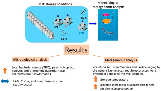
2.1. Milk Sampling and Storage Conditions

The raw milk came from farms located in the geographical area of origin, from cows fed in accordance with the provisions of the PV specification, and mechanically milked. Bulk raw milk samples from eleven producers of pPV cheese were collected and carried at 4 °C to the laboratory within one hour. All raw milk samples were collected in three aliquots (1 L each) in sterilized glass bottles. Upon arrival, one milk aliquot (named t0) was analyzed immediately. The other 2 aliquots (named T10 and T12) were respectively incubated in thermo-regulated water baths (Gheat Bath Control AG-System, Fratelli Galli, Milan, Italy) at 10 °C and 12 °C for 15 h, followed by refrigeration at 4 °C for 45 h.

2.2. Microbiological Analysis

Milk samples before storage (t0) and after T10 and T12 cold storage (as described above) were serially diluted (10-fold) in 1/4 strength Ringer solution (Oxoid Thermofisher, Rodano, Italy), and plated in duplicate on agar media for bacterial enumeration. Total mesophilic and psychrophilic bacteria were counted on Milk Plate Count Agar (Oxoid) at 30 °C for 72 h and 7 °C for 7 days, respectively; lipolytic and proteolytic bacteria were enumerated at 30 °C for 72 h on Tributyrin Agar (Oxoid) and milk agar (1% w/v skim milk powder), respectively, by considering colonies surrounded by a clear zone; coliforms and Escherichia coli were enumerated on ChromID Coli agar (Biomerieux, Bagno a Ripoli, Italy) at 37 °C for 24 h, according to manufacturer instructions; lactic acid bacteria were counted on MRS agar (Oxoid) at 37 °C for 48 h; Pseudomonas spp. were counted on Pseudomonas Agar Base with CFC (Cetrimide–Fucidin–Cephalosporin) supplement (Oxoid) at 30 °C for 48 h; coagulase-positive Staphylococcus were counted on Baird Parker-RPF Agar (Biomerieux) at 37 °C for 24 h. The count of butyric clostridia spores was carried out by a miniaturized MPN method using the selective medium AmpMedia 666 (SY-Lab, Neupurkersdorf, Austria) following the manufacturer procedures. After anaerobic incubation at 37 °C for 48 h, a color change of the chromogenic medium indicated a positive result [20]. By the number of positive vials per sample, the spore count per liter of milk was determined by the MPN table provided. Listeria monocytogenes was determined as follows: 25 mL of milk samples were incubated in 225 mL of Buffered Listeria Enrichment Broth (BLEB, Oxoid) with Listeria Selective Supplement at 30 °C. After 24 h, 0.1 mL of the first enrichment were incubated in 10 mL fresh BLEB at 30 °C. First and second enrichment, after 48 and 24 h, respectively, were streaked out on ALOA agar (Biolife, Milan, Italy) and incubated at 37 °C for 48 h. Presumptive L. monocytogenes colonies were isolated and cultivated in Tryptone Soya Broth (TSB, Oxoid) at 37 °C for 24 h. Confirmation was carried out by using the Singlepath® mono immunochromatographic rapid test (Merck, Darmstadt, Germany), according to manufacturer instructions. STEC E. coli was determined as follows: 25 mL of milk samples were incubated in 225 mL of mTSB Broth with Novobiocin (Merck) at 37 °C for 24 h. The presumptive presence of STEC E. coli in the enrichment culture was determined by using the Singlepath® E. coli O157 immunochromatographic rapid test (Merck), according to manufacturer instructions. In case of a positive result, the enrichment culture was streaked out on Agar ChromID® EHEC (Biomerieux) and incubated at 37 °C for 24 h. Presumptive E. coli O157 colonies were isolated, cultivated in TSB broth at 37 °C for 24 h, and confirmed by the Singlepath® E. coli O157 test.

2.3. Total DNA Extraction

Fifty milliliters of raw milk were centrifuged at 14,400× g for 10 min at 4 °C to separate the upper fatty phase. The pellet was then washed twice with PBS at pH 7.5, followed by centrifugation (8700× g for 7 min at 4 °C). Finally, the pellet was resuspended in TE 0.1 M at pH 8 and the total dsDNA was extracted by the QIAcube HT automated station (Qiagen, Milan, Italy) using QIAamp 96 QIAcube HT kit (Qiagen). Total dsDNA was quantified fluorometrically (QubitTM, Life Technologies, Monza, Italy).

2.4. Metagenomic Analysis

Total DNA of each sample was subjected to NGS analysis at IGATech laboratories (Udine, Italy). Bacterial diversity was evaluated by sequencing of amplified V3-V4 regions of the 16S rRNA gene using an Illumina MiSeq platform. A 2 × 300 bp of MiSeq amplicon library was prepared using the Nextera XT Index Kit (Illumina Inc, San Diego, CA, USA). Amplicons were then ligated by Illumina adaptors, further amplified, and gel-purified as per the standard Illumina protocols.

2.5. Bioinformatic and Data Analysis

Reads were de-multiplexed based on Illumina indexing system. Following the QIIME pipelines, the USEARCH algorithm (version 8.1.1756, 32-bit) (https://drive5.com/usearch/, accessed on 2 February 2021) allowed the following steps: chimera filtering; grouping of replicate sequences; sorting sequences per decreasing abundance; and identification of the Operational Taxonomic Unit (OTU), with a species-level taxonomic resolution. When the taxonomy assignment did not reach the species level, the genus or family name were reported. OTUs < 5 reads were removed as suggested by Probst et al. [21]. On the finoutOTU table, the alpha (α) diversity (richness and Shannon indexes) and the rarefaction curves were assessed by R software (http://www.r-project.org/index.html, accessed on 26 February 2021), using “vegan” [22] and “agricolae” [23] packages. Relative abundance foroutch OTU across the samples was calculated, and “subdominant” and “dominant” OTUs were discriminated as 0.1–1% and ≥1% of relative abundance, respectively. Taxonomic analysis of the bacterial communities was performed and visualized by using “reshape2” and “ggplot2” packages, respectively [24,25]. To investigate possible correlations between samples t0, T10, and T12, the relative abundance (%) of the genera Acinetobacter, Pseudomonas, and Lactococcus was multiplied by log CFU mL−1 of TBC to obtain the absolute value of each genus expressed as log OTU mL−1) as described by Oliveira et al. [26]. The Spearman correlation was also used to determine the log OTU mL−1 distribution of OTU-species of the three most abundant genera (Acinetobacter, Pseudomonas, and Lactococcus) across t0, T10, and T12 milk samples.

2.6. Statistic Analysis

Colony Forming Units (CFU) counts and spore counts were converted into log10 for statistical analysis. To calculate averages, the values below the detection limit were assigned to a value corresponding to half of the detection limit. Analysis of variance was carried out using Microsoft Excel software. To evaluate the association between psychrotrophic and other bacterial populations, Pearson correlation was performed using R software [27].

3. Results and Discussion

3.1. Characterization of the Raw Milk Microbiota

3.1.1. Microbiological Analysis

The effects of the two modified cold storage conditions studied (i.e., 15 h at 10 °C and 12 °C then 45 h at 4 °C, group samples called T10 and T12, respectively) on the bacterial loads of raw milk was evaluated by culture-dependent methods. The mean value of the total mesophilic bacterial (TMB) count of not-stored milk samples (t0) was within the limit of 105 CFU mL−1 established by EU Regulation 853/2004 [11] for raw cow’s milk collected after milking. After storage, TMB counts increased significantly in both T10 and T12 (Table 1); however, they were always below the limit of 3.0 × 10⁵ CFU mL−1 (corresponding to 5.48 log CFU mL−1) established for raw milk after transport and storage at the dairy plant, just before processing (Table 1). Other bacterial groups, such as psychrotrophic, proteolytic, lipolytic, pseudomonads, and coliforms (these latter only in T12) significantly increased after cold storage (Table 1).
Psychrotrophic bacteria generally represent less than 10% of the TMB. They grow at ≤7 °C and, depending on temperature and time of storage, may dominate the microbiota of refrigerated raw milk, reaching 70% to 90% of the bacterial population. They belong to many genera, both Gram-negative and Gram-positive, and Pseudomonas spp. are those most frequently found in refrigerated raw milk [28,29]. Similar to other studies, the mean percentage of the psychrotrophic bacteria out of the TMB in not stored milk (t0) was 65.6% (as log10 cfu mL−1) [4,30]. This proportion increased to 93.9 and 93.6% in T10 and T12 milk samples, respectively. Pseudomonas counts overlapped those of psychrotrophs, representing the 69.8% of the TMB in not stored milk (t0) and reaching 94.3% and 96.7% in T10 and T12 milk samples, respectively. After storage under T10 and T12 conditions, significant correlations between psychrotrophic bacteria and mesophilic, lipolytic, Pseudomonas spp. (r ≥ 0.89, p < 0.001), total coliforms (r = 0.80–0.83, p < 0.01) and, to a lesser extent, proteolytic bacteria (r = 0.63–0.71, p < 0.05) were found (Figure S1). Among psychrotrophic bacteria, which dominate the microbiota of refrigerated raw milk, Pseudomonas are known for the ability to produce extracellular enzymes [17,28,31,32]. In raw milk samples stored at T10 and T12, the psychrotrophic and Pseudomonas loads overlapped with those of lipolytic bacteria, while a similar trend was not shown with proteolytic bacteria. These differences could be due to a different microbiota of the raw milk associated with the 11 PV farms. Even though the initial loads were similar, there can be, within the different bacterial groups, a variety of strains with different growth capabilities [30,33]. Different farm-associated, growth rates during milk storage were reported for lipolytic and proteolytic bacteria [14,34].
Lactic acid bacteria (LAB) enumerated on MRS agar, slowly increased during both storage conditions, although the growth was not statistically significant (Table 1). Additionally, pH of raw milk samples (n = 11; t0 = 6.73 ± 0.04) did not change after storage (T10 = 6.74 ± 0.04; T12 = 6.74 ± 0.04), thus confirming the lack of growth of acidifying microbiota, such as LAB, after cold storage. Following incubation at 8–10 °C and 13–15 °C, Malacarne et al. [4] showed a significant LAB increase after 24 and 12 h, respectively. In the present study, however, milk remained at 4 °C (a temperature very far from the optimum growth range of LAB) for 45 out of the overall 60 h of cold storage in both treatments applied. Among hygiene or spoilage bacterial indicators, such as coagulase positive staphylococci, butyric clostridia spores, E. coli, and coliforms, only these latter significantly increased in T12 samples.
Although the storage at temperatures >4 °C is considered a risky practice for safety in all milk samples of the present study pathogens like L. monocytogenes and E. coli STEC were not detected, both before and after cold storage. To this regard, studies carried out so far to evaluate the potential development of pathogenic microorganisms along the processing of Grana Padano cheese, which shows many similarities with that of pPV cheese, confirmed that the storage conditions of raw milk, and those of processing and ripening, are effective in controlling pathogens along the cheese production chain [35].

3.1.2. DNA Metabarcoding Data

In the t0, T10, and T12 milk samples, a total of 5,373,219 paired end reads with an average of 162,825 reads per sample (range 100,872–334,572) were sequenced, which allowed to identify 533 OTUs having at least >5 reads. Two hundred twenty-seven OTUs were further split up into dominant (66; ≥1% total reads) and subdominant (161; 0.1–1% total reads) taxa (Figure 1; Table S1, respectively). With relative abundance values between 3% and 8%, the top five dominant genera, i.e., Acinetobacter, Lactococcus, Pseudomonas, Corynebacterium, and Streptococcus, prevailed in not stored milk (t0); in T10 and T12 samples, the % relative abundance of Acinetobacter, Lactococcus, and Pseudomonas increased, while that of Corynebacterium and Streptococcus decreased (Figure 1).
Alpha diversity indexes showed no statistical differences in the number (Richness index) and uniformity of distribution (Shannon index) of taxa found in the T10 and T12 samples (Figure S2). Similarly, the ß-dispersion analysis showed that the T10 and T12 samples did not separate across the plot (Figure S3). Interestingly, TMB count was positively correlated with the relative abundance of Pseudomonas-OTUs (r = 0.66, p =0.0002), Lactococcus-OTUs (r = 0.82, p < 0.0001), and Acinetobacter-OTUs (r = 0.77, p < 0.0001), and negatively correlated with the relative abundance of Corynebacterium-OTUs (r = −0.75, p < 0.0001). TMB count was not correlated with Psychrobacter-OTUs (r = −0.06, p = 0.72). Violin plot confirmed an increase of OTUs (expressed as log OTU mL−1), but with a different frequency distribution, of Acinetobacter, Lactococcus, and Pseudomonas, the three most abundant genera, across t0, T10, and T12 samples (Figure 2). Acinetobacter and Lactococcus showed a normal data distribution with the large part of DNA milk samples having a value around median at T12 and a right-skewed value distribution, respectively (Figure 2a,b). Pseudomonas showed a higher log OTU mL−1 value at T10; the violin plot underlined a right-skewed OTU distribution (Figure 2c).
Within Lactococcus spp., Lc. raffinolactis prevailed at the two temperatures of storage while Lactococcus garviae showed a lower relative abundance, with a decreasing trend from 10 to 12 °C (Figure 3a). Interestingly, Lc. raffinolactis has been proposed as a dairy starter and candidate probiotic [36,37]. Within Streptococcus spp, Streptococcus thermophilus was the most abundant species while other streptococcal species, which may be linked to poor hygienic conditions, were considerably less represented. Specifically, S. equinus, S. agalactiae, S. dysgalactiae, S. devriesei, and S. parapneumoniae showed a decrease in the relative abundance from t0 to both T10 and T12 (Figure 3b).
By preventing the growth of the mesophilic microbiota, in particular the lactose fermenting bacteria responsible for the acidification process, refrigeration has considerably reduced the deterioration of raw milk. However, a part of the mesophilic microbiota consisted of psychrotrophic bacteria, capable of developing even at temperatures below the optimal ones (25–35 °C) for mesophiles. Bovine milk always contains a significant amount of lactic acid bacteria (LAB), with considerably variable quantitative ranges, belonging mainly to the genera Lactococcus, Streptococcus, and Lactobacillus. Numerous other bacterial groups, generally psychrotrophic (Pseudomonas, Acinetobacter, and Aeromonas) are detectable and, generally, tend to prevail during milk refrigeration [8,38]. Metagenomic studies on hundreds of milk samples have describe the finding of many bacterial species belonging to several different genera. S. thermophilus and Lactococcus lactis, with about 40 and 20% of sequences, respectively, are the dominant species followed by Acinetobacter, Aeromonas, Brevibacterium, Corynebacterium, Lactobacillus, Pseudoalteromonas, Pseudomonas, and Staphylococcus [6]. In our study, the microbial composition inferred from the metagenomics data appeared to be quite in line with the literature data, with prevalence, in decreasing order, of Acinetobacter spp., Lactococcus spp., Pseudomonas spp., Corynebacterium spp., and Streptococcus spp. Notably, a lower incidence of Lactobacillus spp. was observed in all the analyzed samples. The cold storage at 10 and 12 °C differently affected the relative abundance and frequency distribution of the OTUs within the dominant bacterial genera, as shown by the different shape of the violin plots of Acinetobacter-OTUs, Lactococcus-OTUs, and Pseudomonas-OTUs. This variable behavior can be explained by the possible prevalence, within the three genera, of species or strains with different optimal growth temperatures, which may have been affected by the two cold storage conditions applied in this study.
Acinetobacter spp., Pseudomonas spp., and Psychrobacter spp. as well as many streptococcal species (e.g., S. agalactiae and S. dysgalactiae) can be involved in food spoilage or are recognized as indicators of poor hygienic conditions in food production or processing environments [39]. Although these bacterial groups were found to be abundant in most milk samples and increased in both T10 and T12 samples, no apparent symptoms of spoilage (i.e., appreciable sensory alterations) were found after cold storage at 10 or 12 °C. Together with the absence of pathogenic microbiota, evaluated by both culture-dependent and metagenomic analysis, it can be stated that the storage treatments applied to milk for pPV cheese would not negatively impact on its microbiological and hygienic quality. The increase in the relative abundance of Lactococcus spp. and the positive correlation of Lactococcus-OTUs with the TMB count in both T10 and T12 samples underlined that the cold storage treatments applied could be assimilated to a cold prematuration, resulting in an increase of the indigenous pro-technological microbiota of raw milk. Even if cold maturation brings many advantages to the cheese making process, it may also be a source of variation in yield and quality if not adequately controlled by the cheese maker. Cold maturation is generally followed by thermization or pasteurization to remove psycrotrophic bacteria. This is not the case for pPV, which is produced from raw milk. Therefore, depending on the microbiological quality and variability of milk, cold maturation can positively or negatively affect the final quality of cheese. To circumvent these limits and reduce batch to batch variations, the addition of pre-maturing cultures could be an option [40]. Some Parmigiano Reggiano cheesemakers have introduced the practice of adding a portion of natural whey starter culture to the evening milk to favor its “maturation” and counteract the tendency to reduce the content of mesophilic lactic acid bacteria, which hinders an increase in acidity during the spontaneous milk creaming [41].

4. Conclusions

Raw milk is held under refrigerated conditions in the farm bulk tank before delivering to dairy processing plants. The extent of time that raw milk is stored prior to processing may vary depending on its bacterial composition and hygienic quality, the milk collection intervals, and transport distances. Raw milk can also be stored under refrigerated conditions at the plant prior to processing. These practices can lead to changes of bacterial populations in raw milk both attributable to the differences in the farm management systems, to suboptimal conditions that may occur during transport, and to extended storage in processing plants. Farm and industrial practices, which are rarely homogeneous and univocal in terms of storage times and temperatures of milk along the entire supply chain, can certainly contribute to this microbial variability. Our study provides interesting findings on the possibility to modify the cold storage conditions of raw milk to be used for production of a hard-cooked, long-ripened cheese, such as the “spicy” variety of PV cheese. This prolonged cold storage does not significantly affect the qualitative-quantitative composition of the raw milk microbiota and its hygienic quality but could favor an enrichment in pro-technological bacteria. The possibility of modifying the storage conditions would make it possible to rationalize the management, timing, and costs of the phases of collection, transport, and storage of dairy raw milk, with advantages also for the sustainability of the supply chain.

Supplementary Materials

The following supporting information can be downloaded at: https://www.mdpi.com/article/10.3390/dairy3040048/s1, Figure S1: Dynamics of bacterial populations in raw milk samples (n = 11) stored under T10 (A) and T12 (B) conditions. Correlation among bacterial counts evaluated by the Pearson test (“r” is indicated by numbers; “P” as *** < 0.001, ** < 0.01, * < 0.05). Red line indicated the regression curve, dot indicated the samples and bars indicated the frequence distribution of each variable (bacterial species); Figure S2: Alpha diversity indexes of raw milk samples at t0 (pink bar) and at the two different temperatures of storage (T10, T12) (violet and blue bar, respectively); Figure S3: Beta-dispersion plot of raw milk samples at t0 (black line) and at the two different temperatures of storage (T10, T12) (red and green line, respectively). Lines represented convex hulls enclosing each group’s data points.; Table S1: Subdominant genera ordered for relative abundance from the most to the less abundant.

Author Contributions

Conceptualization, M.Z., D.C. and G.G.; methodology, D.C., B.B. and L.R.; software, M.Z.; formal analysis: M.Z. and F.T.; investigation, B.B., L.R. and G.F.; writing—original draft preparation, M.Z., D.C. and G.G.; writing—review and editing, M.Z., D.C., F.T. and G.G.; supervision, M.Z., D.C. and G.G.; funding acquisition, G.G. and D.C. All authors have read and agreed to the published version of the manuscript.

Funding

This research was funded by the Italian Ministry of Agricultural, Food and Forestry Policies (MIPAAF), Project TEMPRO (grant number D.M. 16837/7100/2019 of 11.04.2019).

Informed Consent Statement

Not applicable.

Data Availability Statement

Data is contained within the article or Supplementary Material.

Conflicts of Interest

The authors declare no conflict of interest.

References

  1. Murphy, S.C.; Boor, K.J. Trouble-shooting sources and causes of high bacteria counts in raw milk. Dairy Food Environ. Sanit. 2000, 20, 606–611. [Google Scholar]
  2. Franciosi, E.; Settanni, L.; Cologna, N.; Cavazza, A.; Poznanski, E. Microbial analysis of raw cows’ milk used for cheese-making: Influence of storage treatments on microbial composition and other technological traits. World J. Microbiol. Biotechnol. 2011, 27, 171–180. [Google Scholar] [CrossRef]
  3. Falardeau, J.; Keeney, K.; Trmčić, A.; Kitts, D.; Wang, S. Farm-to-fork profiling of bacterial communities associated with an artisan cheese production facility. Food Microbiol. 2019, 83, 48–58. [Google Scholar] [CrossRef] [PubMed]
  4. Malacarne, M.; Summer, A.; Franceschi, P.; Formaggioni, P.; Pecorari, M.; Panari, G.; Vecchia, P.; Sandri, S.; Fossa, E.; Scotti, C.; et al. Effects of storage conditions on physico-chemical characteristics, salt equilibria, processing properties and microbial development of raw milk. Int. Dairy J. 2013, 29, 36–41. [Google Scholar] [CrossRef]
  5. Mankai, M.; Boulares, M.; Ben Moussa, O.; Karoui, R.; Hassouna, M. The effect of refrigerated storage of raw milk on the physicochemical and microbiological quality of Tunisian semihard Gouda-type cheese during ripening. Int. J. Dairy Technol. 2012, 65, 250–259. [Google Scholar] [CrossRef]
  6. Quigley, L.; O’Sullivan, O.; Stanton, C.; Beresford, T.P.; Ross, R.P.; Fitzgerald, G.F.; Cotter, P.D. The complex microbiota of raw milk. FEMS Microbiol. Rev. 2013, 37, 664–698. [Google Scholar] [CrossRef]
  7. von Neubeck, M.; Baur, C.; Krewinkel, M.; Stoeckel, M.; Kranz, B.; Stressler, T.; Fischer, L.; Hinrichs, J.; Scherer, S.; Wenning, M. Biodiversity of re-frigerated raw milk microbiota and their enzymatic spoilage potential. Int. J. Food Microbiol. 2015, 211, 57–65. [Google Scholar] [CrossRef]
  8. Vithanage, N.R.; Dissanayake, M.; Bolge, G.; Palombo, E.A.; Yeager, T.R.; Datta, N. Microbiological quality of raw milk attributable to prolonged refrigeration conditions. J. Dairy Res. 2017, 84, 92–101. [Google Scholar] [CrossRef]
  9. Ministero delle Politiche Agricole Alimentari Forestali e del Turismo. Modifica minore del disciplinare di produzione della denominazione «Provolone Valpadana» registrata in qualità di denominazione di origine protetta in forza al regolamento (CE) n. 1107 del 12 giugno 1996. In Gazzetta Ufficiale della Repubblica Italiana n. 91; Istituto Poligrafico e Zecca dello Stato: Roma, Italy, 2019; pp. 17–20. [Google Scholar]
  10. Council of the European Union. Regulation (EC) No 853/2004 of 29 April 2004 laying down specific hygiene rules for food of animal origin. Off. J. Eur. Union. 2004, 50, 55–205. [Google Scholar]
  11. Franciosi, E.; Gardini, F.; Monfredini, L.; Tabanelli, G.; Fabris, A.; Endrizzi, I.; Poznanski, E.; Gasperi, F.; Cavazza, A. Does milk treatment before cheesemaking affect microbial and chemical traits of ripened cheese? Grana Trentino as a case study. J. Dairy Sci. 2012, 95, 5485–5494. [Google Scholar] [CrossRef]
  12. Gatti, M.; Bottari, B.; Lazzi, C.; Neviani, E.; Mucchetti, G. Microbial evolution in raw-milk, long-ripened cheeses produced using undefined natural whey starters. J. Dairy Sci. 2014, 97, 573–591. [Google Scholar] [CrossRef] [PubMed]
  13. Morandi, S.; Battelli, G.; Silvetti, T.; Goss, A.; Cologna, N.; Brasca, M. How the biodiversity loss in natural whey culture is affecting ripened cheese quality? The case of Trentingrana cheese. LWT-Food Sci. Technol. 2019, 115, 1084802. [Google Scholar] [CrossRef]
  14. Celestino, E.L.; Iyer, M.; Roginski, H. The effects of refrigerated storage on the quality of raw milk. Aust. J. Dairy Technol. 1996, 51, 59–63. [Google Scholar]
  15. Lafarge, V.; Ogier, J.-C.; Girard, V.; Maladen, V.; Leveau, J.-Y.; Gruss, A.; Delacroix-Buchet, A. Raw cow milk bacterial population shifts attributable to refrigeration. Appl. Environ. Microbiol. 2004, 70, 5644–5650. [Google Scholar] [CrossRef] [PubMed]
  16. Raats, D.; Maya, O.; Dror, M.; Malka, H. Molecular analysis of bacterial communities in raw cow milk and the impact of refrigeration on its structure and dynamics. Food Microbiol. 2011, 28, 465–471. [Google Scholar] [CrossRef]
  17. Vithanage, N.R.; Dissanayake, M.; Bolge, G.; Palombo, E.A.; Yeager, T.R.; Datta, N. Biodiversity of culturable psychrotrophic microbiota in raw milk attributable to refrigeration conditions, seasonality and their spoilage potential. Int. Dairy J. 2016, 57, 80–90. [Google Scholar] [CrossRef]
  18. Gobbetti, M.; Neviani, E.; Fox, P. The Cheeses of Italy: Science and Technology, 1st ed.; Springer International Publishing: Cham, Switzerland, 2018. [Google Scholar]
  19. Malacarne, M.; Summer, A.; Formaggioni, P.; Franceschi, P.; Sandri, S.; Pecorari, M.; Vecchia, P.; Mariani, P. Dairy maturation of milk used in the manufacture of Parmigiano-Reggiano cheese: Effects on physico-chemical characteristics, rennet-coagulation aptitude and rheological properties. J. Dairy Res. 2008, 75, 218–224. [Google Scholar] [CrossRef]
  20. Brändle, J.; Heinzle, L.; Fraberger, V.; Berta, J.; Zitz, U.; Schinkinger, M.; Stocker, W.; Kneifel, W.; Domig, K.J. Novel approach to enumerate clostridial endospores in milk. Food Control. 2018, 85, 318–326. [Google Scholar] [CrossRef]
  21. Probst, M.; Gómez-Brandón, M.; Bardelli, T.; Egli, M.; Insam, H.; Ascher-Jenull, J. Bacterial communities of decaying Norway spruce follow distinct slope exposure and time-dependent trajectories. Environ. Microbiol. 2018, 20, 3657–3670. [Google Scholar] [CrossRef]
  22. Oksanen, J.; Blanchet, G.F.; Friendly, M.; Kindt, R.; Legendre, P.; McGlinn, D.; Minchin, P.R.; O’Hara, R.B.; Simpson, G.L.; Solymos, P.; et al. Vegan: Community Ecology Package. R Package Version 2.5-5. 2019. Available online: https://cran.r-project.org/ (accessed on 26 February 2021).
  23. De Mendiburu, F. Package ‘Agricolae’. R Package Version 1.2-8. Available online: http://CRAN.R-project.org/package=agricolae (accessed on 4 July 2019).
  24. Caporaso, J.; Kuczynski, J.; Stombaugh, J.; Bittinger, K.; Bushman, F.D.; Costello, E.K.; Fierer, N.; Peña, A.G.; Goodrich, J.K.; Gordon, J.I.; et al. QIIME allows analysis of high-throughput community sequencing data. Nat. Meth. 2010, 7, 335–336. [Google Scholar] [CrossRef]
  25. Wickham, H. Reshape2: Flexibly Reshape Data: A Reboot of the Reshape Package. R Package Version. 2012. Available online: https://cran.r-project.org/ (accessed on 26 February 2021).
  26. Oliveira Dumont, A.C.; Nogueira Souza, F.; Machado de Sant’Anna, F.; Faula, L.L.; Gasparotto Chande, C.; Cortez, A.; Melville Paiva Della Libera, A.M.; Costa, M.; Resende Souza, M.; Heinemann, M.B.; et al. Temporal and geographical comparison of bulk tank milk and water microbiota composition in Brazilian dairy farms. Food Microbiol. 2021, 98, 103793. [Google Scholar] [CrossRef] [PubMed]
  27. R Core Team. R: A Language and Environment for Statistical Computing. Available online: http://www.sthda.com/english/wiki/correlation-matrix-an-r-function-to-do-all-you-need (accessed on 2 March 2020).
  28. Decimo, M.; Morandi, S.; Silvetti, T.; Brasca, M. Characterization of Gram-negative psychrotrophic bacteria isolated from Italian bulk tank milk. J. Food Sci. 2014, 79, M2081–M2090. [Google Scholar] [CrossRef] [PubMed]
  29. De Jonghe, V.; Coorevits, A.; Van Hoorde, K.; Messens, W.; Van Landschoot, A.; De Vos, P.; Heyndrickx, M. Influence of storage conditions on the growth of Pseudomonas species in refrigerated raw milk. Appl. Environ. Microbiol. 2011, 77, 460–470. [Google Scholar] [CrossRef] [PubMed] [Green Version]
  30. Hantsis-Zacharov, E.; Halpern, M. Culturable psychrotrophic bacterial communities in raw milk and their proteolytic and lipolytic traits. Appl. Environ. Microbiol. 2007, 73, 7162–7168. [Google Scholar] [CrossRef]
  31. Hahne, J.; Isele, D.; Berning, J.; Lipski, A. The contribution of fast growing, psychrotrophic microorganisms on biodiversity of refrigerated raw cow’s milk with high bacterial counts and their food spoilage potential. Food Microbiol. 2019, 79, 11–19. [Google Scholar] [CrossRef]
  32. Narvhus, J.A.; Bækkelund, O.N.; Tidemann, E.M.; Østlie, H.M.; Abrahamsen, R.K. Isolates of Pseudomonas spp. from cold-stored raw milk show variation in proteolytic and lipolytic properties. Int. Dairy J. 2021, 123, 105049. [Google Scholar] [CrossRef]
  33. Guinot-Thomas, P.; Al Ammoury, M.; Laurent, F. Effects of storage conditions on the composition of raw milk. Int. Dairy J. 1995, 5, 211–223. [Google Scholar] [CrossRef]
  34. Paludetti, L.F.; Jordan, K.; Kelly, A.L.; Gleeson, D. Evaluating the effect of storage conditions on milk microbiological quality and composition. Irish J. Agric. Food Res. 2018, 57, 52–62. [Google Scholar] [CrossRef]
  35. Giraffa, G. The Microbiota of Grana Padano Cheese. A Review. Foods 2021, 10, 2632. [Google Scholar] [CrossRef]
  36. Kimoto-Nira, H.; Aoki, R.; Mizumachi, K.; Sasaki, K.; Naito, H.; Sawada, T.; Suzuki, C. Interaction between Lactococcus lactis and Lactococcus raffinolactis during growth in milk: Development of a new starter culture. J. Dairy Sci. 2012, 95, 2175–2185. [Google Scholar] [CrossRef]
  37. Jung, M.J.; Lee, C.; Seo, M.; Roh, S.W.; Lee, S.H. Characterization of a potential probiotic bacterium Lactococcus raffinolactis WiKim0068 isolated from fermented vegetable using genomic and in vitro analyses. BMC Microbiol. 2020, 20, 136. [Google Scholar] [CrossRef] [PubMed]
  38. Ribeiro, J.C., Jr.; de Oliveira, A.M.; Silva, F.D.G.; Tamanini, R.; de Oliveira, A.L.M.; Beloti, V. The main spoilage-related psychrotrophic bacteria in refrigerated raw milk. J. Dairy Sci. 2017, 101, 75–83. [Google Scholar] [CrossRef] [PubMed]
  39. Doulgeraki, A.I.; Ercolini, D.; Villani, F.; Nychas, G.J.E. Spoilage microbiota associated to the storage of raw meat in different conditions. Int. J. Food Microbiol. 2012, 157, 130–141. [Google Scholar] [CrossRef] [PubMed]
  40. Lane, C.N.; Sousa-Gallagher, M.J.; McSweeney, P.L.H. Effect of prematuration conditions on the proteolytic and rheological properties of cheesemilk. Lait 2001, 81, 415–427. [Google Scholar] [CrossRef]
  41. Coloretti, F.; Chiavari, C.; Nocetti, M.; Reverberi, P.; Bortolazzo, E.; Musi, V.; Grazia, L. Whey starter addition during maturation of evening milk: Effects on some characteristics of cheese milk and Parmigiano–Reggiano cheese. Dairy Sci. Technol. 2015, 96, 185–197. [Google Scholar] [CrossRef] [Green Version]
Figure 1. Average values of % relative abundance of the 66 dominant taxa retrieved in raw milk samples. t0: milk before storage; T10 and T12: milk after cold storages (i.e., 15 h at 10 °C and 12 °C then 45 h at 4 °C, respectively).
Dairy 03 00048 g001
Figure 2. Distribution of OTUs (expressed as log OTU mL−1) within the three most abundant genera: Acinetobacter (a), Lactococcus (b), and Pseudomonas (c). t0 (blue plot): milk before storage. T10 (yellow plot) and T12 (pink plot): milk after cold storage (i.e., 15 h at 10 °C and 12 °C then 45 h at 4 °C, respectively).
Dairy 03 00048 g002
Figure 3. Average values of relative abundance of Lactococcus (a) and Streptococcus (b) species in t0, T10, and T12 samples. t0 (red bar): milk before storage. T10 (blue bar) and T12 (green bar): milk after cold storage (i.e., 15 h at 10 °C and 12 °C then 45 h at 4 °C, respectively).
Dairy 03 00048 g003
Table 1. Bacterial counts before (t0) and after (T10 and T12) cold storage of raw milk for PV cheese from 11 cheese producers. Values are given as means ± standard deviation.
No Stored MilkStored Milk 1
Bacterial Populations 2t0T10T12Significance
Total mesophilic bacteria4.13 ± 0.56 a5.12 ± 0.70 b5.23 ± 0.87 b**
Total psychrotrophic bacteria2.77 ± 1.05 a4.82 ± 0.89 b4.94 ± 1.09 b**
Proteolytic bacteria2.63 ± 0.60 a3.72 ± 0.61 b3.99 ± 0.68 b**
Lipolytic bacteria3.56 ± 0.51 a4.64 ± 0.87 b4.94 ± 0.88 b**
Pseudomonas spp.2.92 ± 1.02 a4.85 ± 1.01 b5.08 ± 0.95 b**
Lactic acid bacteria2.99 ± 0.57 a3.14 ± 0.48 a3.15 ± 0.52 a-
Total coliforms2.29 ± 0.64 a3.17 ± 1.09 a3.48 ± 0.95 b*
E. coli1.70 ± 1.02 a 1.93 ± 1.11 a2.15 ± 1.09 a-
Coagulase positive staphylococci1.10 ± 0.59 a1.08 ± 0.58 a1.13 ± 0.62 a-
Butyric clostridia spores1.92 ± 0.53 a 1.89 ± 0.43 a1.90 ± 0.43 a-
Values in the same row with different superscript letters differ significantly (* p < 0.05; ** p < 0.01). 1 Milk storage conditions: T10 = milk after 15 h at 10 °C and 45 h at 4 °C; T12 = milk after 15 h at 12 °C and 45 h at 4 °C. 2 Bacterial plate counts are expressed as log10 CFU mL−1; butyric clostridia spores count is expressed as log10 MPN L−1.
Publisher’s Note: MDPI stays neutral with regard to jurisdictional claims in published maps and institutional affiliations.

