Article

Comparative Analysis of Intraoral Scanner Accuracy in a Six-Implant Complete-Arch Model: An In Vitro Study

by
Francesco Ferrini
1,2,*,
Federica Mazzoleni
1,
Matteo Barbini
3,
Carlotta Coppo
3,
Giovanna Laura Di Domenico
3 and
Enrico Felice Gherlone
1,2
1
Dental School, IRCCS San Raffaele, Vita-Salute San Raffaele University, 20132 Milan, Italy
2
Department of Dentistry, IRCCS San Raffaele Hospital and Dental School, Vita-Salute San Raffaele University, 20123 Milan, Italy
3
Oral Surgery School, IRCCS San Raffaele, Vita-Salute San Raffaele University, 20132 Milan, Italy
*
Author to whom correspondence should be addressed.
Prosthesis 2024, 6(2), 401-412; https://doi.org/10.3390/prosthesis6020030
Submission received: 8 March 2024 / Revised: 1 April 2024 / Accepted: 17 April 2024 / Published: 22 April 2024

Abstract

:
(1) Background: Since new intraoral scanner (IOS) versions are introduced to the market and software continues to advance, there is an ongoing need to assess the accuracy of newer IOS models. (2) Methods: Four types of IOSs and one laboratory scanner (used as a reference) were used to scan an edentulous model with six parallel implants and their respective scan bodies, which were connected to each other. Using dedicated software, the distances between all scan bodies were calculated, generating a total of 540 measurements. Trueness (comparisons to the reference model) and precision (intragroup comparisons) were statistically compared with ANOVA and Tukey tests. (3) Results: When considering trueness values, statistically significant differences were observed between the tested scanner for all subgroups considered (p < 0.05). By contrast, no statistically significant differences were reported for precision values. (4) Conclusions: Within the limitations of the present in vitro study, it can be concluded that all tested IOSs were similar in terms of precision, while Trios and i700W yielded the worst trueness values. Nevertheless, increasing the measuring distance leads to a decrease in both trueness and precision.

1. Introduction

The dental field, with all its subareas, is becoming more and more dominated by the digital world. The computerized CAD/CAM system was first used in other fields in 1960, and the dental industry adopted it ten years later [1,2,3].
Obtaining a correct impression that is not altered throughout the time between taking the impression and having it cast by the laboratory technician has always been crucial to producing a correct prosthesis [4,5].
Each prosthodontist, or rather any dentist, must be aware of the best ways to capture the anatomical/mechanical information of each treated patient because both the traditional and the digital impressions are operator-dependent [6,7].
Nowadays, in the digital era, thanks to improvements in the reading precision and accuracy of scanners, the prosthetic manufacturers are able to shape the tissues and fit them as perfectly as possible on the teeth/implant abutments [8,9].
For both the operator and the patient, a digital flow offers a number of benefits [5,6]. For instance, in comparison to the analog technique, which requires the utilization of specific materials that allow the operator to “impress” the teeth, the digital flow eliminates the gag reaction for the patient [10].
Continual improvements in these devices, with a focus on size, design, and scanning speed, have been driven by the pursuit of operational ease by manufacturers [11]. Recently, in fact, wireless technology has received a lot of attention due to its benefits [12,13,14].
Additionally, since every enhancement should be considered in light of the final outcome, accuracy is also considered an important characteristic [15].
Accuracy includes trueness and precision, according to the International Organization for Standardization. Trueness refers to how closely a measurement matches the real value of an object. To assess the accuracy of an IOS, it is essential to compare its measurements to those obtained from a highly accurate reference measurement machine. Usually, laboratory scanners are highly accurate and have been used as reference models in various in vitro studies [16,17,18,19]. Precision, on the other hand, refers to the ability to consistently produce repeatable measurements. Precision evaluation of IOSs involves comparing repeated measurements taken with the same IOS and analyzing the variation between those measurements [20].
Data from the literature indicate that different brands of IOS devices vary significantly in trueness [21,22,23] and that precision notably decreases with greater distances between implants and scan bodies [24,25]. Several factors influence digital impression accuracy, including IOS hardware, software, operator experience, scan body characteristics, and clinical factors [26]. Among these factors, IOS hardware and software have undergone significant changes in recent years, highlighting the importance of updating our knowledge with the latest available devices.
In order to study full-arch rehabilitations, it is necessary to understand how edentulism may occur and what rehabilitative solutions are currently available.
The term edentulism is defined as the loss of one or more dental elements [27].
Edentulism can either be single (if it concerns a single dental element), partial (if it affects several dental elements), or total (if it involves the totality of the dental elements).
The loss of dental elements, which contributes to a clinical picture of edentulism, can be caused by multiple factors, as described in a study conducted by P.S. Hull et al. [28] in which the main causes of this disorder in developed countries, where patients have access to dental care, are cariogenic pathology and periodontal disease.
The prevalence of this phenomenon is mainly related to a segment of the population over 60 years of age, which is confirmed by an ISTAT survey conducted in 2013 [29] that reported the prevalence of edentulism in Italy according to age. This study showed that edentulism is prevalent in individuals over 75 years of age (50.7%), whereas its prevalence is 0.2% in individuals up to 44 years of age and 2.2% in individuals between 45 and 54 years of age.
However, an evaluation of the data from 2005 to 2013 revealed that there has been a gradual improvement in these statistics, especially in the population over 75 years old.
The edentulous condition mainly leads to a change in the bone structure of the jaw, which is caused by a lack of functional stimulation.
The volumetric magnitude of resorption, however, does not have a precise mode or amount but instead varies depending on the individual, systemic or local pathologies, and the type of dental therapies adopted to treat this condition.
Mucosa-supported prostheses represent an alternative option for edentulism patients but play an important role in determining the degree of atrophy they experience.
Following the loss of dental elements, physiological healing processes begin at the extraction site, leading to bone remodeling and subsequent progressive volumetric reduction [30].
In addition to changes in the alveolar portion in both the upper and lower jaws, mechanisms of remodeling in an involutional sense intervene in the basal bone; this pathophysiological mechanism is referred to as post-extraction atrophy [31].
Evolution can proceed to the point of compromising the structure of the alveolar portions by completely disrupting the relationship between the lower and upper jaw and by exacerbating the discrepancy between the two arches both sagittally and transversally.
Several studies, including one conducted by Schropp et al. [32], described post-extraction atrophy by showing that the biggest loss of crestal bone components occurs in the first three months after avulsion of the dental elements.
Bone resorption in the maxillae occurs differently. In the upper jaw, the maxilla decreases volumetrically by contracting centripetally, which results in the disappearance of the alveolar portion and pneumatization of the maxillary sinuses. On the other hand, in the lower jaw, the mandible undergoes a centrifugal resorption process, which is the opposite of what occurs in the upper jaw.
The consequence of these two patterns of resorption is a relationship between the two edentulous ridges that shows a marked mandibular prognathism, particularly due to a hypomaxillary condition; therefore, it is common to observe a third skeletal class scenario even in patients who naturally did not have this type of skeletal class.
Moreover, this type of resorption can lead to a crestal atrophic condition, called “knife blade”, in which the crestal height is partially maintained but the thickness is limited almost exclusively to the palatine cortical component [33].
In aged patients, a high degree of atrophy may lead to morphological changes in the cranial structure and facial bones, which is due to continuous remodeling of the skeleton that results from a functional stimulus.
The main aesthetic changes resulting from an edentulous situation mainly affect the middle third and the lower third of the face.
Changes in these two portions of the face are due to the failure of the atrophic bone structures to support the overlying muscles and tissues [32,34].
All these consequences confirm the need for an edentulous individual to avail himself or herself of prosthetic rehabilitation.
Various prosthetic rehabilitation options designed to treat edentulism exist, each with specific advantages and disadvantages.
Relevant to this study are solutions without bone grafts, the all-on-4 protocol, the all-on-6 protocol, rehabilitations using axial implants (Toronto), and the use of zygomatic implants [35].
In addition to the advantage of having a fixed solution, immediate loading is possible in all these types of rehabilitation, with lower economic and biological costs than solutions involving bone grafts.
Another advantage is having a standardized surgical and prosthetic protocol, which makes this solution predictable and repeatable [34].
The goal of these rehabilitations, particularly those involving the use of inclined implants (all-on-4 and all-on-6), is to use a limited number of implants for the purpose of rehabilitating an entire arch.
Implants are placed axially or at an incline of 30°–45°, thus avoiding the need to place implants in the posterior sectors where bone density is often poor and inadequate to ensure good primary implant stability [36].
The use of inclined implants also makes it possible to safeguard important anatomical structures, such as the inferior alveolar vascular bundle and the maxillary sinus [37].
Implant surgery is a feasible alternative in cases of edentulousness, whether single, partial, or total.
However, it must be kept in mind that for implant surgery, it is necessary to evaluate and know the possible adverse scenarios that may arise [38,39].
First, the presence of possible red lights must be considered, which should not be overlooked at all. These can be identified through a proper, careful, and periodic patient medical review. Contraindications to surgery are divided into local contraindications and systemic contraindications.
Local contraindications are represented by strong risk factors that may adversely affect the survival of the implant and include smoking, as it alters postoperative wound healing by changing the peripheral circulation and hindering osseointegration; poor oral hygiene, which causes a buildup of bacterial plaque and leads to the development of inflammation of the gingival and bone tissue, which promotes resorption of the bone itself with consequent loss of support for the implant; and periodontal disease, which is a contraindication to implantology in cases where it is not treated and controlled but is in an active phase.
Systemic contraindications are divided into two subgroups: absolute and relative.
Absolute systemic contraindications prohibit implant placement completely and include subjects with disabling systemic diseases, such as rheumatoid arthritis, osteogenesis imperfecta, and osteomalacia, subjects who are immunocompromised, HIV-positive, or on an immunosuppressant therapy, drug abusers, individuals under 16 years of age, uncooperative persons with mental disorders, and patients who had a myocardial stroke that occurred less than 3 months earlier.
The second subgroup is the relative systemic contraindications; in these cases, implant insertion is possible, but only after remission of the pathology has been achieved or after appropriate precautions have been taken.
Surgical complications that may be related to implant surgery can be identified as intraoperative complications, early complications, and late complications [40,41].
Intraoperative complications are identified as all complications that may occur during the surgical act and include incorrect implant fixture position, bleeding, lack of primary stability, injury to the elements adjacent to the area of implant placement, soft tissue injury [42], fixture displacement, dehiscence and fenestrations, neurological injuries, aspiration and swallowing of surgical instruments, and mandibular fracture [43].
In conclusion, two other classes of complications can occur: early and late complications. These concern the possible adverse events following surgery that can occur late or early after surgery. These include, edema, emphysema, implant fracture, mucosal dehiscence, and infection.
The use of the latest digital technologies has given the clinician an opportunity to avail himself of tools that can support him during all phases of rehabilitation, starting with the collection of the patient’s data to the design of the surgical procedure and prosthetic finalization, making these rehabilitation phases more predictable and accurate [44,45].
The use of state-of-the-art intraoral scanners (IOSs) has been proven to be valuable in terms of precision and accuracy in reading and detecting soft tissues [46], which makes this type of technology comparable to traditional impressions for the fabrication of prosthetic restorations in full-arch rehabilitations [3].
The aim of this study was to evaluate the accuracy of four intraoral scanners using a complete-arch implant model, which is preferred over the complete-arch dentate model due to its regular geometric shapes.

2. Materials and Methods

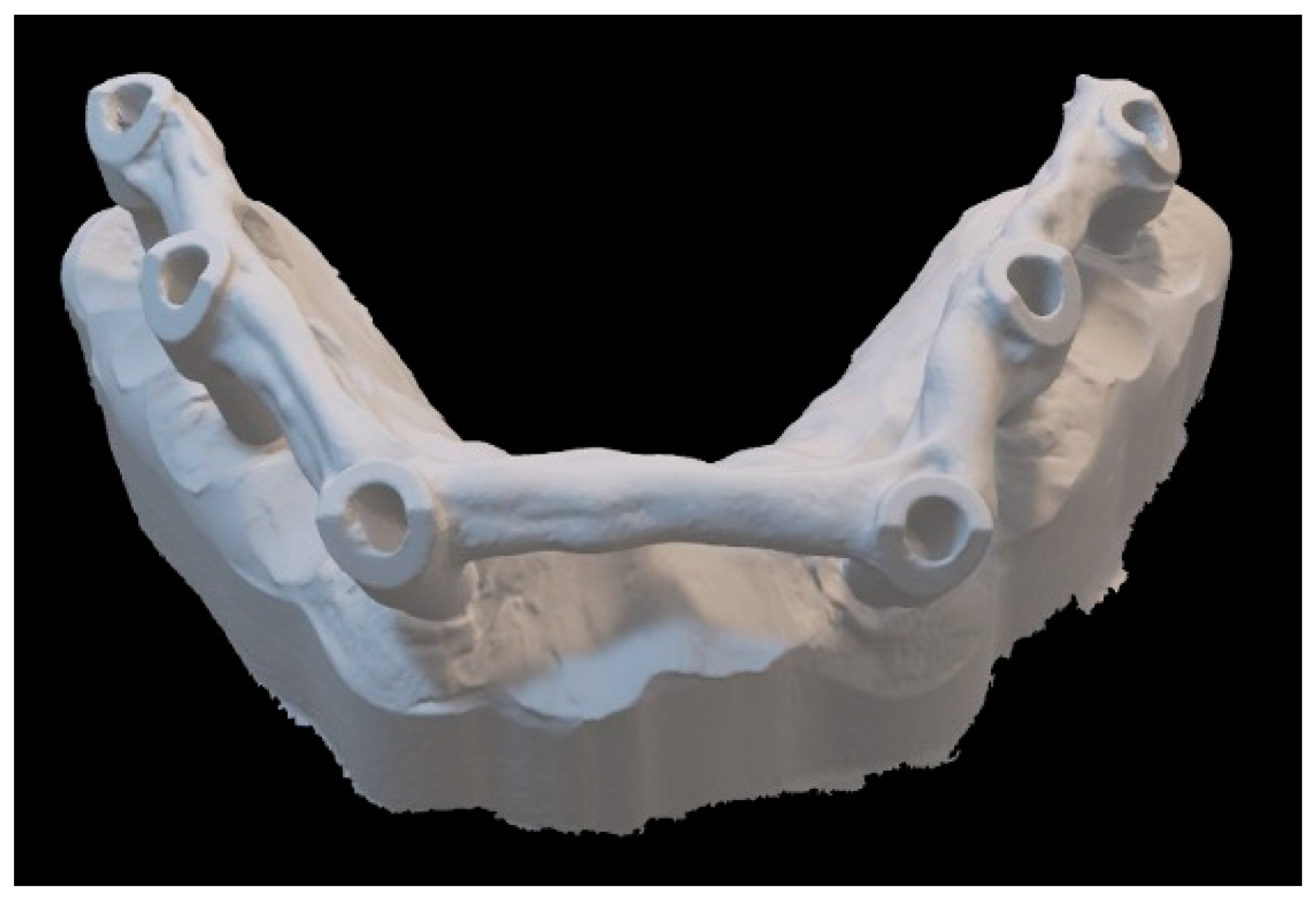
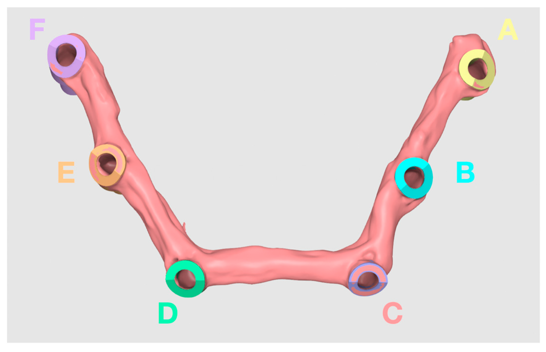
2.1. Fabrication of Master Model

A titanium model representing an edentulous dental arch was created from a scan made with a high-precision scanner with non-contact mechanical-optical technology (Alicona; Alicona Imaging GmbH, Raaba/Graz, Austria) and then processed by the laboratory.
Six implants (Winsix TTi 3.8x11 mm, Biosaf In s.r.l., Trezzano Rosa, Milan, Italy) were placed in the model, and the corresponding polyether ether ketone (PEEK) abutments were screwed in. The six scan bodies were splinted with each other according to the prosthetic protocol: dental floss was placed on all the abutments, and light-curing resin (3M RelyX Unicem 2, 3M, St. Paul, MN, USA) was then placed on this floss and polymerized. To reduce the polymerization shrinkage of the resin splint, the resin splint was sectioned and reconnected (Figure 1).

2.2. Measurements of Distances between the Abutments

The master model was scanned with an Alicona scanner (Alicona Imaging GmbH, Raaba/Graz, Austria) in order to obtain the reference distances between the abutments.
The STL file that was acquired was imported into a dedicated software program (Medit Design software, ML subgroup, version 3.0; Seoul, Republic of Korea). Subsequently, the scan bodies that were acquired were superimposed on the scan bodies using the specific mathematics of the Medit Design software (Figure 2). For each implant scan body, 3 points were collected according to the manual alignment system (listed in the software).
A 4-micron sphere was placed in the exact center of each of the scan bodies using the mathematics of the Medit Design software to obtain a reference point with which to start measuring the distance from one implant to another. Through the creation of this sphere, it was possible to measure the exact distances between implants in a repeatable way.
The distances were assigned to three groups arbitrarily: the short-distance group (under 15 mm), the medium-distance group (between 15 mm and 40 mm), and the long-distance group (over 40 mm) (Figure 2 and Table 1).
The same operator conducted all the measurements of the STL files of the 4 intraoral scanners, and the respective distances were calculated using the same process that was described for the master model.

2.3. Intraoral Scanners

The following IOSs were used according to the scanning protocol indicated by each manufacturer to obtain 9 experimental scans per group:
-
Group 1: Medit i700 wired (Medit, Seoul, Republic of Korea)
-
Group 2: Medit i700 wireless (Medit, Seoul, Republic of Korea)
-
Group 3: CS3800 wireless (Carestream Health, Rochester, NY, USA)
-
Group 4: Trios4 wireless (3Shape, Copenhagen, Denmark)
One expert prosthodontist (F.F.) performed all the scans during the same day and in the same room at the Department of Dentistry of San Raffaele Hospital (Milan, Italy) under similar light and environmental conditions.
For standardization and subsequent digital processing, datasets from each scan were converted to a standard tessellation language (STL) file format [47,48,49,50,51].
For each scanner, the trueness was calculated as the mean of the error between the distance measured by each scanner and its corresponding reference distance. By contrast, the precision was evaluated as the mean of the SD values of 9 repetitive measurements for each scanner [52,53,54,55].

2.4. Statistical Analysis

Statistical evaluation was performed using the STATA analysis software program (StataCorp 2021; Stata Statistical Software: Release 17, StataCorp LLC, College Station, TX, USA). The Shapiro–Wilk test confirmed the presence of a normal data distribution.
The one-way ANOVA test was used to assess the trueness and precision between the four impression groups, considering three different subgroups on the basis of the distance of the scan bodies.
Trueness was determined by calculating the error between the distance measured by each scanner and its corresponding reference distance. The precision of each scanner was determined by calculating the error between the mean of 9 repetitive measurements and a single measurement with the same scanner.
The distances between the scan bodies were categorized as follows: distances less than 15 mm were considered short, distances between 15 and 40 mm were classified as medium, and distances greater than 40 mm were labeled long.
The significance level was set at α = 0.05.

3. Results

3.1. Short Distances

The mean distance between points B–C and E–F measured by the Alicona machine was 14.507 mm (Table 1). Analyses of trueness and precision of the scanner in measuring these distances revealed no statistically significant differences between the tested scanners (Table 2 and Table 3).

3.2. Medium Distances

The mean distance between points A–B, A–C, B–D, B–E, C–D, C–E, D–E, and D–F measured by the Alicona machine was 28.085 mm (Table 1). Analyses of trueness and precision of the scanner in measuring these distances revealed no statistically significant differences between the tested scanners (Table 2 and Table 3).

3.3. Long Distances

The mean distance between points A–D, A–E, A–F, B–F, and C–F measured by the Alicona machine was 44.796 mm (Table 1). Analyses of trueness and precision of the scanner in measuring these distances revealed no statistically significant differences between the tested scanners (Table 2 and Table 3).

4. Discussion

The present in vitro study evaluated the accuracy of four different IOSs, both with wireless and wired technology. The null hypothesis, suggesting no difference in terms of the trueness and precision among the tested scanners, was accepted.
For the present investigation, a titanium model simulating a maxillary edentulous jaw was chosen. Traditionally, research has mainly employed stone or resin casts, both of which are prone to breaking, wearing down, and undergoing deformation during the experimental period [47,48]. On the contrary, a titanium model has superior characteristics, including improved strength, fracture resistance, dimensional stability, and resistance to stress corrosion, rendering it an ideal material for master models.
Moreover, while previous studies typically involved embedding substitutes into the master model to replicate a patient’s intraoral implant condition, the current investigation diverged by directly inserting dental implants into the baseline cast, thus better reflecting real clinical scenarios.
Furthermore, scan bodies with straightforward geometric shapes were used, simplifying measurements between well-defined points and reducing errors associated with landmark identification.
Another important issue is the connection of scan bodies and the misfit between the prosthesis and the dental implant; this value, according to Di Fiore et al., should be around 30–50 μm to avoid mechanical and biological complications [54].
Some recent studies demonstrated that the complete-arch intraoral digital impression with scan body splinting of the implant improved image stitching, resulting in a more easily trackable scanning route and increased overall accuracy of complete-arch digital impressions, as described by Mandelli et al. [4], Mizumoto et al. [50], Imburgia et al. [23], and Cheng et al. [14].
Various techniques have been proposed for splinting scan bodies, such as the use of thermoplastic resin or dental floss. However, due to differences in study design and methods, direct comparisons with the current study are difficult.
In another study, Mizumoto et al. [50] examined the impact of various scan bodies and scanning methods, including adhesive glass fiduciary markers, a pressure-indicating paste applied on the ridge and palate, and floss tied between scan bodies, on both accuracy and scanning duration. Notably, the floss that was tied between scan bodies exhibited notably inferior linear accuracy and poorer angular precision compared to other techniques.
Imburgia et al. [23] conducted a clinical investigation of the efficacy of partial- and complete-arch implant-supported zirconia prostheses obtained with intraoral scanning following splinting of implant scan bodies using thermoplastic resin and flowable composite. This technique reported a prosthetic success rate of 93.3% over a two-year follow-up period.
Cheng et al. [14] conducted an in vitro study to compare the trueness and precision of complete-arch implant impressions using conventional impression, intraoral scanning with and without splinting, and stereophotogrammetry. They concluded that the incorporation of splinting into the scan body led to a slight reduction in three-dimensional and linear deviations at the beginning of the scanning process. However, it did not yield a significant improvement in overall intraoral scanning accuracy.
Currently, the superimposition of a reference STL model on those from tested scanners is predominantly performed using a best-fit algorithm from dedicated software. This superimposition technique can show deflections in scan posts (or implants) relative to the reference model, indirectly indicating changes in the distance between adjacent or cross-arch implants. To minimize algorithm errors, we adopted the linear evaluation approach [55,56,57,58,59] for the superimposition, and for subsequent analyses of the scans, we used Medit Design software. Within the application, the “manual pairing” function was used to superimpose the various scans. This coupling method requires accurately using three reference points for the reference scan and the other scans to be superimposed on the first reference scan. The reference points that were chosen in this study to implement such coupling were the 4-micron sphere placed in the center of the scan body and the two points that were placed on the more angular geometric portions of the scan bodies. Once the software implemented the superposition of the various scans, the correct coupling was checked for added safety (Figure 3).
Multiple studies have examined full-arch digital impressions using models that simulate the creation of an implant-supported superstructure for edentulous jaws. The findings indicate that the digital impressions exhibited trueness and precision comparable to or greater than those of conventional impressions [3].
A similar study was performed by Revell et al. in 2022 [60] in which a total implant arch scanned with five different scanners using a non-solidarized method was examined. That study, however, cannot be compared with the following one because it used a different scanning method; moreover, an older model of the Medit scanner (Medit i500) was used in the study by Revell et al. [60] It was replaced in this study by the i700 and i700 wireless version. However, there are no studies, so far, in which the scanners examined in this study and the solidarized method are evaluated.
Another study, similar to the previous one, was performed by Di Fiore et al. [61] in which scans were performed, again, using the non-splinting technique.
These studies, all of which include a clinical situation in which there is an edentulous arch with an implant fixture placement but a different scanning method, may serve as a starting point for future studies.
Moreover, all of them exhibit the same trend: there is a more pronounced decline in both trueness and precision as the distance between the scan bodies increases. This observation is consistent with findings from other studies [62,63,64,65,66,67,68].
This study has several limitations, with a notable emphasis on the inherent constraints of in vitro experiments. The utilization of a titanium model in this investigation deviates from the anatomical realities present in actual patient arches, potentially limiting the extent to which the findings can be extrapolated to clinical contexts. Moreover, the scope of this research does not encompass an examination of various factors that could potentially influence impression accuracy, including operator skills, the conditions in which the scanning is performed, the lights, the saliva, mouth opening, and the location of the area in which the prosthesis is fabricated. Consequently, it is essential for future research efforts to thoroughly explore and comprehend the multifaceted effects of various factors—including, but not limited to, different implant brands, angular orientations, levels of ambient illumination, and the presence of biologic fluids—on the overall accuracy and efficacy of intraoral scanners.

5. Conclusions

Within the limitations of the present in vitro investigation, the following conclusions can be drawn: (1) there was an interaction between accuracy and the measured distances (in other words, as the distance increased, there was a decrease in the accuracy), and (2) all tested IOSs had similar trueness and precision values.

Author Contributions

Conceptualization, F.F.; methodology, G.L.D.D.; data curation, M.B.; writing—review and editing, F.M.; visualization, C.C.; supervision, E.F.G. All authors have read and agreed to the published version of the manuscript.

Funding

This research received no external funding.

Institutional Review Board Statement

Not applicable.

Informed Consent Statement

Not applicable.

Data Availability Statement

The data presented in this study are available on request from the corresponding author.

Conflicts of Interest

The authors declare no conflicts of interest.

References

  1. Erozan, Ç.; Ozan, O.E. Evaluation of the Precision of Different Intraoral Scanner-Computer Aided Design (CAD) Software Combination in Digital Dentistry. Med. Sci. Monit. 2020, 26, e918529. [Google Scholar] [PubMed]
  2. Amin, S.; Weber, H.P.; Finkelman, M.; El Rafie, K.; Kudara, Y.; Papaspyridakos, P. Digital vs. conventional full-arch implant impressions: A comparative study. Clin. Oral Implant. Res. 2017, 28, 1360–1367. [Google Scholar] [CrossRef]
  3. Capparè, P.; Sannino, G.; Minoli, M.; Montemezzi, P.; Ferrini, F. Conventional versus digital impressions for full arch screw-retained maxillary rehabilitations: A randomized clinical trial. Int. J. Environ. Res. Publish Health 2019, 16, 829. [Google Scholar] [CrossRef] [PubMed]
  4. Mandelli, F.; Gherlone, E.F.; Keeling, A.; Gastaldi, G.; Ferrari, M. Full-arch intraoral scanning: Comparison of two different strategies and their accuracy outcomes. J. Osseointegr. 2018, 10, 65–74. [Google Scholar]
  5. Davidowitz, G.; Kotick, P.G. The use of CAD/CAM in dentistry. Dent. Clin. N. Am. 2011, 55, 559–570. [Google Scholar] [CrossRef] [PubMed]
  6. Janeva, N.M.; Kovacevska, G.; Elencevski, S.; Panchevska, S.; Mijoska, A.; Lazarevska, B. Advantages of CAD/CAM versus Conventional Complete Dentures. Open Access Maced. J. Med. Sci. 2018, 6, 1498–1502. [Google Scholar] [CrossRef] [PubMed]
  7. Duret, F. The pratical dental CAD/CAM in 1993. J. Can Dent. Assoc. 1993, 59, 445–446, 448–453. [Google Scholar] [PubMed]
  8. Punj, A.; Bompolaki, D.; Garaicoa, J. Dental Impression Materials and Techniques. Dent. Clin. N. Am. 2017, 61, 779–796. [Google Scholar] [CrossRef] [PubMed]
  9. Ting-Shu, S.; Jian, S.J. Intraoral Digital Impression Technique: A Review. J. Prosthodont. 2015, 24, 313–321. [Google Scholar] [CrossRef]
  10. Dec, L.J.H. Digital Workflow for Establishing the Posterior Palatal Seal on a Digital Complete Denture. J. Prosthodont. 2021, 30, 817–821. [Google Scholar] [CrossRef]
  11. Suese, K. Progress in digital dentistry: The practical use of intraoral scanners. Dent. Mater. J. 2020, 39, 52–56. [Google Scholar] [CrossRef] [PubMed]
  12. Medit i700 Wireless: Scanning Made Easy with Simile Touch. Dental Economics Editors. 2022. Available online: https://www.biospace.com/article/releases/medit-i700-wireless-scanning-made-easy-with-a-simple-touch/?s=71 (accessed on 7 March 2024).
  13. Cheng, J.; Zhang, H.; Liu, H.; Li, J.; Wang, H.L.; Tao, X. Accuracy of maxillary full-arch digital impressions of tooth and implant models made by two intraoral scanners. Clin. Exp. Dent. Res. 2024, 10, e857. [Google Scholar] [CrossRef] [PubMed]
  14. Cheng, J.; Zhang, H.; Liu, H.; Li, J.; Wang, H.L.; Tao, X. Accuracy of edentulous full-arch implant impression: An in vitro comparison between conventional impression, intraoral scan with and without splinting, and photogrammetry. Clin. Oral Implant. Res. 2024; Ahead of print. [Google Scholar] [CrossRef]
  15. Kihara, H.; Hatakeyama, W.; Komine, F.; Takafuji, K.; Takahashi, T.; Yokota, J.; Oriso, K.; Kondo, H.J. Accuracy and practicality of intraoral scanner in dentistry: A literature review. J. Prosthodont. Res. 2020, 64, 109–113. [Google Scholar] [CrossRef] [PubMed]
  16. Nedelcu, R.G.; Persson, A.S. Scanning accuracy and precision in 4 intraoral scanners: An in vitro comparison based on 3-dimensional analysis. J. Prosthet. Dent. 2014, 112, 1461–1471. [Google Scholar] [CrossRef] [PubMed]
  17. Sim, J.Y.; Jang, Y.; Kim, W.C.; Kim, H.Y.; Lee, D.H.; Kim, J.H. Comparing the accuracy (trueness and precision) of models of fixed dental prostheses fabricated by digital and conventional workflows. J. Prosthodont. Res. 2019, 63, 25–30. [Google Scholar] [CrossRef] [PubMed]
  18. Ender, A.; Mehl, A. In-vitro evaluation of the accuracy of conventional and digital methods of obtaining full-arch dental impressions. Quintessence Int. 2015, 46, 9–17. [Google Scholar]
  19. Carbajal, M.J.B.; Wakabayashi, K.; Nakamura, T.; Yatani, H. Influence of abutment tooth geometry on the accuracy of conventional and digital methods of obtaining dental impressions. J. Prosthet. Dent. 2017, 118, 392–399. [Google Scholar] [CrossRef] [PubMed]
  20. ISO 5725-1; Accuracy (Trueness and Precision) of Measurement Methods and Results—Part 1: General Principles and Definitions. International Standards Organization (ISO): Geneva, Switzerland, 1994.
  21. Mangano, F.G.; Admakin, O.; Bonacina, M.; Lerner, H.; Rutkunas, V.; Mangano, C. Trueness of 12 intraoral scanners in the full-arch implant impression: A comparative in vitro study. BMC Oral Health 2020, 20, 263. [Google Scholar] [CrossRef] [PubMed]
  22. Kim, R.J.-Y.; Benic, G.I.; Park, J.-M. Trueness of digital intraoral impression in reproducing multiple implant position. PLoS ONE 2019, 19, e0222070. [Google Scholar] [CrossRef]
  23. Papaspyridakos, P.; Gallucci, G.O.; Chen, C.J.; Hanssen, S.; Naert, I.; Vandenberghe, B. Digital versus conventional implant impressions for edentulous patients: Accuracy outcomes. Clin. Oral Implant. Res. 2016, 27, 465–472. [Google Scholar] [CrossRef]
  24. Flügge, T.V.; Att, W.; Metzger, M.C.; Nelson, K. Precision of Dental Implant Digitization Using Intraoral Scanners. Int. J. Prosthodont. 2016, 29, 277–283. [Google Scholar] [CrossRef]
  25. Giménez, B.; Özcan, M.; Martínez-Rus, F.; Pradíes, G. Accuracy of a digital impression system based on parallel confocal laser technology for implants with consideration of operator experience and implant angulation and depth. Int. J. Oral Maxillofac. Implant. 2014, 29, 853–862. [Google Scholar] [CrossRef] [PubMed]
  26. Rutkunas, V.; Geciauskaite, A.; Jegelevicius, D.; Vaitiekunas, M. Accuracy of digital implant impressions with intraoral scanners. A systematic review. Eur. J. Oral Implantol. 2017, 1, 101–120. [Google Scholar]
  27. Lee, D.J.; Saponaro, P.C. Management of Edentulous Patients. Dent. Clin. N. Am. 2019, 63, 249–261. [Google Scholar] [CrossRef] [PubMed]
  28. Hull, P.S.; Worthington, H.V.; Clerehugh, V.; Tsirba, R.; Davies, R.M.; Clarkson, J.E. The reason for tooth extraction in adults and their validation. J. Dent. 1997, 25, 233–237. [Google Scholar] [CrossRef] [PubMed]
  29. Istat. Il Ricorso Alle Cure Odontoiatriche e la Salute Dei Denti in Italia. 2013. Available online: https://www.istat.it/it/archivio/164054 (accessed on 7 March 2024).
  30. Cardaropoli, G.; Araújo, M.; Lindhe, J. Dynamics of bone tissue formation in tooth extraction sites. An experimental study in dogs. J. Clin. Periodontol. 2003, 30, 809–818. [Google Scholar] [CrossRef] [PubMed]
  31. Araújo, M.G.; Lindhe, J. Dimensional ridge alterations following tooth extraction. An experimental study in the dog. J. Clin. Periodontol. 2005, 32, 212–218. [Google Scholar] [CrossRef] [PubMed]
  32. Schropp, L.; Wenzel, A.; Kostopoulos, L.; Karring, T. Bone healing and soft tissue contour changes following single-tooth extraction: A clinical and radiographic 12-month prospective study. Int. J. Periodontics Restor. Dent. 2003, 23, 313–323. [Google Scholar]
  33. Cawood, J.I.; Howell, R.A. A classification of the edentulous jaws. Int. J. Oral Maxillofac. Surg. 1988, 17, 232–236. [Google Scholar] [CrossRef]
  34. Agliardi, E. Tilted Implants—Riabilitazione Implanto-Protesica del Paziente Atrofico. s.l.; Quintessence Publishing: Batavia, IL, USA, 2018. [Google Scholar]
  35. Aiuto, R.; Barbieri, C.; Garcovich, D.; Dioguardi, M.; Redaelli, M.; De Micheli, L. Rehabilitation of Edentulous Jaws with Full-Arch Fixed Implant-Supported Prostheses: An Approach with Short and Ultrashort Implants and Metal-Free Materials. Case Rep. Dent. 2020, 2020, 8890833. [Google Scholar] [CrossRef]
  36. Crespi, R.; Vinci, R.; Capparé, P.; Romanos, G.E.; Gherlone, E. A clinical study of edentulous patients rehabilitated according to the “all on four” immediate function protocol. Int. J. Oral Maxillofac. Implant. 2012, 27, 428–434. [Google Scholar]
  37. Cattoni, F.; Chirico, L.; Merlone, A.; Manacorda, M.; Vinci, R.; Gherlone, E.F. Digital Smile Designed Computer-Aided Surgery versus Traditional Workflow in “All on Four” Rehabilitations: A Randomized Clinical Trial with 4-Years Follow-Up. Int. J. Environ. Res. Public Health 2021, 18, 3449. [Google Scholar] [CrossRef] [PubMed]
  38. Gómez-de Diego, R.; Mang-de la Rosa Mdel, R.; Romero-Pérez, M.J.; Cutando-Soriano, A.; López-Valverde-Centeno, A. Indications and contraindications of dental implants in medically compromised patients: Update. Med. Oral Patol. Oral Cir. Bucal. 2014, 19, e483–e489. [Google Scholar] [CrossRef] [PubMed]
  39. Kullar, A.S.; Miller, C.S. Are There Contraindications for Placing Dental Implants? Dent. Clin. N. Am. 2019, 63, 345–362. [Google Scholar] [CrossRef] [PubMed]
  40. Turkyilmaz, I. A Proposal of New Classification for Dental Implant Complications. J. Contemp. Dent. Pract. 2018, 19, 1025–1033. [Google Scholar] [CrossRef] [PubMed]
  41. Freymiller, E.G.; Leizerovitz, M. Management of complications of dental implant surgery. Alpha Omegan 2014, 107, 56. [Google Scholar] [PubMed]
  42. Ozcelik, O.; Haytac, M.C.; Akkaya, M. Iatrogenic trauma to oral tissues. J. Periodontol. 2005, 70, 1793–1797. [Google Scholar] [CrossRef] [PubMed]
  43. Chrcanovic, B.R.; Custodio, A.L. Mandibular fractures associated with endosteal implants. Oral Maxillofac. Surg. 2009, 13, 231–238. [Google Scholar] [CrossRef] [PubMed]
  44. Chmielewski, K.; Ryncarz, W.; Yüksel, O.; Goncalves, P.; Baek, K.W.; Cok, S.; Dard, M. Image analysis of immediate full-arch prosthetic rehabilitations guided by a digital workflow: Assessment of the discrepancy between planning and execution. Int. J. Implant. Dent. 2019, 5, 26. [Google Scholar] [CrossRef]
  45. Menchini-Fabris, G.B.; Toti, P.; Crespi, R.; Crespi, G.; Cosola, S.; Covani, U. A Retrospective Digital Analysis of Contour Changing after Tooth Extraction with or without Using Less Traumatic Surgical Procedures. J. Clin. Med. 2022, 11, 922. [Google Scholar] [CrossRef]
  46. Kapos, T.; Evans, C. CAD/CAM technology for implant abutments, crowns, and superstructures. Int. J. Oral Maxillofac. Implant. 2014, 29, 117–136. [Google Scholar] [CrossRef] [PubMed]
  47. Ellakany, P.; Al-Harbi, F.; El Tantawi, M.; Mohsen, C. Evaluation of the accuracy of digital and 3D-printed casts compared with conventional stone casts. J. Prosthet. Dent. 2022, 127, 438–444. [Google Scholar] [CrossRef] [PubMed]
  48. Jin, S.; Kim, D.; Kim, J.; Kim, W. Accuracy of dental replica models using photopolymer materials in additive manufacturing: In vitro three-dimensional evaluation. J. Prosthodont. 2019, 28, e557–e562. [Google Scholar] [CrossRef] [PubMed]
  49. Mizumoto, R.M.; Yilmaz, B.; McGlumphy, E.A.; Seidt, J.; Johnston, W.M. Accuracyof different digital scanning techniques and scan bodies for complete-arch implant- supported prostheses. J. Prosthet. Dent. 2020, 123, 96–104. [Google Scholar] [CrossRef] [PubMed]
  50. Imbrurgia, M.; Kois, J.; Marino, E.; Lerner, H.; Mangano, F.G. Continuous scan strategy (CSS)–A novel technique to improve the accourcie of intraoral digital impressions. Eur. J. Prosthodont. Restor. Dent. 2020, 28, 128–141. [Google Scholar]
  51. Abdel-Azim, T.; Zandinejad, A.; Elathamna, E.; Lin, W.; Morton, D. The influence of digital fabrication options on the accuracy of dental implant-based single units and complete-arch frameworks. Int. J. Oral Maxillofac. Implant. 2014, 29, 1281–1288. [Google Scholar] [CrossRef] [PubMed]
  52. Baghani, M.T.; Shayegh, S.S.; Johnston, W.M.; Shidfar, S.; Hakimaneh, S.M.R. In vitro evaluation of the accuracy and precision of intraoral and extraoral complete-arch scans. J. Prosthet. Dent. 2021, 126, 665–670. [Google Scholar] [CrossRef] [PubMed]
  53. Menini, M.; Setti, P.; Pera, F.; Pera, P.; Pesce, P. Accuracy of multi-unit implant impression: Traditional techniques versus a digital procedure. Clin. Oral Investig. 2018, 22, 1253–1262. [Google Scholar] [CrossRef]
  54. Pesce, P.; Pera, F.; Setti, P.; Menini, M. Precision and Accuracy of a Digital Impression Scanner in Full-Arch Implant Rehabilitation. Int. J. Prosthodont. 2018, 31, 171–175. [Google Scholar] [CrossRef]
  55. García-Gil, I.; Cortés-Bretón-Brinkmann, J.; Jiménez-García, J.; Peláez-Rico, J.; Suárez-García, M.J. Precision and practical usefulness of intraoral scanners in implant dentistry: A systematic literature review. J. Clin. Exp. Dent. 2020, 12, e784–e793. [Google Scholar] [CrossRef]
  56. Chiu, A.; Chen, Y.W.; Hayashi, J.; Sadr, A. Accuracy of CAD/CAM Digital Impressions with Different Intraoral Scanner Parameters. Sensors 2020, 20, 1157. [Google Scholar] [CrossRef] [PubMed]
  57. Papathanasiou, I.; Kamposiora, P.; Papavasiliou, G.; Ferrari, M. The use of PEEK in digital prosthodontics: A narrative review. BMC Oral Health 2020, 20, 217. [Google Scholar] [CrossRef] [PubMed]
  58. Nathaniel, P.; Ann, R. Australas Coll Dent Surg. CAD/CAM Dent. 1996, 13, 99–107. [Google Scholar]
  59. Preston, J.D.; Duret, F. CAD/CAM in dentistry. Oral Health 1997, 87, 17–20, 23–24, 26–27. [Google Scholar] [PubMed]
  60. Revell, G.; Simon, B.; Mennito, A.; Evans, Z.P.; Renne, W.; Ludlow, M.; Vág, J. Evaluation of complete-arch implant scanning with 5 different intraoral scanners in terms of trueness and operator experience. J. Prosthet. Dent. 2022, 128, 632–638. [Google Scholar] [CrossRef] [PubMed]
  61. Di Fiore, A.; Meneghello, R.; Graiff, L.; Savio, G.; Vigolo, P.; Monaco, C.; Stellini, E. Full arch digital scanning systems performances for implant-supported fixed dental prostheses: A comparative study of 8 intraoral scanners. J. Prosthodont. Res. 2019, 63, 396–403. [Google Scholar] [CrossRef]
  62. Natsubori, R.; Fukazawa, S.; Chiba, T.; Tanabe, N.; Kihara, H.; Kondo, H. In vitro comparative analysis of scanning accuracy of intraoral and laboratory scanners in measuring the distance between multiple implants. Int. J. Implant. Dent. 2022, 8, 18. [Google Scholar] [CrossRef]
  63. Zarone, F.; Ruggiero, G.; Ferrari, M.; Mangano, F.; Joda, T.; Sorrentino, R. Comparison of different intraoral scanning techniques on the completely edentulous maxilla: An in vitro 3-dimensional comparative analysis. J. Prosthet. Dent. 2020, 124, 762.e1–762.e8. [Google Scholar] [CrossRef] [PubMed]
  64. Papazoglou, E.; Wee, A.G.; Carr, A.B.; Urban, I.; Margaritis, V. Accuracy of complete-arch implant impression made with occlusal registration material. J. Prosthet. Dent. 2020, 123, 143–148. [Google Scholar] [CrossRef]
  65. Ender, A.; Attin, T.; Mehl, A. In vivo precision of conventional and digital methods of obtaining complete-arch dental impressions. J. Prosthet. Dent. 2016, 115, 313–320. [Google Scholar] [CrossRef]
  66. Mehl, A.; Reich, S.; Beuer, F.; Güth, J.F. Accuracy, trueness, and precision—A guideline for the evaluation of these basic values in digital dentistry. Int. J. Comput. Dent. 2021, 24, 341–352. [Google Scholar] [PubMed]
  67. Nedelcu, R.; Olsson, P.; Thulin, M.; Nyström, I.; Thor, A. In Vivo Trueness and Precision of Full-Arch Implant Scans Using Intraoral Scanners with Three Different Acquisition Protocols. J. Dent. 2023, 128, 104308. [Google Scholar] [CrossRef] [PubMed]
  68. Di Fiore, A.; Graiff, L.; Savio, G.; Granata, S.; Basilicata, M.; Bollero, P.; Meneghello, R. Investigation of the Accuracy of Four Intraoral Scanners in Mandibular Full-Arch Digital Implant Impression: A Comparative In Vitro Study. Int. J. Environ. Res. Public Health 2022, 19, 4719. [Google Scholar] [CrossRef]
Figure 1. Three-dimensional representation of the splinted model used.
Prosthesis 06 00030 g001
Figure 2. Reference points for calculating distances.
Prosthesis 06 00030 g002
Figure 3. Choice of reference points and subsequent superimposition through Medit Design software.
Prosthesis 06 00030 g003
Table 1. Reference measurements.
Short Distances (<15 mm) (n = 3)Medium Distances (15–40 mm) (n = 7)Long Distances (>40 mm) (n = 5)
MeanMin–MaxMeanMin–MaxMeanMin–Max
Alicona14.50713.392–15.73028.08516.579–35.98144.79643.144–48.035
Table 2. Trueness of the tested IOSs.
Short (μm)Medium (μm)Long (μm)
Group 1363883
Group 2455646
Group 3304961
Group 4425885
Table 3. Precision of the tested IOSs.
Short (μm)Medium (μm)Long (μm)
Group 1128160333
Group 2118120322
Group 3126110324
Group 41.22170319
Disclaimer/Publisher’s Note: The statements, opinions and data contained in all publications are solely those of the individual author(s) and contributor(s) and not of MDPI and/or the editor(s). MDPI and/or the editor(s) disclaim responsibility for any injury to people or property resulting from any ideas, methods, instructions or products referred to in the content.

