
Modelling Prospects of Bio-Electrochemical Immunosensing Platforms

1
Institute of Chemistry, Hebrew University of Jerusalem, Jerusalem 9190401, Israel
2
School of Innovation in Biodesign, Translational Health Science and Technology Institute, Faridabad 121001, India
Electrochem 2024, 5(2), 146-161; https://doi.org/10.3390/electrochem5020010
Submission received: 23 February 2024
/
Revised: 10 April 2024
/
Accepted: 15 April 2024
/
Published: 24 April 2024
Abstract
:Electrochemistry is a hotspot in today’s research arena. Many different domains have been extended for their role towards the Internet of Things, digital health, personalized nutrition, and/or wellness using electrochemistry. These advances have led to a substantial increase in the power and popularity of electroanalysis and its expansion into new phases and environments. The recent COVID-19 pandemic, which turned our lives upside down, has helped us to understand the need for miniaturized electrochemical diagnostic platforms. It also accelerated the role of mobile and wearable, implantable sensors as telehealth systems. The major principle behind these platforms is the role of electrochemical immunoassays, which help in overshadowing the classical gold standard methods (reverse transcriptase polymerase chain reaction) in terms of accuracy, time, manpower, and, most importantly, economics. Many research groups have endeavoured to use electrochemical and bio-electrochemical tools to overcome the limitations of classical assays (in terms of accuracy, accessibility, portability, and response time). This review mainly focuses on the electrochemical technologies used for immunosensing platforms, their fabrication requirements, mechanistic objectives, electrochemical techniques involved, and their subsequent output signal amplifications using a tagged and non-tagged system. The combination of various techniques (optical spectroscopy, Raman scattering, column chromatography, HPLC, and X-ray diffraction) has enabled the construction of high-performance electrodes. Later in the review, these combinations and their utilization will be explained in terms of their mechanistic platform along with chemical bonding and their role in signal output in the later part of article. Furthermore, the market study in terms of real prototypes will be elaborately discussed.
1. Introduction
Electroanalytical techniques are involved based on the interplay between electricity and chemistry/chemical reactions. They involve the measurements of electrical counterparts such as the current, potential, charges, and their dependency relationships with the chemical parameters. The subsequent use of electrical counterparts can be extended for a vast range of applications, i.e., environmental monitoring, food analysis, biomedical analysis, industrial quality control, and as a platform for diagnostic purposes [1]. They are quite powerful, versatile, analytical systems with attributes of high sensitivity and precision with a large linear dynamic range and relatively low-cost instrumentation setups [2]. Some of the most useful electroanalytical techniques are based on the concept of continuously changing applied potentials to the electrode/electrolyte interface and their resulting current outputs. In the past three decades, major advances have been witnessed in the fields of electroanalysis technologies, with innovations in the design of electrodes, tailored interfaces, molecular monolayers, transducer types, green electrode materials, ionophore and biomimetic receptors, tags-added nanomaterials, disposable strip electrodes, and flexible skin-worn or wristband wearable platforms for multiplexed bioelectronic assays [2,3,4,5]. On the contrary, there are various immunochemistry or immunohistochemical tools available in the market (distributed by companies like Abbott and Siemens) that claim to be highly sensitive (1–100 pg/mL detection range). However, these clinical lab-scale instruments have limitations in terms of their regimes, such as expensive reagents, bulky set-up with high costs incurred for each sample analysis, and trained professionals for performing tests and off-site samplings. Moreover, the system involves several hazardous chemicals for their detection. In contrast, in many chemical measurements (involving bulk homogenous solutions), electrochemical processes take place at the electrode/electrolyte interface. Electrochemical reactions occur either at the electrode or at the electrolyte phase boundaries. Thus, the properties of the phase boundaries play a pivotal role in the overall cell reaction. The electrode/electrolyte interface is quite a crucial parameter, affected by the immunosensor formation at the electrode surface, thus leading to an increment in the resistance at the junction and thereby obstructing the diffusion of the electrons. This property is exploited to study the electrochemical impedance spectroscopy of the electrodes and further extended to determine the LOD (limit of detection) and linear range. A simple scheme representing electrochemical biosensors is portrayed in Figure 1. This distinction between the wide range of electro-analytical techniques reflects the basis for the various types of electrical signals used for quantification. The two major types of electrochemical measurements are potentiostatic and potentiometric, which are elaborately discussed in Section 3 of this review. In continuation, many modern nanotechnology efforts have allowed for the development of innovative electrochemical biosensors with high sensitivity by employing various nanomaterials that facilitate the electron transfer and carrying capacity of signal tracers, which will be described later in Section 3 of this article. Furthermore, the signal amplification is explained, wherein the use of an electrode material can act as a supporting matrix (first option), while the employment of various nanomaterial labelling approaches, i.e., carrier of enzymes, can be a second option [6]. An electrochemical immunosensor employs antibodies as a capture and detection means to produce electrical charges for the quantitative analysis of target molecules [7]. The study of real prototypes of currently available systems on the market is discussed in Section 4. In this review, we primarily focus on different technologies explored for electrochemical analysis for immunosensing and their respective whereabouts. Convention systems that lag in the rapid analysis of clinical samples without enrichment, purification, and/or the addition of reagents remain elusive. Hence, the comparative analysis of classical methods concerning new electrochemical operations is discussed in Section 2.
2. Principle of an Electrochemical Immunosensor
The biosensor or the electrochemical platform involves an antibody (Ab) as a capturing probe and quantitatively measures the electrical signal based on the antigen’s presence, i.e., target molecule or protein of interest and antibody complex binding molecule. Table 1 details a comparative analysis of traditional vs. electrochemical approaches for immunosensor-based analyte detection and their respective properties. The basic principle of the sensor is similar to that of an ELISA (direct, indirect, and sandwich), the most common format being sandwich electrochemistry. The immunosensor distinguishes in cases of unbound materials that do not participate in the binding event. The signals are observed due to the catalytic reaction of an enzyme molecule labelled as a signal tracer with detection-antibody only. The products lead to a chemical reaction via the involvement of traducers, thus enabling a sensor device measurement for POCT testing, as shown in Figure 2. Electrochemical immunosensors have a vivid scope of applications in the realms of medical, food, environment, and quality testing. Their underlying sensing principles can be categorized as follows: amperometry, potentiometry, conductometry, and impedance depending on signal output [8,9]. One of the most prominent real-time monitoring setups is paper and microfluidic-based multiplexed platforms.
3. Designing of Diagnostic/Sensor Platform
The main question that comes to any researcher or enthusiast is how the diagnostic platform is designed. What are the layers? How are they aligned for a well-defined signal transduction pathway? Will the design be responsible for the efficiency and selectivity of a specific protein of interest? This section is dedicated to answering such questions in a proper stepwise manner. The design of the electrochemical sensor constitutes various subparts such as the electrode, substrate, transducer element, supporting materials (nanomaterials), enzymes/proteins, and detector element. The electrode in the sensor plays a pivotal role in the immobilization of the capture system (i.e., protein Ab or molecule of interest). It also acts as a transducer element used for the flow of the electrons produced/transferred in the biological reaction system. Thus, the choice of appropriate electrodes with surface modifications (using nanomaterials) is critical to enhance the performance and analytical sensitivity [21,22]. Nanomaterials contribute by enhancing the electrical signals (electron transfer ability), biocompatibility with biomolecules, electro-catalytic traits, higher surface area, and thus the improved loading capacity of proteins, adding a synergistic effect for signal amplification [23,24,25,26]. Nanomaterials have the potential to exhibit biocompatibility, wherein they exhibit biocidal activity against bacteria, cancerous cells, and many others [27,28]. The choice of electrodes and their supporting matrix is quite a culminated setup. Thus, the electrode and the nanomaterials are described in the next section.
3.1. Nanomaterials-Based Modified Platform
The choice of nanomaterial plays an important role in tailoring the efficiency and specificity of an immunosensor. Usually, a carbon electrode or the glassy carbon electrode is most prominently employed as a base to carry out various redox systems, while others involve indium tin oxide, boron-doped diamond electrodes, Au, Pt electrodes, and so forth [29,30,31,32,33,34,35]. Nanomaterials have multiple features, such as biocompatibility, electroactive properties with high attributes of a high surface-to-volume ratio, superb conductivity, and electrical attributes. They are classified as zero (0D)-, one (1D)-, two (2D)- and three-dimensional (3D) based on how large they are (<100 nm). For example, fullerene (0-dimensional with a size of 1.1 nm), an allotrope of carbon, consists of single and double bonds, wherein its molecules can be tubular, ellipsoidal, hollow spheres, or other shapes. Importantly, magnetic nanoparticles, quantum dots, metallic nanoparticles, fullerene, graphene, carbon nanotubes, etc., are a few examples of things that are extensively involved [36].
The glassy carbon electrode and screen-printed carbon electrodes are two of the classical electrode types. Carbon elements are extremely versatile and amenable to various scientific applications. Various carbon nanomaterials like nanotubes, dots, spheres, graphene, nanowires, etc., are extended for various empirical advances. They are electrode scaffolds with exceptionable mechanical and chemical properties. The binder molecules are immobilized on the carbon surface with amide and carboxylic functional groups [26].
Several systems involve Pt, Au, Pd, Ag, Cu, etc., as electrode materials. However, despite their numerous characteristics, they are not suitable for sufficient signal amplification by themselves. Thus, they are combined with various other nanostructures like CNT, C60, and conductive polymers, obtaining remarkable synergetic effects [37]. The concept of synergistic effects will be elaborated on in Section 3.2. In continuation, multi-metallic nanoparticle applications have been employed as electrocatalytic labels in sandwich-type immunoassays. Multi-metal nanoparticles, which have superior electrocatalytic performance, are used due to their unique electronic effects between all metals, forming alloys [38].
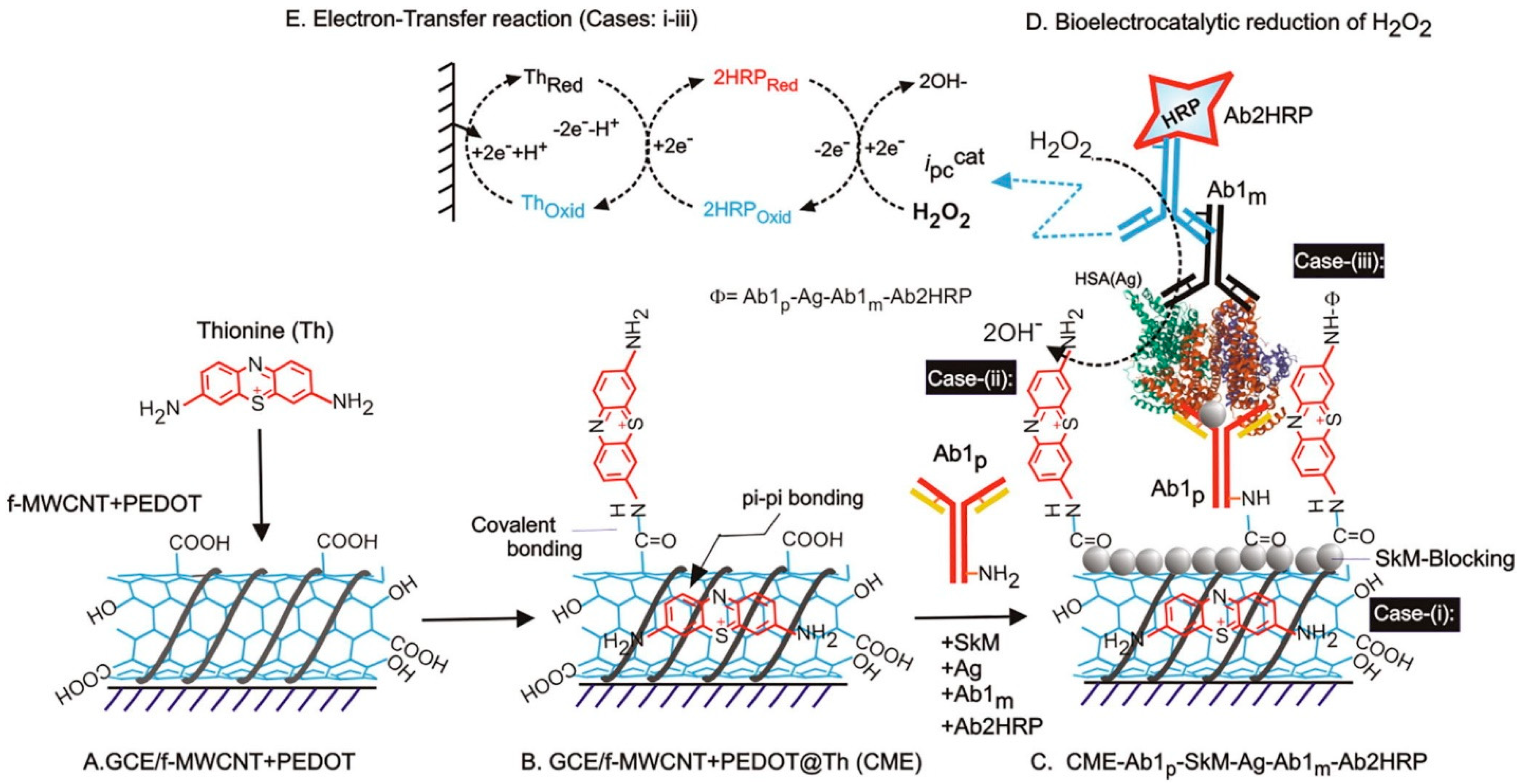
According to the fabrication of immunosensors, they are used for electron transfer enhancement and fabrication of protein layer loading in the case of label-free sensing. In the case of sandwich immunosensors, they can be used as electroactive and electrocatalytic tracers and nanocarriers [39]. Figure 3 accounts for the development of an electrochemical immunosensor for the detection of human serum albumin (HSA) in real urine samples with the involvement of labelled Ab for their well-defined electrocatalytic response. This figure accounts for a thionine-modified electrochemical platform that acts as a redox transducer involving covalent and pi-pi bonding between the GCE-modified functionalized (multi-walled carbon nanotube) MWCNT + PEDOT (poly (3,4 ethylenedioxythiophene) polystyrene sulfonate). This modified matrix was further extended for the detection of HSA after modifying the platform with primary Ab1p (polyclonal), skimmed milk (SkM) blocking agent, primary Ab1m (monoclonal), and Ab2 HRP (secondary antibody linked to HRP) [40].
3.2. Functional Model of Bonding between the Protein and the Underlying Electrode Surface
Immunosensors have specific immunochemical reactions with transducing elements for subsequent techniques like capacitative, potentiometric, impedance, conductometric, amperometric, optical and microgravimetric, etc., analysis [41]. With the breakthrough which has occurred through the determination of plausible reaction dynamics underlying immunosensors, there is corresponding potential to revolutionize traditional procedures. The rapid advancement in this domain means that it has become quite easy to use, with a reliable output in diverse screening [42]. Yet, complications associated with immune-inactive components, their immobilization, non-specific adsorption, and background noise are a few practical complications which mean that they should be dealt with precaution. The sample system for any immunosensor can be blood or its components, sweat, urine, tears, or similar body fluids. Thus, the current modernization of the immunosensor domain has been quite enhanced using an electrochemical approach to solving the complications of clinical analysis in medicine and veterinary sciences. Thus, the upgradation of electro-immunoassays is likely to be driven by the analytical practice and efficiency of the microfluidic analysis. For example, sweat consists of a range of biomarkers and a mixture of metabolites, electrolytes, urea, etc. The other interfering elements can be prevented from coming into contact with the sensor by using a layer of filter systems. A sweat-based microfluidic electrochemical integrated device has been developed by Cao et al., wherein they have elaborately discussed the sweat collector, followed by the vertical channel, transverse channel, electrochemical sensor, and finally sweat evaporator [43]. Figure 4 displays a schematic illustration of a sweat collector, wherein the yellow colours are hydrophobic in nature and formed by wax-screen printing, while the coloured systems are hydrophilic areas. Similarly, Bilbao et al. published a collection of microfluidic-based electrochemical platforms for various analytes [44]. In particular, sandwich immunoassays involving enzyme-functionalized liposomes were used as their catalytic label to obtain a substantially improved assay, which has been validated in one of the reports by Alfonta et al. for the determination of cholera toxin [45].
This paradigm shift shows the exploited arena of nanomaterials for sensor design [46]. Recently, miniaturized automated immunosensors have become a trend to facilitate a shortened analysis duration and faster analytical approach with simplicity. Recently, a sensor has been proposed to facilitate immunophenotyping for various leukaemias with a cluster of differential antibody microarrays [47]. Subsequently, variations in the immunosorbent assays can be extended for the autoimmune diagnosis of rheumatic diseases with high throughput [48]. Thus, different sensors are functionalized for commercial aspects with integrated multiple processors in a single device unit for elevated reliability with features of automation and less reagent consumption. One of these is the micro-total analysis system (μTas), i.e., the future of immune tests [49]. Another article was published by Hui et al. on the high performance of electrochemical heavy metal sensors for non-invasive detection in human fluids. High throughput systems allow for the very sensitive detection of analytes at the ppb level, i.e., 0.1 ppb and 0.5 ppb for copper and zinc heavy metals [50]. Additionally, integrating multiple processors like scanning electrochemical probe microscopy (SPECM) enables the better spatial resolution of imaging. These next-generation systems put enormous emphasis on big data and its analysis, storage, curation, and parallelization. Thus, these intelligent instruments and experiments have active control of nanoscale systems and the integration of nano-electrochemistry and nanoscale micro spectroscopy too [50]. The summing of immunosensors is one of the most frequently used analytical prototypes embracing a vast repertoire of analyte detections by a diverse range of transducers. Its enormous potential has been accepted in clinics, the environment, biological processes, food and diets, and even medical research. It has become an inevitable powerful technological prototype for analytical domains.
There are various ways to achieve antibody–nanoparticle bioconjugation, which is divided into physical and chemical methods. In the case of metallic nanoparticles, the physical methods are based on the following: (a) the spontaneous adsorption of Ab onto metallic nanoparticles through the hydrophobic interaction of the Ab lipophilic part and the metallic surface; (b) the electrostatic interaction between them. On the contrary, the chemical methods involve covalent strategies, the most common being attachment via the thiol group.
Other strategies involve bifunctional linkers (carboxyl-thiol, amine-thiols) or adapter molecules such as biotin and streptavidin, making them react with antibodies via EDC/NHS linkers. The bonding is generally covalent and non-covalent. One of the most crucial covalent interactions in immunosensors is crosslinking (the covalent linkage sites at the transducer substrates which attach glutaraldehyde, carbodiimide succinimide ester, etc.). Coating material systems like tri methoxy silane, polyethyleneimine, etc. [51], are initiators for immunoactivity molecule immobilization. Few reports of self-assembled monolayers are one of the promising alternatives for protein (antigen or antibody, protein of interest) immobilization [52,53]. Sulphur donor atoms strongly coordinate on various noble substrates such as sulphides, disulfides, and thiols, which are perfectly organized. A recent article published in 2022 scientific reports involved the anchoring of biological elements due to the strong and stable gold–sulphur chemistry [54]. In continuation, Shoute et al. reported an impedimetric sensor for COVID-19 detection antibodies using a gold (interdigitated microelectrode array) IMA sensor chip-based microfluidic platform [55]. These sulphur bonds are composed of thiol, with COOH and OH groups ensuring stable affinity for a specific interface. They even help in ensuring the perfect orientation and alignment of protein molecules on the electrode surface to have specific lock and key bonding. Gandhi et al. reported COOH–Cytochrome c bonding, or ‘-CO-NH-’ molecular wiring, to enable the perfect electron transfer mechanism for the efficient electrocatalytic reduction of hydrogen peroxide [56]. Alshanski and coworkers have reported enzymatic sialylation using impedimetric biosensing, which is a label-free biosensor based on interface properties. The biosensor surface consisted of neutral, positive, negative, and zwitter ionic functional groups. Each group had a profound effect, providing signals which were directly related to enzymatic sialylation [57]. Thus, these bonds enable the retention of antibody activities for successful applications in immunosensor designs. In addition, this has led to low background noise and higher sensitivity as a favourable alternative for interfacial design. An interesting report was published in 2003, wherein an epitope of foot and mouth disease was detected using a penta-peptide functionalized CNT [58]. Covalent interaction allows for the stability and repeatability of immunosensor applications. On the contrary, crosslinking leads to multilayer interaction, followed by the creation of diffusion barriers and transport limitations, leading to enhanced time frames with incomplete interactions [59]. An elaborate study of the enzymes on both components of substrate-on-a-nanoparticle configuration has been performed by Algar et al. [57]. An electrochemical quartz crystal microbalance-based technique using amine-terminated PPF for well-defined antibody immobilization has been studied in this respect [60]. Thus, the ideal immobilization inherent characteristics are (A) sufficient loading amount of protein on the transducer element; (B) stability of the immobilized protein for the course of the reaction; (C) sensing or the response of electrochemical systems should be independent of the immobilization process and transducer involved; (D) sensor regeneration ability.
The non-covalent interactions are in general hydrophobic, electrostatic, Van der Walls, and hydrogen interactions. They constitute both the physical and chemical interactions, which are highly dependent on the substrate element. Recently, Gajos et al. in 2023, reported a non-covalent inflow biofunctionalization for capture assays for IgG antibodies. One of the most typical layer-by-layer techniques for self-assembly for various biomolecular immobilization [61]. Caruso et al. introduced the study of a polyamine hydrochloride/polystyrene sulphonate layer for SAM mercaptopropionic acid, enabling a charged modification on the transducer element [62]. The molecules involved electrostatic interactions. In continuation, various antigens and antibodies have been entrapped in film or polymeric systems. Sol–gel can encapsulate distinct bio-moieties in tailored conditions with optimized characteristics (strength, stability, transparency, physical tenability, etc.) [63,64]. An article by Zhan et al. involved ZnO sol as an electrochemical immunosensor for the determination of clenbuterol [62]. Thus, the aforementioned physical interaction-based immunoassays are quite simple and rapid, but their immobilization stability is highly influenced by bioactivity, protein denaturation, and the ionic strength of the solution. This may cause a low-reproducibility constraint of their usage in the mainstream.
In extension on the various interactions, the synergistic effect observed between the CNTs and other nanoparticles helps in improving the parameters of the immunosensor. The synergistic effect helps enhance the sensitivity of the sensor as the nanocomposite has a higher surface area than load-abundant immobilized biomolecules, which improves their electron transfer process. For example, Sun et al. have reported an electrochemical immunosensor for the screening of carcinoembryonic antigen with a redox matrix consisting of AuNP’s, MWCNT, and Azure I, a self-assembling nanocomposite [27], wherein Azure I formed a nanostructural membrane that was entrapped in the MWCNT matrix. Later, negatively charged gold nanoparticles were assembled into the interface via an electrostatic interaction. Thus, anti-CEA antibodies, followed by CEA, can be accommodated on the sensor for a well-defined sensing that has enhanced current sensitivity due to the synergistic effect of the system [27]. Many other papers are described in this regard that help to synergistically amplify the result for the better performance of immunosensors [65,66].
Another type of interaction is based on adsorbate binding, which is responsible for mass loading and interfacial properties, namely roughness and viscoelasticity. They are observed in the case of the piezoelectric response of microgravimetric immunosensors, wherein the shift in the oscillation frequency is studied [67]. They have attributes of real-time analysis, simplicity, ease of usage, use in both gas and solution phase systems, reliability, and reproducibility. Their sensitivity is quite enhanced, and one of these systems is named quartz crystal microbalance (QCM). A review has been published in this regard involving epitome-based imprinted sensors for electrochemical-based timely disease monitoring, using EQCM as one of the potential techniques [67]. A recent report accounts for the detection of Salmonella typhimurium in chicken meat by the immobilization of the protein A-based antibody immunosensor [68]. In continuation, an electrode Au/SAM/antigen was fabricated involving covalent linkages for the detection of anti-sperm antibodies [69]. Another crucial type of gravimetric immunosensor is based on immunological agglutination, wherein the antibody-bearing suspensions help in inducing the corresponding change in the characteristics, thereby altering the interfacial properties of the crystal [69,70]. EQCM has add-on advantages compared to conventional piezoelectrical assays, such as the easy immobilization of biomolecules on the crystal, sensitivity, and feasibility with a wide range of targets [69]. In this context, potentiometric transducers are quite a thing for the commercial market. In general, a local equilibrium is established at the transducer interface, especially for the membrane potential where ‘it is α proportional to specific ion activity’. This relationship has led to the fundamental basis for ion-selective electrodes. These groups of biosensors are characterized based on their analytical performance in distinct domains. One of the first immune electrodes was proposed by Janta and his co-workers for the screening of Concanavalin A [71]. The bond for the attachment was via a covalent bond on the platinum electrode. In continuation, other potentiometric types are pH and gas sensing electrodes, which help to cope with the issues observed for traditional approaches. An ion-based field effect transistor (ISFET) is usually a semiconductor device formed by substituting an ion-sensing membrane for the metal gate for an FET. An ISFET responds to the potential change in the specific immunochemical reaction for the immobilized Ab and Ag mechanism. A few pH-dependent electrochemical sensors have varied enzyme labels, such as glucose oxidases, horse radish peroxidase, urease, etc. Nevertheless, there are only a small number of immunosensors reported in these arenas. A silicon-based ISFET has been reported for the detection of urinary albumin [72]. Herein, Saengdee has developed a low-cost immunosensor based on the binding event between the recombinant Ag85B antigen and anti-Ag85B antibody on the ISFET surface by monitoring the gate potential change at a constant drain current. There is another set of conductometric immunosensors which are quite old but are limited due to their poor selectivity issues (sometimes false negative/positive response). A conductometric immunosensor for E.coli 0157:H7 has been fabricated using a polyaniline/zinc oxide nanocomposite and has been extended to real samples such as skimmed milk [73]. Hence, other capacitance-based or impedance-based options are the most widely used immunosensors, wherein the capacitance is studied, i.e., the principle involves the capacitance of an electrode, which is proportional to the thickness and the dielectric behaviour of the electrode surface and the solid/solution interface. The most critical part during fabrication is the immobilization process. Thus, the sensitivity attributes along with the efficiency of the sensor depend on the coupling/layer of the interaction for various techniques. In the case of capacitive-based immunosensors, the capacitance ↑ is the thickness ↓ of the insulating layers. An additional benefit in the case of a capacitance- or impedance-based sensor is the ability to directly investigate the lock and key interaction (Menton’s Michael Interaction) without involving any reagent or separation step. Another important type is amperometric-based immunosensors, which help in the determination of currents resulting from electrochemical oxidation and reduction-based systems, i.e., the electroactive species at a constant voltage. Mansi and her co-workers reported a well-defined amperometric determination of white spot syndrome virus (vp-28 protein) in penaeid shrimp using a sesamol-based redox transducer on a carbon black/GCE surface in pH 7 PBS media [7]. These types of sensors are usually based on label profiling (examples—oxidases, peroxidases, cytochrome c, etc.), which helps in enhancing their sensitivities inherited by enzyme catalysis [74]. Hence, the amperometric immunosensors can obtain a much better response compared to a classical ELISA. Figure 5 encompasses a pictorial illustration of an amperometric sensor. The only drawback in these is due to their incapable surface renewability. On the other hand, anodic stripping voltammetry is an assay well adapted for measurements of heavy metals, such as copper, and can be a future for immunosensor prototyping. Immunosensors, in this respect, have been reported in various detections of human immunoglobulin (LOD—4.9 fg mL−1), human carcinoembryonic antigen (LOD—3 fg mL−1), human α-fetoprotein (LOD—4.9 fg mL−1), and thrombin (LOD—0.9 fg) on a screen-printed electrode using CdS quantum dots [75]. Out of all the various types of immunosensors, the impedimetric and amperometric immunosensors are widely accepted and used for prototype fabrications. Out of these, the amperometry ones are more selective but are equally difficult to set using a redox transducer.
4. Applications of Real-Time Electrochemical Screening of Protein of Interest: Market Study
The electrochemical instrument market has segmented a compound annual growth rate expanding ~4%, with the domains of products, end users, and region. The projected market is supposed to increase to a valuation of ~USD 3 billion by 2026 [76]. This enlargement is majorly due to rising consumer demand for multi-parameter testing and the need for safe drinking water and environment across regions. The key companies are Dkk-Toa Corporation, Yokogawa Electric Corporation, India; Horiba Ltd., Kyoto, Japan; Hanna Instruments Inc., Italy; Mettler-Toledo International Inc., Danaher Corporation, USA; Xylem Inc., USA; Metrohm AG, Swizerland; Endress+ Hauser AG, Germany, which are the major leading electrochemical profiles. It is anticipated that a wide number of biotechnological and pharmaceutical firms will be the driving factor for the electrochemical advancement, which has led to an increment in the development of spending in the power and energy sector. Battery systems are another significant element anticipated to foster market upliftment. The prevalence of illness brought about by air pollution has the demand for environmental monitoring due to the rise in legislation and awareness campaigns, thereby increasing the spending efficiency for electrochemical-based devices market prototypes. Figure 6 encompasses an electrochemical sensor market overview for the case of the user, product, and end-user profile. For prognosis during the COVID-19 pandemic, prototypes have been built for the monitoring of antibody and antigen levels, with commercially available examples being BinaxNOW, a test developed by Abbot, and the InteliSwab, developed by OvaSURE [77,78,79]. However, due to their low sensitivity and high false negative drawbacks, electrochemical detection techniques can be used to overcome these. Proper avenue development for accurate POC technology requires several stages of development, with the integration of reagent delivery and sample collection, in addition to the diagnostic test and end user comfort. A screen-printed system has been developed for the detection of nucleocapsid protein to quantify severe acute respiratory syndrome-coronavirus-2 (SARS-CoV-2) involving chronoamperometry [80]. The developed biosensor was studied with cross-reactivity interferences to ensure the selectivity of the system, followed by a proof-of-concept study. Later, a similar study was performed using a smartphone-based inexpensive serological diagnosis of SARS-CoV-2 using a ferro-ferricyanide-modified electrode with a square wave voltammetry approach [81]. Figure 7 illustrates the smartphone-based prototype for the same.
The major restraint for these electrochemical systems is their short shelf life, which is projected to hamper the growth of the market. An electrochemical sensor has a shelf life of six months to a year depending on the analyte to be detected in parallel to the protein modifications in a particular environment. This is a limiting factor for global electrochemical sensors. Another big challenge is the limited or narrow temperature range and its sensitivity towards temperature, which is usually internally adjusted. Hence, it is preferable to keep the temperature as stable as possible.
Few of the market products are, MiniMed 770G System, launched by Medtronic in 2020, a hybrid closed loop system. It has the benefits of smartphone connectivity for recent insulin pump systems with sophisticated smart guard technology for automatic glucose measurements, especially for patients with type 1 diabetes mellitus [82].
In 2020, a hand-held structured DNA assembly strategy and a dual-mode electrochemical-fluorescent-based sensor were developed for circulating tumour DNA based on methylene blue- and red-emissive carbon nanodots [83].
Still, this market has risen substantially due to the increased need for diagnostic procedures and improvements in micro-fabrication methodology, which has resulted in the creation of sensitive, selective, but effective biochemical sensors for clinical analysis. The use of an electrochemical–molecular basis for point-of-care has helped to improve sensitivity and quick testing, with expansion capabilities in hospital labs, outpatient clinics, path labs, university and school medical rooms too.
5. Conclusions, Discussion, and Future Outlook
The initial journey of sensors started with the most famous sensor, Clark’s glucose sensor, and now well-developed smart gadgets and skin-based sensor systems exist [84]. Electrochemical immunosensors have emerged as a versatile and robust sensor technology. With the combination of highly specific biorecognition elements and electrical readout, the mapping of the protein of interest can be performed for up to femtomolar levels when compared with the classical techniques. There is the integration of reaction–diffusion and effective media theories to derive a generalized scaling model for an arbitrary immunosensor that relates to the relative change in the redox current to the corresponding change in the antigen concentration (based on faradaic and non-faradaic currents) through scaling exponents related to the geometry of biomolecules diffusion and measurement resolution. The immunosensors are validated via sensor-agnostic scaling formula and the cross-calibration of instruments with a defined physics standardized methodology to compare the performance metrics. This article, on the electrochemical modelling of immunosensors, provides us with proper insights into the modelling of electrode layering, along with a reaction dynamics study (based on bonding, kinetics, and reaction details). In addition, the choice of nanomaterials enhances the analytical character of the fabricated biosensor, as discussed in detail in the paper. The emphasis has been laid on the commercialization of real-time prototypes in the section of market study. We propose a bright future for an electro-immunosensor array-based system for the multiplex sensing of disease biomarkers. Thus, this paper can provide help for further designing prototypes and the proper controls and calibrations required for the selective sensing of a targeted antigen or protein of interest. With the aim of making new advancements and minimizing the loopholes of electrochemical sensors, the vision is to commercialize the point of care devices in rural and technologically backward locations.
Funding
This research received no external funding.
Conflicts of Interest
The author declares no conflicts of interest.
References
- Gandhi, M.; Khairunnisa, A. Electrochemical Profiling of Plants. Electrochem 2022, 3, 434–450. [Google Scholar] [CrossRef]
- Kubota, L.T.; da Silva, J.A.F.; Sena, M.M.; Alves, W.A. Tools and Trends in Bioanalytical Chemistry; Springer International Publishing: Cham, Switzerland, 2021. [Google Scholar] [CrossRef]
- Goldfarb, J.L.; Dou, G.; Salari, M.; Grinstaff, M.W. Biomass-Based Fuels and Activated Carbon Electrode Materials: An Integrated Approach to Green Energy Systems. ACS Sustain. Chem. Eng. 2017, 5, 3046–3054. [Google Scholar] [CrossRef]
- Wu, H.; Gong, Y.; Yu, Y.; Huang, K.; Wang, L. Superior “green” electrode materials for secondary batteries: Through the footprint family indicators to analyze their environmental friendliness. Environ. Sci. Pollut. Res. 2019, 26, 36538–36557. [Google Scholar] [CrossRef] [PubMed]
- Min, J.K.; Jung, Y.; Ahn, J.; Lee, J.G.; Lee, J.; Ko, S.H. Recent Advances in Biodegradable Green Electronic Materials and Sensor Applications. Adv. Mater. 2023, 35, 2211273. [Google Scholar] [CrossRef] [PubMed]
- Xu, R.; Ouyang, L.; Chen, H.; Zhang, G.; Zhe, J. Recent Advances in Biomolecular Detection Based on Aptamers and Nanoparticles. Biosensors 2023, 13, 474. [Google Scholar] [CrossRef] [PubMed]
- Gandhi, M.; Rajagopal, D.; Parthasarathy, S.; Raja, S.; Huang, S.T.; Senthil Kumar, A. In Situ Immobilized Sesamol-Quinone/Carbon Nanoblack-Based Electrochemical Redox Platform for Efficient Bioelectrocatalytic and Immunosensor Applications. ACS Omega 2018, 3, 10823–10835. [Google Scholar] [CrossRef] [PubMed]
- Vishnu, N.; Gandhi, M.; Rajagopal, D.; Kumar, A.S. Pencil graphite as an elegant electrochemical sensor for separation-free and simultaneous sensing of hypoxanthine, xanthine and uric acid in fish samples. Anal. Methods 2017, 9, 2265–2274. [Google Scholar] [CrossRef]
- Gandhi, M.; Vishnu, N.; Katari, N.K. Nanomaterial-Modified Pencil Graphite Electrode as a Multiplexed Low-Cost Point of Care Device. In Smart Nanodevices for Point-of-Care Applications; CRC Press: Boca Raton, FL, USA, 2022. [Google Scholar]
- Haseeb, A. Monoplex and multiplex immunoassays: Approval, advancements, and alternatives. Comp. Clin. Pathol. 2022, 31, 333–345. [Google Scholar]
- Yeonjeong, H.; Ijung, K. Recent Developments in Innovative Magnetic Nanoparticles-Based Immunoassays: From Improvement of Conventional Immunoassays to Diagnosis of COVID-19. BioChip J. 2022, 16, 351–365. [Google Scholar]
- Fowler, J.M.; Danny, K.Y.W.; Halsall, H.B.; Heineman, W.R. Recent developments in electrochemical immunoassays and immunosensors. In Electrochemical Sensors, Biosensors and their Biomedical Applications; Academic Press: Cambridge, MA, USA, 2008; pp. 115–143. [Google Scholar]
- Guidi, A.; Laricchia-Robbio, L.; Gianfaldoni, D.; Revoltella, R.; Bono, D.G. A Comparison of a conventional immunoassay (ELISA) with a surface plasmon resonance-based biosensor for IGF-1 detection in cows’ milk. Biosens. Bioelectron. 2001, 16, 971–977. [Google Scholar] [CrossRef]
- Samper, I.C.; McMahon, C.J.; Schenkel, M.S.; Clark, K.M.; Khamcharoen, W.; Anderson, L.B.R.; Terry, J.S.; Gallichotte, E.N.; Ebel, G.D.; Geiss, B.J.; et al. Electrochemical Immunoassay for the Detection of SARS-CoV-2 Nucleocapsid Protein in Nasopharyngeal Samples. Anal. Chem. 2022, 94, 4712–4719. [Google Scholar] [CrossRef] [PubMed]
- Zhang, J.; Li, Y.; Chai, F.; Li, Q.; Wang, D.; Liu, L.; Tang, B.Z.; Jiang, X. Ultrasensitive point-of-care biochemical sensor based on metal-AIEgen frameworks. Sci. Adv. 2022, 8, eabo1874. [Google Scholar] [CrossRef] [PubMed]
- Zhao, Y.; Song, X. An Electrochemical-Based Point-of-Care Testing Methodology for Uric Acid Measurement. J. Anal. Methods Chem. 2022, 2022, 8555842. [Google Scholar] [CrossRef] [PubMed]
- Macovei, D.G.; Irimes, M.B.; Hosu, O.; Cristea, C.; Tertis, M. Point-of-care electrochemical testing of biomarkers involved in infammatory and infammatory-associated medical conditions. Anal. Bioanal. Chem. 2023, 415, 1033–1063. [Google Scholar] [CrossRef] [PubMed]
- Wu, J.; Ju, H. Clinical Immunoassays and Immunosensing. In Comprehensive Sampling and Sample Preparation; Elsevier: Amsterdam, The Netherlands, 2012; Volume 3, pp. 143–167. [Google Scholar]
- Ozer, T.; Geiss, B.J.; Henry, C.S. Review—Chemical and Biological Sensors for Viral Detection. J. Electrochem. Soc. 2020, 167, 037523. [Google Scholar] [CrossRef] [PubMed]
- Siqi Zhao, S.; Huang, J.; Li, D.; Yang, L. Aptamer-based chemiluminescent optical fiber immunosensor with enhanced signal amplification for ultrasensitive detection of tumor biomarkers. Biosens. Bioelectron. 2022, 214, 114505. [Google Scholar]
- Osaki, S.; Saito, M.; Nagai, H.; Tamiya, E. Surface Modification of Screen-Printed Carbon Electrode through Oxygen Plasma to Enhance Biosensor Sensitivity. Biosensors 2024, 14, 165. [Google Scholar] [CrossRef]
- Limthin, D.; Leepheng, P.; Tunhoo, B.; Onlaor, K.; Klamchuen, A.; Phromyothina, D.; Thiwawong, T. Preparation of surface-modified electrode of copper(II) oxide mixed with the molecularly imprinted polymer for enhancement of melamine detection with photochemical technique. RSC Adv. 2023, 13, 14729. [Google Scholar] [CrossRef] [PubMed]
- Li, L.; Wang, T.; Zhong, Y.; Li, R.; Deng, W.; Xiao, X.; Xu, Y.; Zhang, J.; Hu, X.; Wang, Y. A review of nanomaterials for biosensing applications. J. Mater. Chem. B 2024, 12, 1168–1193. [Google Scholar] [CrossRef]
- Azad, U.P.; Chandra, P. Handbook of Nanobioelectrochemistry-Application in Devices and Biomolecular Sensing; Springer: Berlin/Heidelberg, Germany, 2023; ISBN 978-981-19-9437-1. [Google Scholar]
- Kumar, S.; Wang, Z.; Zhang, W.; Liu, X.; Li, M.; Li, G.; Zhang, B.; Singh, Z. Optically Active Nanomaterials and Its Biosensing Applications—A Review. Biosensors 2023, 13, 85. [Google Scholar] [CrossRef]
- Murjani, B.O.; Kadu, P.S.; Bansod, M.; Vaidya, S.S.; Yadav, M.D. Carbon nanotubes in biomedical applications: Current status, promises, and challenges. Carbon Lett. 2022, 32, 1207–1226. [Google Scholar] [CrossRef]
- Sun, A.L.; Chen, G.R.; Sheng, Q.L.; Zheng, J.B. Sensitive label-free electrochemical immunoassay based on a redox matrix of gold nanoparticles/Azure I/multi-wall carbon nanotubes composite. Biochem. Eng. J. 2011, 57, 1–6. [Google Scholar] [CrossRef]
- Karthikeyan, C.; Tharmalingam, N.; Varaprasad, K.; Mylonakis, E.; Yallapu, M.M. Biocidal and biocompatible hybrid nanomaterials from biomolecule chitosan, alginate and ZnO. Carbohydr. Polym. 2021, 274, 118646. [Google Scholar] [CrossRef] [PubMed]
- McLaughlin, M.H.S.; Pakpour-Tabrizi, A.C.; Jackman, R.B. A detailed EIS study of boron doped diamond electrodes decorated with gold nanoparticles for high sensitivity mercury detection. Sci. Rep. 2021, 11, 9505. [Google Scholar] [CrossRef] [PubMed]
- Matvieiev, O.; Šelešovská, R.; Marton, M.; Hatala, M.; Metelka, R.; Weis, M.; Vojs, M. Effect of different modification by gold nanoparticles on the electrochemical performance of screen-printed sensors with boron-doped diamond electrode. Sci. Rep. 2023, 13, 21525. [Google Scholar] [CrossRef] [PubMed]
- Minenkov, A.; Hollweger, S.; Duchoslav, J.; Erdene-Ochir, O.; Weise, M.; Ermilova, E.; Hertwig, A.; Schiek, M. Monitoring the Electrochemical Failure of Indium Tin Oxide Electrodes via Operando Ellipsometry Complemented by Electron Microscopy and Spectroscopy. ACS Appl. Mater. Interfaces 2024, 16, 9517–9531. [Google Scholar] [CrossRef] [PubMed]
- Huang, J.; Xie, Z.; Luo, S.; Li, M.; Xie, L.; Fan, Q.; Zeng, T.; Zhang, Y.; Zhang, M.; Wang, S.; et al. A sandwich amperometric immunosensor for the detection of fowl adenovirus group I based on bimetallic Pt/Ag nanoparticle-functionalized multiwalled carbon nanotubes. Sci. Rep. 2024, 14, 261. [Google Scholar] [CrossRef] [PubMed]
- Brazaca, L.C.; Imamura, A.H.; Gomes, N.O.; Almeida, M.B.; Scheidt, D.T.; Raymundo-Pereira, P.A.; Oliveira, O.N., Jr.; Janegitz, B.C.; Scheidt, D.T.; Machado, S.A.S.; et al. Electrochemical immunosensors using electrodeposited gold nanostructures for detecting the S proteins from SARS-CoV and SARS-CoV-2. Anal. Bioanal. Chem. 2022, 414, 5507–5517. [Google Scholar] [CrossRef] [PubMed]
- Jeong, B.; Akter, R.; Oh, J.; Lee, D.G.; Ahn, C.G.; Choi, J.S.; Rahman, M.A. Novel electrochemical PMI marker biosensor based on quantum dot dissolution using a double-label strategy. Sci. Rep. 2022, 12, 8815. [Google Scholar] [CrossRef]
- Ishii, K.; Ogata, G.; Yamamoto, T.; Sun, S.; Shiigi, H.; Einaga, Y. Designing Molecularly Imprinted Polymer-Modified Boron-Doped Diamond Electrodes for Highly Selective Electrochemical Drug Sensor. ACS Sens. 2024, 9, 1611–1619. [Google Scholar] [CrossRef]
- Nirmala, A.; Sonar, G.P.; Bhardwaj, P.; Chakravorty, A. Materials Horizons: From Nature to Nanomaterials Handbook of Porous Carbon Materials; Springer: Berlin/Heidelberg, Germany, 2023. [Google Scholar] [CrossRef]
- Gandhi, M.; Amreen, K. Emerging Trends in Nanomaterial-Based Biomedical Aspects. Electrochem 2023, 4, 365–388. [Google Scholar] [CrossRef]
- Kumar, A.S.; Gandhi, M.; Saikrithika, S.; Dinesh, B.; Shafeeq, S.; Ganesh, V. Localized formation of highly surface-active gold nanoparticle on intrinsic Nickel containing carbon black and its scanning electrochemical microscopy interrogation and electrocatalytic oxidation of hydrazine. Electrochim. Acta 2023, 443, 141937. [Google Scholar] [CrossRef]
- Wang, Y.; Zhang, Y.; Wu, D.; Ma, H.; Pang, X.; Fan, D.; Wei, Q.; Du, B. Ultrasensitive Label-free Electrochemical Immunosensor based on Multifunctionalized Graphene Nanocomposites for the Detection of Alpha Fetoprotein. Sci. Rep. 2017, 7, 42361. [Google Scholar] [CrossRef] [PubMed]
- Gandhi, M.; Indiramma, J.; Jayaprakash, N.S.; Kumar, A.S. An efficient electrochemical sandwich ELISA for urinary human serum albumin-biomarker based on highly redox-active thionine surface-confined MWCNT/PEDOT.PSS platform. J. Electroanal. Chem. 2022, 906, 116018. [Google Scholar] [CrossRef]
- Stefan, R.-I.; van Staden, J.F.; Aboul-Enein, H.Y. Immunosensors in clinical analysis. Fresenius J. Anal. Chem. 2000, 366, 659–668. [Google Scholar] [CrossRef]
- Morgan, C.L.; Newman, D.J.; Price, C.P. Immunosensors: Technology and opportunities in laboratory medicine. Clin. Chem. 1996, 42, 193–209. [Google Scholar] [CrossRef]
- Cao, Q.; Liang, B.; Tu, T.; Wei, J.; Fang, L.; Ye, X. Three-dimensional paper-based microfluidic electrochemical integrated devices (3D-PMED) for wearable electrochemical glucose detection. RSC Adv. 2019, 9, 5674–5681. [Google Scholar] [CrossRef]
- Bilbao, E.; Garate, O.; Campos, T.R.; Roberti, M.; Mass, M.; Lozano, A.; Longinotti, G.; Monsalve, L.; Ybarra, G. Electrochemical Sweat Sensors. Chemosensors 2023, 11, 244. [Google Scholar] [CrossRef]
- Alfonta, L.; Willner, I.; Throckmorton, D.J.; Singh, A.K. Electrochemical and quartz crystal microbalance detection of the cholera toxin employing horseradish peroxidase and GM1-functionalized liposomes. Anal. Chem. 2001, 73, 5287–5295. [Google Scholar] [CrossRef]
- Martin, C.R.; Mitchell, D.T. Nanomaterials in analytical chemistry. Anal. Chem. 1998, 70, 322–327. [Google Scholar] [CrossRef]
- Sánchez, M.; Almeida, J.; Vidriales, B.; López-Berges, M.; García-Marcos, M.; Moro, M.; Corrales, A.; Calmuntia, M.; Miguel, J.S.; Orfao, A. Incidence of phenotypic aberrations in series of 467 patients with B chronic lymphoproliferative disorders: Basis for the design of specific four-color stainings to be used for minimal residual disease investigation. Leukemia 2002, 16, 1460–1469. [Google Scholar] [CrossRef] [PubMed]
- Joos, T.O.; Schrenk, M.; Höpfl, P.; Kröger, K.; Chowdhury, U.; Stoll, D.; Dürr, M.; Herick, K.; Rupp, S.; Sohn, K.; et al. A microarray enzyme-linked immunosorbent assay for autoimmune diagnostics. Electrophoresis 2000, 21, 2641–2650. [Google Scholar] [CrossRef] [PubMed]
- Lee, S.J.; Lee, S.Y. Micro total analysis system (μ-TAS) in biotechnology. Appl. Microbiol. Biotechnol. 2004, 64, 289–299. [Google Scholar] [CrossRef] [PubMed]
- Park, J.Y.; Hui, X.; Sharifuzzaman, M.; Sharma, S.; Xuan, X.; Zhang, S.; Ko, S.G.; Yoon, S.H.; Park, J.Y. High-performance flexible electrochemical heavy metal sensor based on layer-by-layer assembly of Ti3C2Tx/MWNTs nanocomposites for noninvasive detection of copper and zinc ions in human biofluids. ACS Appl. Mater. Interfaces 2020, 12, 48928–48937. [Google Scholar] [CrossRef]
- Kiinig, B.; Grgtzel, M. Development of a Piezoelectric Immunosensor for the Detection of Human Erythrocytes; Elsevier Science Publishers B.V.: Amsterdam, The Netherlands, 1993; Volume 276. [Google Scholar]
- Ulman, A. Formation and Structure of Self-Assembled Monolayers. Chem. Rev. 1996, 96, 1533–1554. [Google Scholar] [CrossRef]
- Redondo-Gómez, C.; Parreira, P.; Martins, M.C.L.; Azevedo, S.H. Peptide-based self-assembled monolayers (SAMs): What peptides can do for SAMs and vice versa. Chem. Soc. Rev. 2024, 53, 3714–3773. [Google Scholar] [CrossRef]
- Abdulkarim, H.; Siaj, M. Label-free multiplex electrochemical immunosensor for early diagnosis of lysosomal storage disorders. Sci. Rep. 2022, 12, 9334. [Google Scholar] [CrossRef]
- Shoute, L.C.T.; Abdelrasoul, G.N.; Ma, Y.; Duarte, P.A.; Edwards, C.; Zhuo, R.; Zeng, J.; Feng, Y.; Charlton, C.L.; Kanji, J.N.; et al. Label-free impedimetric immunosensor for point-of-care detection of COVID-19 antibodies. Microsyst. Nanoeng. 2023, 9, 3. [Google Scholar] [CrossRef]
- Gandhi, M.; Rajagopal, D.; Senthil Kumar, A. Molecularly wiring of Cytochrome c with carboxylic acid functionalized hydroquinone on MWCNT surface and its bioelectrocatalytic reduction of H2O2 relevance to biomimetic electron-transport and redox signalling. Electrochim. Acta 2021, 368, 137596. [Google Scholar] [CrossRef]
- Alshanski, I.; Shitrit, A.; Sukhran, Y.; Unverzagt, C.; Hurevich, M.; Yitzchaik, S. Effect of Interfacial Properties on Impedimetric Biosensing of the Sialylation Process with a Biantennary N-Glycan-Based Monolayer. Langmuir 2022, 38, 849–855. [Google Scholar] [CrossRef]
- Pantarotto, D.; Partidos, C.D.; Graff, R.; Hoebeke, J.; Briand, J.-P.; Prato, M.; Bianco, A. Synthesis, structural characterization, and immunological properties of carbon nanotubes functionalized with peptides. J. Am. Chem. Soc. 2003, 125, 6160–6164. [Google Scholar] [CrossRef]
- Fung, Y.S.; Wong, Y.Y. Self-assembled monolayers as the coating in a quartz piezoelectric crystal immunosensor to detect Salmonella in aqueous solution. Anal. Chem. 2001, 73, 5302–5309. [Google Scholar] [CrossRef] [PubMed]
- Wang, H.; Liu, Y.; Yang, Y.; Deng, T.; Shen, G.; Yu, R. A protein A-based orientation-controlled immobilization strategy for antibodies using nanometer-sized gold particles and plasma-polymerized film. Anal. Biochem. 2004, 324, 219–226. [Google Scholar] [CrossRef]
- Erkmen, C.; Selcuk, O.; Unal, D.N.; Kurbanoglu, S.; Uslu, B. Layer-by-layer modification strategies for electrochemical detection of biomarkers. Biosens. Bioelectron. X 2022, 12, 100270. [Google Scholar] [CrossRef]
- Zhang, L.; Zheng, M.; Liu, X.; Sun, J. Layer-by-layer assembly of salt-containing polyelectrolyte complexes for the fabrication of dewetting-induced porous coatings. Langmuir 2011, 27, 1346–1352. [Google Scholar] [CrossRef]
- Lingling, S.; Ooi Kiang, T.; Baowei, M.; Lay Im, T.; Leong Huat, G.; Yik Yuen, G. A novel impedimetric immunosensor based on sol-gel derived Barium Strontium Titanate composite filnm. In Proceedings of the SENSORS, Daegu, Republic of Korea, 22–25 October 2006. [Google Scholar]
- Shaabani, N.; Chan, N.W.C.; Jemere, A.B. A molecularly imprinted sol-gel electrochemical sensor for naloxone determination. Nanomaterials 2021, 11, 631. [Google Scholar] [CrossRef]
- Naeem, N.A.; Guldin, S.; Ghoreishizadeh, S. Electrochemical Sensors for Cortisol: A Review. IEEE Sens. J. 2024, 24, 5746–5758. [Google Scholar] [CrossRef]
- Zhan, K.; Chen, L.; Li, S.; Yu, Q.; Zhao, Z.; Li, J.; Xing, Y.; Ren, H.; Wang, N.; Zhang, G. A novel metal–organic framework based electrochemical immunosensor for the rapid detection of Salmonella typhimurium detection in milk. Food Chem. 2024, 444, 138672. [Google Scholar] [CrossRef] [PubMed]
- Srivastava, J.; Singh, R.S.; Singh, M. Design of EQCM-MIP sensing matrix for highly specific and sensitive detection of thyroglobulin. Biosens. Bioelectron. X 2022, 11, 100154. [Google Scholar] [CrossRef]
- Nugaeva, N.; Gfeller, K.Y.; Backmann, N.; Lang, H.P.; Düggelin, M.; Hegner, M. Micromechanical cantilever array sensors for selective fungal immobilization and fast growth detection. Biosens. Bioelectron. 2005, 21, 849–856. [Google Scholar] [CrossRef]
- Shen, G.; Wang, H.; Tan, S.; Li, J.; Shen, G.; Yu, R. Detection of antisperm antibody in human serum using a piezoelectric immunosensor based on mixed self-assembled monolayers. Anal. Chim. Acta 2005, 540, 279–284. [Google Scholar] [CrossRef]
- Goulet, D.R.; Atkins, W.M. Considerations for the Design of Antibody-Based Therapeutics. J. Pharm. Sci. 2020, 109, 74–103. [Google Scholar] [CrossRef] [PubMed]
- Janata, J. Immunoelectrode. J. Am. Chem. Soc. 1957, 97, 2914–2916. [Google Scholar] [CrossRef]
- Saengdee, P.; Thanapitak, S.; Ongwattanakul, S.; Srisuwan, A.; Pankiew, A.; Thornyanadacha, N.; Chaisriratanakul, W.; Jeamsaksiri, W.; Promptmas, C. A silicon nitride ion sensitive field effect transistor-based immunosensor for determination of urinary albumin. Electrochem. Sci. Adv. 2022, 2, e2100078. [Google Scholar] [CrossRef]
- Mutlaq, S.; Albiss, B.; Al-Nabulsi, A.A.; Jaradat, Z.W.; Olaimat, A.N.; Khalifeh, M.S.; Osaili, T.; Ayyash, M.M.; Holley, R.A. Conductometric immunosensor for escherichia coli o157:H7 detection based on polyaniline/zinc oxide (pani/zno) nanocomposite. Polymers 2021, 13, 3288. [Google Scholar] [CrossRef]
- Patil, A.V.; Chuang, Y.S.; Li, C.; Wu, C.C. Recent Advances in Electrochemical Immunosensors with Nanomaterial Assistance for Signal Amplification. Biosensors 2023, 13, 125. [Google Scholar] [CrossRef] [PubMed]
- Qin, X.; Wang, L.; Xie, Q. Sensitive bioanalysis based on in-situ droplet anodic stripping voltammetric detection of cds quantum dots label after enhanced cathodic preconcentration. Sensors 2016, 16, 1342. [Google Scholar] [CrossRef] [PubMed]
- Precision Reports—Worldwide Market Research Report, Analysis & Consulting. Boston Research 2022. 1–157.
- Weishampel, Z.A.; Young, J.; Fischl, M.; Fischer, R.J.; Donkor, I.O.; Riopelle, J.C.; Schulz, J.E.; Port, J.R.; Saturday, T.A.; van Doremalen, N.; et al. OraSure InteliSwabTM Rapid Antigen Test Performance with the SARS-CoV-2 Variants of Concern—Alpha, Beta, Gamma, Delta, and Omicron. Viruses 2022, 14, 543. [Google Scholar] [CrossRef]
- Pollock, N.R.; Jacobs, J.R.; Tran, K.; Cranston, A.E.; Smith, S.; O’Kane, C.Y.; Roady, T.J.; Moran, A.; Scarry, A.; Carroll, M.; et al. Performance and Implementation Evaluation of the Abbott BinaxNOW Rapid Antigen Test in a High-Throughput Drive-Through Community Testing Site in Massachusetts. J. Clin. Microbiol. 2021, 59, 83–104. [Google Scholar] [CrossRef]
- Pilarowski, G.; Lebel, P.; Sunshine, S.; Liu, J.; Crawford, E.; Marquez, C.; DeRisi, J.; Rubio, L.; Chamie, G.; Martinez, J. Performance Characteristics of a Rapid Severe Acute Respiratory Syndrome Coronavirus 2 Antigen Detection Assay at a Public Plaza Testing Site in San Francisco. J. Infect. Dis. 2021, 223, 1139–1144. [Google Scholar] [CrossRef]
- Mugweru, A.; Clark, B.L.; Pishko, M.V. Electrochemical Redundant Microsensor Arrays for Glucose Monitoring with Patterned Polymer Films. Electroanalysis 2007, 9, 453–458. [Google Scholar] [CrossRef]
- Zeng, R.; Qiu, M.; Wan, Q.; Huang, Z.; Liu, X.; Tang, D.; Knopp, D. Smartphone-Based Electrochemical Immunoassay for Point-of-Care Detection of SARS-CoV-2 Nucleocapsid Protein. Anal. Chem. 2022, 94, 15155–15161. [Google Scholar] [CrossRef] [PubMed]
- Technology That Automatically Adjusts Insulin * MiniMed TM 770G System. 2020. FDA Approves Medtronic MiniMed 780g System—World’s First Insulin Pump with Meal Detection Technology Featuring 5-minute Auto Corrections. Medtronic News. 21 April 2020. Available online: https://news.medtronic.com/2023-04-21-FDA-Approves-Medtronic-MiniMed-TM-780G-System-Worlds-First-Insulin-Pump-with-Meal-Detection-Technology-Featuring-5-Minute-Auto-Corrections (accessed on 24 April 2023).
- Liu, G.; Chai, H.; Guo, Z.; Wang, Z.; Tang, Y.; Miao, P. Hand-in-hand structured DNA monolayer for dual-mode analysis of circulating tumor DNA. Chem. Eng. J. 2022, 450, 138069. [Google Scholar] [CrossRef]
- Clark, L.C.; Lyons, C. Electrode Systems For Continuous Monitoring In Cardiovascular Surgery. Ann. N. Y. Acad. Sci. 1962, 102, 29–45. [Google Scholar] [CrossRef]
Figure 1.
Accounts for the basic format of an electrochemical biosensor. The biorecognition element in the presence of the specific analyte leads to the unique property of a biological recognition event on the transducing device. The bioreceptor signals are converted into a suitable output that is easily readable by the display setup.

Figure 2.
Accounts for the fabrication of an electrochemical immunosensor. The constituents of the electrode fabrication account for suitable nanomaterial modification, followed by the capture antibody, analyte, or the protein of interest, the labelled detection antibody that gives the signal only after the ELISA process is complete.
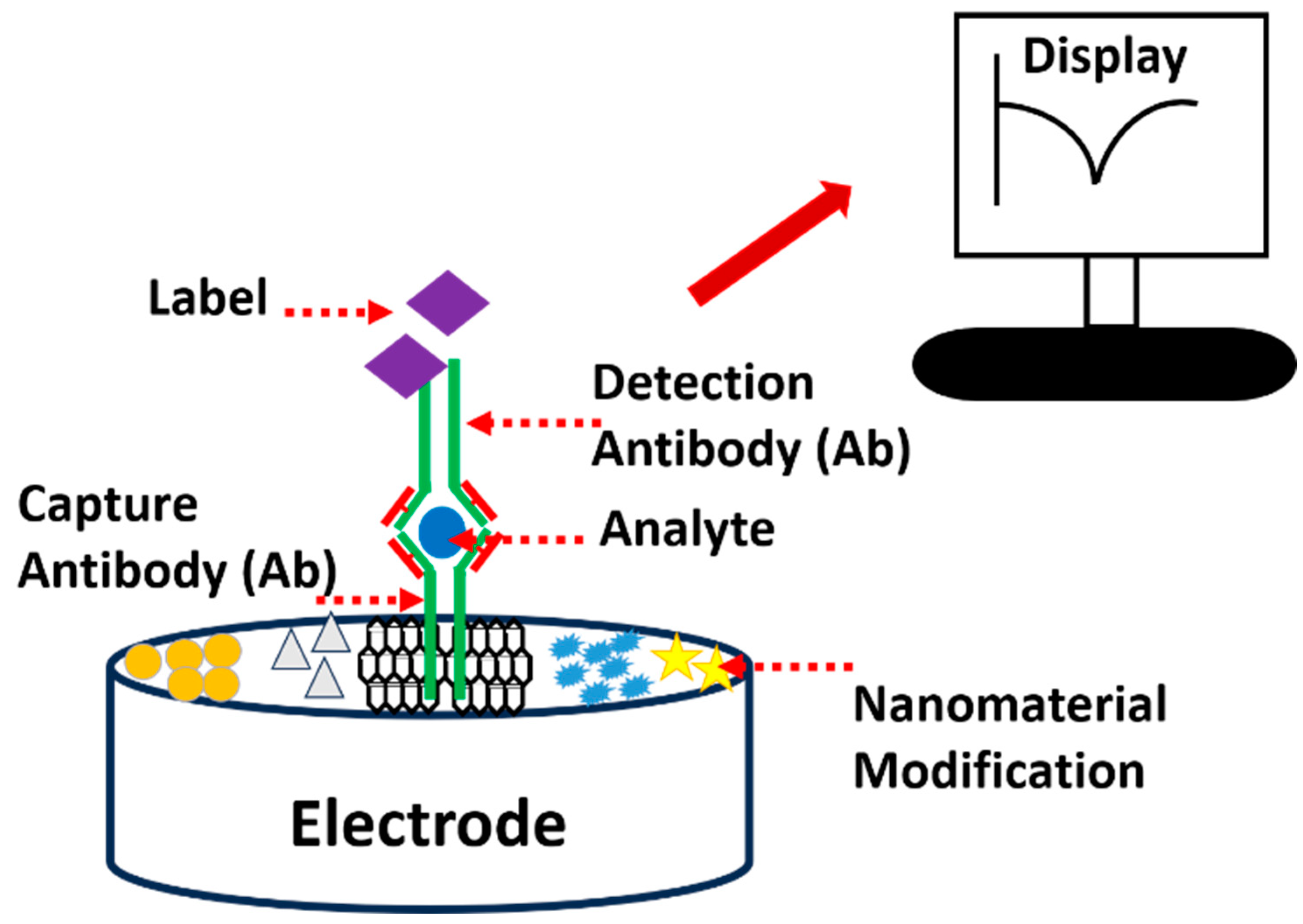
Figure 3.
Illustration of immunosensor configurations in terms of bonding, electron transfer enhancement, signal amplification, bioelectrocatalytic reduction of the substrate, and sensing fabrication. Permission and copyright, JEAC 2022 [40]. The notations are as follows: (A,B) GCE/f―MWCNT + PEDOT@Th (CME) and (C) CME―Ab1p―SkM―Ag(HSA)―Ab1m―Ab2HRP-modified electrodes and the (D) bioelectrocatalytic reduction of H2O2 and (E) electron transfer reaction mechanism. Ab1p—polyclonal primary antibody, Ab1m—monoclonal primary antibody, SkM—skim milk power as a blocking agent, Ag = HSA, Ab2HRP = horseradish peroxide enzyme tagged Ab1p. Cases (i–iii) are possible routes for molecular orientations of surface confined Th and its interactions with Ab2HRP. CME = GCE/f―MWCNT + PEDOT@Th.

Figure 4.
Schematic illustration of a sweat-based electrochemical microfluidic integrated device, which allows the sweat to pass through different filters to ensure the removal of inactive systems and interfering systems that might add complications to the analysis (A). While (B) corresponds to the attachment of the sensor on the body part and cross-sectional illustration for the sweat glands that secrete sweat, which passes through a channel into the electrode and finally gets evaporated after analysis. Copyright, RSC Advances, 2019 [43].
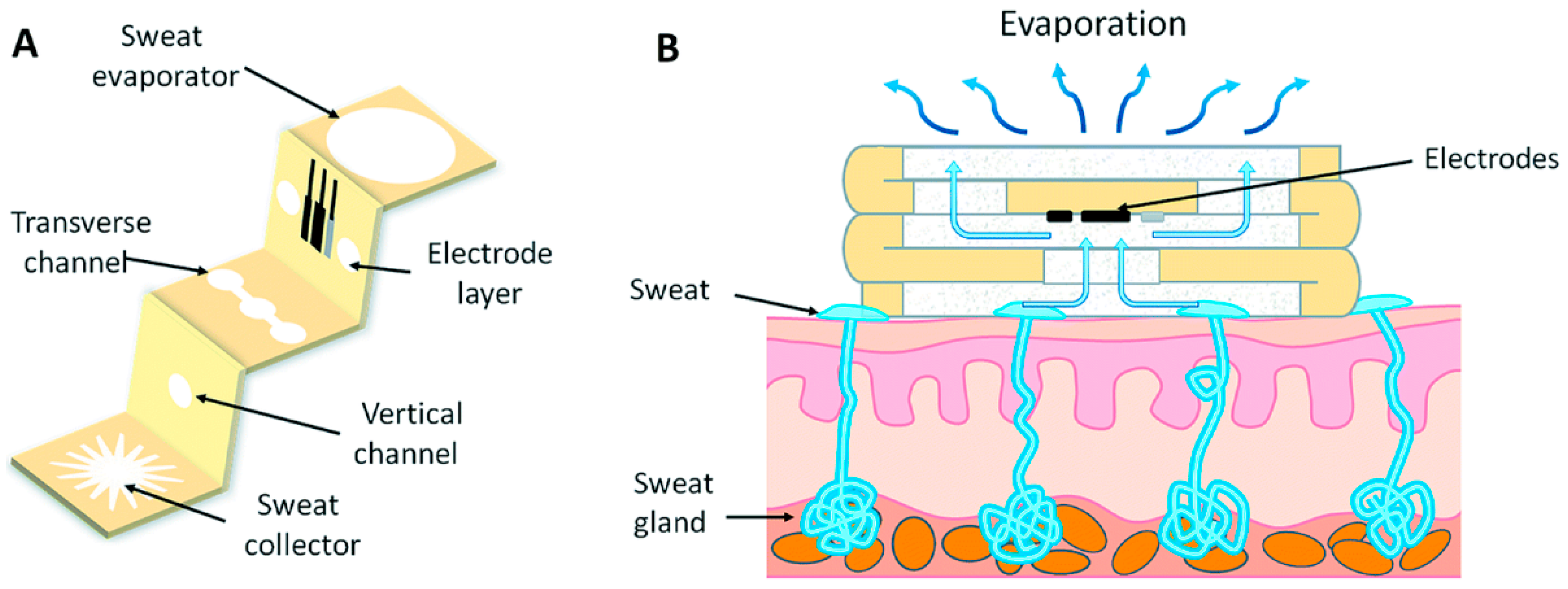
Figure 5.
Different examples of amperometric immunosensors. There are various types, such as label-free-, sandwich-, enzymatic-, and non-enzymatic-based sensors. Based on varied properties of the protein of interest, they are aptly fabricated for defined signal output.
Figure 5.
Different examples of amperometric immunosensors. There are various types, such as label-free-, sandwich-, enzymatic-, and non-enzymatic-based sensors. Based on varied properties of the protein of interest, they are aptly fabricated for defined signal output.
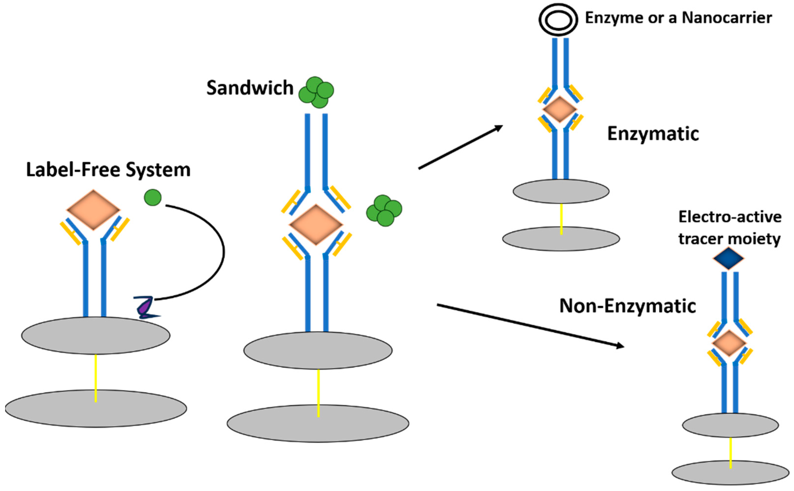
Figure 6.
Illustration of electrochemical sensor market overview.
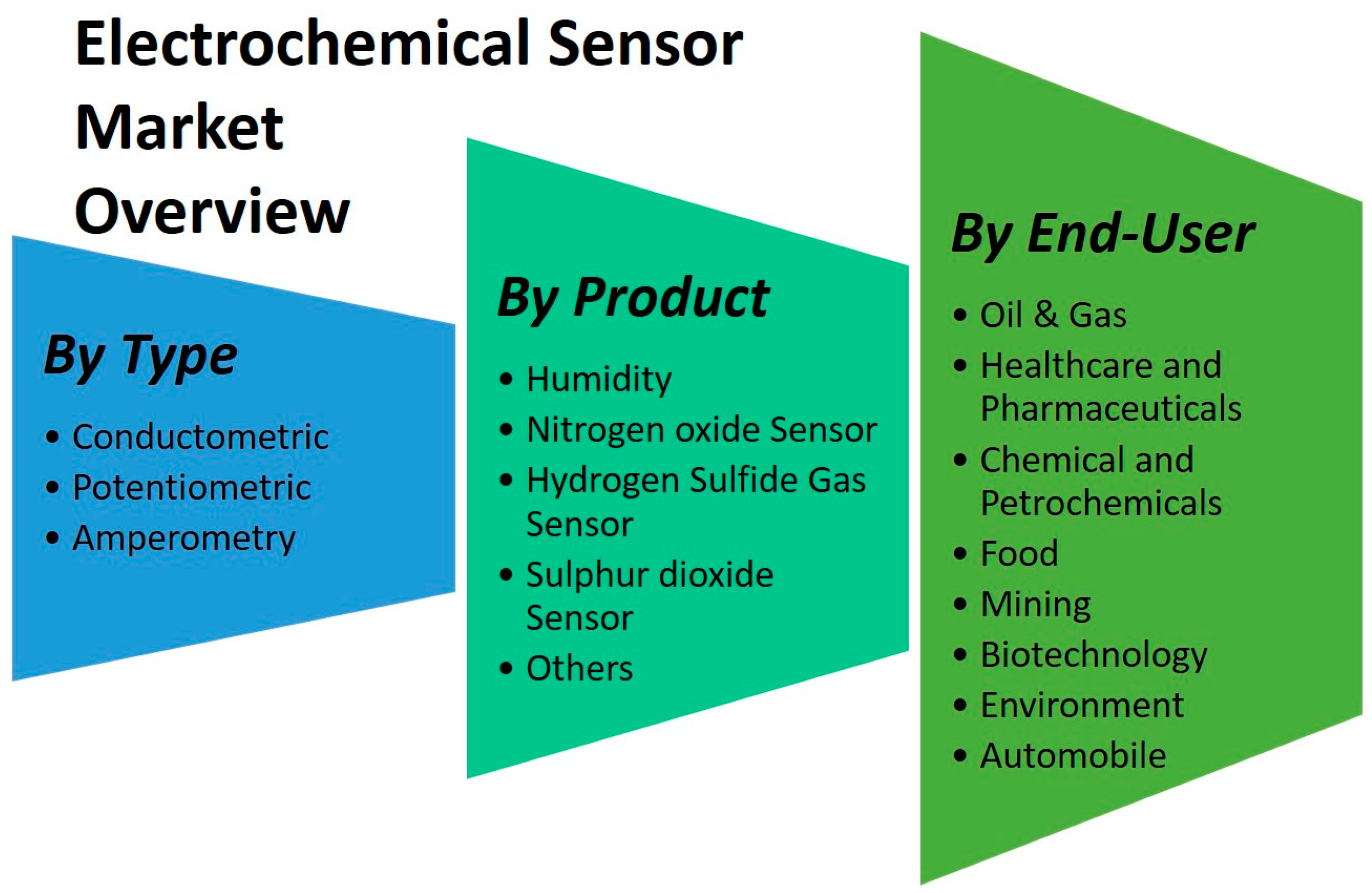
Figure 7.
Illustration of smartphone-enabled point-of-care electro-immunosensor SARS-CoV-2 N protein detection prototype. Copyright and preprinted Anal. Chem. 2022 [81].
Figure 7.
Illustration of smartphone-enabled point-of-care electro-immunosensor SARS-CoV-2 N protein detection prototype. Copyright and preprinted Anal. Chem. 2022 [81].


Table 1.
Tabulation of various conventional vs. electrochemical techniques for immunosensor fabrication and their properties.
Table 1.
Tabulation of various conventional vs. electrochemical techniques for immunosensor fabrication and their properties.
| S. No. | Conventional Immunoassays | Electrochemical Immunoassays | Ref. |
|---|---|---|---|
| 1. | Various techniques are available based on different principles such as fluorescence-based, agglomeration-based, change in optical properties, enzyme immunoassays, etc. They are the gold standards. | They rely on a simple concept of changes in the current, charge transfer or resistance after highly specific antigen–antibody complex (lock and key) formation only. Any variation in the system can change the output signals, and hence the selectivity can be determined. | [10,11,12] |
| 2. | Sophisticated set-up required with skilled technician. Cumbersome instruments A high volume of samples is required | A CHI workstation or a simplistic potentiometer can also be used by semi-skilled personnel for the measurement. Nowadays, small potentiostats (pendrive sizes) are available on the market, which are compatible with mobile phones and tablets. Nanolitres of the sample are sufficient. | [13,14] |
| 3. | Low limit of detection, highly sensitive, and has a wide range of detection. Upto pico or femto molar levels too. | Moderate limit of detection, highly sensitive, and has a moderate range of detection. Upto nano and picomolar levels. | [15,16] |
| 4. | It cannot be extended for on-field POCT devices. | Can be extended as on-field POCT (point of care testing) devices. | [15,16,17] |
| 5. | Pre-sampling procedures are required. Moderate turnaround time. Increased throughput in clinic laboratories. High cost incurred. Plates and vials can be reused. | No pre-sampling is required. Faster turnaround time (~2 to 10 min). Increased throughput. Minimal cost required. Immunosensor-based electrodes are one-time usage only (for the majority of systems). | [18] |
| 6. | Methods involved are as follows: Optical detection; Reflectometry; Ellipsometry; Surface plasmon resonance; Chemiluminescence; Piezoelectric. | Methods involved are as follows: Potentiometry; Amperometry; Electrochemical luminescence; Microgravimetric (EQCM—Electrochemical Quartz Crystal Microbalance); Impedance; FET-based; Bio-resistors-based systems. | [13,19,20] |
Disclaimer/Publisher’s Note: The statements, opinions and data contained in all publications are solely those of the individual author(s) and contributor(s) and not of MDPI and/or the editor(s). MDPI and/or the editor(s) disclaim responsibility for any injury to people or property resulting from any ideas, methods, instructions or products referred to in the content. |
© 2024 by the author. Licensee MDPI, Basel, Switzerland. This article is an open access article distributed under the terms and conditions of the Creative Commons Attribution (CC BY) license (https://creativecommons.org/licenses/by/4.0/).
Share and Cite
MDPI and ACS Style
Gandhi, M. Modelling Prospects of Bio-Electrochemical Immunosensing Platforms. Electrochem 2024, 5, 146-161. https://doi.org/10.3390/electrochem5020010
AMA Style
Gandhi M. Modelling Prospects of Bio-Electrochemical Immunosensing Platforms. Electrochem. 2024; 5(2):146-161. https://doi.org/10.3390/electrochem5020010
Chicago/Turabian StyleGandhi, Mansi. 2024. "Modelling Prospects of Bio-Electrochemical Immunosensing Platforms" Electrochem 5, no. 2: 146-161. https://doi.org/10.3390/electrochem5020010