
Cytotoxic Activity of Metal Nanoparticle Complexes †
Department of Chemistry, Isabella Thoburn College Lucknow, University of Lucknow (U.P.), Lucknow 226007, India
*
Author to whom correspondence should be addressed.
†
Presented at the 4th International Electronic Conference on Applied Sciences, 27 October–10 November 2023; Available online: https://asec2023.sciforum.net/ .
Eng. Proc. 2023, 56(1), 27; https://doi.org/10.3390/ASEC2023-15242
Published: 26 October 2023
(This article belongs to the Proceedings of The 4th International Electronic Conference on Applied Sciences)
Abstract
:Metal complexes are widely used in pharmaceutics, cosmetics, electronics, casting printing and power generation. One of the major challenges due to their long-term use as medicines is their accumulation in the body. This issue needs to be resolved to achieve better results of metal complexes as medicines. The use of metal-nanoparticles (MNPs) can be expected to reduce the toxicity of metals and their accumulation in the body. The aim of this paper is to give an insight into the variation induced in the cytotoxic activity of MNP–ligand complexes by replacing the respective heavy metals with their nanoparticles (NPs).
1. Introduction
The application of metal complexes in multidisciplinary areas of cosmetics, drugs and electricals has been known for a very long time [1]. The discovery of cisplatin in 1965 [2] proved to be a boon for cancer patients. Though the drug had a curative effect, it was associated with long-term side effects on the brain and kidney [3]. To solve the problem of side effects associated with cisplatin, scientists worked on other alternatives, and as a result, carboplatin and oxaliplatin were synthesized, which showed similar drug responses with the additional advantage of limited side effects [4]. Since the exploration of platinum in drugs, scientists have tried to explore other metal atoms like iron, cobalt, gold, titanium, ruthenium etc., as anticancer drugs, which proved to be effective as well [5,6]. But these drugs were also associated with long-term side effects [5]. The major reason for heavy metal toxicity was probably related to either Fenton’s reaction pathway (Figure 1) [7] followed by heavy metal metabolism or their selective binding.
Thus, to solve the problem of toxicity of heavy metals in drugs, MNPs came up as a better alternative. Recent advances in nanomedicines indicate that a smaller drug dosage is required, thereby helping to reduce the metal load or metal toxicity in the body. MNP complexes are reported to have much less impact compared to metal atom complexes, when used in gene and medication delivery in biosystems [8].
2. Metal-Nanoparticle Complexes
An increased ligand density on the surface of MNPs, over a single ligand coordinated with metal, enhance the binding and cellular absorption through multivalent interactions. MNPs can be functionalized with organic ligands generally via two methods. The two processes are the introduction of ligands to NPs prior to the fabrication of NPs and the adsorption of the ligand on the surface of NPs after the fabrication NPs [9].
A literature survey reveals that the bonding of MNPs and synergistic effects improve the activity and reduce the side effects of MNP complexes. Around two decades ago, scientists initiated the work on MNP complexes. They suggested that MNPs provide a route for the sustained release of encapsulated drugs, thereby decreasing the accumulation of drugs in tissues and hence resulting in reduced toxicity. Various MNP complexes have been proven to favor targeted drug delivery as well [10,11]. Cancer cells can be targeted by active targeting moieties like glucuronic acid, folic acid, hyaluronic acid and biotin, as well as macromolecules such as monoclonal antibodies, that specifically bind to receptors on cancerous cells. Ligands with active targeting molecules can significantly enhance the concentration of drugs in the target area, improve the cellular uptake of drug-modified NPs, and significantly decrease the adverse effects of the drug, thus effectively improving the therapeutic effects [12]. Size distribution and coherent functionalization of NPs affect the activity of the complex. Ligand coordination with NPs displayed enhanced activities compared to ligand coordination with metal.
Several studies on metal complexes have been accomplished using transition metals, which have shown good promise against different cell lines. However, long-term use leads to the accumulation of these drugs in the body, which seems to be controlled by the use of NPs. In search of safer drugs much is to be explored with p-block metal complexes, as these metals are less toxic in terms of their accumulation in the body. For example, Bismuth(Bi), known as a semimetal or metalloid due to its varied applications, can be considered as a good representative of the p-block elements. Its multidimensional activity has generated interest for research in the exploration of Bismuth-NP complexes [13]. Compounds of Bi-NP with chalcogens and oxyhalides have been reported to show good therapeutic and diagnostic properties [14]. Bi-selenide NPs (Bi2Se3 NPs) have been explored and are established to have theragnostic potentials [15]. Studies have reported the multiple roles of Bi-sulfide NPs and have established this as an efficient theragnostic for malignancies [16].
3. Anticancer Activity of MNP Complexes
Various groups of scientists have worked on the synthesis of MNP complexes of cobalt, nickel, copper, cadmium, chromium, manganese, gold, ruthenium and lanthanum and demonstrated the activity of these complexes against various cancer cell lines [17,18,19]. Some of these are described in Table 1.
M. Jarestan et al. (2020) synthesized cobalt oxide (Co3O4) NPs functionalized with glutamic acid and conjugated by thiosemicarbazide and investigated their cytotoxicity properties on the gastric cancer (AGS) cell line [27]. N.M. Binu et al. (2021) developed quercetin drug-loaded Nickel oxide NPs, surface modified with polydopamine and folic acid to target breast cancer cells [28]. O. Aktruk (2022) synthesized AuNP complexes conjugated with doxorubicin and capped with levan polysaccharide and determined its cytotoxic activity against the MCF-7 cancer cell line. The physicochemical experiments showed that increasing the levan quantity greatly improved the colloidal stability and drug encapsulation efficacy [29].
4. Targeting Strategies of Drug-Loaded NPs
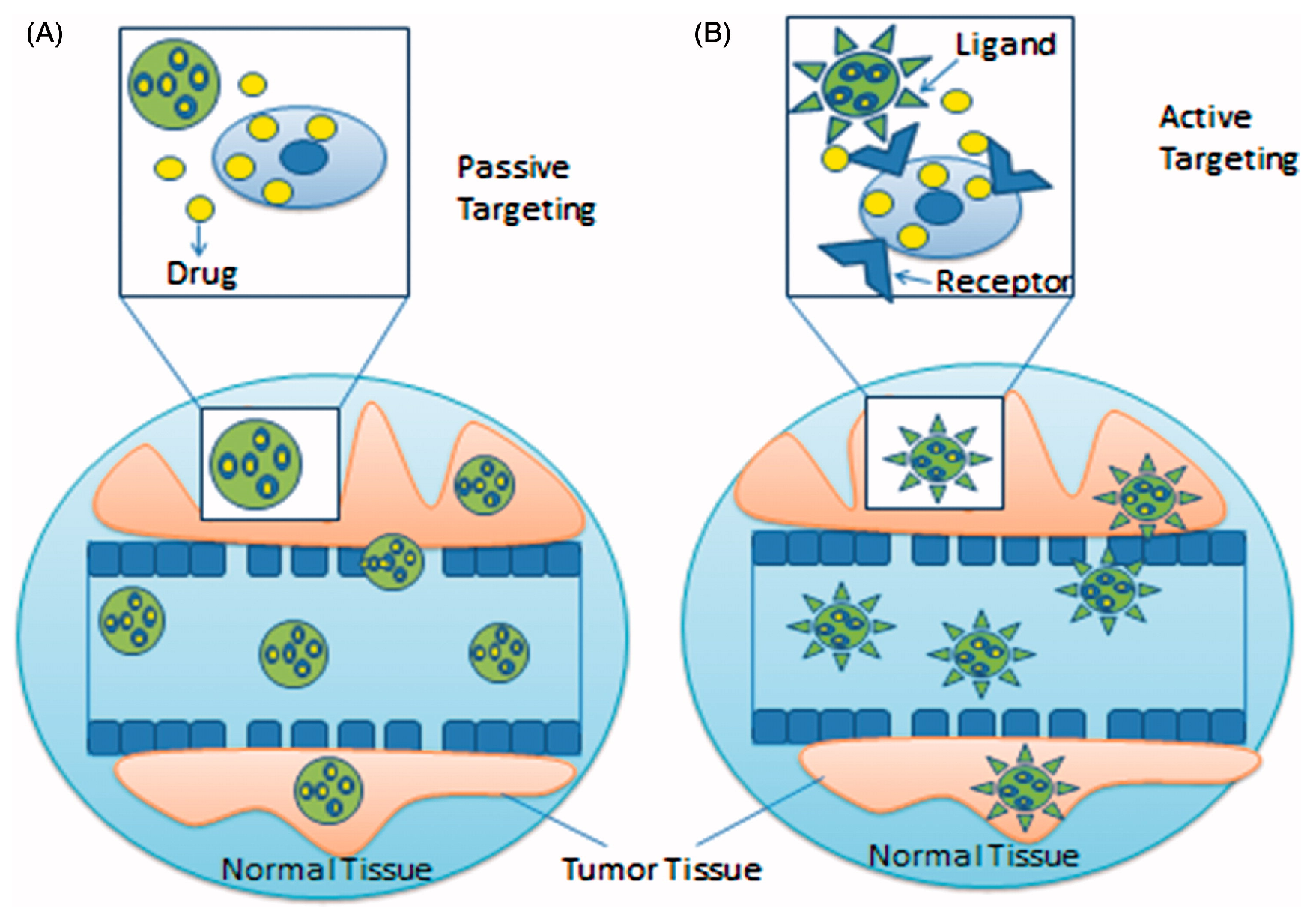
Because of their large surface area, small size and functionalization capacity with targeting ligands and therapeutic molecules, NPs are able to penetrate many body parts without interfering with their regular processes. Generally, drug molecules are released at the target site after being adsorbed onto the surface of NPs. The two main methods through which NPs target cancer cells are passive targeting and active targeting [30].
4.1. Passive Targeting
In contrast to the normal cells, many solid tumors exhibit distinct physical traits such as reduced lymphatic drainage and hyperpermeable vasculature. Nanocarriers enter the circulatory system after being ingested or injected into the body. Then, they extravasate the vasculature to reach the target and release their payload. Nanocarriers have the advantage of being incapable of passing across the tightly closed connection between normal vascular linings; however, the defective vasculature of the cancerous area allows for an increase in the amount of nanocarrier within the target cell. The accumulation of nanocarriers inside the tumor cell is significantly increased by the impaired lymphatic drainage. This phenomenon is the fundamental impetus for passive targeting and known as the EPR (Enhanced Permeability and Retention) effect [30].
Through the EPR effect, drug-loaded NPs could target tumor cells in a passive manner. The pore size of angiogenic tumor vessels between adjacent vascular endothelial cells has generally been found to be larger compared to those in the blood vessels found in normal cells, and therefore, a higher concentration of the loaded drug is released specifically within the extracellular tumor cells by the angiogenic tumor vessels. This enables an effective anticancer therapy with less harmful effects [31]. Figure 2A [32] shows the passive targeting strategy of drug-loaded NPs.
For example, the passive targeting approach has been effectively employed with polylactic-co-glycolic acid-based NPs functionalized with several anticancer agents like paclitaxel, cisplatin and doxorubicin to improve the time of circulation of blood, drug stability and cytotoxic activity [33].
4.2. Active Targeting
Although passive targeting may make it easier for NPs to be effectively localized in the tumor tissue, it cannot further encourage cellular uptake by cancerous cells. NPs functionalized with ligands including nucleic acid aptamers, peptides, antibodies, and small molecules can be dynamically targeted on cancer cells to induce cellular uptake. This phenomenon distinguishes the cancerous cells and normal cells. The active targeting strategy can be generally classified into two sub-categories. First is receptor-mediated endocytosis, in which drug-loaded nanocarriers are surface modified with specific ligands to identify the specific receptor in the tumor sites for its binding and subsequent cellular uptake. Second is the stimuli-responsive intracellular drug delivery, in which the release of the drug depends on a small alteration in the microenvironment of the affected region [30]. Figure 2B [32] shows the active targeting strategy of drug-loaded NPs.
For example, to specifically target colorectal cancer cells, folic acid(FA)-modified polyethylene glycol (PGE) polymer-based drug delivery systems (PLGA-PEG-FA) containing oxaliplatin were developed. This system when functionalized with NPs showed greater cellular uptake in comparison to untargeted molecules and the free drug and increased cell death by receptor-mediated endocytosis [12]. To deliver oxaliplatin to HCT-116 colon cancer cells, Tabasi et al. synthesized CD44(Cluster of differentiation)-conjugated Fe3O4 NPs. Using the MTT assay, they observed that increased intracellular absorption of CD44-conjugated oxaliplatin-loaded NPs produces a considerable decrease in the IC50 measurement of oxaliplatin against HCT-116 cells [34].
5. Conclusions
A review of the literature showed that several metal complexes have been successfully developed as anticancer agents. But these complexes also showed heavy metal toxicity and long-term side effects. To resolve the side effects of metal complexes for the treatment of cancer, scientists have explored new ideas day-by-day. Recent advances in nano drug delivery systems to selectively target cancerous cells have helped to reduce the metal load and accumulation of drugs in the body. The use of NPs improves cell uptake efficiency as well as drug delivery and are safe. MNPs functionalized with ligands are reported to have less harmful effects compared to the metal complexes, when used for the treatment of cancer. Generally, drug molecules with active targeting moieties are released at the target site after being adsorbed by the surface of NPs, and hence, increase the concentration of the drug in the target area, which improves the cellular uptake of drug-loaded NPs. There are the two main targeting strategies, passive targeting and active targeting, through which NPs are highly absorbed by cancerous cells. This review summarizes ligand-functionalized MNP complex inhibitors to resolve the challenges of drug resistance and of metal toxicity due to the overloading of metal in the drug.
Author Contributions
Conceptualization, S. and S.J.; resources, S.J.; data curation, N.S., S. and S.J.; writing-original draft preparation, N.S., S., K.H., S.G. and S.J.; writing-review and editing, N.S. and S.J.; visualization, N.S., S., K.H., S.G. and S.J.; supervision, S.J. All authors have read and agreed to the published version of the manuscript.
Funding
This research received no external funding.
Institutional Review Board Statement
Not applicable.
Informed Consent Statement
Not applicable.
Data Availability Statement
Data are contained within the article.
Conflicts of Interest
The authors declare no conflict of interest.
References
- Mudi, S.Y.; Usman, M.T.; Ibrahim, S. Clinical and Industrial Application of Organometallic Compounds and Complexes: A Review. Am. J. Chem. Appl. 2015, 2, 151–158. [Google Scholar]
- Rosenberg, B.; Van Camp, L.; Krigas, T. Inhibition of Cell Division in Escherichia coli by Electrolysis Products from a Platinum Electrode. Nature 1965, 205, 4972. [Google Scholar] [CrossRef]
- Arany, I.; Safirstein, R.L. Cisplatin nephrotoxicity. Semin. Nephrol. 2003, 23, 460–464. [Google Scholar] [CrossRef]
- Meng, R.D.; Shelton, C.C.; Li, Y.M.; Qin, L.X.; Notterman, D.; Paty, P.B.; Schwartz, G.K. Gamma-Secretase inhibitors abrogate oxaliplatin-induced activation of the Notch-1 signaling pathway in colon cancer cells resulting in enhanced chemosensitivity. Cancer Res. 2009, 69, 573–582. [Google Scholar] [CrossRef]
- Allardyce, C.S.; Dyson, P.J. Medicinal Properties of Organometallic Compounds. In Bioorganometallic Chemistry, 1st ed.; Simonneaux, G., Ed.; Springer: Berlin/Heidelberg, Germany, 2006; Volume 17, pp. 177–210. [Google Scholar]
- Hashmi, K.; Satya; Gupta, S.; Siddique, A.; Khan, T.; Joshi, S. Medicinal Applications of Vanadium Complexes with Schiff Bases. J. Trace Elem. Med. Biol. 2023, 79, 127245. [Google Scholar] [CrossRef]
- Satya; Hashmi, K.; Gupta, S.; Singh, N.; Khan, T.; Joshi, S. Nanofabrication of Metals and Their Compounds for Effective Medicinal and Environmental Applications (A Review). Russ. J. Gen. Chem. 2023, 93, 635–665. [Google Scholar] [CrossRef]
- Chandrakala, V.; Aruna, V.; Angajala, G. Review on metal nanoparticles as nanocarriers: Current challenges and perspectives in drug delivery systems. Emergent Mater. 2022, 5, 1593–1615. [Google Scholar] [CrossRef]
- Bahrami, B.; Hojjat-Farsangi, M.; Mohammadi, H.; Anvari, E.; Ghalamfarsa, G.; Yousefi, M.; Jadidi-Niaragh, F. Nanoparticles and targeted drug delivery in cancer therapy. Immunol. Lett. 2017, 190, 64–83. [Google Scholar] [CrossRef]
- Barry, N.P.E.; Sadler, P.J. Challenges for Metals in Medicine: How Nanotechnology May Help To Shape the Future. ACS Nano 2013, 7, 5654–5659. [Google Scholar] [CrossRef]
- Maldonado, C.R.; Salassa, L.; Gomez-Blanco, N.; Mareque-Rivas, J.C. Nano-functionalization of metal complexes for molecular imaging and anticancer therapy. Coord. Chem. Rev. 2013, 257, 2668–2688. [Google Scholar] [CrossRef]
- Ebrahimnejad, P.; Taleghani, A.S.; Asare-Addo, K.; Nokhodchi, A. An updated review of folate-functionalized nanocarriers: A promising ligand in cancer. Drug Discov. Today 2022, 27, 471–489. [Google Scholar] [CrossRef] [PubMed]
- Balva, M.; Legeai, S.; Garoux, L.; Leclerc, N.; Meux, E. Dismantling and chemical characterization of spent Peltier thermoelectric devices for antimony, bismuth and tellurium recovery. Environ. Technol. 2016, 38, 791–797. [Google Scholar] [CrossRef] [PubMed]
- Li, Y.K.; Yang, M.; Li, M.X.; Yu, H.; Wu, H.C.; Xie, S.Q. Synthesis, crystal structure and biological evaluation of a main group seven-coordinated bismuth (III) complex with 2-acetylpyridine N4-phenylthiosemicarbazone. Bioorganic Med. Chem. Lett. 2013, 23, 2288–2292. [Google Scholar] [CrossRef] [PubMed]
- Islam, A.; Rodrigues, B.L.; Marzano, I.M.; Perreira-Maia, E.C.; Dittz, D.; Paz Lopes, M.T.; Ishfaq, M.; Frézard, F.; Demicheli, C. Cytotoxicity and apoptotic activity of novel organobismuth(V) and organoantimony(V) complexes in different cancer cell lines. Eur. J. Med. Chem. 2016, 109, 254–267. [Google Scholar] [CrossRef]
- Božić, A.R.; Bjelogrlić, S.K.; Novaković, I.T.; Filipović, N.R.; Petrović, P.M.; Marinković, A.D.; Todorović, T.R.; Cvijetić, I.N. Antimicrobial Activity of Thiocarbohydrazones: Experimental Studies and Alignment-Independent 3D QSAR Models. ChemistrySelect 2018, 3, 2215–2221. [Google Scholar] [CrossRef]
- El-Wakiel, N. Nano-sized metal complexes of azo L-histidine: Synthesis, characterization and their application for catalytic oxidation of 2-amino phenol. Appl. Organomet. Chem. 2018, 32, e4229. [Google Scholar] [CrossRef]
- Jawoor, S.S.; Patil, S.A.; Kumbar, M.; Ramawadgi, P.B. Green synthesis of nano sized transition metal complexes containing heterocyclic Schiff base: Structural and morphology characterization and bioactivity study. J. Mol. Struct. 2018, 1164, 378–385. [Google Scholar] [CrossRef]
- Mahmoud, W.; Refaat, A.M.; Mohamed, G.G. Nano Schiff Base and Its Metal Complexes: Synthesis, Characterization Tools, Biological Applications and Molecular Docking Studies. Egypt. J. Chem. 2020, 63, 2157–2176. [Google Scholar]
- Chattopadhyay, S.; Chakraborty, S.P.; Laha, D.; Baral, R.; Pramanik, P.; Roy, S. Surface-modified cobalt oxide nanoparticles: New opportunities for anti-cancer drug development. Cancer Nanotechnol. 2012, 3, 13–23. [Google Scholar] [CrossRef]
- Adwin Jose, P.; Sankarganesh, M.; Dhaveethu Raja, J.; Senthilkumar, G.S.; Nandini Asha, R.; Raja, S.J.; Sheela, C.D. Bio-inspired nickel nanoparticles of pyrimidine-Schiff base: In vitro anticancer, BSA and DNA interactions, molecular docking and antioxidant studies. J. Biomol. Struct. Dyn. 2022, 40, 10715–10729. [Google Scholar] [CrossRef]
- Jose, P.A.; Raja, J.D.; Sankarganesh, M.; Rajesh, J. Evaluation of antioxidant, DNA targeting, antimicrobial and cytotoxic studies of imine capped copper and nickel nanoparticles. J. Photochem. Photobiol. B Biol. 2018, 178, 143–151. [Google Scholar] [CrossRef] [PubMed]
- Harish, R.; Nisha, K.D.; Prabakaran, S.; Sridevi, B.; Harish, S.; Navaneethan, M.; Ponnusamy, S.; Hayakawa, Y.; Vinniee, C.; Ganesh, M.R. Cytotoxicity assessment of chitosan coated CdS nanoparticles for bio-imaging applications. Appl. Surf. Sci. 2020, 499, 143817. [Google Scholar] [CrossRef]
- Sadalage, P.S.; Patil, R.V.; Havaldar, D.V.; Gavade, S.S.; Santos, A.C.; Pawar, K.D. Optimally biosynthesized, PEGylated gold nanoparticles functionalized with quercetin and camptothecin enhance potential anti-inflammatory, anti-cancer and anti-angiogenic activities. J. Nanobiotechnology 2021, 19, 84. [Google Scholar] [CrossRef] [PubMed]
- Zhou, Y.; Yu, Q.; Qin, X.; Bhavsar, D.; Yang, L.; Chen, Q.; Zheng, W.; Chen, L.; Liu, J. Improving the anticancer efficacy of laminin receptor-specific therapeutic ruthenium nanoparticles (RuBB-loaded EGCG-RuNPs) via ROS-dependent apoptosis in SMMC-7721 cells. ACS Appl. Mater. Interfaces 2017, 8, 15000–15012. [Google Scholar] [CrossRef] [PubMed]
- Nosrati, H.; Charmi, J.; Salehiabar, M.; Abhari, F.; Danafar, H. Tumor targeted albumin coated bismuth sulfide nanoparticles (Bi2S3) as radiosensitizers and carriers of curcumin for enhanced chemoradiation therapy. ACS Biomater. Sci. Eng. 2019, 5, 4416–4424. [Google Scholar] [CrossRef] [PubMed]
- Jarestan, M.; Khalatbari, K.; Pouraei, A.; Sadat Shandiz, S.A.; Beigi, S.; Hedayati, M.; Majlesi, A.; Akbari, F.; Salehzadeh, A. Preparation, characterization, and anticancer efficacy of novel cobalt oxide nanoparticles conjugated with thiosemicarbazide. 3 Biotech 2020, 10, 230. [Google Scholar] [CrossRef]
- Binu, N.M.; Prema, D.; Prakash, J.; Balagangadharan, K.; Balashanmugam, P.; Selvamurugan, N.; Venkatasubbu, G.D. Folic acid decorated pH sensitive polydopamine coated honeycomb structured nickel oxide nanoparticles for targeted delivery of quercetin to triple negative breast cancer cells. Colloids Surf. A Physicochem. Eng. Asp. 2021, 630, 127609. [Google Scholar] [CrossRef]
- Akturk, O. The anticancer activity of doxorubicin-loaded levan-functionalized gold nanoparticles synthesized by laser ablation. Int. J. Biol. Macromol. 2022, 196, 72–85. [Google Scholar] [CrossRef]
- Dutta, B.; Barick, K.C.; Hassan, P.A. Recent advances in active targeting of nanomaterials for anticancer drug delivery. Adv. Colloid Interface Sci. 2021, 296, 102509. [Google Scholar] [CrossRef]
- Zhou, Q.; Zhang, L.; Wu, H. Nanomaterials for cancer therapies. Nanotechnol. Rev. 2017, 6, 473–496. [Google Scholar] [CrossRef]
- Dadwal, A.; Baldi, A.; Kumar Narang, R. Nanoparticles as carriers for drug delivery in cancer. Artif. Cells Nanomed. Biotechnol. 2018, 46, 295–305. [Google Scholar] [CrossRef] [PubMed]
- Moreno, D.; Zalba, S.; Navarro, I.; de Ilarduya, C.T.; Garrido, M.J. Pharmacodynamics of cisplatin-loaded PLGA nanoparticles administered to tumor-bearing mice. Eur. J. Pharm. Biopharm. 2010, 74, 265–274. [Google Scholar] [CrossRef] [PubMed]
- Tabasi, H.; Mosavian, M.H.; Sabouri, Z.; Khazaei, M.; Darroudi, M. pH-responsive and CD44-targeting by Fe3O4/MSNs-NH2 nanocarriers for oxaliplatin loading and colon cancer treatment. Inorg. Chem. Commun. 2021, 125, 108430. [Google Scholar] [CrossRef]
Figure 1.
Fenton’s reaction pathway.
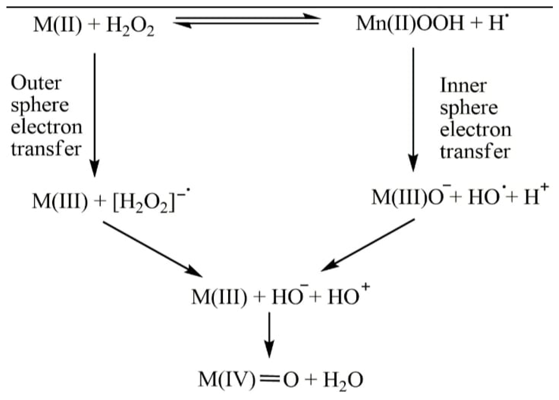
Figure 2.
(A) Passive targeting and (B) active targeting strategy of drug-loaded NPs (open access).

Table 1.
Anticancer activity of some MNP complexes for various cancer cell lines.
| MNPs | Functionalized Ligand | Cancer Cell Lines | Reference |
|---|---|---|---|
| CoONPs | phosphonomethyl iminodiacetic acid | Jurkat (T-cell lymphoma) and KB (oral carcinoma) cell lines | [20] |
| NiNPs | 2-(4,6-dimethoxypyrimidin-2-ylimino)methyl)-6-methoxyphenol (DMPMM) | human breast cancer (MCF-7), human lung cancer (A549) and human liver cancer(HepG2) cell lines | [21] |
| CuNPs | DMPMM | human breast cancer cell line (MCF-7) | [22] |
| CdSNPs | Chitosan | Human Jurkat cell and erythrocytes cell lines | [23] |
| AuNPs | quercetin | human breast cancer cell line (MCF-7) | [24] |
| RuNPs | epigallocatechin gallate | human liver cancer(HepG2) cell line | [25] |
| BiSNPs | Curcumin | Mouse breast cancer cell line(4T1) | [26] |
Disclaimer/Publisher’s Note: The statements, opinions and data contained in all publications are solely those of the individual author(s) and contributor(s) and not of MDPI and/or the editor(s). MDPI and/or the editor(s) disclaim responsibility for any injury to people or property resulting from any ideas, methods, instructions or products referred to in the content. |
© 2023 by the authors. Licensee MDPI, Basel, Switzerland. This article is an open access article distributed under the terms and conditions of the Creative Commons Attribution (CC BY) license (https://creativecommons.org/licenses/by/4.0/).
Share and Cite
MDPI and ACS Style
Singh, N.; Satya; Hashmi, K.; Gupta, S.; Joshi, S. Cytotoxic Activity of Metal Nanoparticle Complexes. Eng. Proc. 2023, 56, 27. https://doi.org/10.3390/ASEC2023-15242
AMA Style
Singh N, Satya, Hashmi K, Gupta S, Joshi S. Cytotoxic Activity of Metal Nanoparticle Complexes. Engineering Proceedings. 2023; 56(1):27. https://doi.org/10.3390/ASEC2023-15242
Chicago/Turabian StyleSingh, Nidhi, Satya, Kulsum Hashmi, Sakshi Gupta, and Seema Joshi. 2023. "Cytotoxic Activity of Metal Nanoparticle Complexes" Engineering Proceedings 56, no. 1: 27. https://doi.org/10.3390/ASEC2023-15242