Proceeding Paper

A Methodical Review of Iridology-Based Computer-Aided Organ Status Assessment Techniques †

by
Suja Alphonse
,
Ramachandran Venkatesan
and
Theena Jemima Jebaseeli
*
Division of Computer Science and Engineering, Karunya Institute of Technology and Sciences, Coimbatore 641114, India
*
Author to whom correspondence should be addressed.
Presented at the International Conference on Recent Advances on Science and Engineering, Dubai, United Arab Emirates, 4–5 October 2023.
Eng. Proc. 2023, 59(1), 9; https://doi.org/10.3390/engproc2023059009
Published: 11 December 2023
(This article belongs to the Proceedings of Eng. Proc., 2023, RAiSE-2023)

Abstract

:
The pseudoscience known as iridology makes the unsubstantiated claim that it can identify medical disorders by examining the iris, the colored portion of the eye. Iridology does not provide a reliable means of diagnosis, and there is no scientific proof to back up its claims. To find patterns that are connected to particular medical conditions, computerized iris analysis software may need to examine thousands of iris images. A method of iridology known as Computer-Aided Iridology (CAI) uses software to study the iris. CAI still is not a medically accepted diagnostic technique and is not any more trustworthy than conventional iridology. Applying technology in medical science had a great impact on diagnosing diseases. Decision making is the most critical task in computer-aided applications. Computer vision and deep learning make this task more accurate and are widely used in many applications, mainly in diagnosing diseases. The methodologies, data acquisition source, and volume of data used for both training and testing in the pre-diagnosis of human organs utilizing iris patterns are thoroughly studied. Understanding its limitations allows researchers to concentrate on creating and evaluating improvements in technology that could boost its accuracy and usefulness. Iridology has been considered as having no use for years and becomes effective when combined with technology. This study includes various technical factors used in iridology for the pre-diagnosing of diseases. Recognizing the limitations of iridology allows healthcare providers to avoid errors in diagnosis and prevent individuals from undergoing redundant procedures or therapies based solely on iridology assessments.

1. Introduction

Iridologists frequently make diagnoses based on personal assessments of iris patterns and colors. Due to this subjective nature, it is prone to bias and error, since various professionals might come to distinct conclusions when investigating the same iris. Because it does not follow the scientific approach, lacks controlled research, and is unable to offer reliable diagnoses, many doctors consider it a pseudoscience. Iridologists frequently make contradictory assertions about the meaning of specific iris characteristics. One practitioner might state that a specific iris marking represents a particular health issue, while another could interpret this in different ways. This lack of standardization erodes iridology’s credibility even more. Needle phobia is a type of anxiety condition that affects between 3.5% and 10% of the entire population [1]. Those suffering from the illness frequently avoid medical care that involves needles, potentially leading to greater health concerns. Iridology may be the best choice for such people.
Iridology is surrounded by skepticism because there is little solid scientific evidence to back up its claims, which limits its application in real life until it is combined with cutting-edge technological approaches. According to skeptics, iridology may cause false positives, which signal a problem when there is not one, and false negatives, which fail to identify a health issue that already exists. Its practical utility in healthcare is called into question because of this unreliability. It is an intriguing idea that iridology might benefit more from being combined with cutting-edge technology [2].

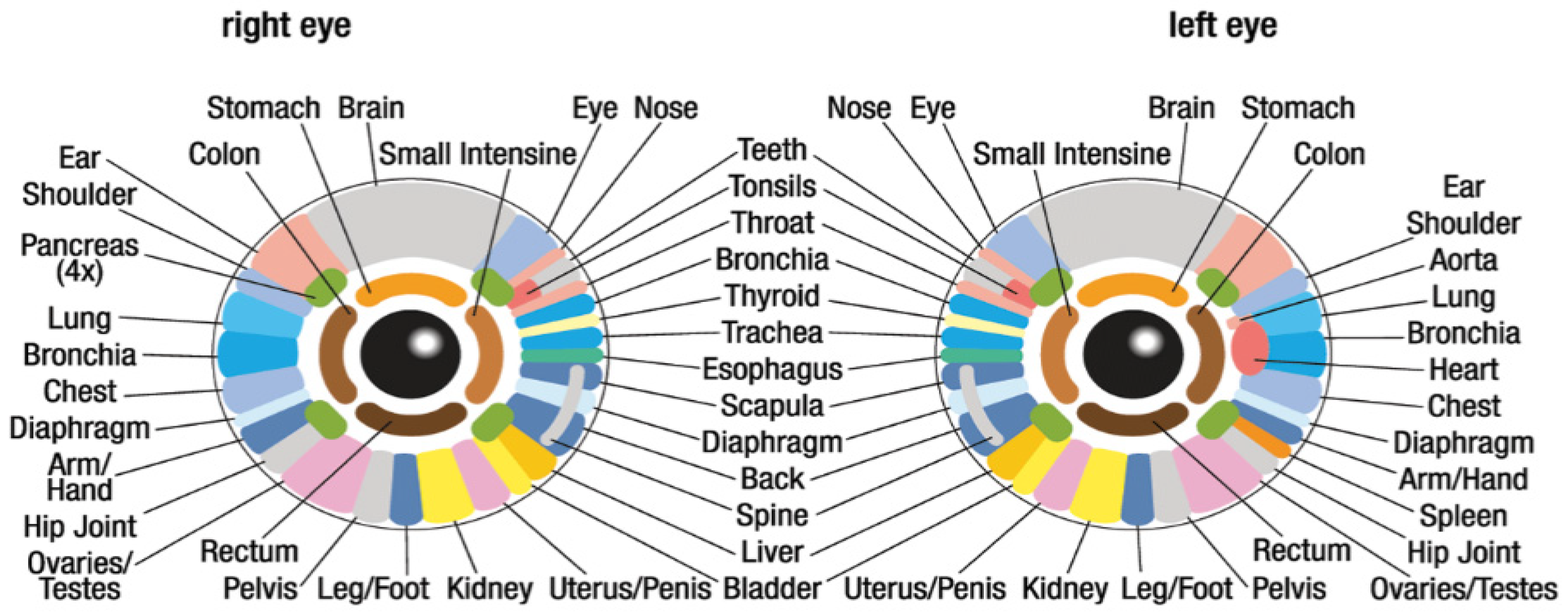
1.1. Overview of Iridology

The eyes do not serve as a light source for the body; rather, they are organs that receive and process light to allow for vision. The iris has a diameter of 12 to 13 millimeters and a thickness of three-tenths of a millimeter. Iridology mainly comprises three types of signs: curved structures that can be found in the stroma, pigments, and vessels. It is a supplementary medicine that examines structures, colors, and other iris features to determine the condition [3]. The brain and nervous system link the iris to all the tissues and organs in the body. Nature has equipped us with a small monitor that shows the most distant parts of the body via nerve reflex reactions in this way [4]. The iridology chart in Figure 1 shows the connections between the organs and the iris.
Researchers and iridologists can only analyze a single section of the iris to determine the state of that organ [5]. The iris, when considering the chart used by iridologists, is separated into seven equal rings that are further divided into 60 zones, each of which is related to an organ. For instance, to know the status of the heart, the region to be considered is in the left iris between the 2 o’clock and 3 o’clock positions. Since it cannot identify the majority of current illnesses, it has long been thought to be of little utility. With high-resolution cameras and computer-aided decision-support systems, iridology can be considered as an alternative method of pre-diagnosis without harm.
Any computer-aided system, including CAI, is highly dependent on the accuracy of the input data. Iridology’s initial data, or iris images, are naturally arbitrary because they require interpretation by humans. The software will produce incorrect results if the input data is inaccurate or biased. Similar to traditional iridology, CAI software struggles with inconsistent iris pattern interpretations. It is challenging to reach an agreement among practitioners and the software itself because there are no standardized guidelines for interpreting iris images, which results in a variety of frequently contradictory diagnoses [6]. Medical professionals in the mainstream do not recognize CAI as a reliable diagnostic tool. It is not accepted within evidence-based medicine because it has not undergone the stringent testing and validation required.
There are several causes for the revival of iridology practice. The emergence of alternative medicine is a general term for medical procedures that do not fall under the purview of traditional medicine. One kind of Complementary and Alternative Medicine (CAM) practice is iridology. Iridology has seen a resurgence in attention as CAM has grown in popularity in recent years [7]. On the internet, people may now discover iridologists and learn more about iridology.

1.2. Challenges

Iridology is discussed on numerous websites and online discussion boards, which has aided in spreading knowledge about the discipline. Many people who are interested in alternative treatment approaches see it as a natural therapy. Still, the following challenges exist:
i.
The notion that a person’s iris may be utilized to identify particular medical disorders is unsupported by scientific research.
ii.
Iridology chart reading is a subjective process that varies from practitioner to practitioner.
iii.
It might be challenging to spot the small changes in the iris that are allegedly linked to health issues.
iv.
Even seasoned iridologists make imprecise diagnoses.

2. Materials

The following data sources were used in pre-diagnoses:
i.
Patients’ medical histories, lab results, and all other diagnostic information included in patient records.
ii.
Various tools, such as digital cameras, slit lamps, and fundus cameras, were used to take iris images.
iii.
Genetic information: this includes details about the patient’s DNA that could be used to pinpoint genetic risk factors for particular diseases.
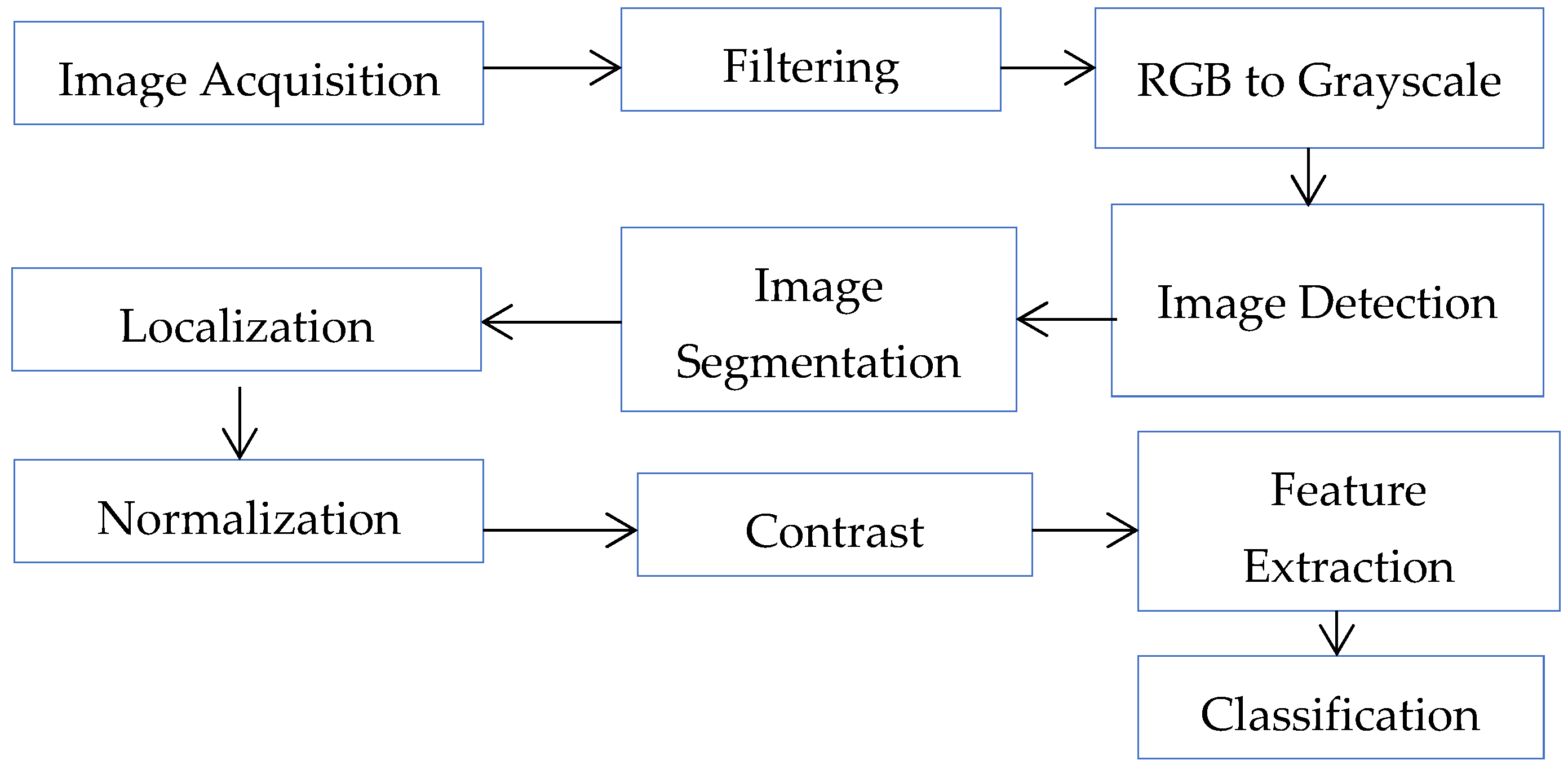
The computer may need to be trained on a sizable dataset of iris images and medical records to perform machine learning. Certain images lose quality due to a lack of texture as well as poor resolution. It would not retain these features in non-ideal or slightly loosened image capture circumstances. Capturing the iris with a high-resolution camera helps to diagnose more accurately. Because of the iris’s tiny size, it fails to detect the distinguishing characteristics, necessitating extreme accuracy. Certain image-capturing devices used by various researchers and the resolution of the image used are given in Table 1.
According to iridology, only the specific region is to be analyzed. The task of iris localization is critical, as it always serves as an essential component in Information Technology applications [8]. The patients were surveyed about regarding their diabetic complication types, methods of supervision, and their levels of diabetic retinal degeneration (if they existed) for further research as part of the project “Finding Informative Regions in Iris Images for Predicting Diabetes”. The corresponding dataset is available at the URL given in [9].

3. Methodology

Iridology becomes a potent tool in the pre-diagnosis of diseases when combined with cutting-edge technology, with a focus on illnesses like heart disease. This groundbreaking investigation delves deeply into the complex technical elements that enable and enhance the effectiveness of this integration. The iris can be captured in incredibly fine detail using modern digital cameras and imaging methods [10]. This degree of accuracy is necessary for reliable analysis. AI systems can analyze iris patterns using machine learning algorithms at a scale and speed that are simply not possible for human analysts [11]. These systems recognise subtle heart disease risk factors and anticipate potential problems long before the classic symptoms appear. AI systems can evaluate iris patterns on a scale and with precision by using machine learning algorithms. In deep learning, to build a model, training a network and making predictions on new data are crucial steps. The phases include image quality, contrast, intensity, image enhancement, transformation, and deciding which area of the image to view. This improves the initial image’s brightness and decides on an effective outcome.
Iridologists claim the lower portion of the iris close to the pupil represents the heart. To evaluate the condition of the heart, they observe changes in this region’s color, texture, and indications [6]. For instance, a cloudy or dark iris here could indicate heart disease. A weak or paper-thin iris may also indicate heart issues. Other indications of heart disease may also be sought by iridologists, such as the presence of iris crypts as small pits in the iris or iris furrows as ridges in the iris. The proposed study’s goal is to examine the technical factors used in iridology for disease prediction.

3.1. Image Pre-Processing

Based on the quality of the data and the methodology used, the steps in pre-processing may be skipped. Some studies claim that the dataset under consideration plays a major role in determining the data pre-processing strategy to use, while other research claims that the choice should be made based on research. The various technical methods that are available for analyzing iris patterns are shown in Figure 2.

3.2. Filtering an Image

This modifies the pixel density of an image to transform it into its intended shape by utilizing various visual editing techniques used by graphic designers and program editors [12].

3.3. RGB to Grayscale

To decrease the complications of computers, an image must be converted to grayscale. During this conversion, the hue and saturation are removed by holding the luminance.

3.4. Image Detection

Computer vision uses the object-detection technique to find occurrences of things in pictures. To retrieve an object, there are several deep learning and machine learning techniques available. Here, an image of the iris should be detected. In most of the applications, it combines localization and classification.

3.5. Image Segmentation

In iridology, segmentation entails dividing the iris into several zones or regions, each of which is thought to represent a different organ or bodily system. These zones correspond to various body parts, including the heart, liver, kidneys, etc. It is believed that by examining particular iris regions, professionals can evaluate the health of corresponding organs or systems. For instance, a certain marking in a particular iris segment might be interpreted as a sign of a heart condition. The entire image after detection is segmented or masked so that the required segments alone are examined instead of the entire image. By keeping the precise resolution, the iris image-segmentation technique attempts to normalize the image in a novel way [13].

3.6. Localization

Drawing the bounding box on the region of interest is known as localization. Predicting various coordinates helps to identify the region of interest.

3.7. Normalization

Normalization in iridology refers to the process of attempting to spot variations in what is considered a normal or healthy iris pattern. A few variations in iris patterns are linked to potential health problems, according to practitioners. This entails contrasting the iris’s observed characteristics with a reference set of iris patterns that are thought to be indicative of health. It is believed that variations in this reference pattern may be a sign of potential health issues. In particular, when the ranges of the characteristics are diverse, the normalization procedure’s objective is to prevent the influence of a few of the examined features from masking that of others. For two images of the same iris obtained under different conditions to display discernible traits at equal spatial places, normalization must yield iris regions with the same fixed dimensions [14].

3.8. Contrast Enhancement

Image contrast enhancement is a technique for increasing the contrast quality of intensity variations in a picture. High-performance image-enhancing algorithms improve system efficiency dramatically [15].

3.9. Feature Extraction

Features in iridology refer to the physical traits or qualities seen in the iris, such as hues, patterns, lines, spots, and other distinctive markings [16]. For instance, specific pigmentations or variations in iris fibers may be interpreted as warning signs for potential health issues. Iridologists frequently base their evaluations and diagnoses on features. The same feature may be interpreted differently by different practitioners, resulting in differences in their assessments.

3.10. Constructing a Model

To create new diagnostic tools that may be more accurate than iridology, computer-vision deep learning methods are being used. The patterns and hues of the iris can be examined using these methods in much greater detail compared to using the human eye [17]. The goal of feature extraction is to quantify each pixel’s characteristics, such as medians, standard deviations, coefficients of variations, the Signal-to-Noise Ratio (SNR), etc. Additionally, they are frequently used to spot patterns that are hidden from view. Computer-aided applications, particularly those used in medical diagnosis, require decision making as a key component. The pixel ratio aids in categorization. Various classifiers and their accuracies are described in Table 2.
To find patterns that are connected to particular medical conditions, computerized iris analysis software may need to examine thousands of iris images. However, the sheer amount of data can make it challenging for computers to independently make wise decisions. Healthcare professionals can use computer-aided applications, such as diagnostic algorithms powered by artificial intelligence (AI), to make diagnoses that are more accurate and trustworthy [18]. These tools examine a sizable amount of patient data, such as imaging studies, laboratory findings, and patient histories, to spot patterns and anomalies that may be difficult for humans to detect. Despite their extensive training, healthcare professionals are still susceptible to mistakes brought on by cognitive biases or fatigue. By providing objective data, computer-aided applications help reduce these errors.

3.11. Limitations

Understanding the limitations and pseudoscientific nature of iridology is critical when discussing its potential integration with technology for enhanced diagnoses [19]. To guarantee accuracy and reliability, any integration with technology should be subjected to the same high standards of evidence as mainstream medical practices. Relying on iridology assessments without solid scientific support may result in postponed or ineffective medical treatment, potentially jeopardizing patient health. Integrating technology into iridology requires an open and critical mind [20].

4. Conclusions

The footpath of iridology began 3000 years ago in various countries like China, India, and Egypt as reported in archaeological data. It is included as a substitute for diagnosing diseases that are scientifically not proven. Even though iridology is considered harmful and useless, many researchers have proven its accuracy in predicting a disease at between 80–97%. It depends on the quality of the data captured or the pre-processing mechanism applied to it and the methodology used to classify. It is quite a challenging and interesting task to determine if any disease can be pre-diagnosed with a scan.

Author Contributions

Conceptualization, S.A. and R.V.; methodology, S.A.; formal analysis, S.A.; investigation, S.A. and R.V.; resources, T.J.J.; writing—original draft preparation, S.A., R.V. and T.J.J.; writing—review and editing, S.A.; visualization, S.A.; supervision, R.V.; project administration, T.J.J.; funding acquisition, S.A., R.V. and T.J.J. All authors have read and agreed to the published version of the manuscript.

Funding

This research received no external funding.

Institutional Review Board Statement

Not applicable.

Informed Consent Statement

Not applicable.

Data Availability Statement

Data are contained within the article.

Acknowledgments

The authors would like to thank the Karunya Institute of Technology and Sciences for all the support to complete this research.

Conflicts of Interest

The authors declare no conflict of interest.

References

  1. Aminah, R.; Saputro, A.H. Application of machine learning techniques for diagnosis of diabetes based on iridology. In Proceedings of the 2019 International Conference on Advanced Computer Science and Information Systems (ICACSIS), Bali, Indonesia, 12–13 October 2019; pp. 133–138. [Google Scholar]
  2. Barden, A. Can Iridology Really Detect Health Conditions by Analyzing the Iris? Available online: https://www.allaboutvision.com/eye-care/eye-exams/what-is-iridology/ (accessed on 2 November 2023).
  3. Esteves, R.B.l.; Morero, J.A.; de Souza Pereira, S.; Mendes, K.D.; Hegadoren, K.M.; Cardoso, L. Parameters to increase the quality of iridology studies: A scoping review. Eur. J. Integr. Med. 2021, 43, 101311. [Google Scholar]
  4. Simon, A.; Worthen, D.M.; Mitas, J.A. An evaluation of iridology. Jama 1979, 242, 1385–1389. [Google Scholar]
  5. Ramlee, R.A.; Ranjit, S. Using iris recognition algorithm, detecting cholesterol presence. In Proceedings of the 2009 International Conference on Information Management and Engineering, Kuala Lumpur, Malaysia, 3–5 April 2009; pp. 714–717. [Google Scholar]
  6. Permatasari, L.I.; Novianty, A.; Purboyo, T.W. Heart disorder detection based on computerized iridology using support vector machine. In Proceedings of the 2016 International Conference on Control, Electronics, Renewable Energy and Communications (ICCEREC), Bandung, Indonesia, 13–15 September 2016; Volume 13, pp. 157–161. [Google Scholar]
  7. Crone, S.F.; Lessmann, S.; Stahlbock, R. The impact of preprocessing on data mining: An evaluation of classifier sensitivity in direct marketing. Eur. J. Oper. Res. 2006, 173, 781–800. [Google Scholar]
  8. Singh, B.K.; Verma, K.; Thoke, A.S. Investigations on impact of feature normalization techniques on classifier’s performance in breast tumor classification. Int. J. Comput. Appl. 2015, 116, 11–15. [Google Scholar]
  9. Discovering Informative Regions in Iris Images to Predict Diabetes. Available online: https://github.com/NaghmeNazer/diabetes-iridology/tree/master (accessed on 2 November 2023).
  10. El-Sisi, H.O.; El-Gamal, F.E.; Hikal, N.A. Iridology-Based Human Health Examination. In Proceedings of the 2021 17th International Computer Engineering Conference (ICENCO), Cairo, Egypt, 29–30 December 2021; pp. 7–13. [Google Scholar]
  11. Ilin, R.; Watson, T.; Kozma, R. Abstraction hierarchy in deep learning neural networks. In Proceedings of the 2017 International Joint Conference on Neural Networks (IJCNN) 2017, Anchorage, AK, USA, 14–19 May 2017; pp. 768–774. [Google Scholar]
  12. Buchanan, T.J.; Sutherland, C.J.; Strettle, R.J.; Terrell, T.J.; Pewsey, A. An investigation of the relationship between anatomical features in the iris and systemic disease, with reference to iridology. Complement. Ther. Med. 1996, 4, 98–102. [Google Scholar]
  13. Donoghue, S. Modern Iridology: A Holistic Guide to Reading the Eyes; Aeon Books: Columbia, MD, USA, 2023. [Google Scholar]
  14. Sruthi, K.; Vijayakumar, J.; Thavamani, S. Deep Learning-Based Verification of Iridology in Diagnosing Type II Diabetes Mellitus. Int. J. Pattern Recognit. Artif. Intell. 2022, 36, 2252017. [Google Scholar]
  15. Alshdaifat, E.A.; Alshdaifat, D.A.; Alsarhan, A.; Hussein, F.; El-Salhi, S.M.D.F.S. The Effect of Preprocessing Techniques, Applied to Numeric Features, on Classification Algorithms. Performance 2022, 6, 11. [Google Scholar]
  16. Önal, M.N.; Güraksin, G.E.; Duman, R. Convolutional neural network-based diabetes diagnostic system via iridology technique. Multimed. Tools Appl. 2023, 82, 173–194. [Google Scholar]
  17. Rende, U.; Guller, A.; Goldys, E.M.; Pollock, C.; Saad, S. Diagnostic and prognostic biomarkers for tubulointerstitial fibrosis. J. Physiol. 2023, 601, 2801–2826. [Google Scholar]
  18. Özbilgin, F.; Kurnaz, Ç.; Aydın, E. Prediction of Coronary Artery Disease Using Machine Learning Techniques with Iris Analysis. Diagnostics 2023, 13, 1081. [Google Scholar]
  19. Perner, P. IRIS acquisition and detection for computer-assisted iridiology. In Proceedings of the 22nd Signal Processing and Communications Applications Conference, SIU 2014—Proceedings, Trabzon, Turkey, 23–25 April 2014; pp. 2291–2295. [Google Scholar]
  20. Lekarskie, W. Iridiology and Iridodiagnostics. Wiad. Lek. 1984, 37, 163–167. [Google Scholar]
Figure 1. Connections between the organs and iris [2].
Engproc 59 00009 g001
Figure 2. General disease diagnosis steps in computer-aided iridology.
Engproc 59 00009 g002
Table 1. Data-collection devices.
YearData-Collection DeviceResolution
2023Nikon D3300 DSLR camera6000 × 4000
2022Digital Camera0–255 pixels
2020Slit lamp device-
201912.8-megapixel back-illuminated camera-
2018Digital camera (1.75 m pixel resolution, 0.5 × digital zoom, and LED flash)-
Table 2. Processes and statistical detail used in Iridology assisted by computers.
YearImage Pre-ProcessingFeature ExtractionData SourceTotal Data UsedAccuracy %
2023Iris localization—Daugman’s Integral DDiscrete Wavelet Transform (DWT)Nikon D3300198104—Normal93
DSLR camera94—Abnormal
2022Filtering—Gaussian filterGLCMIndia Institute Delhi Database (IITD)5027—Normal95.96
23—Abnormal
2022RGB to Gary scaleGLCMDigital camera250125—NormalLinear—87
Edge detection and Circle Hough Transform
NormalizationPolynomial kernal—89
125—AbnormalGaussian kernel—91
2020Daugman’s circular edge detection operatorGLCMSlit lamp device 4015—Normal81
25—Abnormal
2019CLAHEPCAFrom the previous researcher11055—Normal95.45
55—Abnormal
2018CLAHEPCAFrom the previous researcher9040—NormalPCA—90
GLCM50—AbnormalGLCM—77.5
2018CLAHEPCA 9092.5
Disclaimer/Publisher’s Note: The statements, opinions and data contained in all publications are solely those of the individual author(s) and contributor(s) and not of MDPI and/or the editor(s). MDPI and/or the editor(s) disclaim responsibility for any injury to people or property resulting from any ideas, methods, instructions or products referred to in the content.

