Review

Fungal Necrotrophic Interaction: A Case Study of Seed Immune Response to a Seed-Borne Pathogen

by
Mailen Ortega-Cuadros
1,2,
Sophie Aligon
2,
Tatiana Arias
3,
Aída M. Vasco-Palacios
4,
Cassandre Rosier--Pennevert
2,
Natalia Guschinskaya
2,
Aurélia Rolland
2 and
Philippe Grappin
2,*
1
Instituto de Biología, Facultad de Ciencias Exactas y Naturales, Universidad de Antioquia, Calle 67 No. 53–108, Medellín 050010, Colombia
2
Institut Agro Rennes-Angers, University Angers, INRAE, IRHS, SFR 4207 QuaSaV, F-49000 Angers, France
3
Marie Selby Botanical Gardens, Downtown Sarasota Campus, 1534 Mound Street, Sarasota, FL 34236, USA
4
Grupo de Microbiología Ambiental—Grupo BioMicro, Escuela de Microbiología, Universidad de Antioquia, Calle 70 No. 52–21, Medellín 050010, Colombia
*
Author to whom correspondence should be addressed.
Seeds 2024, 3(2), 216-227; https://doi.org/10.3390/seeds3020017
Submission received: 29 December 2023 / Revised: 7 April 2024 / Accepted: 11 April 2024 / Published: 22 April 2024

Abstract

:
Seeds play a vital role in the perpetuation of plant species, both in natural environments and agriculture. However, they often face challenges from biotic stresses, such as seed-borne pathogenic fungi. The transgenerational transmission of these seed-borne fungi, along with their dissemination during seed commercialization, can contribute to the emergence of global epidemic diseases, resulting in substantial economic losses. Despite the recognized impact of seed-borne pathogens on agriculture, our understanding of seed–pathogen interactions remains limited. This review establishes parallels between the current state of knowledge regarding seed responses to pathogen interactions and well-established plant defense models, primarily derived from typical physiological conditions observed during leaf infections. Examining fragmented results from various pathosystems, this review seeks to offer a comprehensive overview of our current understanding of interactions during seed development and germination. The necrotrophic interactions in Brassicaceae are described using recent transcriptomic and genetic studies focused on the Arabidopsis/Alternaria pathosystem, which illustrates original response pathways in germinating seeds that markedly differ from the general concept of plant–pathogen interactions. The co-existence of regulatory mechanisms affecting both seed resistance and susceptibility, potentially promoting fungal colonization, is examined. The vulnerable response during germination emerges as a crucial consideration in the context of sustainable plant health management in agriculture.

1. Introduction

Seeds are the fundamental reproductive structure and play a crucial role in the propagation and survival of flowering plants and gymnosperms, including many economically important species [1,2,3]. The seed is composed of an embryo (sporophyte), reserve tissues (cotyledons, endosperm, or perisperm), and often a covering tissue, known as the testa [1,2]. After seed development, the mature seed is metabolically equipped to optimize germination and seedling establishment upon dissemination in a stressful environment [1]. Seed germination is controlled by complex regulation integrating environmental signals, such as light, temperature, nitrate sources, and endogenous hormonal signals, such as the well-described abscisic acid and gibberellin pathways [1,3,4]. Germination begins with water imbibition that induces a burst of respiratory and energetic metabolism, activating translation from stored RNA and RNA neo-syntheses. Environmental factors will interfere with hormonal metabolism and seed responsiveness, controlling embryo growth potential and radicle protrusion through weakened endosperm and testa rupture. Hydrolysis and mobilization of the storage nutriment are concomitant with this step of germination completion to support the transition to an autotrophic development phase signaling seedling establishment [1,5].
In addition to its physical and chemical attributes, the seed also hosts a diverse microbial community, called a microbiome, which can influence its ecological and biological functions. This seed-associated microbiota is acquired during various stages of the seed’s life cycle [6,7]. The community of seed-borne microorganisms, including bacteria, fungi, viruses, and oomycetes, which can colonize the surface of the seed, are referred to as the epiphytic microbiota. Microorganisms colonizing internal tissues, such as the embryo and endosperm, are known as the endophytic microbiota. The microbiota can be transmitted from the mother plant to the seedling through seed germination. This phenomenon is known as vertical transmission and provides a way for the microbiome to spread into plant communities [6,7]. The beneficial microbiota contributes significantly to seed nutrient uptake, disease resistance, and adaptation to environmental changes [7]. Moreover, some seed-borne microbial agents are pathogenic and can affect seed viability and germination vigor. Their transmission to seedlings is a major cause of disease outbreaks and crop yield loss [8,9]. This is particularly critical in the case of endophytic localization of pathogens because seed treatments are not effective. Moreover, the increasingly restrictive ban on the use of pesticides makes it necessary to seek alternative solutions to control the sanitary quality of the seed [8,9,10,11].
A wide variety of plant microorganisms, including bacteria, nematodes, viruses, and fungi, are described as plant pathogenic agents [12,13] (Figure 1a). These cause significant losses in valuable crops, such as brassicas [14]. An important group of phytopathogens that strongly affect agricultural production are fungi [11,15]. According to their nutritional strategies and host interactions, fungi can be classified as biotrophs, necrotrophs, and hemibiotrophs [16]. Biotrophs rely on living host cells for nutrition, necrotrophs use hydrolytic enzymes and toxins to induce host cell death and subsequently feed on dead tissues, while hemibiotrophs alternate between biotrophic and necrotrophic phases [16,17,18]. Plants have developed complex defense responses to protect against these pathogens. Defense mechanisms differ according to pathogen lifestyles [12,13]. They involve different physical barriers, such as cell wall appositions, hormonal signaling, and antimicrobial metabolites, such as phytoalexins or reactive oxygen species (ROS) and proteins [12,13,19,20] (Figure 1b). While the significance of seed pathogen transmission in crop diseases is acknowledged and it is hypothesized that seed pathogen interactions differ markedly from models commonly described in pathosystems using leaf infections [21], mechanisms of seed response to a pathogenic agent remain poorly documented. Recent studies employing the pathosystem Arabidopsis thaliana/Alternaria brassicicola [22] have detailed non-canonical defense responses in germinating seeds [23]. This is in contrast to the established model describing defense mechanisms against necrotrophic agents [20,24] based on the physiological model of rosette leaf inoculation [21]. Notably, the response of the germinating seed not only expressed resistance mechanisms but also identified a susceptible response that could potentially facilitate the spread of the pathogen [23].
This review assesses general models of pathogenic plant interactions, encompassing plant defense mechanisms and providing examples to illustrate susceptible responses. The aim is to contextualize recent knowledge pertaining to seed immune responses [23]. Regarding seed responses, predominant insights have been gained from studying necrotrophic interactions [22,23,25,26], primarily using the Arabidopsis thaliana/Alternaria brassicicola pathosystem. However, due to the fragmentary knowledge of interactions controlling seed pathogen transmission, this perspective is complemented by examples borrowed from other pathosystems. These novel working hypotheses of biotic interactions during germination open avenues for considering innovative crop protection strategies.

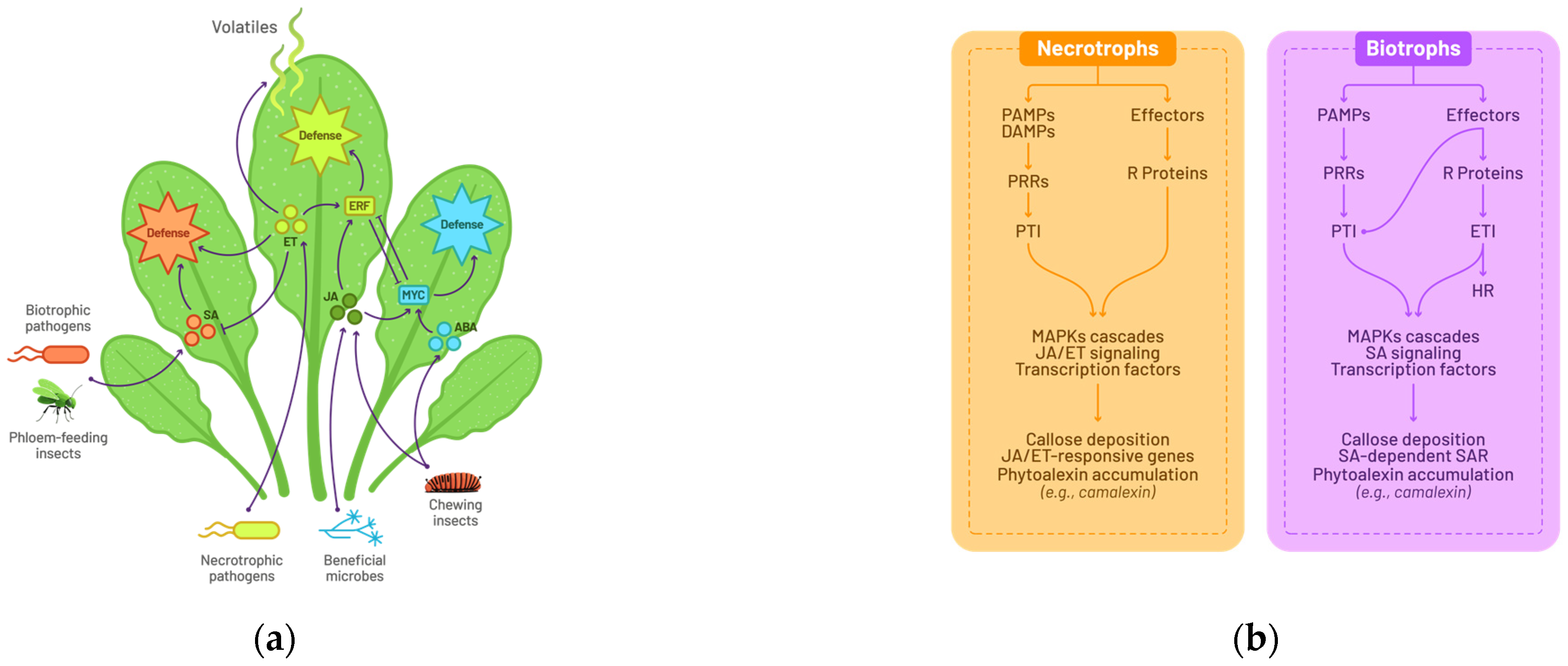
2. Plant Immune Response

2.1. A General Model for Plant–Pathogen Interactions

To safeguard themselves against attack from phytopathogens, plants have developed a sophisticated immune system. This can be classified as either innate immunity or induced immunity [12,13,27]. In host plants, the innate immune response is the first line of defense. Upon contact with external pathogens, the plant utilizes specialized Pathogen Recognition Receptors (PRRs) to identify specific molecules from the infectious agent, known as Pathogen/Microbe-Associated Molecular Patterns (P/MAMPs), as well as damage-associated molecular patterns (DAMPs). This process initiates a swift response designed to impede the pathogen’s proliferation, referred to as P/MAMP-triggered Immunity (PTI) [12,13]. However, if the pathogen manages to evade PTI through the deployment of virulent effector molecules, an induced immune response is activated. This response involves the recognition of the pathogen’s effector molecules by plant resistance proteins, such as nucleotide-binding site leucine-rich repeat (NBS-LRR) proteins. Subsequently, the Effector-Triggered Immunity (ETI) mechanism is activated to effectively detect and target the pathogen [12,18,19].
Once plant immunity is triggered, physical barriers are reinforced [12,20,24], such as callose deposition or cuticular wax accumulation. Subsequently, a complex network of signaling pathways orchestrated by the phytohormones salicylic acid, jasmonic acid, and ethylene comes into play [16,19,28]. These signaling pathways lead to an oxidative burst [16,19], the production of antimicrobial peptides (AMPs) [29], and the biosynthesis of secondary metabolites, such as indole-derived metabolites, including phytoalexins [19,30] (Figure 1b). The salicylic acid (SA) pathway has been associated with plant response to biotrophic microorganisms [16,31] (Figure 1b). The accumulation of SA induces ROS production, molecules that usually trigger the hypersensitive response (HR), a type of programmed cell death targeting infected cells, thus limiting the spread of pathogens [19,31,32]. At this stage, when a plant successfully overcomes an infection, it could develop systemic acquired resistance (SAR), making the plant ready to induce a resistance mechanism in case of a pathogenic attack. This phenomenon works like a memory, providing heightened resistance to subsequent attacks and ensuring extended and more robust protection against phytopathogens [33,34].
Other hormones such as ethylene and jasmonic acid, along with secondary metabolites, such as camalexin, brassinin, and glucosinolates (GSL), are produced in response to infection by necrotrophic and hemibiotrophic pathogens. These hormones act as signaling molecules to activate plant defenses, while the secondary metabolites directly inhibit pathogens or produce toxic derivatives crucial for combating the pathogens [18,28,30,35] (Figure 1b).
To gain a comprehensive understanding of plant defense mechanisms against microbial pathogens, the research emphasis has predominantly centered on plant susceptibility [12,13]. However, the intricate defense mechanisms implicated in a non-host plant remain unknown until now [27,36]. The crucial distinction in these mechanisms lies in the specificity of resistance, where host resistance is specifically tailored to particular pathogens, while non-host resistances (NHR) provide a more generalized defense. Although they share common mechanisms, such as the activation of signaling pathways and the production of antimicrobial compounds, host resistance is highly adaptive, providing specific immune responses, while NHR acts using broader defenses against various threats [36,37]. NHR comes down to a capability to recognize diverse microbial agents as non-hosts, granting plants broad and persistent resistance against a wide range of pathogens [27,36]. This phenomenon is believed to originate from a coevolutionary dynamic between plants and pathogens [13]. Plant immune resistance is influenced by a combination of abiotic factors, pathogen characteristics, host physiology, and plant tissues under attack. Certain studies proposed that this event arises either due to the inability of pathogens to suppress the PTI [13] or because one of the plant’s NB-LRR receptors recognizes the effector molecules by the pathogen, thus triggering ETI [13,27,37,38]. However, detailed NHR mechanisms are still not fully understood. Consequently, it is imperative to conduct further research to elucidate the molecular foundations of plant resistance and achieve a more comprehensive grasp of plant defense mechanisms [36].

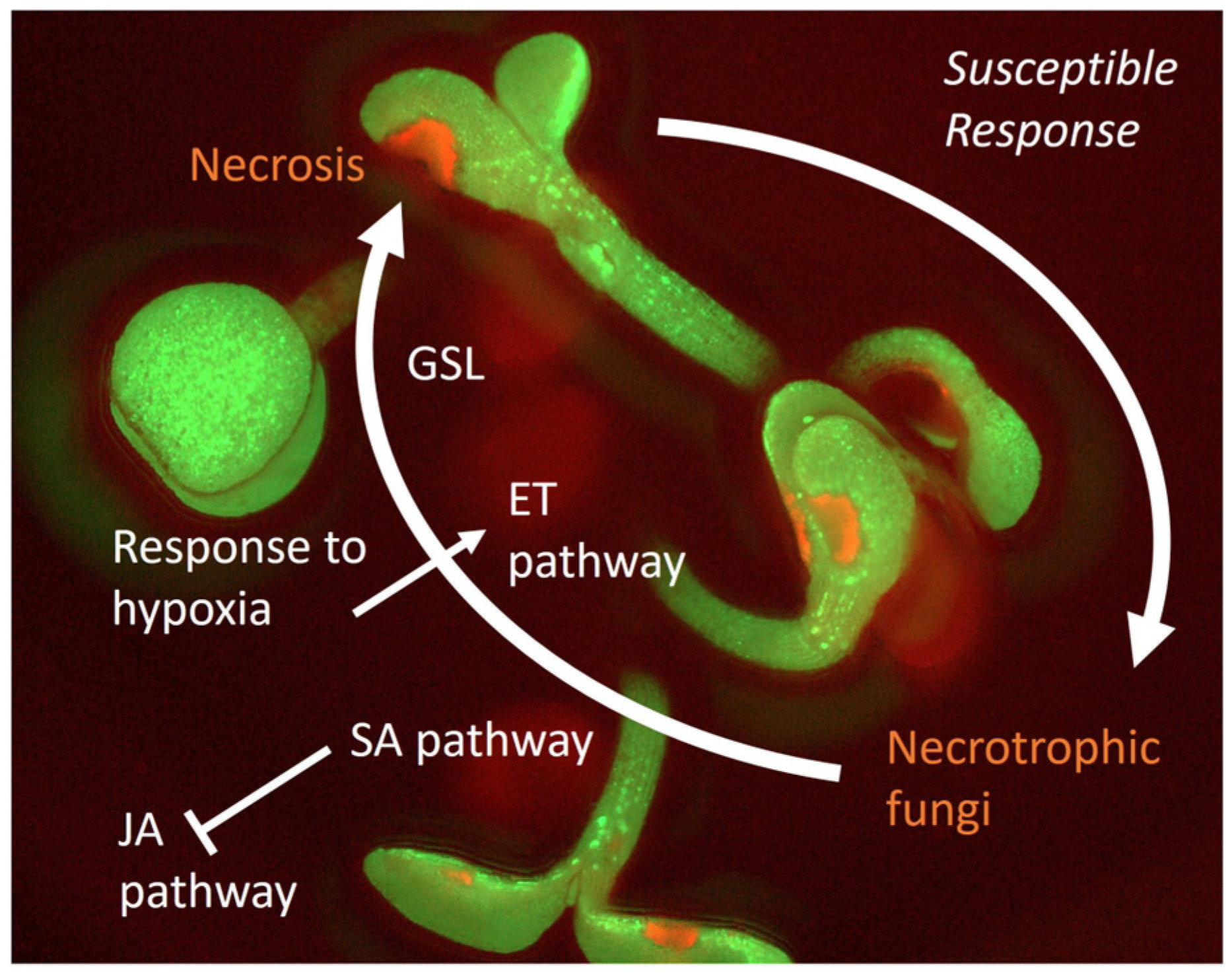
2.2. Susceptible Response and Plant Tolerance

Furthermore, the endophytic condition assumed by specific pathogenic agents in non-host plants engenders considerable scientific interest, given its potential association with the elicitation of susceptible responses (SRs) [27]. This scenario is marked by the host’s constrained capacity to counteract infection, fostering an environment conducive to pathogen proliferation and establishment inside the plant.
A pathogen strategy encompasses the manipulation of plant defenses and metabolic processes to its advantage. The pathogen must employ factors, such as effectors, phytohormones, and phytotoxins, to activate the so-called SR genes within the plant. This enables the pathogen to control both the plant’s defense mechanisms (physical and physiological) and metabolic processes, such as the reallocation of energy-rich compounds and the regulation of metal equilibrium, for its own benefit. Particularly in the case of phytopathogenic fungi, it is well documented that they have evolved not only to counteract the plant’s immune response but also to manipulate it for their own benefit. This includes their capacity to recognize plant-derived factors, such as chemical signals in the cuticle, leaf topology, and trichomes, thereby facilitating their successful colonization of the host [20]. Furthermore, certain seed-borne necrotrophic fungi, like Botrytis cinerea (B. cinerea), employ sophisticated systems to elude host recognition. They achieve this by hijacking or disrupting plant defense signaling and disabling defense mechanisms through the secretion of proteinaceous effectors and sRNA effectors, all geared towards promoting their own benefit (Table 1) [39,40]. In particular, B. cinerea adopts a multifaceted approach in hosts like tomato, tobacco, and Arabidopsis. It deploys effectors, such as BcSpl, from the Cerato-platanin protein family, and BcGs1, a glycoproteinaceous elicitor, to induce necrosis and programmed cell death (PCD) [41,42] with the purpose of generating dead tissue to grow through its necrotrophic properties. It is also described in the tomato host that B. cinerea manipulates SA accumulation via β-(1,3)(1,6)-d-glucan exopolysaccharide, suppressing JA-mediated signaling pathways and promoting disease development [43]. Moreover, it is documented in Arabidopsis that B. cinerea hijacks auxin metabolism through GH3.2 expression and Auxin Indole-3-Acetic Acid–Aspartic Acid–IAA–Aspartate (IAA-Asp) production, altering hormone dynamics to favor colonization and virulence [44]. In a strategic extension of its arsenal, B. cinerea also utilizes small RNAs to manipulate the RNA interference (RNAi) system in Arabidopsis and tomato. This tactic specifically targets key immune defense genes, such as peroxiredoxin (associated with oxidative stress), mitogen-activated protein kinases (MPK1, MPK2, MPKKK4), and a cell wall-associated kinase (WAK), silencing genes critical for immune defense and further enhancing its pathogenic capabilities [45].
Identifying genes that facilitate pathogen proliferation contributing to disease development is crucial for implementing genetic strategies focused on suppressing symptom-associated genes, leading to plants being better adapted to infections. Tolerant plants would be able to maintain their productivity levels, remain symptom-free, and potentially even benefit from the presence of endogenous pathogens [46]. Nevertheless, it is important to acknowledge that immunity defense mechanisms remain effective, and their efficacy may vary depending on the plant and pathogen species, as well as other environmental factors.

3. Alternaria brassicicola: An Important Seed-Borne Pathogen in Brassicaceae and a Model for Studying Pathogen Interactions with Seeds

Brassicaceae (Cruciferae) comprise approximately 4140 species, including the model species Arabidopsis thaliana (Arabidopsis) and economically important brassica vegetables, such as Brassica oleracea (broccoli, cauliflower, kale, brussels sprouts, and cabbage), Brassica rapa (vegetables, oilseeds, and forage), Brassica juncea (vegetables and mustard seeds), and Brassica napus (oilseeds) [47,48]. Brassicaceae species are susceptible to the seed-borne necrotrophic fungi Alternaria brassicicola (Berk.) Sacc. (referred to as Alternaria hereinafter), the causal agent of black spot disease. This seed-borne pathogen Alternaria (Ascomycota, Dothideomycetes, Pleosporales, and Pleosporaceae) is a common genus that frequently occurs as a saprotroph of decaying organic matter, but few species are plant pathogens [49,50]. Diseases caused by Alternaria species are widespread and can affect other crops, such as tomatoes, apples, carrots, and potatoes [51,52]. The economic impact on agricultural production is significant, as it affects the plant at all stages of development, from seed germination to post-harvest [22,50,53].
The infection process begins with fungal penetration into the plant through wounds or stomata [20]. Once inside the plant, the fungus secretes secondary metabolites, toxins [54,55], and a series of hydrolytic enzymes, such as proteases, lipases, and Cell Wall-Degrading Enzymes (CWDEs), leading to the death of plant tissues and facilitating the entry of the fungus into plant cells [49,51,56]. In leaf tissue, disease symptoms include leaf spots, yellow chlorotic halos, necrotic lesions, and leaf size reduction, resulting in a decrease in leaf photosynthetic capacity. In seedlings, a symptom known as damping off occurs and is characterized by the rotting of the plant’s stem base and roots, wilting, and causing the death of young plants as a consequence of the infection [49,51,56].
Alternaria is transmitted to the seed and is considered a seed-borne pathogen. At the seed level, Alternaria infection can adversely affect seed quality, germination rates, and crop yield. Several factors influence disease development and severity, including moisture, low light intensity, temperature, plant age, and conidia density [50,53]. The impact of infection on agricultural production and the search for solutions to reduce its harmful effects motivate the study of the mechanisms of interaction between plants of agronomic interest and Alternaria.

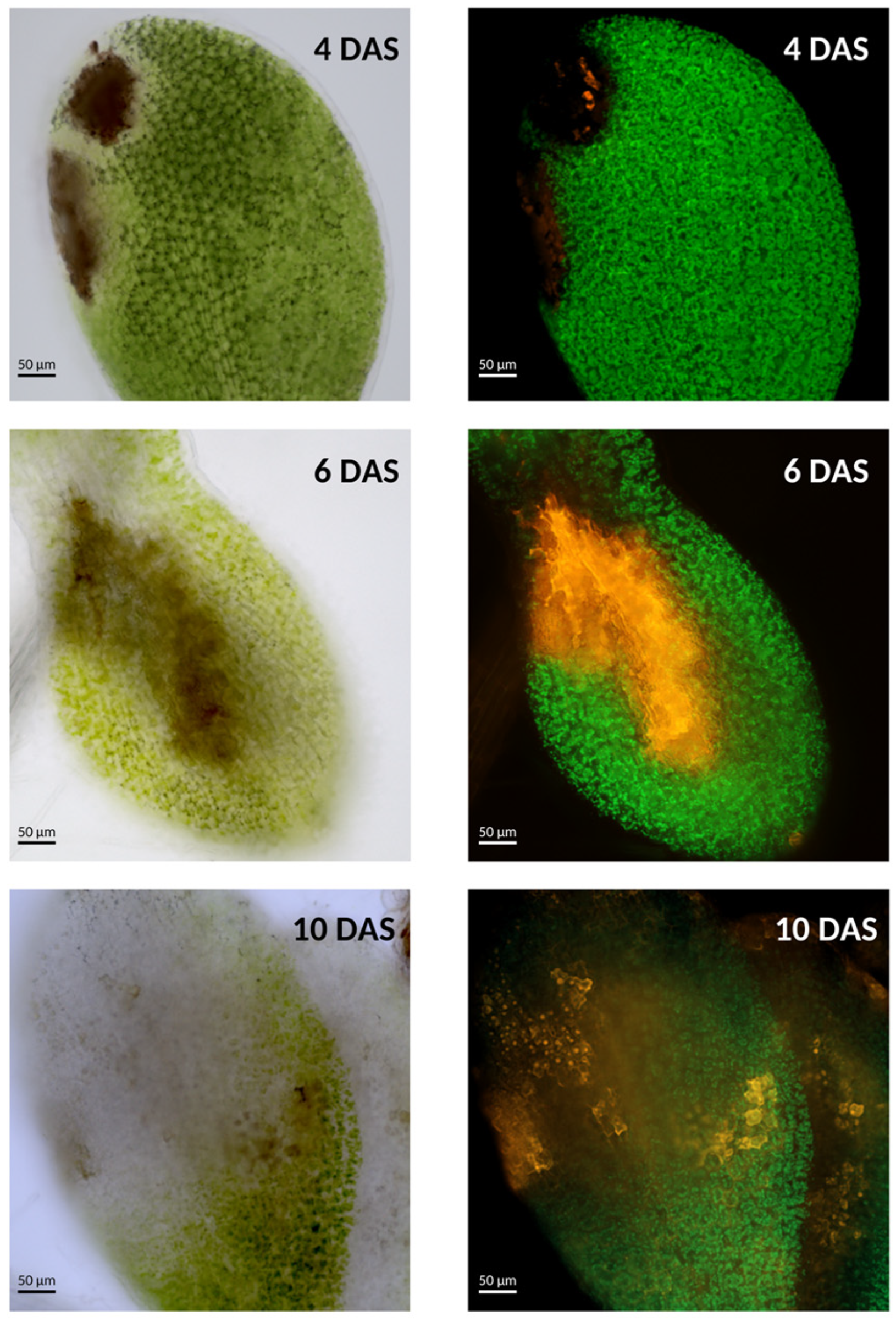
4. Seed Defense Mechanisms and Interactions with the Necrotrophic Seed-Borne Fungi Alternaria brassicicola

4.1. A silent Interaction with the Seed

Mechanisms of the transgenerational transmission of pathogens are poorly documented because few infection symptoms can be detected in seeds. Many seed-borne pathogens are considered non-hosts during seed development [57,58]. A striking example is the bacterial pathogen Xanthomonas campestris pv. campestris, known for attacking cruciferous plants [57]. In beans, this pathogen is transmitted from seed to seedling. Interestingly, its growth in these plants is like how it behaves in plants contaminated with X. citri pv. phaseoli var. fuscans (a bean-related pathogen). This does not significantly affect seed germination or seedling development, nor does it cause visible disease symptoms [57].

4.2. Interaction with the Necrotrophic Seed-Borne Fungi Alternaria brassicicola

Because seeds contribute to reproductive success, it is hypothesized that seed protection mechanisms would be part of their adaptive traits [25]. At the time of dissemination, the mature seed has acquired structural and metabolic properties [4,25] that could contribute to non-host resistance. The accumulation of secondary metabolites, such as phenolic compounds, in the mature seed [26] and the development of physical barriers, such as the seed coat [4,25], include some of those mechanisms. Also, pending seed germination and seedling establishment, these properties enable seeds to maintain their survival when challenged by abiotic and biotic pressures in their environment [7,25,26,59,60].
The endosperm acts not only in the control of seed germination but also as a protective barrier during germination [4,61]. Genes encoding detoxifying enzymes, such as glutathione S-transferases and peroxidases, play a crucial role in protecting the embryo against ROS that could result from pathogen interaction during seed germination. Genes related to plant–fungus interactions and hormonal metabolism also hold crucial roles in enhancing immunity and regulating plant growth. A transcriptomic study showed an over-expression of genes responsible for synthesizing salicylic acid (SA), indole-3-acetic acid (IAA), and amino synthetases (GH3) [61]. At the transcriptome level, the activation of secondary metabolites and defense response in the endosperm illustrates the activation of defense pathways during germination [61]. It is quite remarkable that certain mechanisms in seeds seem to differ from those in plants. Nevertheless, there are also instances of similar defense mechanisms being observed. For example, plant defense through polyphenol oxidase (PPO), a group of enzymes that catalyze the oxidation of hydroxy phenols, yielding products with antimicrobial properties. In dormant wild oats (Avena fatua L.), seeds attacked by Fusarium avenaceum trigger post-translational activation of the PPO, demonstrating the seed’s ability to induce enzymatic biochemical defenses against pathogenic fungi. This PPO activity was also found to be induced in non-living caryopsis cover tissues, such as lemma and pale [26].
The mature seed has developed resistance properties to face biotic and abiotic stress after dispersal. However, the immune responses of seeds to biotic interactions during germination have been infrequently studied, although this step is crucial to control seed-borne pathogen transmission to the new plant [22,25]. The model pathosystem Arabidopsis thaliana/Alternaria brassicicola has been recently used to describe gene regulation induced by the necrotrophic interaction during germination and seedling establishment [22,23,62].
RNA-seq data from Alternaria-infected and healthy Arabidopsis seeds were compared at different time points of the germination kinetics until the stage of seedling establishment. Although a transcriptomic study carried out on inoculated rosette leaves [21] highlighted an induction by Alternaria of the jasmonate pathway and camalexin metabolism (Figure 1b), the response of the germinating seed rather reflected an activation of the SA and ET pathways to the detriment of the jasmonate pathway [23]. It is documented that this type of unexpected response could constitute a diversion towards inappropriate defense pathways of the plant by the fungus to induce an SR favoring its development [39,40]. Additionally, genes related to response to hypoxia and indole-derived metabolites, such as GSL, were induced by Alternaria during germination [23]. Gene ontology enrichment analysis showed a stronger and more significant association among genes related to the response to hypoxia at an early development stage. Their fold enrichment decreased as seedling establishment progressed (3 Days After Sowing (DAS): 7.08, 6 DAS: 4.29, and 10 DAS: 3.07) [23]. This response is surprising, but it illustrates a competition for oxygen, which benefits the physiology of the early imbibed seed. Seed germination is well adapted to low oxygen levels [63], and these low oxygen levels limit the growth of the fungus. The induction at the RNA level of the indole GSL metabolism that was observed in Arabidopsis seeds [23] is well described [64].
Phenotyping analyses of mutant seeds deficient in the identified pathways via RNA-seq showed that the mutants deficient in ethylene response ein2 and etr1 exhibited a 21.9% and 55.4% lower rate of necrosis than the WT control, respectively. Also, GSL-deficient mutants (qk0 and gtr1gtr2) exhibited a 65.9% and 73.6% lower rate of necrosis than those in the control group, respectively. Noticeably, the GSL-deficient mutants displayed also a reduced Alternaria overgrowth [23], indicating that the lack of necrosis, usually mediated by the Alternaria-induced GSL pathway in WT, would be limiting colonization of the necrotrophic fungus. In this context, the induction of GSL metabolism could also contribute to the SR of the infected seed. The identified changes in gene regulation induced by Alternaria in germinating seeds provide an original working hypothesis where SR and defense mechanisms co-exist (Figure 2).
Scientific evidence reinforces the hypothesis that the seed–pathogen interactions differ from models described for the whole plants. It is notable that the transcriptome of infected tissues differed drastically between 6-day-old seedlings and 10-day-old seedlings [23,62]. Unpublished data obtained in our laboratory have shown that necrosis symptoms (Figure 3) in newly germinated seedlings (4 DAS) exhibit more distinct and localized patterns compared to older seedlings (after 10 days), where necrosis becomes more diffuse and widespread in the infected tissue.
Microscopic view (objective 20×) of necrotic symptoms on Alternaria-infected Arabidopsis cotyledons in visible and fluorescent light. The necrotic area is shown in red (false color) using a long-pass GFP filter. At an early germinative stage (4 DAS) necrosis occurs in well-defined areas, whereas at later stages (6 and 10 DAS), necrosis is diffuse. GSL mutants show no necrosis at 3 DAS. Scale bar: 50 µm.
It is hypothesized that at the germinative stage, the young seedling reacts to infection with a well-circumscribed HR. GLS-deficient mutants did not show any necrosis symptoms at the young seedling stage (4 DAS), suggesting that the observed HR would be mediated by the activation of the plant’s GSL pathway, whereas after 6 and 10 days of development, new necrosis was observed, including GSL mutants. The diffused appearance of the latter is attributed to cell death directly induced by the fungal penetration of plant tissues (unpublished data).

5. Conclusions and Future Perspectives

Despite significant progress in seed biology, there are still notable gaps in our understanding of seed immunity. Recent molecular and genetic analyses of Arabidopsis interactions with Alternaria during germination and seedling emergence have highlighted a particular defense mechanism and adaptive strategies employed when competitiveness is crucial for the survival of both the fungus and the germinating seedling [23]. The Arabidopsis thaliana/Alternaria brassicicola pathosystem illustrates that the fungus controls the hormonal response, while the seed attempts to kill the fungus by reducing oxygen. This type of model provides a basis for unraveling the intricate molecular dynamics of their interactions. This model not only provides a basis for unraveling the intricate molecular dynamics of their interactions but also represents a valuable framework for understanding the complex mechanisms of pathogen resistance and host response [22,23]. Specifically, further detailing the contribution of GSL metabolism to necrosis symptoms and Alternaria colonization in germinating seeds [23] is necessary. This exploration could offer a strategic lever for controlling seed tolerance to necrotrophic attacks and effectively managing transgenerational pathogen propagation.
Arabidopsis is a widely studied model species that presents advantages of a wide diversity, including well-characterized genetic, genomic, and phenotypic resources [65,66,67]. While Arabidopsis may not hold direct economic significance, its close phylogenetic relationship and shared genetic and molecular traits with Brassica species [68] facilitate the transfer of valuable knowledge regarding seed–fungal interactions to benefit Brassica crops. To further explore this, we plan to leverage Arabidopsis gene expression profiles using the Weighted Gene Correlation Network Analysis (WGCNA) approach [69]. Future analyses should involve conducting transcriptomic studies on deficient mutants in GSL and camalexin within the context of seeds infected by Alternaria during germination. These upcoming investigations aim to unveil co-expression networks, grouping genes based on similar expression patterns and phenotypic contributions. The WGCNA approach would be helpful to identify master regulatory genes controlling seed tolerance and provide candidate genes to search orthologs in brassica crops, which contribute to seed resistance in crops, such Brassica vegetables.
The investigation of seed defense mechanisms against pathogenic fungi has become essential in the pursuit of strategies to ensure seed health and mitigate extensive losses in agricultural fields. Comparative genomics will provide deeper insights into plant–pathogen interactions. This knowledge is needed to develop genetic resistance or to better manage diseases in brassica crops.

Author Contributions

Conceptualization, P.G., T.A., A.M.V.-P., A.R. and N.G.; methodology, P.G., T.A., A.M.V.-P., S.A., A.R., N.G., C.R.--P. and M.O.-C.; validation, P.G. and T.A.; formal analysis, M.O.-C., S.A. and C.R.--P.; investigation, P.G., A.R., C.R.--P., N.G. and M.O.-C.; writing—original draft preparation, M.O-C.; writing—review and editing, P.G., T.A. and M.O.-C.; visualization, P.G., T.A. and A.M.V.-P.; supervision, P.G., T.A. and A.M.V.-P.; project administration, P.G.; funding acquisition, P.G. All authors have read and agreed to the published version of the manuscript.

Funding

This research was funded by the Programme Prioritaire de Recherche (PPR) de l’ANR, Cultiver et Protéger Autrement (CPA), project number IA-20-PCPA-0009. More details can be found at: SUCSEED, https://anr.fr/ProjetIA-20-PCPA-0009 (accessed on 10 April 2024).

Acknowledgments

We would like to thank the FUNGISEM team for their support in this investigation and thank Luz Marina Melgarejo, Adriana Tofiño, and Nubia Velazquez for their productive discussion about this project. We would like to express our gratitude to Jerome Verdier for his invaluable assistance in conducting experimental analyses and interpreting data. We thank Fabienne Simonneau of the IMAC imaging plateau of the SFR 4207 QUASAV.

Conflicts of Interest

The authors declare no conflicts of interest. The funders had no role in the design of the study; in the collection, analyses, or interpretation of data; in the writing of the manuscript; or in the decision to publish the results.

References

  1. Bewley, J.D.; Bradford, K.J.; Hilhorst, H.W.M.; Nonogaki, H. Seeds: Physiology of Development, Germination and Dormancy, 3rd ed.; Springer: New York, NY, USA, 2013. [Google Scholar] [CrossRef]
  2. Gupta, S.; Van Staden, J.; Doležal, K. An Understanding of the Role of Seed Physiology for Better Crop Productivity and Food Security. Plant Growth Regul. 2022, 97, 171–173. [Google Scholar] [CrossRef]
  3. Nonogaki, H. Seed Biology Updates–Highlights and New Discoveries in Seed Dormancy and Germination Research. Front. Plant Sci. 2017, 8, 524. [Google Scholar] [CrossRef]
  4. Nonogaki, H. Seed Germination and Dormancy: The Classic Story, New Puzzles, and Evolution. J. Integr. Plant Biol. 2019, 61, 541–563. [Google Scholar] [CrossRef] [PubMed]
  5. Nonogaki, H. Seed dormancy and germination—Emerging mechanisms and new hypotheses. Front. Plant Sci. 2014, 5, 233. [Google Scholar] [CrossRef]
  6. Nelson, E.B. The Seed Microbiome: Origins, Interactions, and Impacts. Plant Soil. 2018, 422, 7–34. [Google Scholar] [CrossRef]
  7. War, A.F.; Bashir, I.; Reshi, Z.A.; Kardol, P.; Rashid, I. Insights into the Seed Microbiome and Its Ecological Significance in Plant Life. Microbiol. Res. 2023, 269, 127318. [Google Scholar] [CrossRef]
  8. Buitink, J.; Leprince, O. Advances in Seed Science and Technology for More Sustainable Crop Production, 1st ed.; Buitink, J., Leprince, O., Eds.; Burleigh Dodds Series in Agricultural Science; Burleigh Dodds Science Publishing: Cambridge, UK, 2022. [Google Scholar] [CrossRef]
  9. Dell’Olmo, E.; Tiberini, A.; Sigillo, L. Leguminous Seedborne Pathogens: Seed Health and Sustainable Crop Management. Plants 2023, 12, 2040. [Google Scholar] [CrossRef]
  10. Cram, M.M.; Fraedrich, S.W. Seed Diseases and Seedborne Pathogens of North America. Tree Plant. Notes 2010, 53, 35–44. [Google Scholar]
  11. Kumar, R.; Gupta, A. Seed-Borne Diseases of Agricultural Crops: Detection, Diagnosis & Management; Kumar, R., Gupta, A., Eds.; Springer: Singapore, 2020. [Google Scholar] [CrossRef]
  12. Dodds, P.N.; Rathjen, J.P. Plant Immunity: Towards an Integrated View of Plant–Pathogen Interactions. Nat. Rev. Genet. 2010, 11, 539–548. [Google Scholar] [CrossRef]
  13. Jones, J.D.G.; Dangl, J.L. The Plant Immune System. Nature 2006, 444, 323–329. [Google Scholar] [CrossRef]
  14. Roberts Steven, J.; Aoife, O.; John, W. Brassica Diseases. Agriculture and Horticulture Development Board. Available online: https://bit.ly/3uOmXi9 (accessed on 26 February 2024).
  15. Gullino, M.L.; Munkvold, G. (Eds.) Global Perspectives on the Health of Seeds and Plant Propagation Material; Springer: Dordrecht, The Netherlands, 2014; Volume 6. [Google Scholar] [CrossRef]
  16. Glazebrook, J. Contrasting Mechanisms of Defense against Biotrophic and Necrotrophic Pathogens. Annu. Rev. Phytopathol. 2005, 43, 205–227. [Google Scholar] [CrossRef] [PubMed]
  17. Divon, H.H.; Fluhr, R. Nutrition Acquisition Strategies during Fungal Infection of Plants. FEMS Microbiol. Lett. 2007, 266, 65–74. [Google Scholar] [CrossRef] [PubMed]
  18. Ghozlan, M.H.; EL-Argawy, E.; Tokgöz, S.; Lakshman, D.K.; Mitra, A. Plant Defense against Necrotrophic Pathogens. Am. J. Plant Sci. 2020, 11, 2122–2138. [Google Scholar] [CrossRef]
  19. Bolton, M.D. Primary Metabolism and Plant Defense—Fuel for the Fire. Mol. Plant-Microbe Interact. 2009, 22, 487–497. [Google Scholar] [CrossRef] [PubMed]
  20. Łaźniewska, J.; Macioszek, V.K.; Kononowicz, A.K. Plant-Fungus Interface: The Role of Surface Structures in Plant Resistance and Susceptibility to Pathogenic Fungi. Physiol. Mol. Plant Pathol. 2012, 78, 24–30. [Google Scholar] [CrossRef]
  21. van Wees, S.C.M.; Chang, H.-S.; Zhu, T.; Glazebrook, J. Characterization of the Early Response of Arabidopsis to Alternaria Brassicicola Infection Using Expression Profiling. Plant Physiol. 2003, 132, 606–617. [Google Scholar] [CrossRef] [PubMed]
  22. Pochon, S.; Terrasson, E.; Guillemette, T.; Iacomi-Vasilescu, B.; Georgeault, S.; Juchaux, M.; Berruyer, R.; Debeaujon, I.; Simoneau, P.; Campion, C. The Arabidopsis Thaliana-Alternaria Brassicicola Pathosystem: A Model Interaction for Investigating Seed Transmission of Necrotrophic Fungi. Plant Methods 2012, 8, 16. [Google Scholar] [CrossRef] [PubMed]
  23. Ortega-Cuadros, M.; De Souza, T.L.; Berruyer, R.; Aligon, S.; Pelletier, S.; Renou, J.-P.; Arias, T.; Campion, C.; Guillemette, T.; Verdier, J.; et al. Seed Transmission of Pathogens: Non-Canonical Immune Response in Arabidopsis Germinating Seeds Compared to Early Seedlings against the Necrotrophic Fungus Alternaria Brassicicola. Plants 2022, 11, 1708. [Google Scholar] [CrossRef] [PubMed]
  24. Łaźniewska, J.; Macioszek, V.K.; Lawrence, C.B.; Kononowicz, A.K. Fight to the Death: Arabidopsis Thaliana Defense Response to Fungal Necrotrophic Pathogens. Acta Physiol. Plant 2010, 32, 1–10. [Google Scholar] [CrossRef]
  25. Dalling, J.W.; Davis, A.S.; Arnold, A.E.; Sarmiento, C.; Zalamea, P.-C. Extending Plant Defense Theory to Seeds. Annu. Rev. Ecol. Evol. Syst. 2020, 51, 123–141. [Google Scholar] [CrossRef]
  26. Fuerst, E.P.; Okubara, P.A.; Anderson, J.V.; Morris, C.F. Polyphenol Oxidase as a Biochemical Seed Defense Mechanism. Front. Plant Sci. 2014, 5, 117840. [Google Scholar] [CrossRef]
  27. Panstruga, R.; Moscou, M.J. What Is the Molecular Basis of Nonhost Resistance? Mol. Plant-Microbe Interact. 2020, 33, 1253–1264. [Google Scholar] [CrossRef]
  28. Macioszek, V.K.; Jęcz, T.; Ciereszko, I.; Kononowicz, A.K. Jasmonic Acid as a Mediator in Plant Response to Necrotrophic Fungi. Cells 2023, 12, 1027. [Google Scholar] [CrossRef]
  29. Li, J.; Hu, S.; Jian, W.; Xie, C.; Yang, X. Plant Antimicrobial Peptides: Structures, Functions, and Applications. Bot. Stud. 2021, 62, 5. [Google Scholar] [CrossRef]
  30. Stotz, H.U.; Sawada, Y.; Shimada, Y.; Hirai, M.Y.; Sasaki, E.; Krischke, M.; Brown, P.D.; Saito, K.; Kamiya, Y. Role of Camalexin, Indole Glucosinolates, and Side Chain Modification of Glucosinolate-Derived Isothiocyanates in Defense of Arabidopsis against Sclerotinia sclerotiorum. Plant J. 2011, 67, 81–93. [Google Scholar] [CrossRef]
  31. L Schlaich, N. Arabidopsis Thaliana-the Model Plant to Study Host-Pathogen Interactions. Curr. Drug Targets 2011, 12, 955–966. [Google Scholar] [CrossRef]
  32. Balint-Kurti, P. The Plant Hypersensitive Response: Concepts, Control and Consequences. Mol. Plant Pathol. 2019, 20, 1163–1178. [Google Scholar] [CrossRef]
  33. Ádám, A.; Nagy, Z.; Kátay, G.; Mergenthaler, E.; Viczián, O. Signals of Systemic Immunity in Plants: Progress and Open Questions. Int. J. Mol. Sci. 2018, 19, 1146. [Google Scholar] [CrossRef]
  34. Conrath, U. Systemic Acquired Resistance. Plant Signal Behav. 2006, 1, 179–184. [Google Scholar] [CrossRef]
  35. Sellam, A.; Iacomi-Vasilescu, B.; Hudhomme, P.; Simoneau, P. In Vitro Antifungal Activity of Brassinin, Camalexin and Two Isothiocyanates against the Crucifer Pathogens Alternaria Brassicicola and Alternaria Brassicae. Plant Pathol. 2007, 56, 296–301. [Google Scholar] [CrossRef]
  36. Gill, U.S.; Lee, S.; Mysore, K.S. Host Versus Nonhost Resistance: Distinct Wars with Similar Arsenals. Phytopathology 2015, 105, 580–587. [Google Scholar] [CrossRef]
  37. Schulze-Lefert, P.; Panstruga, R. A Molecular Evolutionary Concept Connecting Nonhost Resistance, Pathogen Host Range, and Pathogen Speciation. Trends Plant Sci. 2011, 16, 117–125. [Google Scholar] [CrossRef]
  38. Nürnberger, T.; Lipka, V. Non-host Resistance in Plants: New Insights into an Old Phenomenon. Mol. Plant Pathol. 2005, 6, 335–345. [Google Scholar] [CrossRef]
  39. Patkar, R.N.; Naqvi, N.I. Fungal Manipulation of Hormone-Regulated Plant Defense. PLoS Pathog. 2017, 13, e1006334. [Google Scholar] [CrossRef]
  40. Shao, D.; Smith, D.L.; Kabbage, M.; Roth, M.G. Effectors of Plant Necrotrophic Fungi. Front. Plant Sci. 2021, 12, 995. [Google Scholar] [CrossRef]
  41. Frías, M.; González, C.; Brito, N. BcSpl1, a Cerato-Platanin Family Protein, Contributes to Botrytis Cinerea Virulence and Elicits the Hypersensitive Response in the Host. New Phytol. 2011, 192, 483–495. [Google Scholar] [CrossRef]
  42. Zhang, Y.; Zhang, Y.; Qiu, D.; Zeng, H.; Guo, L.; Yang, X. BcGs1, a Glycoprotein from Botrytis Cinerea, Elicits Defence Response and Improves Disease Resistance in Host Plants. Biochem. Biophys. Res. Commun. 2015, 457, 627–634. [Google Scholar] [CrossRef]
  43. El Oirdi, M.; El Rahman, T.A.; Rigano, L.; El Hadrami, A.; Rodriguez, M.C.; Daayf, F.; Vojnov, A.; Bouarab, K. Botrytis Cinerea Manipulates the Antagonistic Effects between Immune Pathways to Promote Disease Development in Tomato. Plant Cell 2011, 23, 2405–2421. [Google Scholar] [CrossRef]
  44. González-Lamothe, R.; El Oirdi, M.; Brisson, N.; Bouarab, K. The Conjugated Auxin Indole-3-Acetic Acid–Aspartic Acid Promotes Plant Disease Development. Plant Cell 2012, 24, 762–777. [Google Scholar] [CrossRef]
  45. Weiberg, A.; Wang, M.; Lin, F.-M.; Zhao, H.; Zhang, Z.; Kaloshian, I.; Huang, H.-D.; Jin, H. Fungal Small RNAs Suppress Plant Immunity by Hijacking Host RNA Interference Pathways. Science 2013, 342, 118–123. [Google Scholar] [CrossRef]
  46. Gorshkov, V.; Tsers, I. Plant Susceptible Responses: The Underestimated Side of Plant–Pathogen Interactions. Biol. Rev. 2022, 97, 45–66. [Google Scholar] [CrossRef]
  47. German, D.A.; Hendriks, K.P.; Koch, M.A.; Lens, F.; Lysak, M.A.; Bailey, C.D.; Mummenhoff, K.; Al-Shehbaz, I.A. An Updated Classification of the Brassicaceae (Cruciferae). PhytoKeys 2023, 220, 127. [Google Scholar] [CrossRef]
  48. Rakow, G. Species Origin and Economic Importance of Brassica; Springer: Berlin/Heidelberg, Germany, 2004; Volume 54, pp. 3–11. [Google Scholar] [CrossRef]
  49. Cho, Y. How the Necrotrophic Fungus Alternaria Brassicicola Kills Plant Cells Remains an Enigma. Eukaryot. Cell 2015, 14, 335–344. [Google Scholar] [CrossRef]
  50. Dharmendra, K.; Neelam, M.; Yashwant, K.B.; Ajay, K.; Kamlesh, K.; Kalpana, S.; Gireesh, C.; Chanda, K.; Sushil, K.S.; Raj, K.M.; et al. Alternaria Blight of Oilseed Brassicas: A Comprehensive Review. Afr. J. Microbiol. Res. 2014, 8, 2816–2829. [Google Scholar] [CrossRef]
  51. Ali, S.; Tyagi, A.; Rajarammohan, S.; Mir, Z.A.; Bae, H. Revisiting Alternaria-Host Interactions: New Insights on Its Pathogenesis, Defense Mechanisms and Control Strategies. Sci. Hortic. 2023, 322, 112424. [Google Scholar] [CrossRef]
  52. Mamgain, A.; Roychowdhury, R.; Tah, J. Alternaria Pathogenicity and Its Strategic Controls. Res. J. Biol. 2013, 1, 1–9. [Google Scholar]
  53. Nowicki, M.; Nowakowska, M.; Niezgoda, A.; Kozik, E. Alternaria Black Spot of Crucifers: Symptoms, Importance of Disease, and Perspectives of Resistance Breeding. J. Fruit. Ornam. Plant Res. 2012, 76, 5–19. [Google Scholar] [CrossRef]
  54. Meena, M.; Gupta, S.K.; Swapnil, P.; Zehra, A.; Dubey, M.K.; Upadhyay, R.S. Alternaria Toxins: Potential Virulence Factors and Genes Related to Pathogenesis. Front. Microbiol. 2017, 8, 1451. [Google Scholar] [CrossRef]
  55. Pedras, M.S.C.; Chumala, P.B.; Jin, W.; Islam, M.S.; Hauck, D.W. The Phytopathogenic Fungus Alternaria Brassicicola: Phytotoxin Production and Phytoalexin Elicitation. Phytochemistry 2009, 70, 394–402. [Google Scholar] [CrossRef]
  56. Lawrence, C.B.; Mitchell, T.K.; Craven, K.D.; Cho, Y.-R.; Cramer, R.A.; Kim, K.-H. At Death’s Door: Alternaria Pathogenicity Mechanisms. Plant Pathol. J. 2008, 24, 101–111. [Google Scholar] [CrossRef]
  57. Darrasse, A.; Darsonval, A.; Boureau, T.; Brisset, M.-N.; Durand, K.; Jacques, M.-A. Transmission of Plant-Pathogenic Bacteria by Nonhost Seeds without Induction of an Associated Defense Reaction at Emergence. Appl. Environ. Microbiol. 2010, 76, 6787–6796. [Google Scholar] [CrossRef]
  58. Darsonval, A.; Darrasse, A.; Meyer, D.; Demarty, M.; Durand, K.; Bureau, C.; Manceau, C.; Jacques, M.-A. The Type III Secretion System of Xanthomonas Fuscans Subsp. Fuscans Is Involved in the Phyllosphere Colonization Process and in Transmission to Seeds of Susceptible Beans. Appl. Environ. Microbiol. 2008, 74, 2669–2678. [Google Scholar] [CrossRef]
  59. Debeaujon, I.; Léon-Kloosterziel, K.M.; Koornneef, M. Influence of the Testa on Seed Dormancy, Germination, and Longevity in Arabidopsis. Plant Physiol. 2000, 122, 403–414. [Google Scholar] [CrossRef]
  60. Smýkal, P.; Vernoud, V.; Blair, M.W.; Soukup, A.; Thompson, R.D. The Role of the Testa during Development and in Establishment of Dormancy of the Legume Seed. Front. Plant Sci. 2014, 5, 351. [Google Scholar] [CrossRef]
  61. Endo, A.; Tatematsu, K.; Hanada, K.; Duermeyer, L.; Okamoto, M.; Yonekura-Sakakibara, K.; Saito, K.; Toyoda, T.; Kawakami, N.; Kamiya, Y.; et al. Tissue-Specific Transcriptome Analysis Reveals Cell Wall Metabolism, Flavonol Biosynthesis and Defense Responses Are Activated in the Endosperm of Germinating Arabidopsis Thaliana Seeds. Plant Cell Physiol. 2012, 53, 16–27. [Google Scholar] [CrossRef]
  62. Ortega-Cuadros, M.; Chir, L.; Aligon, S.; Arias, T.; Verdier, J.; Grappin, P. Dual-Transcriptomic Datasets Evaluating the Effect of the Necrotrophic Fungus Alternaria Brassicicola on Arabidopsis Germinating Seeds. Data Brief. 2022, 44, 108530. [Google Scholar] [CrossRef]
  63. Chung, H.; Lee, Y.-H. Hypoxia: A Double-Edged Sword During Fungal Pathogenesis? Front. Microbiol. 2020, 11, 1920. [Google Scholar] [CrossRef]
  64. Tao, H.; Miao, H.; Chen, L.; Wang, M.; Xia, C.; Zeng, W.; Sun, B.; Zhang, F.; Zhang, S.; Li, C.; et al. WRKY33-mediated Indolic Glucosinolate Metabolic Pathway Confers Resistance against Alternaria Brassicicola in Arabidopsis and Brassica Crops. J. Integr. Plant Biol. 2022, 64, 1007–1019. [Google Scholar] [CrossRef]
  65. Horton, M.W.; Hancock, A.M.; Huang, Y.S.; Toomajian, C.; Atwell, S.; Auton, A.; Muliyati, N.W.; Platt, A.; Sperone, F.G.; Vilhjálmsson, B.J.; et al. Genome-Wide Patterns of Genetic Variation in Worldwide Arabidopsis Thaliana Accessions from the RegMap Panel. Nat. Genet. 2012, 44, 212–216. [Google Scholar] [CrossRef]
  66. Koornneef, M.; Alonso-Blanco, C.; Vreugdenhil, D. Naturally Occurring Genetic Variation in Arabidopsis Thaliana. Annu. Rev. Plant Biol. 2004, 55, 141–172. [Google Scholar] [CrossRef]
  67. Somerville, C.; Koornneef, M. A Fortunate Choice: The History of Arabidopsis as a Model Plant. Nat. Rev. Genet. 2002, 3, 883–889. [Google Scholar] [CrossRef] [PubMed]
  68. Nikolov, L.A.; Shushkov, P.; Nevado, B.; Gan, X.; Al-Shehbaz, I.A.; Filatov, D.; Bailey, C.D.; Tsiantis, M. Resolving the Backbone of the Brassicaceae Phylogeny for Investigating Trait Diversity. New Phytol. 2019, 222, 1638–1651. [Google Scholar] [CrossRef] [PubMed]
  69. Langfelder, P.; Horvath, S. WGCNA: An R Package for Weighted Correlation Network Analysis. BMC Bioinform. 2008, 9, 559. [Google Scholar] [CrossRef] [PubMed]
Figure 1. General model of plant defense signaling and immune responses. (a) Overview of the plant’s signal detection mechanisms involving interactions with herbivorous, beneficial, and pathogenic microorganisms. (b) The plant immune response against necrotrophs and biotrophs involves diverse defenses, including physical barriers, hormonal signaling, and antimicrobial metabolites. Swift responses, initiated by Pathogen Recognition Receptors (PRRs), progress from P/MAMP-triggered Immunity (PTI) to Effector-Triggered Immunity (ETI) upon evasion by pathogens.
Seeds 03 00017 g001
Figure 2. Working model illustrating a susceptible response (SR) to Alternaria in germinating seeds. Proposed regulations have been provided by transcriptomic and mutant analyses [1]. Alternaria induced at the RNA level salicylic acid (SA) pathway and the indole glucosinolate (GSL) metabolism, as well as ethylene (ET) signaling and the response to hypoxia. The SA pathway is usually induced to the detriment of jasmonic acid (JA) response, which has not been found to be repressed in the transcriptomic data. Mutant analyses illustrated the contribution of GSL and response to ET in the occurrence of necrosis symptoms that are needed for Alternaria development. Responses to hypoxia have been also documented to induce ET signaling. The text with white font describes the responses of the seed illustrated by the transcriptomic analysis.
Seeds 03 00017 g002
Figure 3. Necrosis profiles at 4, 6, and 10 Days After Sowing (DAS).
Seeds 03 00017 g003
Table 1. Plant responses induced by Botrytis cinerea that illustrate susceptible response.
HostPathogen-Induced Plant Response PathwayReference
Solanum lycopersicum (tomato), Nicotiana tabacum (tobacco), and Arabidopsis thaliana (Arabidopsis)Programmed cell death (PCD)Frías et al. [41]
Zhang et al. [42]
Solanum lycopersicum (tomato)SA pathwayEl Oirdi M et al. [43]
Arabidopsis thaliana (Arabidopsis)Auxin pathwayGonzález-Lamothe R et al. [44]
Solanum lycopersicum (tomato) and Arabidopsis thaliana (Arabidopsis)Peroxiredoxin (oxidative stress-related gene), mitogen-activated protein kinases (MPK1, MPK2, MPKKK4), and a cell wall-associated kinase (WAK)Weiberg et al. [45]
Disclaimer/Publisher’s Note: The statements, opinions and data contained in all publications are solely those of the individual author(s) and contributor(s) and not of MDPI and/or the editor(s). MDPI and/or the editor(s) disclaim responsibility for any injury to people or property resulting from any ideas, methods, instructions or products referred to in the content.

