

Topical Advisory Panel applications are now closed. Please contact the Editorial Office with any queries.
Journal Description
Diagnostics
Diagnostics
is an international, peer-reviewed, open access journal on medical diagnosis published semimonthly online by MDPI. The British Neuro-Oncology Society (BNOS), the International Society for Infectious Diseases in Obstetrics and Gynaecology (ISIDOG) and the Swiss Union of Laboratory Medicine (SULM) are affiliated with Diagnostics and their members receive a discount on the article processing charges.
- Open Access— free for readers, with article processing charges (APC) paid by authors or their institutions.
- High Visibility: indexed within Scopus, SCIE (Web of Science), PubMed, PMC, Embase, Inspec, CAPlus / SciFinder, and other databases.
- Journal Rank: JCR - Q1 (Medicine, General and Internal) / CiteScore - Q2 (Internal Medicine)
- Rapid Publication: manuscripts are peer-reviewed and a first decision is provided to authors approximately 21.6 days after submission; acceptance to publication is undertaken in 2.7 days (median values for papers published in this journal in the second half of 2025).
- Recognition of Reviewers: reviewers who provide timely, thorough peer-review reports receive vouchers entitling them to a discount on the APC of their next publication in any MDPI journal, in appreciation of the work done.
- Companion journals for Diagnostics include: LabMed and AI in Medicine.
Impact Factor:
3.3 (2024);
5-Year Impact Factor:
3.3 (2024)
Latest Articles
Predictive Factors of Functional Prognosis in Patients with Rhegmatogenous Retinal Detachment Treated by Pars Plana Vitrectomy—A Retrospective Study
Diagnostics 2026, 16(11), 1696; https://doi.org/10.3390/diagnostics16111696 (registering DOI) - 30 May 2026
Abstract
Background/Objectives: Rhegmatogenous retinal detachment (RRD) is a surgical emergency that causes vision loss if not properly treated. Pars plana vitrectomy (PPV) is the preferred procedure for most complex RRD cases, with a high success rate. This study aimed to evaluate the significance
[...] Read more.
Background/Objectives: Rhegmatogenous retinal detachment (RRD) is a surgical emergency that causes vision loss if not properly treated. Pars plana vitrectomy (PPV) is the preferred procedure for most complex RRD cases, with a high success rate. This study aimed to evaluate the significance of predictive factors and their impact on postoperative best corrected visual acuity (BCVA) in patients undergoing PPV for RRD. Methods: In this retrospective study, 136 eyes from 135 patients who underwent PPV for RRD were examined. A robust fitting algorithm for linear models was used to assess the influence of preoperative patient-related factors on the functional outcomes of PPV for RRD. Results: Various factors were analyzed, ranging from demographic parameters, such as gender and residency, to more complex ocular findings, including BCVA, duration of symptoms, description of the RRD, retinal tears, macular status, presence of proliferative vitreoretinopathy (PVR), epiretinal membrane, or macular hole. Conclusions: Duration of symptoms, phacoemulsification during follow-up, and preoperative BCVA showed a statistically significant correlation with postoperative BCVA. Lens status, macular status, extent of retinal detachment, number of tears, and PVR did not influence the postoperative outcome.
Full article
(This article belongs to the Special Issue Diagnosing, Treating, and Preventing Eye Diseases)
Open AccessReview
First-Trimester Ultrasound: A Comparative Review of Guidelines
by
Eirini Boureka, Ioannis Tsakiridis, Georgios Michos, Anastasios Liberis, Sonia Giouleka, Apostolos Mamopoulos, Ioannis Kalogiannidis and Themistoklis Dagklis
Diagnostics 2026, 16(11), 1695; https://doi.org/10.3390/diagnostics16111695 (registering DOI) - 30 May 2026
Abstract
First-trimester sonographic examination remains a fundamental part of antenatal care, providing crucial information for the well-being of both the mother and fetus and leading to the best possible perinatal outcomes. This study aimed to review and compare the most recently published guidelines on
[...] Read more.
First-trimester sonographic examination remains a fundamental part of antenatal care, providing crucial information for the well-being of both the mother and fetus and leading to the best possible perinatal outcomes. This study aimed to review and compare the most recently published guidelines on first-trimester ultrasound. Therefore, a descriptive review of guidelines from the American Institute of Ultrasound in Medicine (AIUM), the Australasian Society of Ultrasound in Medicine (ASUM), the Association of the Scientific Medical Societies in Germany (AWMF), the International Society of Ultrasound in Obstetrics and Gynecology (ISUOG), the Society of Obstetrician and Gynecologists of Canada (SOGC) and the World Association of Perinatal Medicine (WAPM) regarding first-trimester scans was conducted. There is a consensus regarding the main parameters that should be evaluated, the qualifications of the examiner and specifications of the ultrasound machine, as well as the importance of informed consent. Additionally, the importance of careful visualization of fetal anatomy is discussed, with minor discrepancies regarding the appropriate protocol used. The use of combined first-trimester examination is encouraged by all the reviewed medical societies, although cell-free DNA testing is addressed only by a few, with similar indications. Controversy exists regarding the indications and appropriate gestational age at which the first-trimester scan should be performed, as well as the proper establishment of pregnancy dating. Notably, recommendations regarding fetal growth restriction (FGR) and diagnostic invasive procedures are made only by few medical societies, with the AWMF and SOGC addressing screening for FGR. Furthermore, invasive diagnostic testing is discussed by the AIUM, AWMF and SOGC, with differentiations among them regarding the indications for pursuing such procedures. First-trimester sonographic examination is essential for assessing fetal viability, establishing accurate pregnancy dating, evaluating fetal and maternal anatomy and calculating the risk of various fetal and maternal conditions. The implementation of evidence-based, unified protocols would advance both maternal and fetal outcomes.
Full article
(This article belongs to the Special Issue Advances in Ultrasound Diagnosis in Maternal Fetal Medicine Practice)
Open AccessArticle
Evaluation of Novel Immunohistochemical Biomarkers for the Diagnosis of Celiac Disease Demonstrates the Utility of TCRδ Immunostaining
by
Heeyeon Lee, Vrinda Shenoy, Priyanka Gopalkaje, Sam Parsons, Anuradha Kaistha and Elizabeth J. Soilleux
Diagnostics 2026, 16(11), 1694; https://doi.org/10.3390/diagnostics16111694 (registering DOI) - 30 May 2026
Abstract
Background/Objectives: Celiac disease (CD) is a T-cell-mediated autoimmune condition, triggered by gluten ingestion. Duodenal biopsy is the gold-standard diagnosis for CD, which is often limited by interobserver variability between pathologists. Immunohistochemistry (IHC) is a powerful technique for detecting biomarkers with potential diagnostic
[...] Read more.
Background/Objectives: Celiac disease (CD) is a T-cell-mediated autoimmune condition, triggered by gluten ingestion. Duodenal biopsy is the gold-standard diagnosis for CD, which is often limited by interobserver variability between pathologists. Immunohistochemistry (IHC) is a powerful technique for detecting biomarkers with potential diagnostic significance. This study aims to investigate five candidate biomarkers, BTNL8, NKp46, TdT, THEMIS, and TCRδ, that might improve the reproducibility of the diagnosis of CD. Methods: Formalin-fixed paraffin-embedded material, surplus to diagnostic requirements, was obtained from 46 subjects (untreated CD: n = 21, CD treated with gluten-free diet: n = 5; controls: n = 20) and immunostained for BTNL8, NKp46, TdT, THEMIS and TCRδ. BTNL8 staining was scored on a 0–3 semi-quantitative scale. NKp46, TdT, THEMIS, and TCR delta-positive intra-epithelial lymphocytes (IELs) were quantified as mean counts per 100 epithelial cells (ECs). Results: TCRδ-positive IELs were markedly elevated in CD biopsies (median 9.4 IELs/100 ECs) compared to healthy controls (median 0.5 IELs/100 ECs; p < 0.001), with a threshold of >2.1 TCRδ-positive IELs per 100 ECs yielding an AUC of 0.94 and interobserver agreement of 0.82. NKp46 expression was also increased in CD (median 13.8 IELs/100 ECs) versus controls (median 9.6; p < 0.001), with >12.8 NKp46-positive IELs per 100 ECs achieving an AUC of 0.86 and interobserver agreement of 0.82. Immunostaining for the other biomarkers demonstrated less clear differences between CD and healthy controls. Conclusions: Corroborating several recent publications, TCRδ immunostaining provides high diagnostic accuracy and good interobserver agreement in the diagnosis of CD on duodenal biopsy, even for patients on a gluten-free diet.
Full article
(This article belongs to the Section Pathology and Molecular Diagnostics)
Open AccessSystematic Review
Lipoid Pneumonia: HRCT and MRI Spectrum, Diagnostic Pitfalls, and Imaging-Based Diagnostic Workflow—A Systematic Review
by
Miriam Adorna, Martina Contino, Alessandro Libra, Letizia Antonella Mauro, Davide Giuseppe Castiglione, Claudia Mattina, Claudio Mauceri, Claudia Crimi, Alberto Terminella, Giacomo Cusumano, Alessandra Gurrera, Pietro Valerio Foti, Gianluca Sambataro, Antonio Basile, Carlo Vancheri and Stefano Palmucci
Diagnostics 2026, 16(11), 1693; https://doi.org/10.3390/diagnostics16111693 (registering DOI) - 30 May 2026
Abstract
Background/Objectives: Lipoid pneumonia (LP) is a rare and frequently underdiagnosed pulmonary condition with a broad spectrum of radiological manifestations that can closely mimic infectious, inflammatory, and neoplastic lung diseases. Despite its clinical relevance, no standardized imaging-based diagnostic pathway exists. For this reason,
[...] Read more.
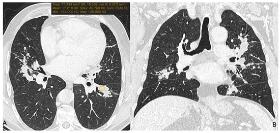
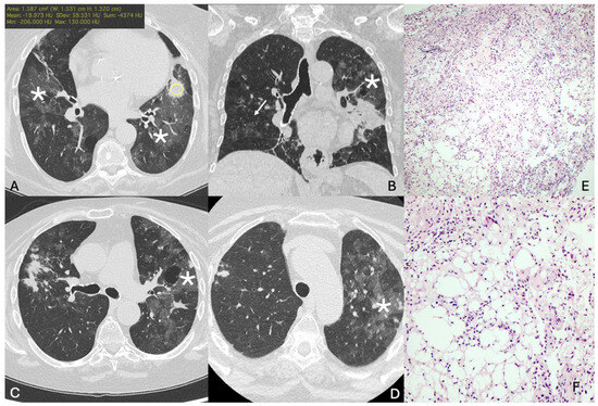
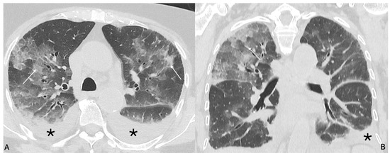
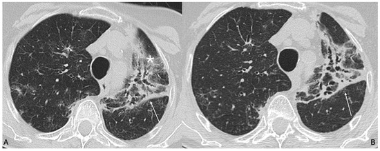
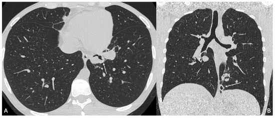
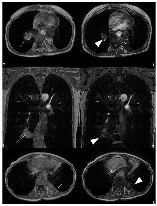
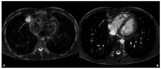
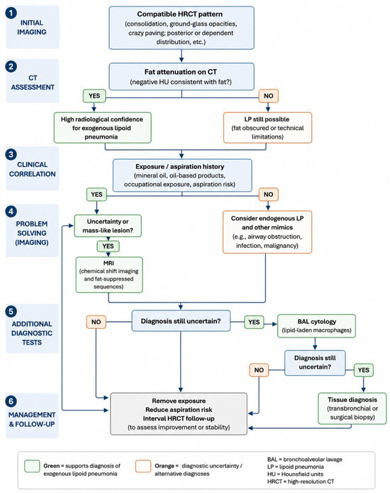
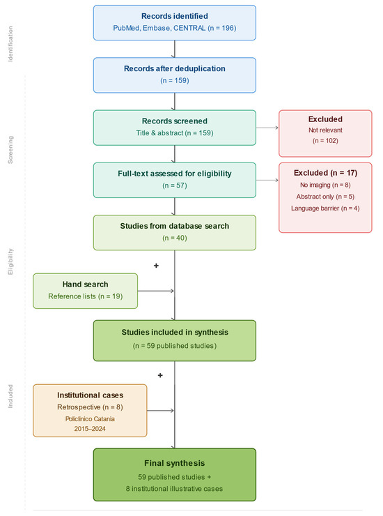
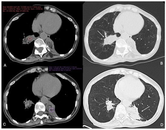
Background/Objectives: Lipoid pneumonia (LP) is a rare and frequently underdiagnosed pulmonary condition with a broad spectrum of radiological manifestations that can closely mimic infectious, inflammatory, and neoplastic lung diseases. Despite its clinical relevance, no standardized imaging-based diagnostic pathway exists. For this reason, this pictorial narrative review aims to provide a structured, imaging-centred synthesis of LP, to characterise the full spectrum of high-resolution CT (HRCT) and magnetic resonance imaging (MRI) findings, and to propose a pragmatic diagnostic workflow. Methods: A systematic literature search was performed in PubMed, MEDLINE, Embase, and the Cochrane Library from January 1950 to February 2025. Search terms combined “lipoid pneumonia” with imaging-related keywords including “HRCT,” “computed tomography,” “MRI,” and “fat attenuation.” After screening 891 deduplicated records, 60 studies were included in the narrative synthesis. Eight illustrative institutional cases with imaging–pathology correlation were additionally selected to demonstrate key imaging phenotypes. Results: HRCT is the cornerstone modality, demonstrating intralesional fat attenuation (typically −30 to −150 HU) in 40–80% of cases depending on series and disease chronicity. Additional patterns include ground-glass opacity, crazy paving, centrilobular nodules, and mass-like consolidation mimicking malignancy. Fat attenuation is absent in up to 60% of cases when inflammatory exudate or fibrosis masks lipid content. MRI, particularly chemical shift imaging, serves as a problem-solving adjunct in pseudotumoral or densitometrically equivocal presentations. A pragmatic diagnostic workflow is proposed, integrating HRCT findings, exposure history, fat-sensitive MRI in selected cases, BAL cytology, and histopathological confirmation when required. Conclusions: A pattern-based radiological approach, anchored on HRCT and integrated with clinical exposure history, BAL cytology, and selective use of fat-sensitive MRI, enables accurate diagnosis of LP in most cases and can prevent unnecessary invasive procedures including surgical resection performed under suspicion of malignancy.
Full article
(This article belongs to the Special Issue Current Trends in Computed Tomography: Optimization and Clinical Practice)
►▼
Show Figures

Figure 1
Open AccessInteresting Images
Sinonasal and Pulmonary Computed Tomography Images Before and After Triple-Combination Therapy in a Patient with Cystic Fibrosis Without ΔF508 Mutations
by
Corrado Tagliati, Giovanna Campagna, Maria Di Sabatino, Giuseppe Lanni, Davide Battista and Pietro Ripani
Diagnostics 2026, 16(11), 1692; https://doi.org/10.3390/diagnostics16111692 (registering DOI) - 30 May 2026
Abstract
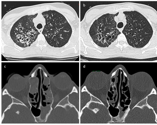
Here, we present the case of a 25-year-old patient with G542X and G85E cystic fibrosis mutations who underwent computed tomography examination before and after triple-combination therapy. Clear improvement in sinonasal and lung involvement is visible two years after modulator treatment initiation. To the
[...] Read more.
Here, we present the case of a 25-year-old patient with G542X and G85E cystic fibrosis mutations who underwent computed tomography examination before and after triple-combination therapy. Clear improvement in sinonasal and lung involvement is visible two years after modulator treatment initiation. To the best of our knowledge, this is the first report about sinonasal improvement demonstrated by computed tomography images in a patient with G542X/G85E mutations.
Full article
(This article belongs to the Collection Interesting Images)
►▼
Show Figures

Figure 1
Open AccessCase Report
Asphyxial Mechanisms in Sand Burial, Findings and Diagnostic Challenges—A Case Report and a Literature Review
by
Donato Morena, Anna Claudia Caruso, Martina Padovano, Matteo Scopetti and Vittorio Fineschi
Diagnostics 2026, 16(11), 1691; https://doi.org/10.3390/diagnostics16111691 (registering DOI) - 30 May 2026
Abstract
Background: Fatal sand burial is a rare and diagnostically challenging entity in forensic practice. In such cases, death may result from thoracic or thoracoabdominal compression, airway obstruction by particulate material, massive inhalation of sand or soil, or a combination of these mechanisms. External
[...] Read more.
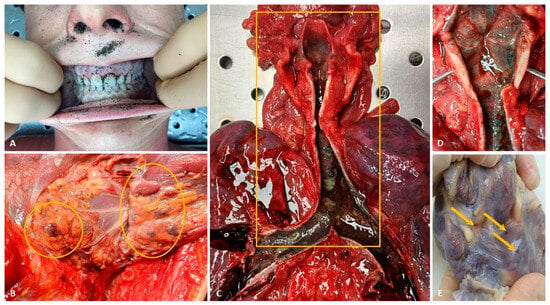
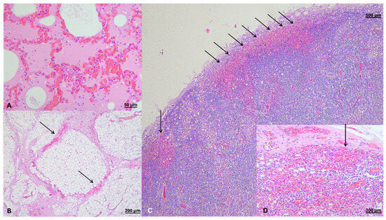
Background: Fatal sand burial is a rare and diagnostically challenging entity in forensic practice. In such cases, death may result from thoracic or thoracoabdominal compression, airway obstruction by particulate material, massive inhalation of sand or soil, or a combination of these mechanisms. External signs may be subtle or absent, making postmortem interpretation highly dependent on a comprehensive, multilevel assessment. Case Presentation and Methods: We report the case of a 17-year-old male who died following accidental sand burial caused by the collapse of a self-excavated beach tunnel. External examination, autopsy, histological and toxicological analyses were performed. A review of the literature was also conducted to identify published forensic cases of fatal sand or soil burial and to compare circumstantial, macroscopic, microscopic, and ancillary findings. Results: Autopsy revealed marked pulmonary edema and congestion, multivisceral congestion, scattered sand granules within the larynx, and epicardial petechiae. Histological examination demonstrated acute pulmonary emphysema, edema, vascular congestion, and hemorrhagic laterocervical lymph nodes. Overall, the findings were considered most consistent with mechanical asphyxia due to thoracic compression. The literature review identified six eligible studies describing eight fatal cases. Despite the limited sample size and marked heterogeneity, two main diagnostic patterns emerged: compression-related deaths, usually associated with tunnel or beach-hole collapse and minimal or absent particulate material within the airways, and aspiration-/obstruction-related deaths, characterized by abundant or compact sand or soil material within the airways. In cases without massive aspiration, mixed mechanisms may coexist. Pulmonary edema and congestion were the most frequently reported autopsy findings. When available, histological examination appeared useful in distinguishing antemortem from postmortem burial. Conclusions: Fatal sand burial should not be regarded as a uniform forensic entity. External examination alone is often insufficient, and accurate diagnosis requires a comprehensive, multidisciplinary approach integrating scene reconstruction, autopsy data, histopathological findings, and ancillary analyses. Hemorrhagic involvement of the laterocervical lymph nodes may represent a potentially relevant but currently underexplored finding, whose diagnostic significance warrants further investigation in analogous cases.
Full article
(This article belongs to the Special Issue Advances in Pathology for Forensic Diagnosis)
►▼
Show Figures

Figure 1
Open AccessArticle
Early Detection of Cerebral Vasospasm Following Aneurysmal Subarachnoid Hemorrhage by IL-6 Electrochemiluminescence Analysis of the Cerebrospinal Fluid
by
Adi Ahmetspahic, Ema Selimovic, Faruk Alagic, Almaida Alagic, Ermin Begovic, Suzana Tihic, Almir Dzurlic, Razim Mahmutagic, Mirza Pojskic and Alberto Feletti
Diagnostics 2026, 16(11), 1690; https://doi.org/10.3390/diagnostics16111690 (registering DOI) - 30 May 2026
Abstract
Background/objectives: Aneurysmal subarachnoid hemorrhage (aSAH) is a neurosurgical emergency associated with cerebral vasospasm, representing an important complication. This study assessed the association between cerebrospinal fluid (CSF) interleukin-6 (IL-6) levels and cerebral vasospasm after aSAH. Methods: This prospective single-center observational study with repeated measurements
[...] Read more.
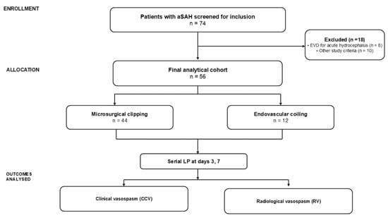
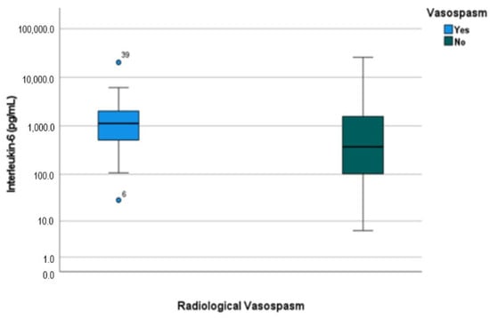
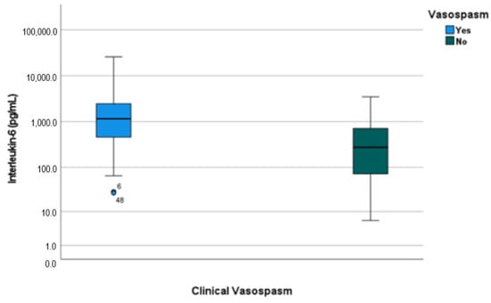
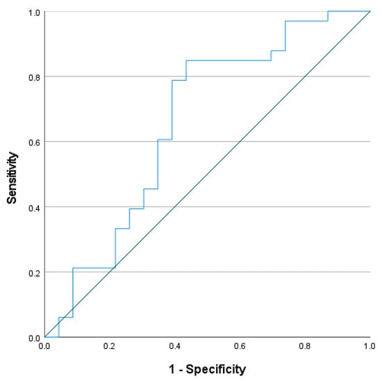
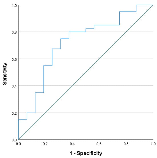
Background/objectives: Aneurysmal subarachnoid hemorrhage (aSAH) is a neurosurgical emergency associated with cerebral vasospasm, representing an important complication. This study assessed the association between cerebrospinal fluid (CSF) interleukin-6 (IL-6) levels and cerebral vasospasm after aSAH. Methods: This prospective single-center observational study with repeated measurements included 56 patients among 74 screened patients with aSAH; 18 patients were excluded according to predefined exclusion criteria, including infection, unavailable CSF samples or requirement for CSF diversion within the first seven days of onset. The samples were obtained via lumbar puncture on post-hemorrhage days 3 and 7. IL-6 was quantified using an electrochemiluminescent immunoassay. Radiological vasospasm (RV) was assessed by computed tomography angiography, while clinical vasospasm (CCV) was defined as new neurological deterioration unexplained by other causes. Results: Median IL-6 levels were 423.5 pg/mL on day 3 and 726.0 pg/mL on day 7. Day 7 IL-6 levels were associated with RV (p = 0.045) and CCV (p = 0.010), while day 3 IL-6 was associated with CCV only (p = 0.035). Day 7 IL-6 showed modest discriminatory performance for CCV: AUC = 0.722, cut-off 460 pg/mL, sensitivity 75.0%, and specificity 68.8%; for RV: AUC = 0.659, cut-off 451 pg/mL, sensitivity 81.8%, and specificity 56.5%. Conclusions: Elevated CSF IL-6 levels were associated with cerebral vasospasm after aSAH, with a more consistent association observed for CCV than for RV; however, these findings are hypothesis-generating and require validation before clinical applicability can be determined.
Full article
(This article belongs to the Special Issue Biochemical Testing Applications in Clinical Diagnosis—2nd Edition)
►▼
Show Figures

Figure 1
Open AccessArticle
Radiograph-Based Deep Learning Model to Support Finger Joint Selection for Ultrasound Examination in Rheumatoid Arthritis
by
Youngjae Park, Keum San Chun, Seungeun Lee, Joon-Yong Jung, Sungwon Lee, Hyemin Park, Tuan Dinh Le, Hyeondeok Choi and Wan-Uk Kim
Diagnostics 2026, 16(11), 1689; https://doi.org/10.3390/diagnostics16111689 - 29 May 2026
Abstract
Background/Objectives: Ultrasound is the standard imaging modality to evaluate the inflammatory changes in hand joints of rheumatoid arthritis (RA) patients. However, it is operator-dependent and takes a long time to examine. In this study, we developed a radiograph-based deep learning (DL) model to
[...] Read more.
Background/Objectives: Ultrasound is the standard imaging modality to evaluate the inflammatory changes in hand joints of rheumatoid arthritis (RA) patients. However, it is operator-dependent and takes a long time to examine. In this study, we developed a radiograph-based deep learning (DL) model to support prioritization of finger joints for ultrasound (US) examination in RA patients. Methods: In this retrospective study, hand radiographs from RA patients who underwent same-day US examination of bilateral finger joints were analyzed. A DL model was developed using hand radiographs from 270 patients (2043 finger joints) to estimate joint-level likelihood of inflammatory activity. US findings served as the reference standard for model training, while clinical findings of joint tenderness and swelling were incorporated as additional tabular inputs. Model performance was evaluated in a temporal-split test cohort consisting of 40 patients (270 joints) and compared with the performance of a clinical-only logistic regression model based on joint tenderness and swelling. Results: In the test set, the DL model demonstrated higher sensitivity (82.1% vs. 38.5%), negative predictive value (96.8% vs. 90.3%), and F1-score (69.6% vs. 48.4%) than the clinical-only model. Although the area under the receiver operating characteristic curve did not differ significantly between models (p = 0.43), precision–recall (PR) analysis showed superior performance of the DL model, with a higher area under the PR curve (0.625 vs. 0.540). At the threshold maximizing the F1-score, DL-assisted triage reduced the number of finger joints selected for US examination by approximately 80%. Conclusions: A radiograph-based DL model can support efficient prioritization of finger joints for US examination in RA, offering a practical approach to enhance joint-level US triage in routine clinical practice.
Full article
(This article belongs to the Special Issue Advances in the Use of Artificial Intelligence for the Diagnosis and Management of Hand Conditions)
Open AccessArticle
A Consistent Landmark for Tibial Tunnel Placement in Arthroscopic Remnant-Preserving Posterior Cruciate Ligament Reconstruction: Use of Champagne-Glass Drop-Off and Lateral Cartilage Point—A Retrospective Case Series
by
Yu-Ze Luan, Wei-Jun Hong, Tzu-Chun Chung and Chien-Sheng Lo
Diagnostics 2026, 16(11), 1688; https://doi.org/10.3390/diagnostics16111688 - 29 May 2026
Abstract
Background/Objectives: Accurate tibial tunnel placement is critical for successful posterior cruciate ligament reconstruction (PCLR), yet remains technically demanding due to limited visualization and anatomic variability. This study aimed to demonstrate the feasibility of an arthroscopic technique for remnant-preserving PCLR using the champagne-glass drop-off
[...] Read more.
Background/Objectives: Accurate tibial tunnel placement is critical for successful posterior cruciate ligament reconstruction (PCLR), yet remains technically demanding due to limited visualization and anatomic variability. This study aimed to demonstrate the feasibility of an arthroscopic technique for remnant-preserving PCLR using the champagne-glass drop-off and lateral cartilage point as consistently identifiable arthroscopic anatomic bony landmarks, and to evaluate the success rate of tibial tunnel placement in targeted position using postoperative magnetic resonance imaging (MRI). Methods: A retrospective review was performed of patients who underwent arthroscopic remnant-preserving PCLR using a trans-septal approach with the described dual-landmark technique between March 2020 and October 2022. Of 31 eligible patients, 20 with complete clinical follow-up and postoperative 1-year MRI were included for analysis. Tibial tunnel position was assessed on MRI to determine success rate of placement in the targeted inferior–lateral tibial footprint based on anatomic reference. Clinical outcomes, including knee range of motion and posterior laxity, were also evaluated. Results: MRI evaluation demonstrated tibial tunnel consistent placement with the predefined targeted zone in all patients (20/20). At a median follow-up of 745 days, the mean knee range of motion was 140.0 ± 12.7 degrees. Posterior stability assessment showed grade 0 laxity in 75% of patients and grade 1 laxity in 25%. No graft failures, neurovascular complications, infections, or revision PCLR procedures were observed. Conclusions: This retrospective case series suggests that the dual-landmark technique (champagne-glass drop-off and lateral cartilage point) may facilitate consistent tibial tunnel placement in remnant-preserving PCLR. Level of Evidence: IV (Retrospective case series).
Full article
(This article belongs to the Special Issue Arthroscopy Techniques in Diagnosis and Treatment 2026)
Open AccessInteresting Images
Primary Pulmonary Artery Sarcoma: Multimodality Imaging of a Rare Intravascular Tumor Mimicking Pulmonary Embolism
by
Dan Li, Zhongyu Liu, Shuo Liang and Hong Zhang
Diagnostics 2026, 16(11), 1687; https://doi.org/10.3390/diagnostics16111687 - 29 May 2026
Abstract
Primary pulmonary artery sarcoma (PPAS) is a mesenchymal tumor originating from the pulmonary artery, accounting for approximately 0.001–0.003% of all sarcomas. The early clinical symptoms are atypical, and diagnosis is often delayed, making the management of this disease challenging. The widespread availability of
[...] Read more.
Primary pulmonary artery sarcoma (PPAS) is a mesenchymal tumor originating from the pulmonary artery, accounting for approximately 0.001–0.003% of all sarcomas. The early clinical symptoms are atypical, and diagnosis is often delayed, making the management of this disease challenging. The widespread availability of multidetector computed tomography (MDCT), 18F-fluorodeoxyglucose positron emission tomography/computed tomography (18F-FDG PET/CT), and high-resolution echocardiography has significantly improved the diagnostic capability for PPAS. We herein report a 74-year-old female patient who presented with a 3-week history of exertional dyspnea without an apparent trigger. She had received anti-inflammatory therapy at another hospital for one week. Five days before admission, she experienced right-sided chest pain without apparent cause, which was respiratory-related. On the day of admission, laboratory tests revealed a slight elevation in D-dimer levels. Echocardiography showed an irregular, moderately echogenic mass at the origin of the right pulmonary artery. Enhanced computed tomography (CT) of the chest revealed a filling defect in the right pulmonary artery accompanied by bilateral pleural effusion. The patient was given heparin anticoagulation therapy. To confirm the nature of these lesions, a PET/CT scan was conducted five days after admission, which indicated hypermetabolism in the right pulmonary artery, suggesting primary pulmonary artery sarcoma. Due to the poor efficacy of anticoagulation therapy, the patient continued to experience breath-holding after physical activity. Subsequently, catheter-guided interventional angiography was carried out for pulmonary artery thrombectomy and biopsy, and histopathological examination revealed pulmonary artery sarcoma. Given the patient’s respiratory failure and heart failure, as well as the uncertain efficacy of radiotherapy and chemotherapy, interventional pulmonary artery thrombectomy alleviated the chest pain. Currently, the patient’s overall condition is stable.
Full article
(This article belongs to the Section Medical Imaging and Theranostics)
Open AccessCase Report
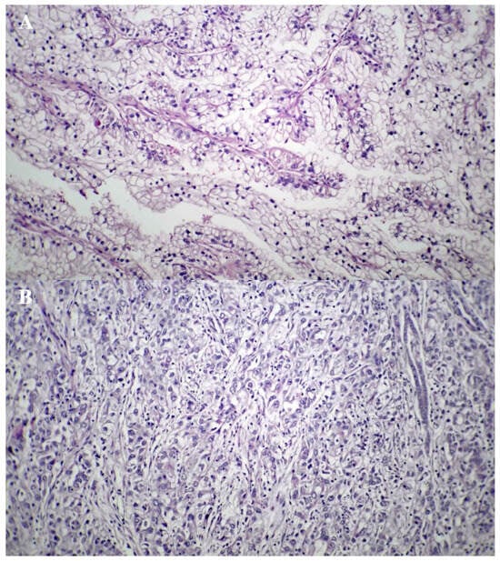
Schwannoma Mimicking Neck Nodal Metastasis on F-18 FDG PET/CT in Cervical Cancer: A Case Report with a Multimodal Approach
by
Seokho Yoon, Hye Jin Baek, Bonghoi Choi and Hyo Jung An
Diagnostics 2026, 16(11), 1686; https://doi.org/10.3390/diagnostics16111686 - 29 May 2026
Abstract
Background: In oncologic patients, hypermetabolic neck lesions identified on F-18 fluorodeoxyglucose (FDG) positron emission tomography/computed tomography (PET/CT) are often assumed to indicate nodal metastasis, especially in advanced disease. However, benign tumors such as schwannomas can also demonstrate avid F-18 FDG uptake, potentially
[...] Read more.
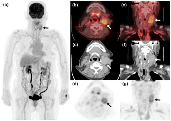
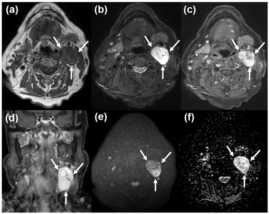
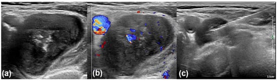
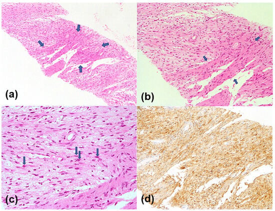
Background: In oncologic patients, hypermetabolic neck lesions identified on F-18 fluorodeoxyglucose (FDG) positron emission tomography/computed tomography (PET/CT) are often assumed to indicate nodal metastasis, especially in advanced disease. However, benign tumors such as schwannomas can also demonstrate avid F-18 FDG uptake, potentially leading to false-positive nodal staging and unwarranted assumptions of metastatic disease. Case Presentation: An 85-year-old woman with advanced uterine cervical cancer (FIGO stage IIB) underwent F-18 FDG PET/CT for staging purposes. Alongside intense uptake in the primary tumor, a hypermetabolic mass was incidentally identified in the left neck, raising concerns about nodal metastasis. Further imaging, including MRI and high-resolution ultrasonography (US), suggested a non-nodal origin, and US-guided core needle biopsy confirmed the diagnosis of a schwannoma, with histopathologic examination demonstrating characteristic Antoni A and B areas with diffuse S-100 positivity. Because the patient was elderly and repeatedly declined aggressive treatment, management was ultimately limited to symptom-directed palliative radiotherapy. Although tissue confirmation did not directly alter the delivered treatment strategy, it clarified the staging and prevented the neck lesion from being misclassified as metastatic disease. Conclusions: This case underscores that not all hypermetabolic neck lesions detected on F-18 FDG PET/CT in oncologic patients indicate metastatic lymphadenopathy. A multimodal imaging approach combined with minimally invasive tissue sampling can provide critical diagnostic clarification, particularly when PET/CT findings might otherwise lead to unsupported nodal upstaging or misguided assumptions in treatment planning.
Full article
(This article belongs to the Special Issue Advances in Head and Neck and Oral Maxillofacial Radiology)
►▼
Show Figures

Figure 1
Open AccessSystematic Review
Predicting 30-Day Readmission After Stroke: A Systematic Review and Meta-Analysis to Inform Predictor Selection
by
Saurabh Kalra, Farya Fakoori, Mohammad Nafeli Shahrestani, Zhaoqianyu Xiong, Hannah Gardener, WayWay M. Hlaing, Carolina Marinovic Gutierrez, Gillian Gordon Perue, Negar Asdaghi, Jose G. Romano, Tatjana Rundek and Emir Veledar
Diagnostics 2026, 16(11), 1685; https://doi.org/10.3390/diagnostics16111685 - 29 May 2026
Abstract
Background: Thirty-day readmission after stroke remains common, yet contemporary readmission rates, prediction model performance, and predictor domains have not been comprehensively synthesized. Methods: Following PRISMA guidelines, we searched PubMed, Embase, Web of Science, Scopus, and Google Scholar for studies published between 1 January
[...] Read more.
Background: Thirty-day readmission after stroke remains common, yet contemporary readmission rates, prediction model performance, and predictor domains have not been comprehensively synthesized. Methods: Following PRISMA guidelines, we searched PubMed, Embase, Web of Science, Scopus, and Google Scholar for studies published between 1 January 2021 and 9 October 2025. Readmission proportions and model discrimination, measured by area under the receiver operating characteristic curve (AUC), were pooled using random-effects meta-analysis. Heterogeneity was assessed using I2. Predictors were summarized across studies by domain. Results: Twenty studies met inclusion criteria: 15 studies comprising 358,434 patients contributed quantitative data, and 5 were included in qualitative synthesis only. The pooled proportion was 12.9% (95% CI: 10.1–15.8%), with subgroup estimates of 14.2% (95% CI: 11.9–16.6%) for all-cause and 3.6% (95% CI: 0.5–6.7%) for stroke-specific readmissions. Study-level AUCs ranged from 0.59 to 0.88, with a pooled AUC of 0.69 (95% CI: 0.64–0.75), indicating moderate discrimination. Substantial heterogeneity was observed (I2 > 98%, p < 0.001), and pooled estimates should be interpreted cautiously. Predictor selection was poorly standardized and largely driven by data availability, with inconsistent inclusion of key clinical and post-discharge domains such as stroke severity, functional status, discharge disposition, post-discharge care, and social determinants of health. Conclusions: Thirty-day readmission after stroke remains common, and currently available models demonstrate modest predictive discrimination; no consistently high-performing, broadly generalizable prediction model has yet emerged. Improving prediction will require broader predictors capturing stroke severity, care transitions, follow-up, and patient context, along with external validation and integration into clinical workflows.
Full article
(This article belongs to the Special Issue Risk Factors, Incidence, and Outcome of Stroke)
Open AccessCase Report
Extraosseous 99mTc-MDP Uptake Guiding Intraoperative Sampling in Severe Inflammatory Myopathy: A Case Report and Literature Review
by
Masha Maharaj, Sanvir Sirriram, Nav Govender, Trisha Govender, Babita D. Bhana and Nisaar Korowlay
Diagnostics 2026, 16(11), 1684; https://doi.org/10.3390/diagnostics16111684 - 29 May 2026
Abstract
Background/Objectives: We report a case of severe dermatomyositis demonstrating characteristic widespread extraosseous uptake on 99mTc-methylene diphosphonate (99mTc-MDP) bone scintigraphy. This study highlights the diagnostic value of this modality in detecting active inflammatory myopathy when conventional muscle biopsy is inconclusive and
[...] Read more.
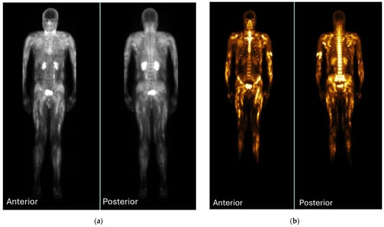
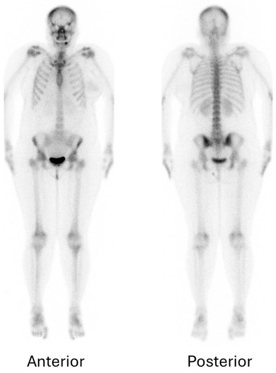
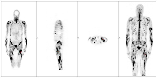
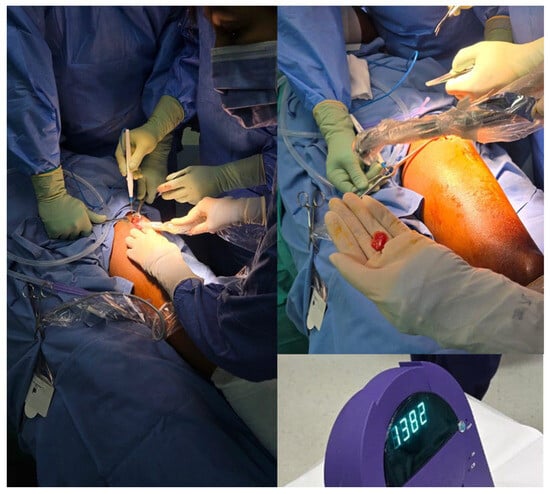
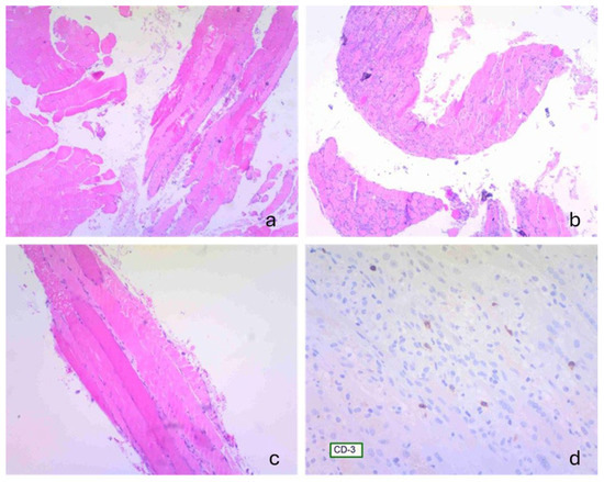
Background/Objectives: We report a case of severe dermatomyositis demonstrating characteristic widespread extraosseous uptake on 99mTc-methylene diphosphonate (99mTc-MDP) bone scintigraphy. This study highlights the diagnostic value of this modality in detecting active inflammatory myopathy when conventional muscle biopsy is inconclusive and introduces its novel use for intraoperative gamma-probe-guided biopsy to precisely target metabolically active muscle. This approach may help target metabolically active muscle in heterogeneous idiopathic inflammatory myopathies (IIMs). Case Presentation: A 49-year-old man developed progressive proximal muscle weakness (Medical Research Council grade 2/5 proximally, 5/5 distally) beginning in June 2025 following influenza infection, accompanied by dysphagia, classic dermatomyositis cutaneous manifestations, back pain, and difficulty standing. Laboratory evaluation revealed elevated inflammatory markers (ESR 55 mm/hr, CRP 20 mg/L), leukocytosis (16.58 × 10⁹/L), markedly raised creatine kinase (19,937 IU/L), and troponin T levels. An initial quadriceps muscle biopsy performed on 29 July 2025 was non-diagnostic. Three-phase 99mTc-MDP scintigraphy (~1110 MBq) demonstrated intense, diffuse extraosseous uptake involving bilateral deltoids (symmetric), biceps and triceps (patchy), paraspinal muscles (longitudinal), gluteal muscles, thighs (quadriceps and hamstrings), and gastrocnemius muscles, with relative suppression of appendicular skeletal uptake on delayed images due to soft-tissue tracer dominance—findings consistent with severe inflammatory myopathy. Following reinjection (~1100 MBq), intraoperative gamma-probe-guided biopsy targeted areas of highest uptake (left quadriceps femoris and distal triceps brachii; intraoperative counts 1300–1400 versus background ~500). Histopathology revealed histiocyte-predominant inflammation with myofibre necrosis and regeneration, sparse CD4+ T-cell infiltrates, and absence of fibrosis, consistent with necrotising myopathy. Positive antinuclear antibodies and strong anti-Mi-2 antibodies confirmed the diagnosis of dermatomyositis. Treatment included pulse methylprednisolone followed by oral prednisone taper, methotrexate, azathioprine, intravenous immunoglobulin, and planned rituximab therapy. Discussion: Whole-body 99mTc-MDP scintigraphy provided a complementary whole-body functional assessment of disease extent, revealing widespread muscular involvement. The novel application of intraoperative gamma-probe-guided biopsy enabled real-time targeting of metabolically active muscle, facilitating targeted sampling after an initial non-diagnostic biopsy and yielding supportive histopathological findings. This dual diagnostic and interventional role demonstrates the technical feasibility of gamma-probe guidance in a diagnostically challenging case of dermatomyositis. Conclusions: In our case, the integration of 99mTc-MDP scintigraphy with gamma-probe-guided biopsy enabled precise targeting of metabolically active muscle following an initial non-diagnostic biopsy. This multimodal approach may be useful in selected diagnostically challenging cases of severe inflammatory myopathy. Larger studies are needed to evaluate its reproducibility and added value.
Full article
(This article belongs to the Special Issue Current Trends and Future Directions in Radiotheragnostic Research: A Special Issue in Collaboration with ISRT 2025)
►▼
Show Figures

Figure 1
Open AccessReview
Hemophilia in the Era of Advanced Therapies: Structural Monitoring, the Role of Musculoskeletal Ultrasound, and a Proposed Multidisciplinary Care Model—A Structured Narrative Review
by
Felipe Querol-Giner, Magdalena Querol-Giner, Sofía Pérez-Alenda and Felipe Querol-Fuentes
Diagnostics 2026, 16(11), 1683; https://doi.org/10.3390/diagnostics16111683 - 29 May 2026
Abstract
Background: Advances in hemophilia treatment, including extended half-life factor concentrates, non-replacement therapies, and gene therapy, have substantially reduced bleeding frequency and improved life expectancy. However, persistent musculoskeletal damage, subclinical bleeding, and residual arthropathy remain important clinical challenges despite improved hematologic control. Objective: We
[...] Read more.
Background: Advances in hemophilia treatment, including extended half-life factor concentrates, non-replacement therapies, and gene therapy, have substantially reduced bleeding frequency and improved life expectancy. However, persistent musculoskeletal damage, subclinical bleeding, and residual arthropathy remain important clinical challenges despite improved hematologic control. Objective: We aimed to analyze recent therapeutic advances in hemophilia, examine persistent musculoskeletal complications, and propose a multidisciplinary care model based on structural monitoring, highlighting the role of musculoskeletal ultrasound. Methods: A structured narrative review with a reproducible search strategy was conducted following PRISMA 2020-informed methodological principles. PubMed/MEDLINE and Scopus were searched for clinically relevant studies published between 2018 and 2026 focusing on advanced hemophilia therapies, musculoskeletal complications, and structural monitoring. A total of 478 records were identified, and 13 studies were included after screening and selection using the Rayyan platform. Results: Modern therapies markedly reduce clinically evident hemarthroses, but structural joint alterations and subclinical disease activity may persist, particularly in patients with pre-existing arthropathy. Imaging-based studies identified persistent synovial and osteochondral alterations despite effective hematologic control. Magnetic resonance imaging remains the reference standard for structural assessment, although its routine use may be limited by accessibility and cost. Musculoskeletal ultrasound emerges as an accessible and reproducible tool for dynamic joint evaluation and early detection of structural alterations, supporting longitudinal monitoring and individualized rehabilitation strategies. Conclusions: In the era of advanced therapies, comprehensive hemophilia management requires not only effective hematologic control but also structured musculoskeletal follow-up. The integration of musculoskeletal ultrasound into multidisciplinary care models may favor earlier detection of joint alterations and more individualized rehabilitation strategies.
Full article
(This article belongs to the Special Issue Recent Advances in Diagnosis and Management of Musculoskeletal Disorders)
Open AccessArticle
Implementation of an AI-Based Clinical Decision Support System Predicting In-Hospital Cardiac Arrest in General Wards: A Multicenter Staggered-Implementation Study in Secondary Hospitals in Korea
by
Minjeong Kim, Dongjoon Yoo, Eunbi Noh, Yongwook Jeong, Minsoo Kim, Kyung-Jae Cho, Mincheol Kim, You Dong Sohn and Gyu Chong Cho
Diagnostics 2026, 16(11), 1682; https://doi.org/10.3390/diagnostics16111682 - 29 May 2026
Abstract
Background/Objectives: In-hospital cardiac arrest (IHCA) remains a devastating event associated with high morbidity and mortality among general ward patients. While Rapid Response Systems (RRS) can help identify deteriorating patients, maintaining these systems in secondary hospitals is frequently hindered by severe fiscal and personnel
[...] Read more.
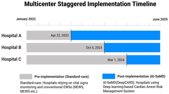
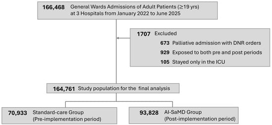
Background/Objectives: In-hospital cardiac arrest (IHCA) remains a devastating event associated with high morbidity and mortality among general ward patients. While Rapid Response Systems (RRS) can help identify deteriorating patients, maintaining these systems in secondary hospitals is frequently hindered by severe fiscal and personnel constraints. Consequently, evidence regarding the real-world clinical effectiveness of artificial intelligence software as a medical device (AI-SaMD) for predicting deterioration in such resource-constrained settings remains limited. Methods: We conducted a retrospective analysis on a multicenter, staggered-implementation study evaluating 164,761 eligible adult general ward admissions across three secondary hospitals in South Korea. The intervention involved deploying an AI-SaMD (DeepCARS), which utilizes four routine vital signs to predict ward IHCA within 24 h. The primary outcome was ward IHCA. Secondary outcomes included in-hospital mortality and length of stay (LOS). Exploratory analyses investigated the mechanisms of clinical outcomes by evaluating lead-times to interventions, outcomes in sepsis subgroups, changes in care directives, and post-arrest neurological outcomes. Results: AI-SaMD implementation was associated with a 21% reduction in ward IHCA incidence (adjusted rate ratio 0.79; 95% CI, 0.65–0.96; p = 0.016) and a 15% reduction in in-hospital mortality (aRR 0.85; 95% CI, 0.79–0.90; p < 0.001), alongside significantly shorter hospital and intensive care unit LOS. These associations were also observed in patients with sepsis (IHCA aRR 0.71; 95% CI, 0.54–0.93; p = 0.013). Lead-times to critical care intervention and to antibiotic escalation were numerically shorter in the AI-SaMD group by 16.3 h (p = 0.066) and 2.6 h (p = 0.523); poor neurological outcome at discharge among ward IHCA cases was 85/108 (78.7%) in the AI-SaMD group versus 63/102 (61.8%) in the standard-care group (aRR 1.13; 95% CI, 0.99–1.33; p = 0.058); and the full-code death rate did not differ between groups (aRR 0.94; 95% CI, 0.76–1.15)—none of these additional analyses reached statistical significance. Conclusions: In secondary hospitals unable to operate an RRS due to fiscal limitations, implementation of an AI-SaMD as an additional informational layer was associated with lower ward IHCA and in-hospital mortality. The AI-SaMD may serve as an actionable and scalable additional safety layer for general-ward patients in resource-constrained environments where RRS infrastructure is not feasible. Although this was a multicenter, large-scale study, the present analysis was retrospective and quasi-experimental in design; rigorous randomized studies are needed to confirm these associations.
Full article
(This article belongs to the Special Issue AI and Digital Health for Disease Diagnosis and Monitoring, 2nd Edition)
►▼
Show Figures

Figure 1
Open AccessArticle
Volumetric Imaging of Ex Vivo Oral Mucosa Specimens with Multi-Scale Wide Field-of-View Optical Coherence Tomography/Microscopy in Near-Infrared-II Window
by
Chuan-Bor Chueh, Shih-Jung Cheng, Hui-Hsin Ko, Ming-Che Tu, Ting-Hao Chen and Hsiang-Chieh Lee
Diagnostics 2026, 16(11), 1681; https://doi.org/10.3390/diagnostics16111681 - 29 May 2026
Abstract
Background/Objectives: Intraoperative margin assessments of oral squamous cell carcinoma (SCC) are fundamentally limited by sampling errors and freezing artifacts inherent to standard frozen section analysis. We developed a mobile, multi-scale, wide field-of-view (FOV) swept-source optical coherence tomography/microscopy (SS-OCT/OCM) system operating in the Near-Infrared-II
[...] Read more.
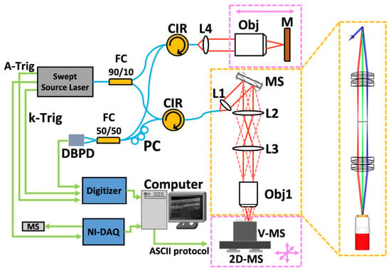
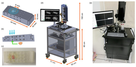
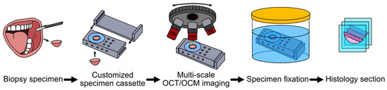
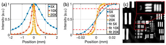
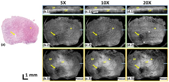
Background/Objectives: Intraoperative margin assessments of oral squamous cell carcinoma (SCC) are fundamentally limited by sampling errors and freezing artifacts inherent to standard frozen section analysis. We developed a mobile, multi-scale, wide field-of-view (FOV) swept-source optical coherence tomography/microscopy (SS-OCT/OCM) system operating in the Near-Infrared-II (NIR-II) window (1.68 μm) to provide a rapid, non-destructive, volumetric evaluation of excised oral mucosal tissues. Methods: To correlate optical images with histopathology, we engineered a custom 3D-printed tissue cassette that physically mitigates macroscopic shrinkage during scanning and subsequent tissue fixation. A three-axis motorized assembly extends the effective imaging FOV without compromising resolution, while a custom 3D multi-resolution pyramid stitching algorithm synthesizes wide-FOV mosaics. Results: The customized cassette enabled precise, one-to-one spatial correlation between optical volumes and histopathology sections. Crucially, a 3 × 3 mosaic scan acquired with a 10× objective balanced imaging resolution and acquisition time, providing sufficient structural clarity to visualize basement membrane loss—a hallmark of SCC invasion. Conclusions: This 1.68 μm, fully automatic, multiscale SS-OCT/OCM platform demonstrates the feasibility of serving as a rapid, three-dimensional imaging tool for potential future use as an adjunct to conventional frozen sections.
Full article
(This article belongs to the Collection Biomedical Optics: From Technologies to Applications)
►▼
Show Figures

Figure 1
Open AccessInteresting Images
High-Risk Thymoma Hidden Within a Cystic Thymic Lesion: A Diagnostic Clue from Diffusion-Weighted MRI
by
Kyungsoo Bae, Hyo Jung An and Kyung Nyeo Jeon
Diagnostics 2026, 16(11), 1680; https://doi.org/10.3390/diagnostics16111680 - 29 May 2026
Abstract
Cyst-predominant anterior mediastinal lesions are commonly attributed to benign thymic cysts. However, thymomas—the most common thymic epithelial tumor—may rarely present as a predominantly cystic mass with only a small solid component, posing a diagnostic challenge. The World Health Organization (WHO) classifies thymomas according
[...] Read more.
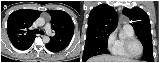
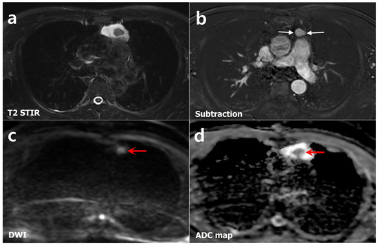
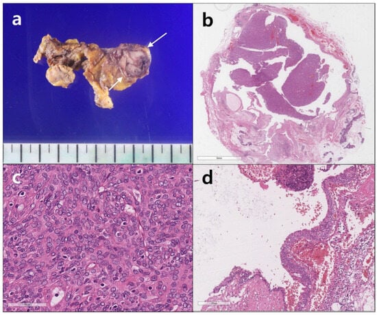
Cyst-predominant anterior mediastinal lesions are commonly attributed to benign thymic cysts. However, thymomas—the most common thymic epithelial tumor—may rarely present as a predominantly cystic mass with only a small solid component, posing a diagnostic challenge. The World Health Organization (WHO) classifies thymomas according to the morphology of tumor epithelial cells and the degree of lymphocytic infiltration, a schema that correlates with biological behavior and prognosis. Unlike low-risk subtypes, high-risk thymomas frequently require multimodal treatment because of their more aggressive clinical course. MRI has increasingly been utilized as a problem-solving modality for indeterminate lesions identified on CT. In particular, diffusion-weighted imaging (DWI) with apparent diffusion coefficient (ADC) measurement has emerged as a potentially useful imaging biomarker for predicting histologic subtypes of thymic epithelial tumors. We report a case of a WHO type B3 thymoma presenting as a predominantly cystic anterior mediastinal lesion, in which MRI findings suggested the possibility of a high-risk subtype. The imaging and corresponding histopathologic findings are presented in this paper.
Full article
(This article belongs to the Section Medical Imaging and Theranostics)
►▼
Show Figures

Figure 1
Open AccessCase Report
Severe Metabolic Decompensation in Metastatic Sarcomatoid Renal Cell Carcinoma During Immune Checkpoint Inhibitor Therapy: A Case Report and Literature Review
by
Lorena Ciumărnean, Cezara Andreea Gerdanovics, Olga Hilda Orășan, Alexandru Gerdanovics, Nicoleta Valentina Leach, Ioana Para and Gabriela Bombonica Dogaru
Diagnostics 2026, 16(11), 1679; https://doi.org/10.3390/diagnostics16111679 - 29 May 2026
Abstract
Background and Clinical Significance: Sarcomatoid renal cell carcinoma is a rare and highly aggressive variant of renal cell carcinoma, frequently associated with advanced-stage disease, early metastatic spread, and poor prognosis. Although immune checkpoint inhibitors have improved outcomes in metastatic renal cell carcinoma,
[...] Read more.
Background and Clinical Significance: Sarcomatoid renal cell carcinoma is a rare and highly aggressive variant of renal cell carcinoma, frequently associated with advanced-stage disease, early metastatic spread, and poor prognosis. Although immune checkpoint inhibitors have improved outcomes in metastatic renal cell carcinoma, particularly in tumors with sarcomatoid differentiation, they may also induce severe immune-related adverse events involving multiple organ systems. Case Presentation: We report the case of a 54-year-old woman with metastatic clear cell renal cell carcinoma with sarcomatoid differentiation, previously treated with nivolumab plus ipilimumab and subsequently with pazopanib, who was admitted with severe dehydration, repeated vomiting, marked asthenia, lower-limb-predominant muscle weakness, and inability to maintain orthostatism. Laboratory investigations revealed severe hyperkalemia, hyponatremia, hypoglycemia, anemia, thrombocytopenia, and prerenal acute kidney injury. The patient had a previous history of severe endocrine immune-related toxicity, including autoimmune hypophysitis and hypothyroidism, which had led to discontinuation of immunotherapy. Following fluid resuscitation, electrolyte correction, and supportive treatment, the metabolic abnormalities resolved and renal function improved significantly. Given the severity of the muscle weakness, a possible immune-mediated neuromuscular adverse event was also considered, although hyperkalemia remained a plausible contributing factor. Conclusions: This case highlights the complex interplay between prior immune checkpoint inhibitor exposure, endocrine dysfunction, metabolic decompensation, and possible neuromuscular involvement in metastatic sarcomatoid renal cell carcinoma. Early recognition, careful differential diagnosis, and multidisciplinary management are essential to prevent rapid clinical deterioration and optimize outcomes in patients with complex immune-related toxicities.
Full article
(This article belongs to the Special Issue Challenges in Urology: From Diagnosis to Management—2nd Edition)
►▼
Show Figures

Figure 1
Open AccessArticle
Association Between Dipstick Hematuria and Elevated Albuminuria in a Hospital-Based Population with Diverse Chronic Conditions
by
Cheewaporn Tanintheerakul, Rungnapa Peerakam, Pharisa Nanthawong, Suwatsin Kittikunnathum and Piyawan Bunpo
Diagnostics 2026, 16(11), 1678; https://doi.org/10.3390/diagnostics16111678 - 29 May 2026
Abstract
Background/Objective: Albuminuria is an early marker of kidney damage and cardiovascular risk. However, data on its prevalence and association with urine dipstick parameters in heterogeneous, hospital-based populations remain limited. The objective of this study was to determine the prevalence of elevated albuminuria
[...] Read more.
Background/Objective: Albuminuria is an early marker of kidney damage and cardiovascular risk. However, data on its prevalence and association with urine dipstick parameters in heterogeneous, hospital-based populations remain limited. The objective of this study was to determine the prevalence of elevated albuminuria measured by the urine albumin-to-creatinine ratio (UACR) in a diverse patient population and evaluate associations with semi-quantitative urine dipstick parameters. Methods: This cross-sectional study included 393 adults recruited from a tertiary-care hospital setting, comprising individuals undergoing clinical evaluation and routine health assessments. Given the hospital-based recruitment and enrichment with chronic conditions, the study population represents a high-risk cohort rather than the general population. Spot urine samples were analyzed for the UACR and dipstick parameters. Elevated albuminuria was defined as a UACR ≥ 30 mg/g. Associations were assessed using chi-square tests and multivariate logistic regression, with adjusted odds ratios (aORs) and 95% confidence intervals (CIs). Results: Elevated albuminuria was observed in 64% of participants. This high prevalence likely reflects the underlying clinical characteristics of the study population, including a substantial burden of chronic kidney disease and other comorbidities, and should not be interpreted as representative of population-level prevalence. Chronic kidney disease was independently associated with elevated albuminuria (aOR 3.14, 95% CI 1.12–8.86, p = 0.030), as was male sex (aOR 1.54, 95% CI 1.01–2.34, p = 0.045). Dipstick-positive blood was significantly associated with elevated albuminuria (85% vs. 60% in those without dipstick blood positivity, p = 0.005). Dipstick albumin strongly correlated with the UACR (p < 0.001), although 22% of individuals with negative dipstick albumin still had an elevated UACR. Other urine dipstick parameters were not significantly associated with elevated albuminuria. Conclusions: In this high-risk, hospital-based cohort, elevated albuminuria was common and associated with kidney disease and male sex. Dipstick blood positivity is significantly associated with albuminuria and may warrant further investigation. However, reliance on dipstick testing alone may underestimate disease burden, supporting the need for broader implementation of UACR-based screening strategies.
Full article
(This article belongs to the Section Clinical Laboratory Medicine)
Open AccessCase Report
Imaging-Guided Surgical Decision-Making and Bone Healing in Mandibular Cystic and Tumor-like Lesions: A Case-Based Radiologic Observation
by
Ömer Uranbey, Filip Kulewicz, Özenç Beste Kandemir, Furkan Özbey, Büşra Ekinci, India Maag, Agata Małyszek, Maciej Janeczek, Maciej Dobrzyński and Kamil Nelke
Diagnostics 2026, 16(11), 1677; https://doi.org/10.3390/diagnostics16111677 - 29 May 2026
Abstract
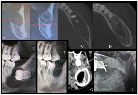
Background and Clinical Significance: Mandibular cystic lesions are heterogeneous in biologic behavior, radiologic appearance, and surgical management. Treatment selection is influenced by lesion extent, cortical bone condition, structural stability, and proximity to adjacent teeth and vital anatomical structures. In large mandibular lesions, case-specific
[...] Read more.
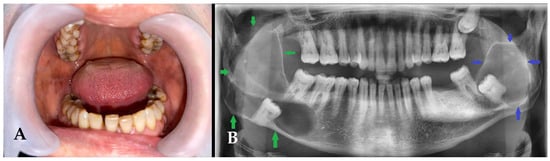
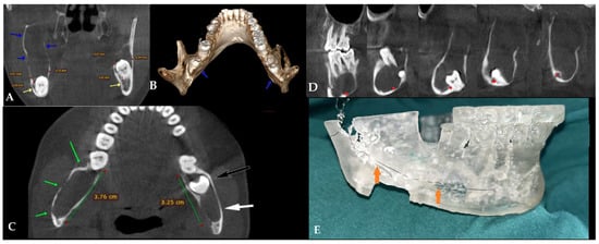
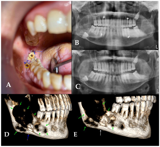
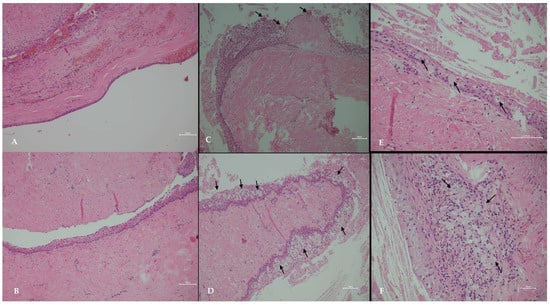
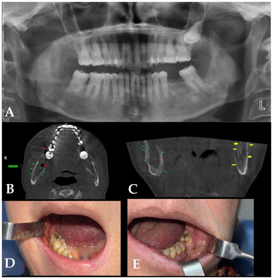
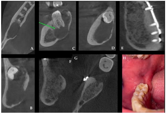
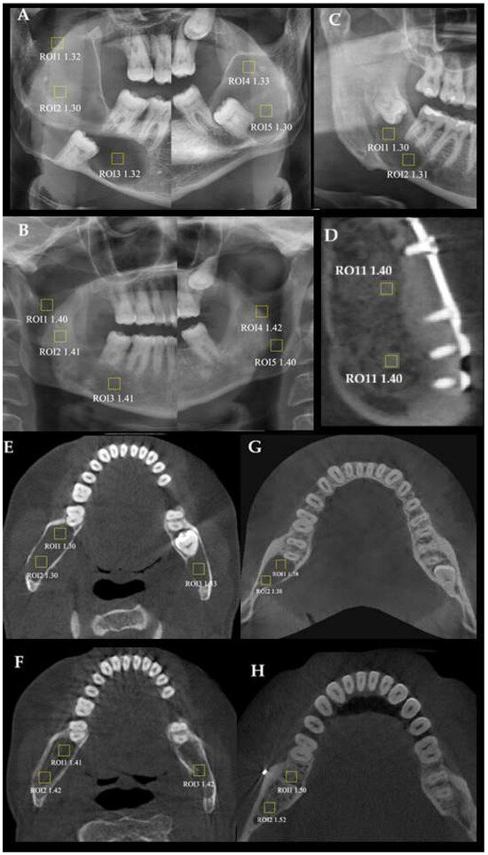
Background and Clinical Significance: Mandibular cystic lesions are heterogeneous in biologic behavior, radiologic appearance, and surgical management. Treatment selection is influenced by lesion extent, cortical bone condition, structural stability, and proximity to adjacent teeth and vital anatomical structures. In large mandibular lesions, case-specific decision-making may range from staged decompression or marsupialization to single-stage enucleation, peripheral ostectomy, graft-assisted reconstruction, and preventive mandibular plating (PMP). Case Presentation: In the first case, a 60-year-old female presented with bilateral mandibular lesions: a dentigerous cyst on the right side, treated with marsupialization followed by enucleation, and a contralateral glandular odontogenic cyst (GOC) managed with primary enucleation alone. The second case involved a large, unilateral odontogenic keratocyst (OKC) managed with a radical approach, including enucleation followed by peripheral ostectomy and PMP. Histopathologic evaluation was performed in both cases to confirm diagnosis and support definitive treatment planning. Radiologic follow-up at 1 year demonstrated bone regeneration in all treated sites. Conclusions: This case report presents two different clinical cases involving three mandibular cystic lesions managed according to case-specific surgical indications and evaluated with standardized postoperative panoramic and CBCT imaging; limited supportive assessment of trabecular bone remodeling was also performed to further characterize radiologic healing patterns. The authors aimed to descriptively document postoperative bone condition, radiologic healing, and patient safety in these cases, and to highlight the most important surgical and radiological findings that may influence case-specific clinical decision-making.
Full article
(This article belongs to the Special Issue Diagnosis and Management of Dental Medicine and Surgery, 2nd Edition)
►▼
Show Figures

Figure 1
Journal Menu
► ▼ Journal Menu-
- Diagnostics Home
- Aims & Scope
- Editorial Board
- Reviewer Board
- Topical Advisory Panel
- Instructions for Authors
- Special Issues
- Topics
- Sections & Collections
- Article Processing Charge
- Indexing & Archiving
- Editor’s Choice Articles
- Most Cited & Viewed
- Journal Statistics
- Journal History
- Journal Awards
- Society Collaborations
- Conferences
- Editorial Office
Journal Browser
► ▼ Journal BrowserHighly Accessed Articles
Latest Books
E-Mail Alert
News
Topics
Topic in
Biology, JCM, Diagnostics, Dentistry Journal
Assessment of Craniofacial Morphology: Traditional Methods and Innovative Approaches
Topic Editors: Nikolaos Gkantidis, Carlalberta VernaDeadline: 1 June 2026
Topic in
Diagnostics, Electronics, Eng, Micro, Micromachines
Micro-Nanoelectronic Systems for Diagnosis and Therapies
Topic Editors: Wubin Bai, Jiho ShinDeadline: 30 June 2026
Topic in
Applied Sciences, Diagnostics, JCM, Clinics and Practice, Therapeutics
Advances in Diagnosis and Treatment of Dental Diseases and Temporomandibular Joints
Topic Editors: Rafał Obuchowicz, Małgorzata Pihut, Karolina Nurzynska, Andrzej UrbanikDeadline: 31 July 2026
Topic in
Cancers, Diagnostics, Gastrointestinal Disorders, JCM, Current Oncology
Metastatic Colorectal Cancer: From Laboratory to Clinical Studies, 2nd Edition
Topic Editors: Ioannis Ntanasis-Stathopoulos, Diamantis I. TsilimigrasDeadline: 20 August 2026
Conferences
Special Issues
Special Issue in
Diagnostics
Trends and Novelties in Bone Imaging
Guest Editor: Kamyar MoradiDeadline: 31 May 2026
Special Issue in
Diagnostics
Advances in the Use of Artificial Intelligence for the Diagnosis and Management of Hand Conditions
Guest Editor: Ishith SethDeadline: 31 May 2026
Special Issue in
Diagnostics
Advanced Diagnosis and Integrated Management of Bone and Soft Tissue Tumors
Guest Editor: Tiao LinDeadline: 31 May 2026
Special Issue in
Diagnostics
Advances in the Diagnosis and Management of Idiopathic Normal Pressure Hydrocephalus
Guest Editors: Elisabeth Kapaki, Efstratios-Stylianos PyrgelisDeadline: 31 May 2026
Topical Collections
Topical Collection in
Diagnostics
Clinical Guidelines/Expert Consensus on Diagnostics
Collection Editor: Zhongheng Zhang
Topical Collection in
Diagnostics
Diagnosis of Endometriosis: Biomarkers and Clinical Methods
Collection Editor: Antonio Simone Laganà
Topical Collection in
Diagnostics
Nuclear Medicine and Molecular Imaging Technology
Collection Editor: Andreas Kjaer