Osteology 2024, 4(2), 53-63; https://doi.org/10.3390/osteology4020005 - 22 Apr 2024
Abstract
A proportion of osteoarthritis (OA) patients are unsatisfied with post-operative outcomes following total joint replacement surgery (TJR), with insufficient pain relief or poor functional improvement. Predicting those who will have poor outcomes would be beneficial for patients and clinicians. The aim of this
[...] Read more.
A proportion of osteoarthritis (OA) patients are unsatisfied with post-operative outcomes following total joint replacement surgery (TJR), with insufficient pain relief or poor functional improvement. Predicting those who will have poor outcomes would be beneficial for patients and clinicians. The aim of this study was to determine the relationship between baseline anthropometric data and the concentration of pre-operative serum and peri-operative synovial fluid (SF) cytokines and 7-month post-operative outcomes in a cohort of knee and hip OA patients. 160 OA patients were recruited who were scheduled for TJR. The concentration of 24 cytokines was measured in blood and SF by multiplex assay. EQ5D index health status was assessed pre-operatively and at 7 months post-operatively. 13% of patients were identified as non-responders based on EQ5D index. Compared to responders, non-responders were of higher body mass index (BMI), had greater waist and hip circumference, and had higher levels of SF leptin but lower levels of SF resistin (p < 0.05). Linear regression analysis found a significant but weak relationship between pre-operative body weight and post-operative response (ΔEQ5D index; r = 0.222, p = 0.049). The combination of body weight with SF amphiregulin and SF IL-6 provided an improved predictive model of post-operative response (r = 0.470, p = 0.035).
Full article
(This article belongs to the Special Issue New Trends in Arthroplasty)
►
Show Figures
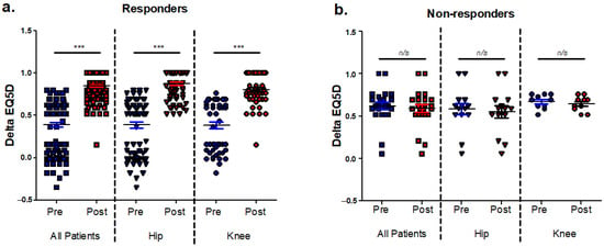
Figure 1


