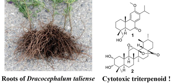
Communication

Cytotoxic Terpenoids from the Roots of Dracocephalum taliense

College of Bioscience and Biotechnology, Shenyang Agricultural University, Shenyang 110866, China
*
Author to whom correspondence should be addressed.
Molecules 2018, 23(1), 57; https://doi.org/10.3390/molecules23010057
Submission received: 5 November 2017 / Revised: 12 December 2017 / Accepted: 14 December 2017 / Published: 27 December 2017
(This article belongs to the Collection Herbal Medicine Research)

Abstract

:
A chemical investigation of methanol extract from the roots of Dracocephalum taliense led to the isolation of a new aromatic abietane diterpenoid, 12-methoxy-18-hydroxy-sugiol (1), and one highly-oxygenated ursane triterpenoid, 2α,3α-dihydroxy-11α,12α-epoxy-urs-28,13β-olide (2), together with 15 known natural products (317). Among these, compounds 113 and 1517 were detected for the first time in the genus of Dracocephalum. The structures of all of these isolates were determined by extensively spectroscopic analyses. In the anti-inflammatory assay, compounds 1 and 2 had no obvious inhibitory activity on the release of cytokine IL-2 in lipopolysaccharide-induced RAW 264.7 macrophages. However, compound 2 exhibited significant cytotoxic activity against cell lines HepG2 (IC50 = 6.58 ± 0.14 μM) and NCI-H1975 (IC50 = 7.17 ± 0.26 μM).

Graphical Abstract

1. Introduction

Terpenoids represent the most widespread and largest class of natural products, with more than 80,000 metabolites in a greater family that also includes steroids and carotenoids [1]. Most terpenoids serve a variety of roles in mediating mutualistic and antagonistic interactions among organisms in the natural world [2]. In flowering plants, terpenoids also constitute a large group of naturally-occurring specialized metabolites, especially as common substances in the Lamiaceae family. For example, abundant diterpenoids with diverse structural scaffolds and important pharmaceutical functions have been discovered in the genus Isodon [3,4,5]. The genus Salvia is a rich source of structurally-diverse abietane and clerodane diterpenoids [6]. A series of uniquely defensive sesterterpenoids have been found in the species of Leucosceptrum canum and Colquhounia coccinea var. mollis by our previous phytochemical investigation [7,8,9,10]. As a result, the chemical and structural diversity of terpenoids and their biological activities are interesting for research.
Dracocephalum is a herbaceous genus belonging to the Lamiaceae family and is widely distributed in China. Some plants in this genus, including D. moldavica and D. tanguticum, are broadly used in traditional medicine for gastropathy, tracheitis, and cardiovascular disease in Xinjiang and Tibet [11,12]. Typical metabolites in the plants of this genus were flavonoids and terpenoids [13,14,15,16]. Monoterpenes limonene and α-terpineol might be responsible for antinociceptive properties in the essential oil of D. kotschyi [17]. Icetexane diterpenoids, as the main constitutes in D. komarovi and D. kotschyi, showed moderate trypanocidal activity [16,18,19]. D. taliense is a perennial plant with thick cylindric roots and bluish violet flowers on short stalks. It grows in sandy deposits, cliffs, and rocky slopes. It is endemic in distribution in Dali and Shangri-la regions of Yunnan Province (Figure 1). The whole plant of D. taliense is used by local people to treat liver disorders, which mainly exhibited good effects on hepatitis and icterus, and also applied for adjusting the stomach [20,21]. However, there is nearly no literature reporting the chemical constituents of this plant. Here, we present the isolation and structural elucidation of the chemical components from the roots of D. taliense and their cytotoxic and anti-inflammatory activities.

2. Results and Discussion

2.1. Structural Elucidation of Compounds

Compound 1 (Figure 2) was obtained as a light yellow oil. Its molecular formula is C21H30O3 according to the 13C NMR spectroscopic and high resolution electrospray ionization mass spectroscopy (HRESIMS) data (found: m/z 353.2092 [M + Na]+; calcd. for 353.2087). The 1H NMR spectra data of 1 (Table 1, Supplementary Materials Figure S1) exhibited signals for a tetra-substituted benzene ring with protons at δH 6.95 and 7.78, two singlet methyls at δH 1.02 and 1.27, two secondary methyls at δH 1.17 (3H, d, J = 7.0 Hz) and 1.19 (3H, d, J = 7.0 Hz), one methoxyl signal at δH 3.94, and other signals between 1.04 and 3.24 ppm. The 13C NMR and distortionless enhanced polarization transfer (DEPT) spectra (Supplementary Materials Figure S2) demonstrated 21 carbon resonances which were assigned to five methyls, five methylenes (with one oxygenated), four methines, and seven quaternary carbons (with one carbonyl carbon at δC 197.2). These 20 typically skeletal carbons indicated that compound 1 was a characteristic abietane-type diterpenoid [22,23]. In the heteronuclear multiple bond coherence (HMBC) spectrum of 1 (Figure 3), the olefinic proton signal at δH 7.78 (H-14) showed correlations to the carbonyl carbon at δC 197.2 (C-7) and the quaternary carbon at δC 162.3 (C-12), and the methoxyl signal exhibited correlation with the quaternary carbon at δC 162.3, indicating the carbonyl moiety and methoxyl group substituted in C-7 and C-12, respectively. The HMBC correlations of the oxygenated methylenes at δH 3.61 and 3.81 to δC 36.2 (C-3), 39.2 (C-4), 51.1 (C-5), and methyl carbon at δC 27.2 indicated oxygenation of either Me-18 or Me-19. From the 2D rotational nuclear Overhauser effect spectroscopy (ROESY) spectrum of 1, the correlation between δH 1.27 (Me-20) and 1.02 (Me-19), δH 3.61 (H-18b) and 1.94 (H-5) were observed that confirmed the oxygenation of Me-18. Thus, the chemical structure of 1 was confirmed and named 12-methoxy-18-hydroxy-sugiol.
Compound 2 was isolated as a white amorphous solid and has a molecular formula of C30H46O5, as determined from its HRESIMS molecular ion at m/z 509.3234 ([M + Na]+; calcd. for 509.3237), accounting to eight double-bond equivalents. The 1H NMR spectrum of 2 (Table 1, Supplementary Materials Figure S7) showed seven methyl signals, including two secondary methyls (δH 1.14 (3H, d, J = 6.6 Hz) and 0.84 (3H, d, J = 7.2 Hz)) and five singlet methyls (δH 1.15, 1.07, 1.05, 1.01, and 0.87), suggesting that compound 2 was an ursane-type triterpenoid [24]. Analysis of 13C NMR (Supplementary Materials Figure S8) and heteronuclear single-quantum correlation (HSQC) spectra revealed 30 carbons corresponding to one carbonyl resonance (δC 179.5), six quaternary carbon (with one oxygenated at δC 89.2), nine methines (with four oxygenated), seven methylenes, and seven methyls. Based on the aforementioned 1H and 13C NMR spectra data indicated that compound 2 was a highly-oxygenated ursane triterpenoid [24]. Comparing the 13C NMR data (Table 1) of 2 with those of 2α,3β-dihydroxy-11α,12α-epoxy-urs-28,13β-olide [24] showed that 2 had the same planar structure. The only difference was that the C-3 (δC 83.7) in 2α,3β-dihydroxy-11α,12α-epoxy-urs-28,13β-olide shifted upfield to δC 78.7, accounting the β orientation of 3-OH changed to α in 2, which was further confirmed by ROESY correlations of H-3 with H-2 and Me-24, Me-25 with H-2 and H-3, and low coupling constant value of 3J2-3 (1.9 Hz). Finally, compound 2 was characterized as shown in Figure 2, and was named as 2α,3α-dihydroxy-11α,12α-epoxy-urs-28,13β-olide.
Twelve known diterpenoids, including sugiol (3) [23], abieta-8,11,13-triene (4) [25], dehydroabietane (5) [26], ferruginol (6) [27], cryptojapanol (7) [28], inuroyleanol (8) [22], callitrisic acid (9) [29], 11,14-dihydroxy-12,19-dimethoxy-7-oxo-8,11,13-abietatrien-19,20-olide (10) [30], totarol (11) [31], 7α-hydroxytotarol (12) [32], sempervirol (13) [33], and cyclocoulterone (14) [16], and three steroids, including (22E)-ergosta-6,9,22-triene-3β,5β,8α-triol (15) [34], (22E)-ergosta-6,22-diene-3β,5β,8α-triol (16) [34], and stigmast-4-en-6β-ol-3-one (17) [35], were also isolated and identified by comparison of their spectroscopic data with the literature.

2.2. Bioactivities

Compounds 1 and 2 were assayed for their cytotoxic activity against different cell lines NCI-H1975, HepG2, and MCF-7, using a previously described MTS (3-(4,5-dimethylthiazol-2-yl)-5(3-carboxymethoxyphenyl)-2-(4-sulfopheny)-2H-tetrazolium) method [36]. It was evident that only compound 2 exhibited significant cytotoxic activity, with IC50 values of 7.17 ± 0.26 and 6.58 ± 0.14 μM against cell lines NCI-H1975 and HepG2, respectively, which were still less active than the positive control (Table 2). In the anti-inflammatory assay, both of the two compounds did not show obvious inhibitory activity on the release production of cytokine IL-2 in lipopolysaccharide-induced RAW 264.7 macrophages.
Abietane diterpenoids, as a large group of secondary metabolites, exhibited diversely biological properties including antitumour, antituberculostatic, antiplatelet aggregation, and anti-inflammatory activities [28]. 11,14-Dihydroxy-12,19-dimethoxy-7-oxo-8,11,13-abietatrien-19,20-olide (10) showed inhibition effects against the secretion of LTC4 which was more potent than the ketotifen used as a positive control [30]. 7α-Hydroxytotarol (12) exhibited cytotoxic activity against wide human cancer cell lines and could also inhibit the growth of Gram-positive bacteria and fungi [32]. The icetexane type diterpenoid, cyclocoulterone (14) showed moderate trypanocidal activity against epmastigotes of Trypanosoma cruzi [16].

3. Experimental

3.1. General

Optical rotations were obtained on a Jasco P-1020 spectropolarimeter (Jasco, Tokyo, Japan). UV spectroscopic data were measured on a Shimadzu-210A double-beam spectrophotometer (Shimadzu, Tokyo, Japan). IR spectra of samples in KBr discs were recorded on a Bruker-Tensor-27 spectrometer (Bruker, Karlsruhe, Germany) with KBr pellets. Mass spectra were obtained on an Agilent Q-TOF 6200 spectrometer (Agilent Technologies, Santa Clara, CA, USA). Column chromatographies were performed on 200–300 mesh silica gel (Qingdao Marine Chemical Factory, Qingdao, China), or Sephadex LH-20 (Amersham Phamacia Biotech, Uppsala, Sweden), or MCI gel CHP-20P (75–150 μm, Mitsubishi Chemical Corp., Tokyo, Japan). NMR spectra were measured on a Bruker Avance-600 spectrometer (Bruker, Karlsruhe, Germany) in deuterated solvent with TMS as the internal standard. Semi-preparative HPLC was performed on an Agilent 1260 series instrument (Agilent, Santa Clara, CA, USA) equipped with a quaternary pump, an autosampler, a vacuum degasser, a thermostatted column compartment, a diode array detector and an Eclipse XDB-C18 column (5 μm, 9.4 × 250 mm). TLC spots were visualized under UV light and by spraying with 5% H2SO4 in EtOH, followed by heating.

3.2. Plant Material

The roots of D. taliense were collected from Shangri-la in Yunnan Province in August 2016 and identified by Dr. Chunlei Xiang. An authentic sample (SYAU-2016-0246) was kept at the College of Bioscience and Biotechnology, Shenyang Agricultural University.

3.3. Extraction and Isolation

Dried and powdered roots of D. taliense (4.0 kg) were extracted with methanol at room temperature. The crude extract was concentrated in vacuo to obtain 40 g of methanol extract. This fraction was chromatographed on a silica gel column, eluting successively with a solvent gradient system (dichloromethane/acetone, 1:0–0:1) to give six fractions (Frs. A–F). Fr. A (3.7 g) was further subjected to MCI gel column chromatography (methanol/water, 60:40–100:0) to obtain five subfractions (Frs. A1–A5). Fr. A4 (0.25 g) was subjected to column chromatography on silica gel, eluting with petroleum ether/acetone (92:8–0:100) and purified by a Sephadex LH-20 (acetone as eluent) to yield 6 (3.2 mg) and 11 (5.2 mg). Fr. A3 (0.42 g) was chromatographed on Sephadex LH-20 column (acetone as eluent) and then purified by semi-preparative HPLC (methanol/water, 75:25, 3 mL/min) to give 4 (3.0 mg, tR 25.9 min), 7 (5.5 mg, tR 14.7 min), and 13 (3.5 mg, tR 17.1 min). Fr. B (1.5 g) was subjected to column chromatography on silica gel, eluting with petroleum ether/ethyl acetate (85:15) and recrystallized with acetone to yield 17 (55.1 mg).
Fr. C (7.9 g) was further subjected to MCI gel column chromatography (methanol/water, 50:50–100:0) to obtain four subfractions (Frs. C1–C4). Fr. C1 was recrystallized with methanol to yield 2 (7.5 mg). Fr. C2 was subjected to Sephadex LH-20 column (methanol as eluent) and then purified by semi-preparative HPLC (methanol/water, 66:34, 3 mL/min) to give 1 (5.0 mg, tR 15.6 min), 3 (3.6 mg, tR 17.6 min), 9 (21.7 mg, tR 21.5 min), 10 (8.6 mg, tR 11.3 min), and 12 (7.8 mg, tR 12.7 min). Fr. C2 was subjected to column chromatography on silica gel, eluting with petroleum ether/acetone (7:1) and then purified by semi-preparative HPLC (methanol/water, 70:30, 3 mL/min), to yield 5 (4.5 mg, tR 15.9 min), 8 (5.3 mg, tR 6.7 min), and 14 (12.5 mg, tR 10.4 min). Subfraction Fr. C3 was applied to a silica gel column eluted with petroleum ether/acetone (9:1) and purified by semi-preparative HPLC (methanol/water, 78:22, 3 mL/min) to yield 15 (5.2 mg, tR 8.9 min) and 16 (9.8 mg, tR 9.6 min).

3.4. Spectroscopic Data

12-Methoxy-18-hydroxy-sugiol (1): light yellow oil; [α]D25 = +55.6 (c = 0.1, MeOH); UV (MeOH) λmax (log ε): nm 215 (4.16), 270 (3.88), 364 (2.81); IR (KBr, cm−1): νmax 3441, 2942, 1637, 1602, 1455, 1375, 1241, 1167, 913; 1H and 13C NMR data, see Table 1; HRESIMS: m/zobsd 353.2092 [M + Na]+ (m/zcalcd [C21H30O3Na]+ = 353.2087).
2α,3α-Dihydroxy-11α,12α-epoxy-urs-28,13β-olide (2): white amorphous solid; [α]D25 = +37.6 (c = 0.1, MeOH); UV (MeOH) λmax (log ε): 202 (3.85) nm; IR (KBr, cm−1): νmax 3443, 2937, 1778, 1633, 1460, 1390, 1141, 1043; 1H and 13C NMR data, see Table 1; HRESIMS: m/zobsd 509.3234 [M + Na]+ (m/zcalcd [C30H46O5Na]+ = 509.3237).
Sugiol (3): light yellow solid; 13C NMR (acetone-d6, 150 MHz) δ: 38.6 (t, C-1), 19.6 (t, C-2), 42.1 (t, C-3), 33.8 (s, C-4), 50.5 (d, C-5), 36.4 (t, C-6), 196.9 (s, C-7), 124.4 (s, C-8), 156.9 (s, C-9), 38.6 (s, C-10), 110.4 (d, C-11), 160.7 (s, C-12), 133.7 (s, C-13), 126.4 (d, C-14), 27.4 (d, C-15), 22.8 (q, C-16), 22.6 (q, C-17), 32.9 (q, C-18), 21.6 (q, C-19), 23.5 (q, C-20).
Abieta-8,11,13-triene (4), yellow solid; 13C NMR (acetone-d6, 150 MHz) δ: 39.7 (t, C-1), 20.0 (t, C-2), 42.4 (t, C-3), 34.0 (s, C-4), 51.5 (d, C-5), 19.8 (t, C-6), 30.5 (t, C-7), 132.6 (s, C-8), 153.0 (s, C-9), 38.1 (s, C-10), 120.9 (d, C-11), 111.3 (d, C-12), 148.8 (s, C-13), 127.1 (d, C-14), 27.4 (d, C-15), 23.0 (q, C-16), 22.9 (q, C-17), 33.6 (q, C-18), 21.9 (q, C-19), 25.2 (q, C-20).
Dehydroabietane (5), yellow oil; 13C NMR (acetone-d6, 150 MHz) δ: 39.4 (t, C-1), 19.4 (t, C-2), 35.9 (t, C-3), 38.5 (s, C-4), 44.3 (d, C-5), 19.4 (t, C-6), 30.7 (t, C-7), 135.6 (s, C-8), 148.4 (s, C-9), 38.0 (s, C-10), 124.4 (d, C-11), 125.0 (d, C-12), 146.0 (s, C-13), 127.4 (d, C-14), 34.2 (d, C-15), 24.4 (q, C-16), 24.3 (q, C-17), 71.5 (t, C-18), 18.0 (q, C-19), 25.6 (q, C-20).
Ferruginol (6), yellow oil; 13C NMR (acetone-d6, 150 MHz) δ: 39.7 (t, C-1), 20.0 (t, C-2), 42.4 (t, C-3), 34.0 (s, C-4), 51.5 (d, C-5), 20.0 (t, C-6), 30.5 (t, C-7), 126.3 (s, C-8), 148.8 (s, C-9), 38.2 (s, C-10), 111.3 (d, C-11), 153.1 (s, C-12), 132.6 (s, C-13), 127.1 (d, C-14), 27.4 (d, C-15), 22.9 (q, C-16), 23.0 (q, C-17), 34.0 (q, C-18), 21.9 (q, C-19), 25.2 (q, C-20).
Cryptojapanol (7), light yellow solid, 13C NMR (methanol-d4, 150 MHz) δ: 37.4 (t, C-1), 20.0 (t, C-2), 42.4 (t, C-3), 34.4 (s, C-4), 52.1 (d, C-5), 36.5 (t, C-6), 201.8 (s, C-7), 129.4 (s, C-8), 140.6 (s, C-9), 41.6 (s, C-10), 149.2 (s, C-11), 152.0 (s, C-12), 140.9 (s, C-13), 117.3 (d, C-14), 27.8 (d, C-15), 23.9 (q, C-16), 23.8 (q, C-17), 33.6 (q, C-18), 21.9 (q, C-19), 17.8 (q, C-20), 61.7 (q, 12-OMe).
Inuroyleanol (8), light yellow solid; 13C NMR (acetone-d6, 150 MHz) δ: 37.2 (t, C-1), 19.7 (t, C-2), 41.9 (t, C-3), 34.0 (s, C-4), 50.7 (d, C-5), 36.3 (t, C-6), 206.9 (s, C-7), 113.0 (s, C-8), 137.5 (s, C-9), 41.2 (s, C-10), 140.6 (s, C-11), 154.2 (s, C-12), 126.8 (s, C-13), 158.4 (s, C-14), 26.4 (d, C-15), 20.8 (q, C-16), 20.7 (q, C-17), 33.4 (q, C-18), 21.8 (q, C-19), 17.7 (q, C-20), 62.1 (q, 12-OMe).
Callitrisic acid (9), yellow solid, 13C NMR (methanol-d4, 150 MHz) δ: 38.8 (t, C-1), 21.2 (t, C-2), 40.8 (t, C-3), 44.8 (s, C-4), 54.4 (d, C-5), 22.4 (t, C-6), 33.2 (t, C-7), 136.1 (s, C-8), 146.8 (s, C-9), 39.5 (s, C-10), 126.5 (d, C-11), 124.9 (d, C-12), 146.7 (s, C-13), 127.7 (d, C-14), 34.8 (d, C-15), 24.5 (q, C-16), 24.5 (q, C-17), 181.5 (s, C-18), 29.3 (q, C-19), 23. 8 (q, C-20).
11,14-Dihydroxy-12,19-dimethoxy-7-oxo-8,11,13-abietatrien-19,20-olide (10), yellow solid; 13C NMR (acetone-d6, 150 MHz) δ: 35.5 (t, C-1), 22.4 (t, C-2), 40.1 (t, C-3), 36.8 (s, C-4), 45.1 (d, C-5), 37.9 (t, C-6), 206.0 (s, C-7), 113.7 (s, C-8), 130.1 (s, C-9), 39.8 (s, C-10), 141.8 (s, C-11), 154.2 (s, C-12), 127.7 (s, C-13), 158.0 (s, C-14), 26.4 (d, C-15), 20.7 (q, C-16), 20.7 (q, C-17), 23.4 (q, C-18), 106.0 (d, C-19), 60.0 (t, C-20), 62.1 (q, 12-OMe), 54.9 (q, 19-OMe).
Totarol (11), yellow oil; 13C NMR (acetone-d6, 150 MHz) δ: 40.4 (t, C-1), 20.2 (t, C-2), 42.3 (t, C-3), 33.8 (s, C-4), 50.7 (d, C-5), 20.1 (t, C-6), 29.5 (t, C-7), 133.8 (s, C-8), 142.4 (s, C-9), 38.3 (s, C-10), 123.5 (d, C-11), 115.0 (d, C-12), 154.2 (s, C-13), 131.3 (s, C-14), 28.1 (d, C-15), 20.5 (q, C-16), 20.5 (q, C-17), 33.6 (q, C-18), 21.9 (q, C-19), 25.6 (q, C-20).
7α-Hydroxytotarol (12), yellow oil; 13C NMR (acetone-d6, 150 MHz) δ: 43.7 (t, C-1), 20.6 (t, C-2), 43.9 (t, C-3), 34.6 (s, C-4), 53.3 (d, C-5), 40.7 (t, C-6), 65.3 (d, C-7), 131.4 (s, C-8), 141.6 (s, C-9), 38.1 (s, C-10), 124.2 (d, C-11), 115.2 (d, C-12), 154.2 (s, C-13), 131.2 (s, C-14), 28.2 (d, C-15), 20.6 (q, C-16), 20.5 (q, C-17), 34.1 (q, C-18), 23.8 (q, C-19), 27.4 (q, C-20).
Sempervirol (13), yellow oil; 13C NMR (acetone-d6, 150 MHz) δ: 39.9 (t, C-1), 20.0 (t, C-2), 42.5 (t, C-3), 33.9 (s, C-4), 51.5 (d, C-5), 19.9 (t, C-6), 30.8 (t, C-7), 133.7 (s, C-8), 141.9 (s, C-9), 38.1 (s, C-10), 122.7 (d, C-11), 132.8 (s, C-12), 152.6 (s, C-13), 115.3 (d, C-14), 27.9 (d, C-15), 23.1 (q, C-16), 23.0 (q, C-17), 33.7 (q, C-18), 21.9 (q, C-19), 25.4 (q, C-20).
Cyclocoulterone (14), light yellow solid, 13C NMR (acetone-d6, 150 MHz) δ: 39.3 (t, C-1), 19.6 (t, C-2), 41.8 (t, C-3), 34.7 (s, C-4), 50.8 (d, C-5), 40.7 (t, C-6), 209.9 (s, C-7), 113.2 (s, C-8), 116.7 (s, C-9), 73.2 (s, C-10), 140.1 (s, C-11), 151.5 (s, C-12), 117.0 (s, C-13), 161.2 (s, C-14), 25.1 (d, C-15), 21.0 (q, C-16), 20.9 (q, C-17), 32.3 (q, C-18), 21.8 (q, C-19), 42.2 (t, C-20), 101.9 (t, –OCH2O–).
(22E)-Ergosta-6,9,22-triene-3β,5β,8α-triol (15), white solid, 13C NMR (CDCl3, 150 MHz) δ: 32.6 (t, C-1), 30.6 (t, C-2), 66.3 (d, C-3), 36.1 (t, C-4), 82.7 (s, C-5), 135.4 (d, C-6), 130.7 (d, C-7), 78.3 (s, C-8), 142.5 (s, C-9), 37.9 (s, C-10), 119.7 (d, C-11), 41.2 (t, C-12), 43.6 (s, C-13), 48.1 (d, C-14), 20.9 (t, C-15), 28.6 (t, C-16), 55.8 (d, C-17), 13.0 (q, C-18), 25.5 (q, C-19), 39.9 (d, C-20), 20.7 (q, C-21), 135.1 (d, C-22), 132.4 (d, C-23), 43.2 (d, C-24), 33.0 (d, C-25), 19.6 (q, C-26), 19.9 (q, C-27), 17.5 (q, C-28).
(22E)-Ergosta-6,22-diene-3β,5β,8α-triol (16), white solid, 13C NMR (CDCl3, 150 MHz) δ: 34.7 (t, C-1), 30.1 (t, C-2), 66.4 (d, C-3), 36.9 (t, C-4), 82.1 (s, C-5), 130.7 (d, C-6), 135.4 (d, C-7), 79.4 (s, C-8), 51.0 (d, C-9), 36.9 (s, C-10), 20.6 (t, C-11), 39.3 (t, C-12), 44.5 (s, C-13), 51.7 (d, C-14), 28.6 (t, C-15), 23.4 (t, C-16), 56.2 (d, C-17), 12.8 (q, C-18), 18.2 (q, C-19), 39.7 (d, C-20), 19.6 (q, C-21), 132.3 (d, C-22), 135.2 (d, C-23), 42.7 (d, C-24), 33.0 (d, C-25), 19.9 (q, C-26), 17.5 (q, C-27), 20.9 (q, C-28).
Stigmast-4-en-6β-ol-3-one (17), colorless oil, 13C NMR (acetone-d6, 150 MHz) δ: 38.0 (t, C-1), 34.8 (t, C-2), 199.4 (s, C-3), 126.3 (d, C-4), 169.2 (s, C-5), 73.4 (d, C-6), 39.9 (t, C-7), 30.6 (d, C-8), 54.7 (d, C-9), 38.7 (s, C-10), 21.7 (t, C-11), 40.5 (t, C-12), 43.2 (s, C-13), 56.7 (d, C-14), 24.8 (t, C-15), 28.9 (t, C-16), 56.9 (d, C-17), 12.2 (q, C-18), 19.6 (q, C-19), 36.9 (d, C-20), 19.3 (q, C-21), 34.6 (t, C-22), 26.7 (t, C-23), 46.7 (d, C-24), 29.9 (d, C-25), 20.1 (q, C-26), 19.1 (q, C-27), 23.7 (t, C-28), 12.3 (q, C-29).

3.5. Cytotoxic Assay

The in vitro cytotoxic activity of compounds 1 and 2 were determined by the MTS method [36]. The tested human cancer cell lines, including human lung adenocarcinoma cell line (NCI-H1975), human hepatocellular carcinoma cell line (HepG2), and human breast adenocarcinoma cell line (MCF-7), were seeded in 96-well plates, and then the plates were incubated for 24 h at 37 °C in a 5% CO2 incubator. Subsequently, the compounds were added at a dosage of 0, 0.128, 0.256, 0.512, 1, 2, 5, 10, 20, 40, and 80 μM. After 72 h, MTS was added to the culture medium and the absorbance at 490 nm using a microplate reader (Bio-Rad, Hercules, CA, USA). Each sample was carried out in triplicate. The evaluation of IC50 values were calculated with the GraphPad Prism 5.01 (GraphPad Software Inc., San Diego, CA, USA) software.

3.6. Anti-Inflammatory Assay

The murine macrophage RAW 264.7 cell line was cultured in DMEM medium supplemented with 10% heated-inactivated fetal bovine serum in a 37 °C, 5% CO2 incubator. Before the anti-inflammatory assay, test compounds were assessed for their cytotoxicity against the RAW 264.7 cell line and were found to be non-toxic at the tested concentrations (40, 20, 10, 5, and 0 μM). Anti-inflammatory activity was assessed by enzyme-linked immunosorbent assay (ELISA, BD Biosciences, Mountain View, CA, USA) using commercial interleukin-2 (IL-2) detecting kits as previously described [37].

3.7. Statistical Analysis

The results were presented as mean values ± SD (standard deviations) of the three replicates.

4. Conclusions

Previous phytochemical investigation has revealed that flavonoids and terpenoids were the major constituents of genus Dracocephalum [11]. Biological studies on these flavonoids have revealed their broad pharmacological activities, especially antioxidant, immunomodulatory, and cytotoxic activities, as well as cardiovascular protective effects [11,13,14,15,38,39]. Trypanocidal diterpenoids with icetexane and octahydroindene skeletons were discovered from the whole plants of D. komarovi [16,19]. In this work, a new aromatic abietane diterpenoid, and one highly-oxygenated ursane triterpenoid, together with 15 known compounds, belonging to abietane (313) and icetexane (14) diterpenoids, and steroids (1517), were isolated from the roots of D. taliense. Based on literature reviews, all the chemical constituents, except compound 14, were detected for the first time in the genus of Dracocephalum. Although, compounds 1 and 2 were inactive on the release of cytokine IL-2 in lipopolysaccharide-induced RAW 264.7 macrophages, compound 2 showed significant cytotoxic activity against cell lines HepG2 and NCI-H1975. The results increase the chemical diversity and bioactive constituents of secondary metabolites produced by D. taliense.

Supplementary Materials

Supplementary materials are available online. NMR spectra data (Figures S1–S12) of compounds 1 and 2 are available in the Supplementary Material.

Acknowledgments

This research was supported financially by the National Key Research and Development (R&D) Program of China (2017YFD0201400, 2017YFD0201403) and the Talent Program of Shenyang Agricultural University (awarded to Shihong Luo).

Conflicts of Interest

The authors declare no conflict of interest.

References

  1. Christianson, D.W. Structural and chemical biology of terpenoid cyclases. Chem. Rev. 2017, 117, 11570–11648. [Google Scholar] [CrossRef] [PubMed]
  2. Gershenzon, J.; Dudareva, N. The function of terpene natural products in the natural world. Nat. Chem. Biol. 2007, 3, 408–414. [Google Scholar] [CrossRef] [PubMed]
  3. Liu, M.; Wang, W.G.; Sun, H.D.; Pu, J.X. Diterpenoids from Isodon species: An update. Nat. Prod. Rep. 2017, 34, 1090–1140. [Google Scholar] [CrossRef] [PubMed]
  4. Dai, L.P.; Li, C.; Yang, H.Z.; Lu, Y.Q.; Yu, H.Y.; Gao, H.M.; Wang, Z.M. Three new cytotoxic ent-kaurane diterpenes from Isodon excisoides. Molecules 2015, 20, 17544–17556. [Google Scholar] [CrossRef] [PubMed]
  5. Wan, J.; Jiang, H.Y.; Tang, J.W.; Li, X.R.; Du, X.; Li, Y.; Sun, H.D.; Pu, J.X. ent-Abietanoids isolated from Isodon serra. Molecules 2017, 22, 309. [Google Scholar] [CrossRef] [PubMed]
  6. Wu, Y.B.; Ni, Z.Y.; Shi, Q.W.; Dong, M.; Kiyota, H.; Gu, Y.C.; Cong, B. Constituents from Salvia species and their biological activities. Chem. Rev. 2012, 112, 5967–6026. [Google Scholar] [CrossRef] [PubMed]
  7. Luo, S.H.; Qian, L.; Niu, X.M.; Xie, M.J.; Xu, Z.; Schneider, B.; Gershenzon, J.; Li, S.H. Glandular trichomes of Leucosceptrum canum harbor defensive sesterterpenoids. Angew. Chem. Int. Ed. 2010, 122, 4471–4475. [Google Scholar] [CrossRef] [PubMed]
  8. Luo, S.H.; Hua, J.; Li, C.H.; Jing, S.X.; Liu, Y.; Li, X.N.; Zhao, X.; Li, S.H. New antifeedant C20 terpenoids from Leucosceptrum canum. Org. Lett. 2012, 14, 5768–5771. [Google Scholar] [CrossRef] [PubMed]
  9. Luo, S.H.; Hugelshofer, C.L.; Hua, J.; Jing, S.X.; Li, C.H.; Liu, Y.; Li, X.N.; Zhao, X.; Magauer, T.; Li, S.H. Unraveling the metabolic pathway in Leucosceptrum canum by isolation of new defensive leucosceptroid degradation products and biomimetic model synthesis. Org. Lett. 2014, 16, 6416–6419. [Google Scholar] [CrossRef] [PubMed]
  10. Li, C.H.; Jing, S.X.; Luo, S.H.; Shi, W.; Hua, J.; Liu, Y.; Li, X.N.; Schneider, B.; Gershenzon, J.; Li, S.H. Peltate glandular trichomes of Colquhounia coccinea var. mollis harbor a new class of defensive sesterterpenoids. Org. Lett. 2013, 15, 1694–1697. [Google Scholar] [CrossRef] [PubMed]
  11. Zeng, Q.; Jin, H.Z.; Qin, J.J.; Fu, J.J.; Hu, X.J.; Liu, J.H.; Yan, L.; Chen, M.; Zhang, W.D. Chemical constituents of plants from the genus Dracocephalum. Chem. Biodivers. 2010, 7, 1911–1929. [Google Scholar] [CrossRef] [PubMed]
  12. Ren, D.M.; Guo, H.F.; Yu, W.T.; Wang, S.Q.; Ji, M.; Lou, H.X. Stereochemistry of flavonoidal alkaloids from Dracocephalum rupestre. Phytochemistry 2008, 69, 1425–1433. [Google Scholar] [CrossRef] [PubMed]
  13. Selenge, E.; Murata, T.; Kobayashi, K.; Batkhuu, J.; Yoshizaki, F. Flavone tetraglycosides and benzyl alcohol glycosides from the Mongolian medicinal plant Dracocephalum ruyschiana. J. Nat. Prod. 2013, 76, 186–193. [Google Scholar] [CrossRef] [PubMed]
  14. Saeidnia, S.; Gohari, A.R.; Ito, M.; Kiuchi, F.; Honda, G. Bioactive constituents from Dracocephalum subcapitatum (O. Kuntze) Lipsky. Z. Naturforsch. C 2005, 60, 22–24. [Google Scholar] [CrossRef]
  15. Jahaniani, F.; Ebrahimi, S.A.; Rahbar-Roshandel, N.; Mahmoudian, M. Xanthomicrol is the main cytotoxic component of Dracocephalum kotschyii and a potential anti-cancer agent. Phytochemistry 2005, 66, 1581–1592. [Google Scholar] [CrossRef] [PubMed]
  16. Uchiyama, N.; Kiuchi, F.; Ito, M.; Honda, G.; Takeda, Y.; Khodzhimatov, O.K.; Ashurmetov, O.A. New icetexane and 20-norabietane diterpenes with trypanocidal activity from Dracocephalum komarovi. J. Nat. Prod. 2003, 66, 128–131. [Google Scholar] [CrossRef] [PubMed]
  17. Golshani, S.; Karamkhani, F.; Monsefesfehani, H.R.; Abdollahi, M. Antinociceptive effects of the essential oil of Dracocephalum kotschyi in the mouse writhing test. J. Pham. Sci. 2004, 7, 76–79. [Google Scholar]
  18. Saeidnia, S.; Gohari, A.R.; Uchiyama, N.; Ito, M.; Honda, G.; Kiuchi, F. Two new monoterpene glycosides and trypanocidal terpenoids from Dracocephalum kotschyi. Chem. Pharm. Bull. 2004, 52, 1249–1250. [Google Scholar] [CrossRef] [PubMed]
  19. Uchiyama, N.; Ito, M.; Kiuchi, F.; Honda, G.; Takeda, Y.; Khodzhimatov, O.K.; Ashurmetov, O.A. A trypanocidal diterpene with novel skeleton from Dracocephalum komarovi. Tetrahedron Lett. 2004, 45, 531–533. [Google Scholar] [CrossRef]
  20. Wu, Z.Y.; Li, X.W. Dracocephalum Linnaeus; Science Press: Beijing, China, 1994; Volume 17, pp. 127–133. [Google Scholar]
  21. Yang, Z.Y. Illustrated Handbook for Medicinal Materials from Nature in Yunnan; Yunnan Science and Technology Press: Yunnan, China, 2007; Volume 4, p. 35. [Google Scholar]
  22. Frontana, B.; Cárdenas, J.; Rodríguez-Hahn, L. Diterpenoids from Salvia coulteri. Phytochemistry 1994, 36, 739–741. [Google Scholar] [CrossRef]
  23. Gao, J.J.; Han, G.Q. Cytotoxic abietane diterpenoids from Caryopteris incana. Phytochemistry 1997, 44, 759–761. [Google Scholar]
  24. Jiao, K.; Li, H.Y.; Zhang, P.; Pi, H.F.; Ruan, H.L.; Wu, J.Z. Three new ursane-type triterpenoids from the aerial parts of Isodon excisoides. J. Asian Nat. Prod. Res. 2013, 15, 962–968. [Google Scholar] [CrossRef] [PubMed]
  25. Corral, J.M.M. D.; Gordaliza, M.; Salinero, M.A.; Feliciano, A.S. 13C NMR data for abieta-8,11,13-triene diterpenoids. Magn. Reson. Chem. 1994, 32, 774–781. [Google Scholar] [CrossRef]
  26. Fraga, B.M.; Hernández, M.G.; Artega, J.M.; Suárez, S. The microbiological transformation of the diterpenes dehydroabietanol and teideadiol by Mucor plumbeus. Phytochemistry 2003, 63, 663–668. [Google Scholar] [CrossRef]
  27. Kelecom, A. An abietane diterpene from the labiate Coleus barbatus. Phytochemistry 1984, 23, 1677–1679. [Google Scholar] [CrossRef]
  28. Rodríguez, B. 1H and 13C NMR spectral assignments of some natural abietane diterpenoids. Magn. Reson. Chem. 2003, 41, 741–746. [Google Scholar] [CrossRef]
  29. Zhang, H.; Sun, H. Diterpenoids from Rabdosia kunmingensis. Phytochemistry 1989, 28, 3405–3409. [Google Scholar]
  30. Furukawa, M.; Makino, M.; Ohkoshi, E.; Uchiyama, T.; Fujimoto, Y. Terpenoids and phenethyl glucosides from Hyssopus cuspidatus (Labiatae). Phytochemistry 2011, 72, 2244–2252. [Google Scholar] [CrossRef] [PubMed]
  31. Ying, B.P.; Kubo, I. Complete 1H and 13C NMR assignments of totarol and its derivatives. Phytochemistry 1991, 30, 1951–1955. [Google Scholar] [CrossRef]
  32. Pettit, G.R.; Tan, R.; Northen, J.S.; Herald, D.L.; Chapuis, J.C.; Pettit, R.K. Antineoplastic agents. 529. Isolation and structure of nootkastatins 1 and 2 from the Alaskan yellow cedar Chamaecyparis nootkatensis. J. Nat. Prod. 2004, 67, 1476–1482. [Google Scholar] [CrossRef] [PubMed]
  33. Mangoni, L.; Caputo, R. Sempervirol, a novel type of diterpene phenol. Tetrahedron Lett. 1967, 8, 673–675. [Google Scholar] [CrossRef]
  34. Song, L.; Shi, J.G.; Lin, S.; Yang, Y.C.; Yao, C.S. Chemical constituents from the linseed meal. Fitoterapia 2014, 97, 15–22. [Google Scholar] [CrossRef] [PubMed]
  35. Niu, X.; Li, S.; Peng, L.; Lin, Z.; Rao, G.; Sun, H. Constituents from Limonia Crenulata. J. Asian Nat. Prod. Res. 2001, 3, 299–311. [Google Scholar] [CrossRef] [PubMed]
  36. Lin, J.; Niu, S.; Ding, Z.; Wang, R.; Dai, Q.; Wei, W.; Luo, R.; Liu, L. Isolation and characterization of aphidicolin derivatives from Tolypocladium inflatum. Molecules 2017, 22, 1168. [Google Scholar] [CrossRef] [PubMed]
  37. Hua, J.; Liu, Y.C.; Jing, S.X.; Luo, S.H.; Li, S.H. Macrocyclic diterpenoids from the latex of Euphorbia helioscopia. Nat. Prod. Commun. 2015, 10, 2037–2039. [Google Scholar] [PubMed]
  38. Selenge, E.; Murata, T.; Tanaka, S.; Sasaki, K.; Batkhuu, J.; Yoshizaki, F. Monoterpene glycosides, phenylpropanoids, and acacetin glycosides from Dracocephalum foetidum. Phytochemistry 2014, 101, 91–100. [Google Scholar] [CrossRef] [PubMed]
  39. Olennikov, D.N.; Chirikova, N.K.; Okhlopkova, Z.M.; Zulfugarov, I.S. Chemical composition and antioxidant activity of Tanara Oto (Dracocephalum palmatum Stephan), a medicinal plant used by the North-Yakutian nomads. Molecules 2013, 18, 14105–14121. [Google Scholar] [CrossRef] [PubMed]
Sample Availability: Samples of the compounds 117 are available from the authors.
Figure 1. The ecological habitats (A) and roots (B) of D. taliense.
Molecules 23 00057 g001
Figure 2. Chemical structures of compounds 117 from roots of D. taliense.
Molecules 23 00057 g002
Figure 3. Key HMBC correlations (H→C) of compounds 1 and 2.
Molecules 23 00057 g003
Table 1. 1H (600 MHz) and 13C (150 MHz) NMR spectral data of compounds 1 and 2.
Position1 a2 b
δH (ppm), (Hz)δC (ppm)δH (ppm), (Hz)δC (ppm)
1a2.44, m38.8, t1.91, m41.5, t
1b1.55, m1.48, m
2a1.79, m19.4, t4.07, m66.0, d
2b1.61, m
3a1.96, m36.2, t3.46, d (1.9)78.7, d
3b1.04, m
4-39.2, s-38.2, s
51.94, m51.1, d1.20, m47.4, d
6a2.70, m36.5, t1.47, m (2H)17.3, t
6b2.64, m
7a-197.2, s1.29, m31.2, t
7b1.14, m
8-124.7, s-41.6, s
9-157.3, s1.70, d (1.5)51.0, d
10-39.0, s-37.7, s
116.95, s106.0, d3.14, dd (1.5, 3.7)54.4, d
12-162.3, s2.94, d (3.7)56.2, d
13-135.4, s-89.2, s
147.78, s125.5, d-41.2, s
15a3.24, m27.1, d1.71, m26.7, t
15b1.54, m
16a1.17, d (3H, 7.0)22.7, q2.22, m22.4, t
16b1.31, m
171.19, d (3H, 7.0)22.8, q-45.0, s
18a3.81, d (10.7)64.7, t1.92, m53.9, d
18b3.61, d (10.7)
191.02, s (3H)27.2, q2.30, m32.9, d
201.27, s (3H)23.9, q1.87, m34.6, d
21a 1.51, m28.2, t
21b 1.05, m
22a 1.68, m25.5, t
22b 1.58, m
23 1.01, s (3H)28.2, q
24 0.87, s (3H)21.3, q
25 1.07, s (3H)18.5, q
26 1.05, s (3H)20.3, q
27 1.15, s (3H)16.0, q
28 -179.5, s
29 1.14, d (3H, 6.6)18.2, q
30 0.84, d (3H, 7.2)11.1, q
12-OMe3.94, s (3H)56.0, q
a recorded in acetone-d6; b recorded in CDCl3.
Table 2. Cytotoxicity and Anti-inflammatory activity of compounds from D. taliense.
NoCytotoxicity to Different Cell Lines/IC50 (μM)Inhibitory Activity on Inflammatory Cytokine (μM)
NCI-H1975HepG2MCF-7IL-2
1>80>80>80>40
27.17 ± 0.266.58 ± 0.14>80>5
PC a(6.82 ± 0.24) × 10−3(34.72 ± 2.31) × 10−3(54.35 ± 7.72) × 10−3(2.38 ± 0.28) × 10−2
a Taxol was used as a positive control (PC) for cytotoxic assay; cyclosporine A was used as a positive control for anti-inflammatory assay. NCI-H1975, human lung adenocarcinoma cell line; HepG2, human hepatocellular carcinoma cell line; MCF-7, human breast adenocarcinoma cell line. Values are mean ± SD (n = 3).