Article

Potent in Vitro α-Glucosidase Inhibition of Secondary Metabolites Derived from Dryopteris cycadina

1
Department of Botany, Islamia College University, Peshawar 25000, Pakistan
2
Department of Chemistry, University of Malakand, Upper Dir 23050, Pakistan
3
Department of Chemistry, University of Swabi, Khyber Pakhtunkhwa, Anbar 23430, Pakistan
4
Department of Pharmacy, Abdul Wali Khan University, Mardan 23200, Pakistan
5
Instituto de Ciencias Químicas Aplicadas, Universidad Autónoma de Chile, Santiago 7500912, Chile
6
Departamento de Química Orgánica, Facultad de Farmacia, Universidad de Santiago de Compostela, 15782 Santiago de Compostela, Spain
7
Instituto de Investigación e Innovación en Salud, Facultad de Ciencias de la Salud, Universidad Central de Chile, Santiago 8330507, Chile
8
Laboratory of Pharmaceutical Chemistry, Faculty of Pharmacy, University of Santiago de Compostela, 15782 Santiago de Compostela, Spain
*
Authors to whom correspondence should be addressed.
Molecules 2019, 24(3), 427; https://doi.org/10.3390/molecules24030427
Submission received: 30 November 2018 / Revised: 19 January 2019 / Accepted: 21 January 2019 / Published: 24 January 2019
(This article belongs to the Section Natural Products Chemistry)

Abstract

:
α-glucosidase is responsible for the hydrolysis of complex carbohydrates into simple absorbable glucose and causes postprandial hyperglycemia. α-glucosidase inhibition is thus the ideal target to prevent postprandial hyperglycemia. The present study was therefore designed to analyze the effects of various compounds isolated from Dryopteris cycadina against α-glucosidase including β-Sitosterol 1, β-Sitosterol3-O-β-d-glucopyranoside 2, 3, 5, 7-trihydroxy-2-(p-tolyl) chorman-4-one 3, Quercetin-3-0-β-d-glucopyranoside (3/→0-3///)- β-d- Quercetin -3-0- β –d-galactopyranoside 4 and 5, 7, 4/-Trihydroxyflavon-3-glucopyranoid 5. The in vitro spectrophotometric method was used for the analysis of test compounds against possible inhibition. Similarly, molecular docking studies were performed using the MOE software. These compounds showed concentration-dependent inhibition on α-glucosidase, and compounds 1 (IC50: 143 ± 0.47 µM), 3 (IC50:133 ± 6.90 µM) and 5 (IC50: 146 ± 1.93 µM) were more potent than the standard drug, acarbose (IC50: 290 ± 0.54 µM). Computational studies of these compounds strongly supported the in vitro studies and showed strong binding receptor sensitivity. In short, the secondary metabolites isolated from D. cycadina demonstrated potent α-glucosidase inhibition that were supported by molecular docking with a high docking score.

Graphical Abstract

1. Introduction

Diabetes is characterized by persistent endocrine disorder of multiple etiologies caused by a comparatively low or complete absence of insulin [1]. It is characterized by hyperglycemia [2] with the irregular metabolism of carbohydrates, proteins, fats, and electrolytes due to a lack of insulin or the insensitivity of target cells to insulin [3], which results the elevation of blood sugar, leading to hyperglycemia [4]. Hyperglycemia is the initial metabolic disorder of type 2 diabetes mellitus and if left untreated may develop into serious complications like damaged organs and systems, particularly the eyes, kidneys, nerves, blood and cardiovascular system [5,6]. The major defect that occurs in early diabetes is postprandial hyperglycemia, which mainly occurs due to excessive eating, obesity, age and lack of exercise [7,8,9]. α-glucosidase (EC 3.2.1.20) is a hydrolytic enzyme, mainly secreted by cells lining in the brush borders of epithelial cells of small intestine [10,11]. α-glucosidase mainly target α-1→4 glycosidic linkages and is responsible for the hydrolysis of complex carbohydrates into simple absorbable glucose. α-glucosidase belong to the sub-subclass hydrolases that cause the hydrolysis of various substances in the body [12,13]. In the 1970s, α-glucosidase hydrolases were discovered that were used to recover postprandial hyperglycemia, and which were officially approved as an antidiabetic drug later in the 1990s [14,15]. In this context, more potent and tolerable agents could account for the effective management of the disease.
Dryopteris cycadina is a medicinal plant from the Dryopteridaceae family. The Dryopteris genus consists of 250 species of ferns and are mostly distributed in the temperate northern hemisphere of Eastern Asia [16]. A literature survey revealed numerous medicinal uses of the Dryopteris species in folk medicine [17]. It is helpful in the treatment of rheumatism, epilepsy, pain [18], and as a remedy for snake bites and fungal infections [19], as well as diabetes management. The isolated compounds from this plant are flavonoid glycosides, which possess antioxidant, antibacterial and antitumor properties, and HIV-1 reverse transcriptase inhibitory activity [20,21]. The chemical constituents in D. cycadina includes steroids, phenols, phenolic glycosides, flavonoid glycosides, flavonoids, terpenoids, phenylpropranoids and others [22,23]. Kaempferol-3, 4/-di-O-α- l-rhamnopyranoside and Kaempferol-3,7-di-O-α- l-rhamnopyranoside isolated from the D. cycadina plant showed remarkable antinociceptive activity [24,25].
Based on the strong pharmacological, phytochemical and traditional uses of different species of Dryopteris, and it’s potential as an antidiabetic, the current study was designed to test compounds 15 isolated from D. cycadina against α- glucosidase for possible inhibition, and computational studies were carried out to test receptor binding sensitivity.

2. Results

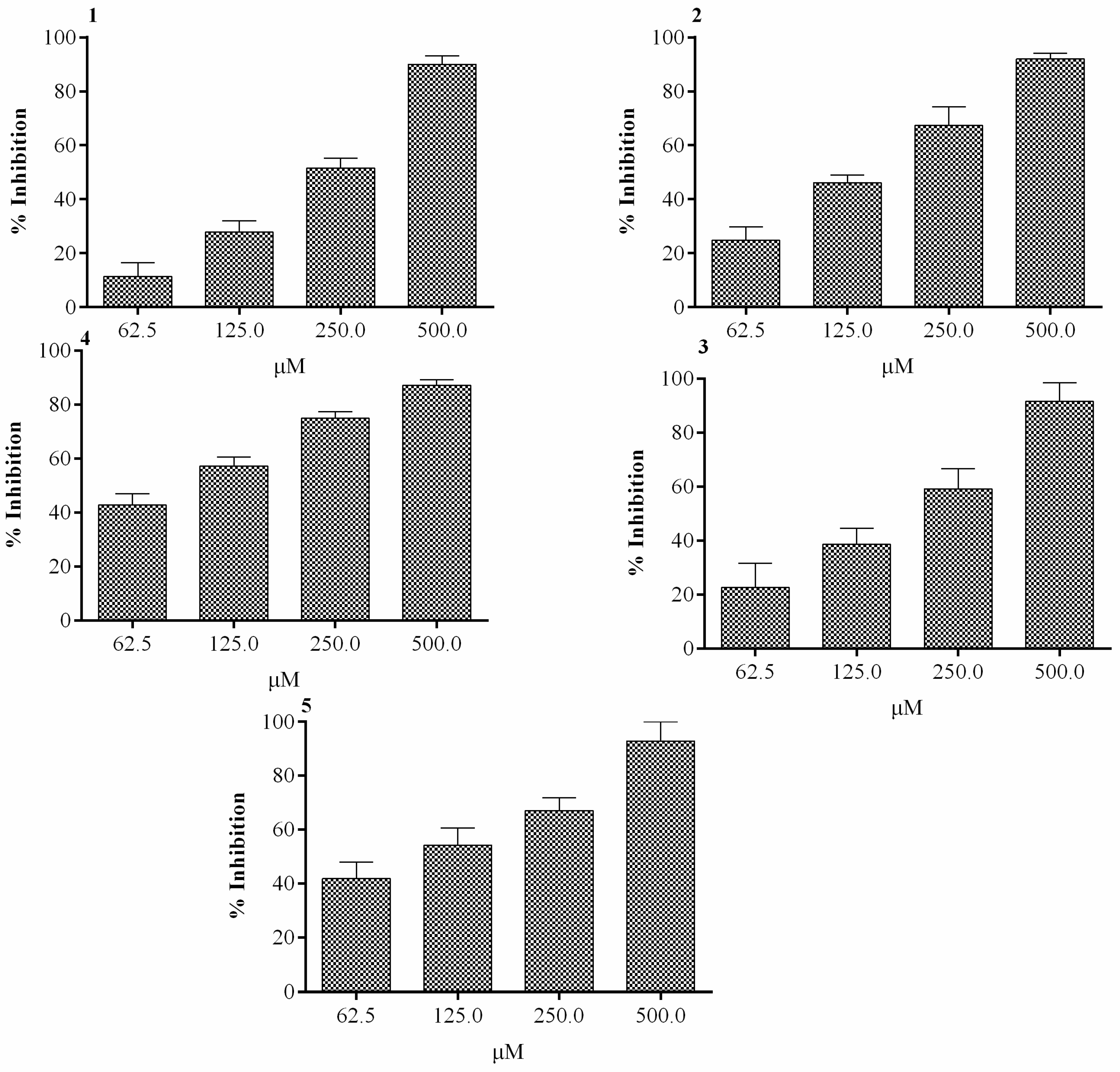
2.1. Effect of In Vitro α-Glucosidase Activity

Compounds 15 isolated from D. cycadina, as seen in Figure 1, were screened for in vitro α-glucosidase inhibitory activity. Compound 1, at a concentration of 500 μM, exhibited the maximum α-glucosidase inhibitory activity of 92.9% (Figure 2), while its half maximal inhibitory concentration was observed as 143 ± 0.47μM, as seen in Table 1. The maximum inhibition of compound 2 was 92% and was observed at 500 µM, as seen in Figure 2, with an IC50 value of 314 ± 4.58 μM, see Table 1. Compound 3 showed marked inhibitory activity against α-glucosidase at various concentrations, with a maximum inhibition of 94% at 500 µM, see Figure 2. The half maximal concentration (IC50) was calculated as 133 ± 6.90 µM, as shown in Table 1. The inhibitory activity showed by compound 4 against α-glucosidase was 87.2%, and was observed at a concentration of 500 µM, see Figure 2, and had an IC50 value of 298 ± 0.67µM, as seen in Table 1. Compound 5 demonstrated excellent inhibition of 97.1% at 500 µM, as seen in Figure 2, and its calculated IC50 was 146 ± 1.93 µM, see Table 1.

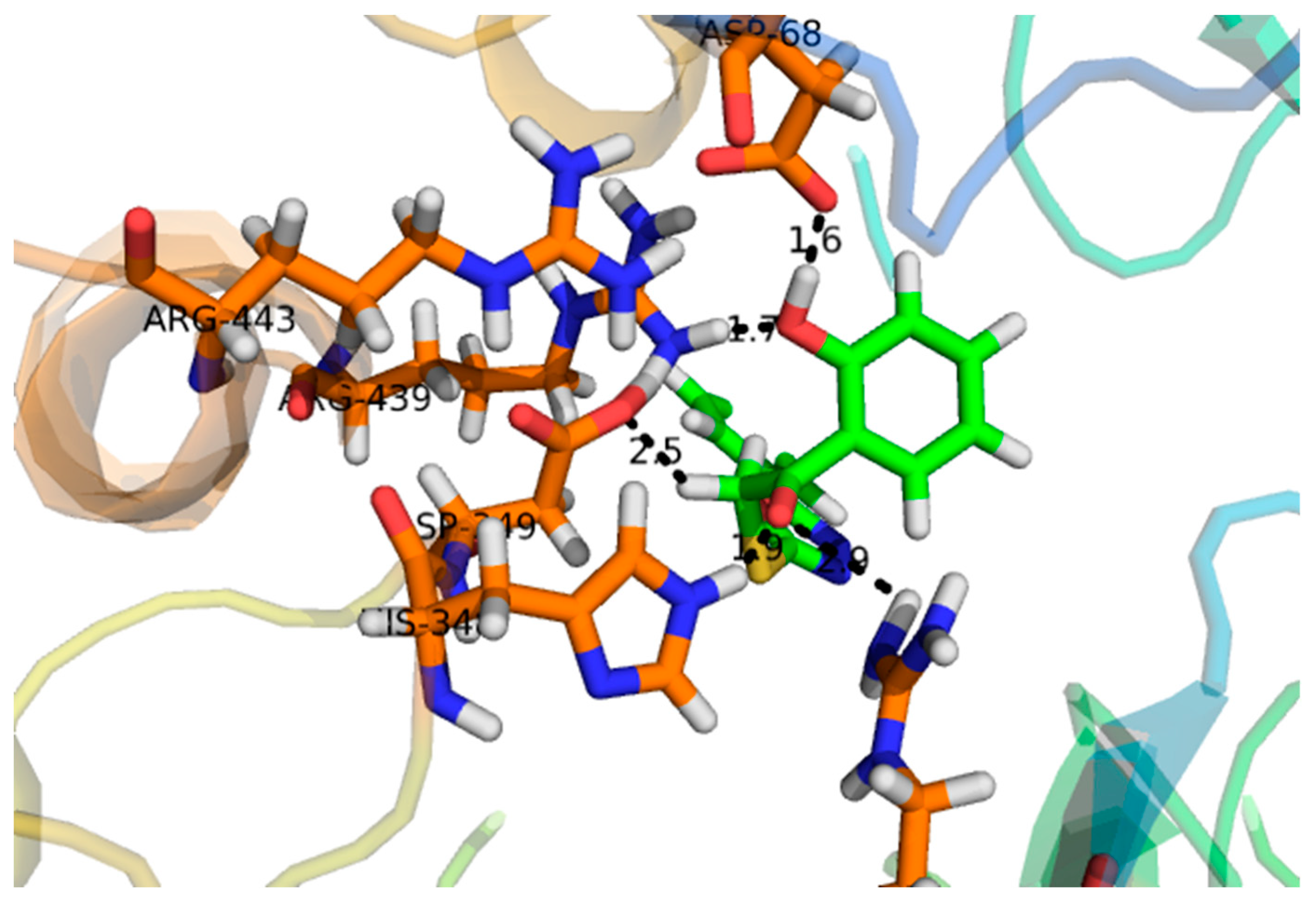
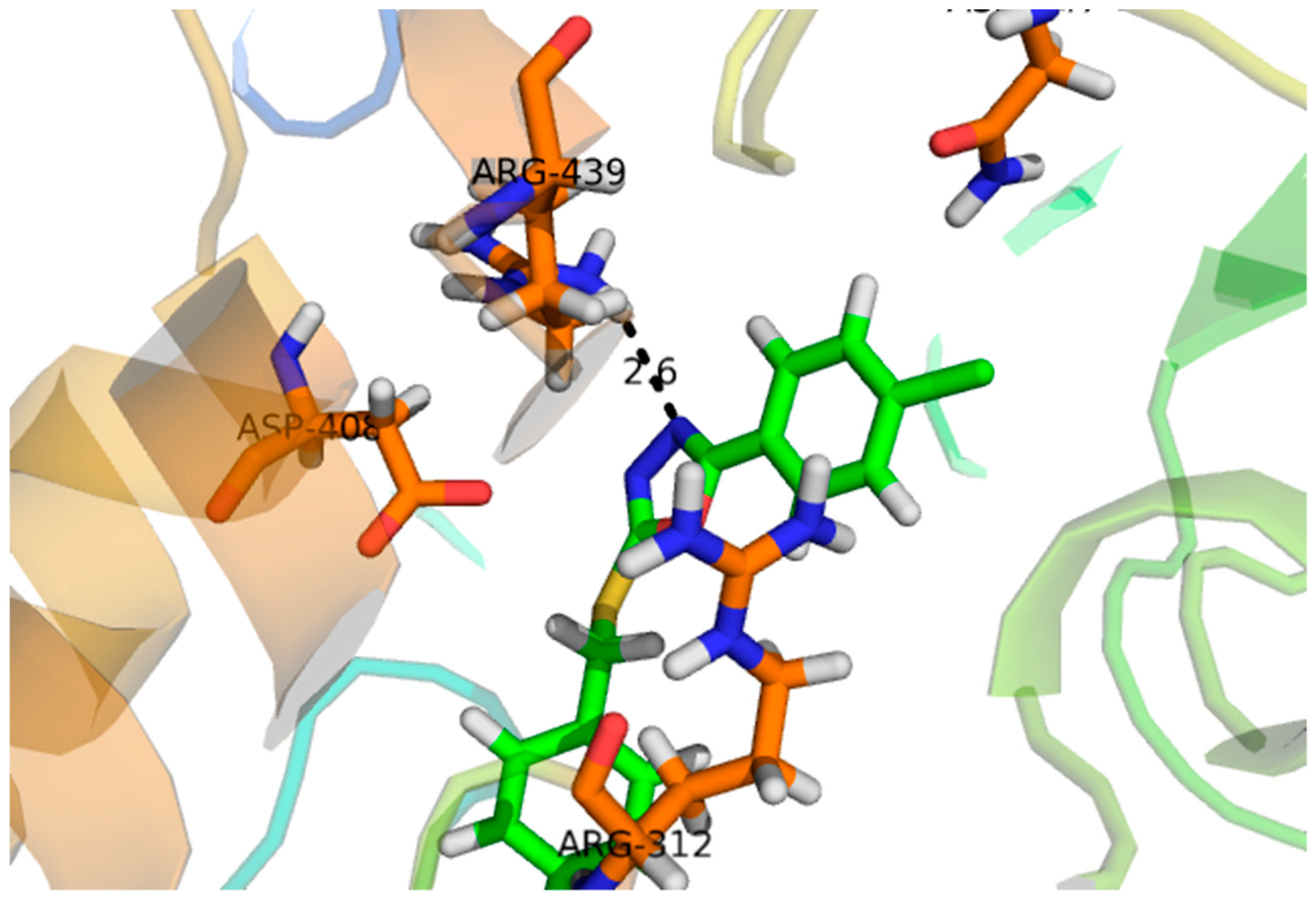
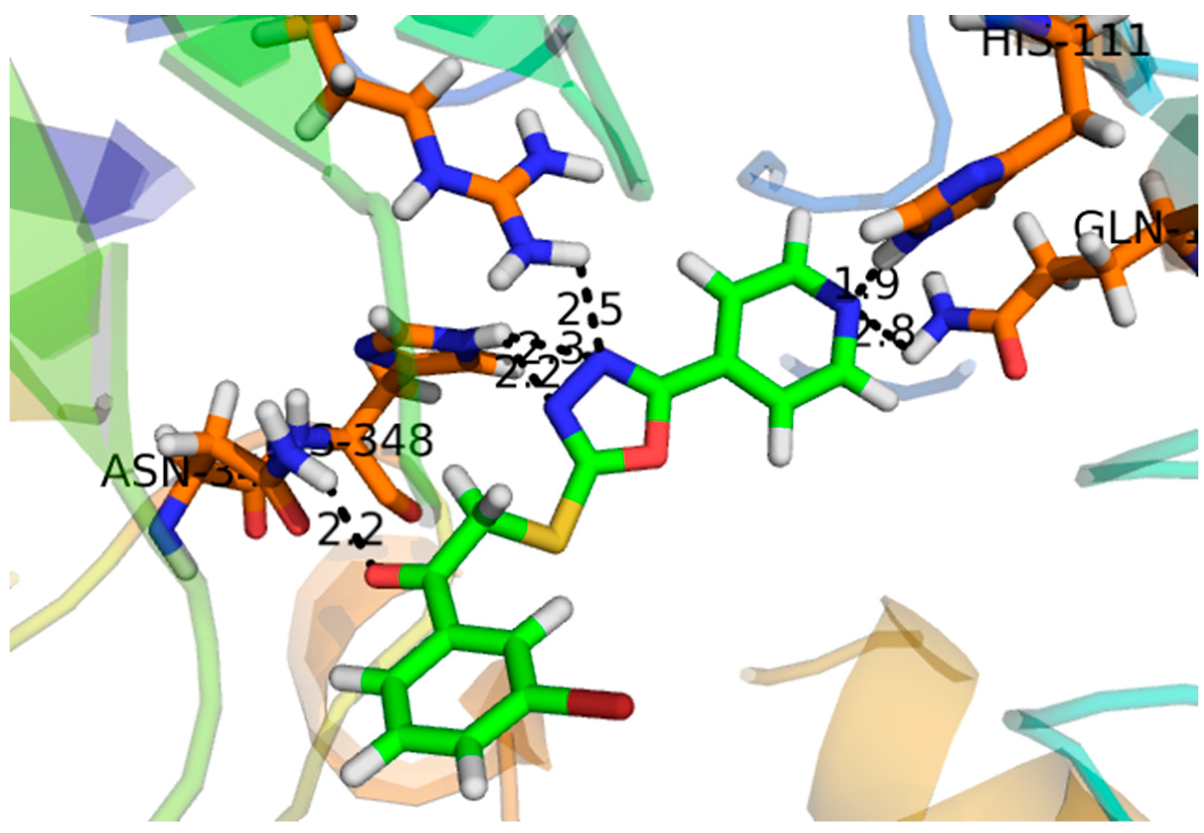
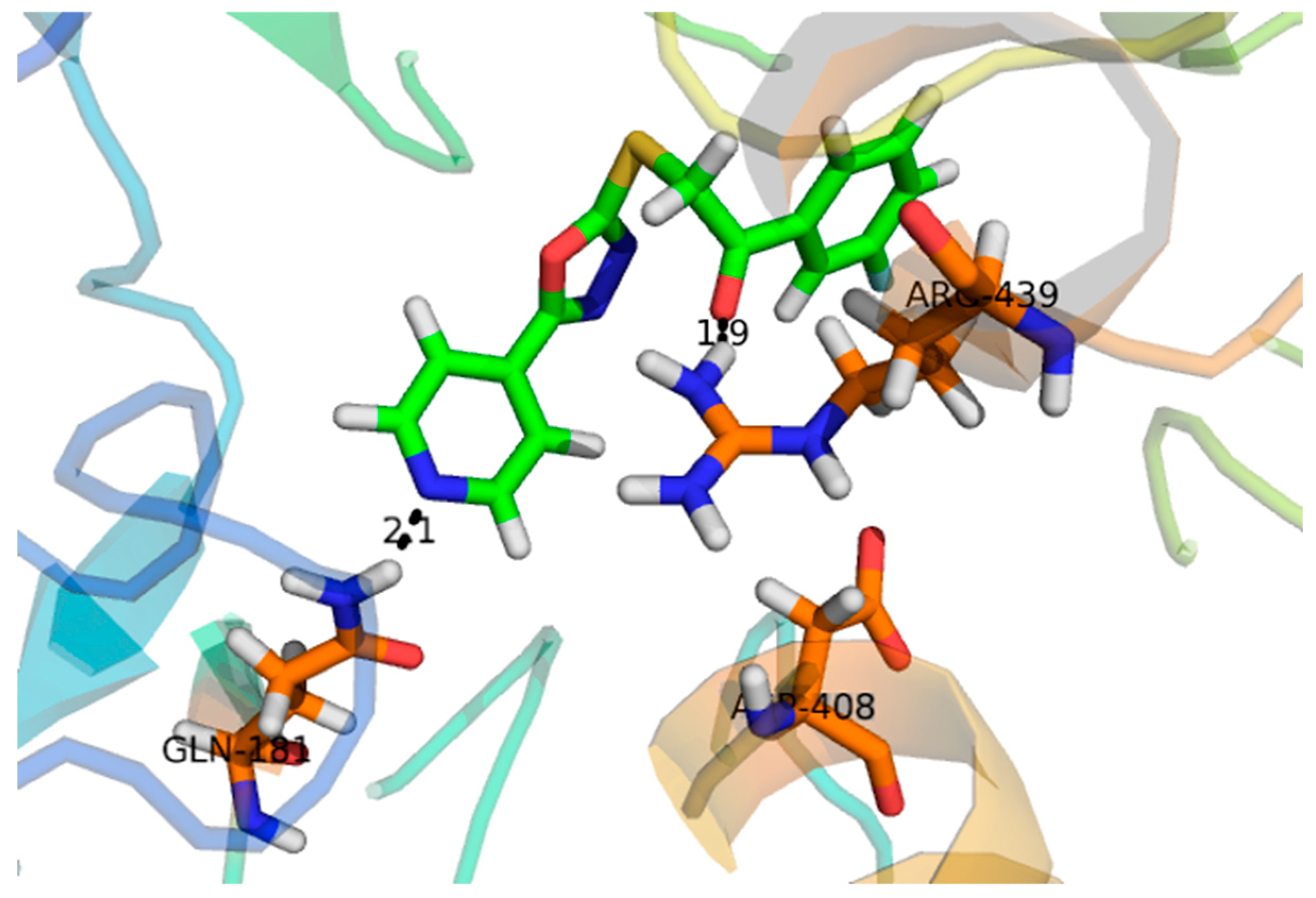
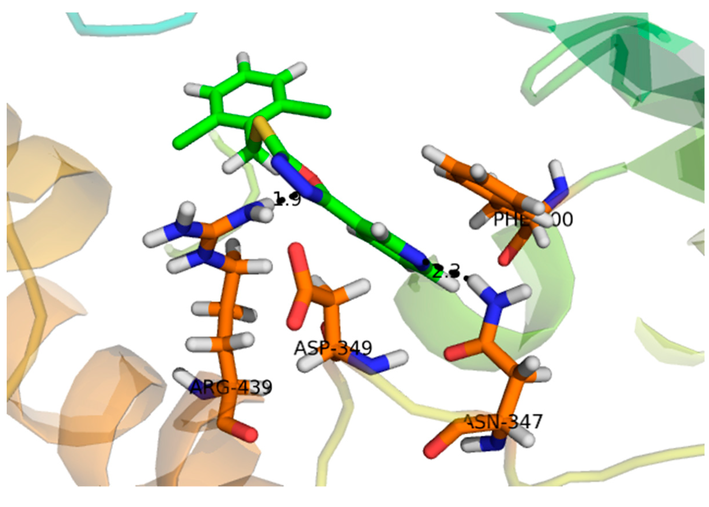
2.2. Effect of Molecular Docking

Compound 1, with a docking score of −16.097 (Table 2) displayed significant hydrogen bonding interactions with the four catalytic residues of Arg-442, Asp-215, Asp-352 and Gln-182 of the receptor α-glucosidase, as seen in Figure 3. Compound 2, with a docking score of −7.756, as seen in Table 2, showed one binding interaction with α-glucosidase. The hydrogen binding interaction was found with amino acid Asn-415 of α-glucosidase, see Figure 4. One arene–arene and eight hydrogen binding interactions were observed in compound 3 with a docking score of −22.480. The active site residues such as Arg-315, Asp-307, His- 280, Lys-156, Ser-240 and Thr-310 indicated hydrogen binding interactions, while Tyr-158 exhibited an arene–arene interaction with α-glucosidase, as seen in Figure 5. The docking score of compound 4 was −12.931, indicating that two different interactions were found, that is, one hydrogen bond and other arene–arene interaction with the active site residue Arg-442 and Tyr 158, as seen in Figure 6. Compound 5, with a docking score of −15.752, had one arene–arene and three hydrogen binding interactions—Asp-242, Lys-156, Pro-312 and Tyr-158—with the active site residue of the receptor (Figure 7).

3. Materials and Methods

3.1. Materials

α-glucosidase (EC3.2.1.20) was obtained from Sigma Aldrich, and acarbose was obtained from Bayer, Pakistan. An ELISA Micro Plate Reader (Emax) from Molecular Devices and isolated compounds 15 from D. cycadina were used.

3.2. Assay Protocol

The α-glucosidase (Saccharomyces cerevisiae) inhibitory assay was carried out with slight modifications according to the method described in [21,26,27]. For the evaluation of α-glucosidase inhibitory activity, 10 µl of freshly prepared phosphate buffer (pH 6.8) were plated in triplicate for a micro-well plate with the help of a micropipette. Then, 30 µL of α-glucosidase enzyme with a concentration of 0.017 units/mL of 70% ethanol and 10 µL of test compound solution were added to the same well plate. The plates were then incubated for 15 min at 37 °C and an initial reading was taken at 405 nm using the ELISA micro plate reader. Furthermore, 100 µl of 0.7mM PNPG substrate was added to each well of the plate and again incubated at 37 °C for 30 min, forming a yellow color. The final reading was taken again at λ max 405 nm using the ELISA plate reader. A total of 70% ethanol was used as a negative control, whereas acarbose was used as a positive control. Each reading was taken in triplicate. The percent inhibition was calculated using the following formula, % inhibition = [(A negative control − A test sample)/A negative control] × 100, where A is absorbance.

3.3. Half-Maximal Inhibitory Concentration of Compounds (IC50)

The compound that exhibited a 50% or greater inhibition on α-glucosidase was subjected to IC50 determination. The half-maximal inhibitory concentration (IC50) of the active compounds was determined by preparing various amounts of test solution—like 500 µM, 250 µM, 125 µM and 62.5 µM—and their inhibitory studies were determined using the method described earlier. The half-maximal inhibitory concentration values were determined using the Graphpad Prism version 7.0 software (San Diego, CA, USA. All values are represented as mean ± SEM.

3.4. Computational Study

The three-dimensional structure for α-glucosidase of Saccharomyces cerevisiae has not yet been solved. Thus, the three-dimensional structure of α-glucosidase was generated using the Molecular Operating Environment (MOE 2010.11) software and the molecular docking study was performed on the same software. The MOE-Dock was used as the docking software implemented in MOE and ligplot was implemented in MOE for the purpose of visualizing the interaction between protein and ligand. The primary sequence of the α glucosidase was retrieved using Uniprot (Universal Protein Resource) (http://www.uniprot.org/) in Federal Acquisition S Streamlining (FASTA) format and the target sequence was then kept in the text-file for further evaluation [28]. The accession number of α glucosidase of Saccharomyces cerevisiae was P07265. Then Protein-BLAST was performed to identify homologs in the PDB (RCSB Protein Databank) [9,29,30]. Hence, the crystal structure of Saccharomyces cerevisiae (PDB Id: 3A47_A), which has 72% sequence identity to the target protein, was selected as the template for the target protein sequence for the prediction of the tertiary structure of the target protein. The amino acid sequence of the target protein in FASTA format was copied and pasted into the sequence editor of the MOE software. Then the template protein was loaded into the same MOE software. Prior to docking, the 2D structures of all inhibitors were drawn using the Cambridge Soft Chem3D Ultra Version 10.0 by Cambridge Soft Corp, MA, USA. Protein-ligand docking studies were performed using the MOE 2009.10 software package. Ligands were optimized using the default parameters of the MOE-DOCK software, including energy minimization, protonation and the removal of nonpolar hydrogens. Now the entire ligand database was docked into the binding pocket of the protein using the triangular matching docking method. Ten different conformations of each ligand–protein complex was generated, each possessing its specific docking score. The docking process was repeated for the validation of the docking method for the type of interaction. Finally, the two- and three-dimensional images of each complex were analyzed and taken.

3.5. Statistical Analysis

All the data are expressed as the mean ± SEM of three independent readings. The IC50 values were calculated using the Graph Pad Prism version 7.0 software (San Diego, CA, USA), while the docking studies were performed using the MOE (2009-10) software.

4. Discussion

The present study revealed a significant in vitro α-glucosidase assay that was strongly complimented by computational studies. In vitro α-glucosidase assay is a simple, economical and time-saving method for the analysis of the inhibitory potential of various compounds. In the present study, the inhibitory activity of isolated bioactive constituents was determined by measuring its spectrophotometric absorbance at a wavelength of 405 nm using ELISA. Besides this, the color change of the reaction mixture from deep yellow to light yellow also indicated significant inhibition of the α-glucosidase [30,31].
Compound 3 was found to be the most active compound, with an IC50 value of 133 ± 6.90 µM, and thus elicited marked inhibitory activity against α-glucosidase. It could be attributed to π-electrons of the hydroxyphenyl ring, and the hydrogen bonding interactions of the two pyran rings were engaged to create strong interactions with the amino acids of the α-glucosidase. Compound 1 exhibited a significant inhibitory effect, with an IC50 value of 143 ± 0.47 µM. However, previous studies showed it to be inactive [32]. Compound 1, which had a docking score of -16.097, exhibited excellent binding interactions with the active residues of the amino acids. The activity of this compound may be attributed to the interaction of the hydroxyl group of the tetrahydropyran group with the active sites of the enzyme, such as Arg-442, Asp-215, Asp-352 and Gln-182, as seen in Figure 3. It is assumed that the strong inhibitory activity of this compound may be attributed to the interaction of the hydroxyl group of the tetrahydropyran group with the active sites of the enzyme. Compound 5, with an IC50 value of 146 ± 1.93 µM, exhibited marked inhibitory activity against α-glucosidase due to the hydrogen bonding interaction of the pyran ring. Moreover, thee chromen ring also showed two hydrogen bonding interactions and a π-electron interaction with the receptor atoms. Compound 4 and 2 showed lesser α-glucosidase inhibitory activity, with an IC50 value of 298 ± 0.67 and 314 ± 4.58 µM, lower than the standard acarbose (290 ± 0.54 µM), respectively. In compound 2, the weak inhibitory activity was due to the single bond interaction of the oxygen atom of the phenanthrene ring with the receptor atoms, while compound 5 exhibited weak α-glucosidase inhibitory activity due to weak bonding interactions of the chromen ring with receptor atoms.
The Molecular Operating Environment (MOE) docking program (Molecular operating environment MOE. 2008. C.C.G.I.M, Quebec Canada, MOE-Dock, Chemical Computing Group. 1998. Inc., Montreal Quebec Canada), was used to examine the binding modes of the test compounds against the α-glucosidase enzyme. The binding modes of all test compounds (15) isolated from D. cycadina against α-glucosidase enzyme was examined by molecular docking studies, and the results are provided in Table 2. The docking studies illustrated that the order of bioactivity of the tested compounds followed the same trend as was observed for the IC50 values. All the compounds interacted chemically with the active site as well as catalytic residues of the α-glucosidase. Based on the docking results, compounds 1, 3 and 5 were considered to be the most active compounds, as seen in Table 2. The most active conformer for each compound was selected based on the (S) score from the docking results. A lower (S) score indicates a stable pose and good interactions [33]. The order of activity of these compounds was 3 > 1 > 5 > 4 > 2, which follows the same trend as the in vitro biological assay, seen in Table 1.
Compound 3 (IC50 136 ± 1.10 µM) −22.480 acted at different interaction sites with α-glucosidase amino acid residues. The residue Tyr-158 showed arene–cation interactions with π-electrons of the hydroxyphenyl ring. Moreover, the amino acid residues Arg-315, Asp-307, His- 280, Lys-156, Ser-240 and Thr-310 of the two pyran rings were engaged in making strong hydrogen binding interactions, as seen in Figure 5. Compound 5 (IC50 143 ± 0.47 µM) −15.752 interacted with the α-glucosidase residue i.e., Pro-312 by a hydrogen bonding interaction of the pyran ring. Moreover, the chromen ring also showed two hydrogen bonding interactions and a π-electron interaction with receptor atoms, such as Asp-242, Lys-156 and Tyr-158, as seen in Figure 7. Compounds 4 (IC50 298 ± 0.67 µM) and 2 (IC50 314 ± 4.58 µM) showed poor α-glucosidase interactions with amino acid residues of α-glucosidase, for example compound 4 −12.931 exhibited a weak interaction with the chromen ring and the active site residue Arg-442 and Tyr 158, whereas compound 2 −7.756 exhibited poor α-glucosidase inhibitory activity and had a single interaction of the phenanthrene ring with Asn-415 of the active site of the enzyme, as seen in Figure 4. Due to the limited quantity of compounds 1–5, kinetic studies were not performed. It is suggested that kinetic studies are conducted in the future to determine the mechanism of these compounds.

5. Conclusions

In short, over the years, natural products have shown outstanding therapeutic potential [34,35,36,37,38,39]. In our study, compounds 15 isolated from D. cycadina possessed strong α-glucosidase inhibition when studied at different concentrations with an overall, concentration-dependent effect. These compounds elicited marked potency in terms of the IC50 values and were better than the standard drug used, acarbose. Similarly, in molecular docking studies these compounds exhibited strong binding potentials, which supported the in vitro experimental findings. In this context, further detailed studies are recommended to explore the mechanisms, safety and clinical aspects of these molecules.

Author Contributions

S.A. and B.U. performed the invitro studies, M.A. and A.R. provided the isolated the compounds, H.K., E.U. and E.S.-S. designed the study and finalized the manuscript.

Funding

This research received no external funding.

Acknowledgments

Miss Surriya Amin is thankful to the Islamia College Peshawar for providing a conducive research environment.

Conflicts of Interest

The authors declare no conflict of interest.

References

  1. Qaisar, M.N.; Chaudhary, B.A.; Sajid, M.U.; Hussain, N. Evaluation of α-Glucosidase Inhibitory Activity of Dichloromethane and Methanol Extracts of Croton bonplandianum Baill. Trop. J. Pharm. Res. 2014, 13, 1833–1836. [Google Scholar] [CrossRef]
  2. Benalla, W.; Bellahcen, S.; Bnouham, M. Antidiabetic Medicinal Plants as a Source of alpha-Glucosidase Inhibitors. Curr. Diabetes Rev. 2010, 6, 247–254. [Google Scholar] [CrossRef] [PubMed]
  3. Ravi, K.; Rajasekaran, S.; Subramanian, S. Antihyperlipidemic Effect of Eugenia jambolana Seed Kernel on Streptozotocin-induced Diabetes In Rats. Food Chem. Toxicol. 2005, 43, 1433–1439. [Google Scholar] [CrossRef] [PubMed]
  4. De Matos, A.M.; de Macedo, M.P.; Rauter, A.P. Bridging type 2 diabetes and alzheimer’s disease: assembling the puzzle pieces in the quest for the molecules with therapeutic and preventive potential. Med. Res. Rev. 2017, 38, 261–324. [Google Scholar] [CrossRef] [PubMed]
  5. Hassan, S.K.; El-Sammad, N.M.; Mousa, A.M.; Mohammed, M.H.; Farrag, A.E.R.H.; Hashim, A.N.E.; Werner, V.; Lindequist, U.; Nawwar, M.A.E.-M. Hypoglycemic and antioxidant activities of Caesalpinia ferrea Martius leaf extract in streptozotocin-induced diabetic rats. Asian Pac. J. Trop. Biomed. 2015, 5, 462–471. [Google Scholar] [CrossRef] [Green Version]
  6. Rosas-Ramírez, D.; Escandón-Rivera, S.; Pereda-Miranda, R. Morning glory resin glycosides as α-glucosidase inhibitors: In vitro and in silico analysis. Phytochemistry 2018, 148, 39–47. [Google Scholar] [CrossRef] [PubMed]
  7. Brinkman, A.K. Management of Type 1 Diabetes. Nurs. Clin. N. Am. 2017, 52, 499–511. [Google Scholar] [CrossRef]
  8. Faggioni, M.; Baber, U.; Sartori, S.; Giustino, G.; Cohen, D.J.; Henry, T.D.; Farhan, S.; Ariti, C.; Dangas, G.; Gibson, M.; et al. Incidence, patterns, and associations between dual-antiplatelet therapy cessation and risk for adverse events among patients with and without diabetes mellitus receiving drug-eluting stents: Results from the PARIS registry. JACC Cardiovasc. Interv. 2017, 10, 645–654. [Google Scholar] [CrossRef]
  9. Chen, Y.-G.; Li, P.; Li, P.; Yan, R.; Zhang, X.-Q.; Wang, Y.; Zhang, X.-T.; Ye, W.-C.; Zhang, Q.-W. α-Glucosidase inhibitory effect and simultaneous quantification of three major flavonoid glycosides in Microctis folium. Molecules 2013, 18, 4221–4232. [Google Scholar] [CrossRef]
  10. Ichiki, H.; Miura, T.; Kubo, M.; Ishihara, E.; Komatsu, Y.; Tanigawa, K.; Okada, M. New antidiabetic compounds, mangiferin and its glucoside. Biolog. Pharm. Bull. 1998, 21, 1389–1390. [Google Scholar] [CrossRef]
  11. Ikeda, K.; Takahashi, M.; Nishida, M.; Miyauchi, M.; Kizu, H.; Kameda, Y.; Arisawa, M.; Watson, A.A.; Nash, R.J.; Fleet, G.W. Homonojirimycin analogues and their glucosides from Lobelia sessilifolia and Adenophora spp. (Campanulaceae). Carbohyd. Res. 1999, 323, 73–80. [Google Scholar] [CrossRef]
  12. Abbas, G.; Al-Harrasi, A.S.; Hussain, H. Discovery and Development of Antidiabetic Agents from Natural Products; Elsevier: Amsterdam, The Netherlands, 2017; pp. 251–269. [Google Scholar]
  13. Kim, S.D. α-Glucosidase Inhibitor Isolated from Coffee. J. Microbiol. Biotechnol. 2015, 25, 174–177. [Google Scholar] [CrossRef]
  14. Hakamata, W.; Kurihara, M.; Okuda, H.; Nishio, T.; Oku, T. Design and screening strategies for α-glucosidase inhibitors based on enzymological information. Curr. Top. Med. Chem. 2009, 9, 3–12. [Google Scholar] [CrossRef] [PubMed]
  15. Samoshin, A.V.; Dotsenko, I.A.; Samoshina, N.M.; Franz, A.H.; Samoshin, V.V. Thio-beta-D-glucosides: Synthesis and evaluation as glycosidase inhibitors and activators. Int. J. Carbohyd. Chem. 2014, 2014, 941059. [Google Scholar] [CrossRef]
  16. Mink, J.N.; Singhurst, J.R.; Holmes, W.C. Dryopteris marginalis (Dryopteridaceae): New to Texas. Phytoneuron 2010, 53, 1–6. [Google Scholar]
  17. Vasudeva, S. Economic importance of pteridophytes. Indian Fern J. 1999, 16, 130–152. [Google Scholar]
  18. Joshi, K.; Joshi, A.R. Ethnobotanical studies on some lower plants of the central development region, Nepal. Ethnobot. Leaflets 2008, 12, 832–840. [Google Scholar]
  19. Gao, Z.; Ali, Z.; Zhao, J.; Qiao, L.; Lei, H.; Lu, Y.; Khan, I.A. Phytochemical investigation of the rhizomes of Dryopteris crassirhizoma. Phytochem. Lett. 2008, 1, 188–190. [Google Scholar] [CrossRef]
  20. Singh, I.P.; Bharate, S.B. Phloroglucinol compounds of natural origin. Nat. Prod. Rep. 2006, 23, 558–591. [Google Scholar] [CrossRef] [PubMed]
  21. Lin, Y.-S.; Lee, S.-S. Flavonol glycosides with α-glucosidase inhibitory activities and new flavone C-Diosides from the leaves of Machilus konishii. Helv. Chim. Acta 2014, 97, 1672–1682. [Google Scholar] [CrossRef]
  22. Han, X.; Li, Z.; Li, C.Y.; Jia, W.N.; Wang, H.T.; Wang, C.H. Phytochemical Constituents and Biological Activities of Plants from the Genus Dryopteris. Chem. Biodives. 2015, 12, 1131–1162. [Google Scholar] [CrossRef] [PubMed]
  23. Liu, Q.; Hu, H.-J.; Li, P.-F.; Yang, Y.-B.; Wu, L.-H.; Chou, G.-X.; Wang, Z.-T. Diterpenoids and phenylethanoid glycosides from the roots of Clerodendrum bungei and their inhibitory effects against angiotensin converting enzyme and α-glucosidase. Phytochemistry 2014, 103, 196–202. [Google Scholar] [CrossRef] [PubMed]
  24. Ali, M.; Khan, S.A.; Rauf, A.; Khan, H.; Shah, M.R.; Ahmad, M.; Mubarak, M.S.; Hadda, T.B. Characterization and antinociceptive activity (in vivo) of kempferol-3, 4′-di-O-α-L-rhamnopyranoside isolated from Dryopteris cycadina. Med. Chem. Res. 2015, 24, 3218–3229. [Google Scholar] [CrossRef]
  25. Ali, M.; Rauf, A.; Ben Hadda, T.; Bawazeer, S.; Abu-Izneid, T.; Khan, H.; Raza, M.; Khan, A.; Shah, S.; Pervez, S. Mechanisms underlying anti-hyperalgesic properties of Kaempferol-3, 7-di-O-α-L-rhamnopyranoside isolated from Dryopteris cycadina. Curr. Top. Med. Chem. 2017, 17, 383–390. [Google Scholar] [CrossRef] [PubMed]
  26. Taukoorah, U.; Mahomoodally, M.F. Crude Aloe vera Gel Shows Antioxidant Propensities and Inhibits Pancreatic Lipase and Glucose Movement In Vitro. Adv. Pharmacol. Sci. 2016, 2016, 1–9. [Google Scholar] [CrossRef] [PubMed]
  27. Jabeen, B.; Riaz, N.; Saleem, M.; Naveed, M.A.; Ashraf, M.; Alam, U.; Rafiq, H.M.; Tareen, R.B.; Jabbar, A. Isolation of natural compounds from Phlomis stewartii showing α-glucosidase inhibitory activity. Phytochemistry 2013, 96, 443–448. [Google Scholar] [CrossRef] [PubMed]
  28. Taha, M.; Ismail, N.H.; Imran, S.; Wadood, A.; Rahim, F.; Ali, M.; Rehman, A.U. Novel quinoline derivatives as potent In vitro α-glucosidase inhibitors: In silico studies and SAR predictions. Med. Chem. Comm. 2015, 2015. [Google Scholar] [CrossRef]
  29. Azam, S.S.; Uddin, R.; Wadood, A. Structure and dynamics of alpha-glucosidase through molecular dynamics simulation studies. J. Mol. Liquids 2012, 174, 58–62. [Google Scholar] [CrossRef]
  30. Rauf, A.; Uddin, G.; Siddiqui, B.S.; Khan, A.; Khan, H.; Arfan, M.; Muhammad, N.; Wadood, A. In-vivo antinociceptive, anti-inflammatory and antipyretic activity of pistagremic acid isolated from Pistacia integerrima. Phytomedicine 2014, 21, 1509–1515. [Google Scholar] [CrossRef]
  31. Ayaz, M.; Junaid, M.; Ullah, F.; Subhan, F.; Sadiq, A.; Ali, G.; Ovais, M.; Shahid, M.; Ahmad, A.; Wadood, A.; et al. Anti-Alzheimer’s Studies on β-Sitosterol Isolated from Polygonum hydropiper L. Front. Pharmcol. 2017, 8, 697. [Google Scholar] [CrossRef]
  32. Atta ur, R.; Zareen, S.; Choudhary, M.I.; Akhtar, M.N.; Khan, S.N. α-Glucosidase inhibitory activity of triterpenoids from Cichorium intybus. J. Nat. Prod. 2008, 71, 910–913. [Google Scholar] [CrossRef] [PubMed]
  33. Taha, M.; Ismail, N.H.; Imran, S.; Wadood, A.; Ali, M.; Rahim, F.; Khan, A.A.; Riaz, M. Novel thiosemicarbazide–oxadiazole hybrids as unprecedented inhibitors of yeast α-glucosidase and in silico binding analysis. RSC Adv. 2016, 6, 33733–33742. [Google Scholar] [CrossRef]
  34. Khan, H.; Saeed, M.; Gilani, A.H.; Muhammad, N.; ur Rehman, N.; Mehmood, M.H.; Ashraf, N. Antispasmodic and antidiarrheal activity of rhizomes of Polygonatum verticillatum maneuvered predominately through activation of K+ channels: Components identification through TLC. Toxicol. Ind. Health 2016, 32, 677–685. [Google Scholar] [CrossRef] [PubMed]
  35. Marya; Khan, H. Anti-inflammatory potential of alkaloids as a promising therapeutic modality. Lett. Drug Des. Discov. 2017, 14, 240–249. [Google Scholar] [CrossRef]
  36. Marya; Khan, H.; Mushtaq, G.; Amjad Kamal, M.; Ahmad, I. Glycosides as possible lead antimalarial in new drug discovery: Future perspectives. Curr. Drug Metab. 2017, 18, 163–171. [Google Scholar] [CrossRef] [PubMed]
  37. Marya; Khan, H.; Nabvi, S.M.; Habtemariam, S. Anti-diabetic potential of peptides: Future prospects as therapeutic agents. Life Sci. 2018, 193, 153–158. [Google Scholar] [CrossRef] [PubMed]
  38. Nabavi, S.F.; Khan, H.; D’Onofrio, G.; Šamec, D.; Shirooie, S.; Dehpour, A.R.; Argüelles, S.; Habtemariam, S.; Sobarzo-Sanchez, E. Apigenin as neuroprotective agent: Of mice and men. Pharmacol. Res. 2018, 128, 359–365. [Google Scholar] [CrossRef] [PubMed]
  39. Patel, S.; Rauf, A.; Khan, H.; Khalid, S.; Mubarak, M.S. Potential health benefits of natural products derived from truffles: A review. Trends Food Sci. Technol. 2017, 70, 1–8. [Google Scholar] [CrossRef]
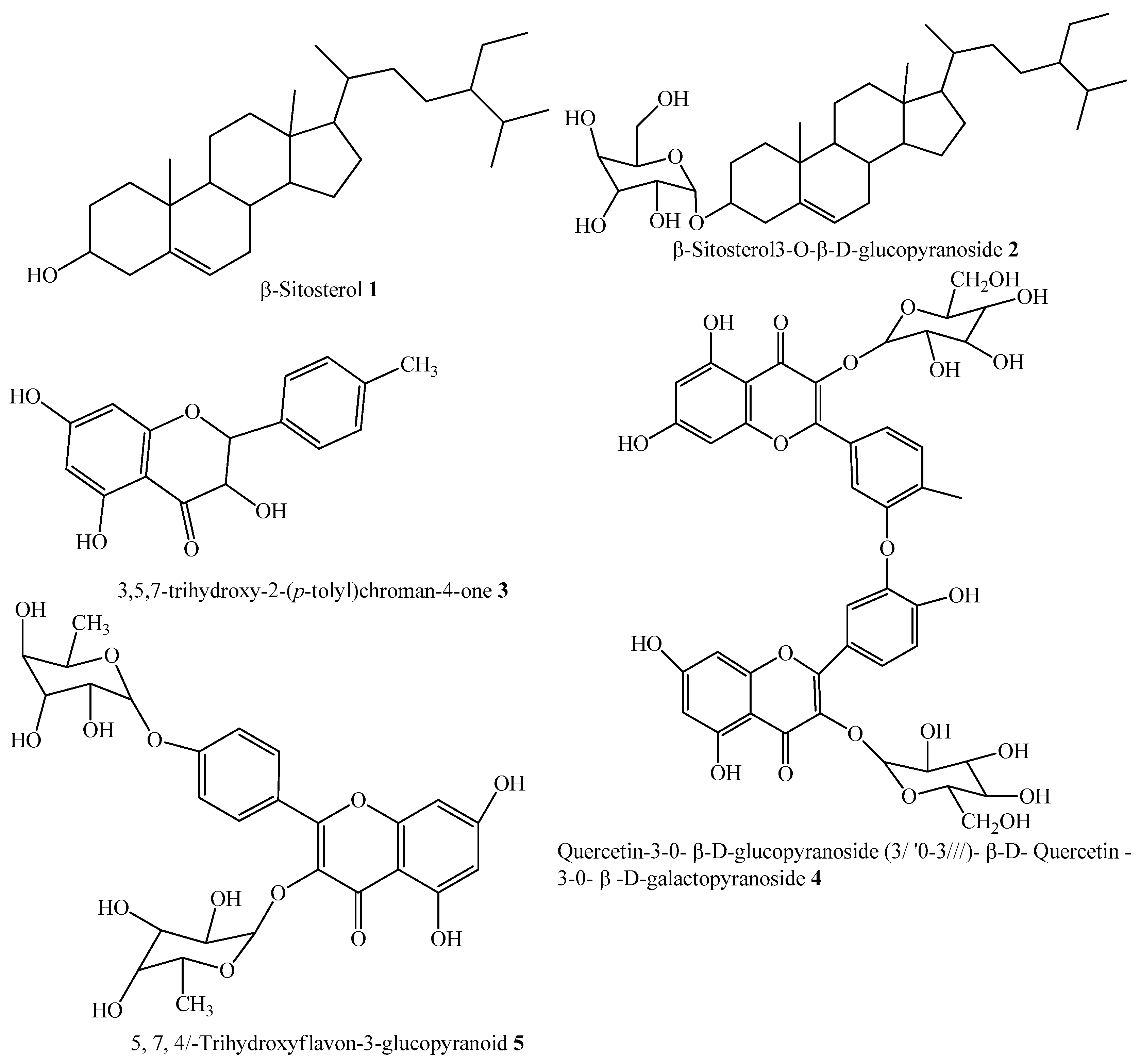
Figure 1. Structure of test compounds 1–5 isolated from Dryopteris cycadina.
Molecules 24 00427 g001
Figure 2. Per cent inhibition of test compounds 15 from Dryopteris cycadina against α-glucosidase at various concentrations. Values are expressed as mean ± SEM of three independent readings.
Molecules 24 00427 g002
Figure 3. Represents the 3D structures of Compound 1 showing binding potential. Red color in the image shows position of oxygen, green benzene ring, white hydrogen atom, brown color indicates amino acid sequence and blue color indicate nitrogen or amino group.
Molecules 24 00427 g003
Figure 4. Represents the 3D structures of Compound 2 showing binding potential. Red color in the image shows position of oxygen, green benzene ring, white hydrogen atom, brown color indicates amino acid sequence and blue color indicate nitrogen or amino group.
Molecules 24 00427 g004
Figure 5. Represent the 3D structures of Compound 3 showing binding potential. Red color in the image shows position of oxygen, green benzene ring, white hydrogen atom, brown color indicates amino acid sequence and blue color indicate nitrogen or amino group.
Molecules 24 00427 g005
Figure 6. Represents the 3D structures of Compound 4 showing binding potential. Red color in the image shows position of oxygen, green benzene ring, white hydrogen atom, brown color indicates amino acid sequence and blue color indicate nitrogen or amino group.
Molecules 24 00427 g006
Figure 7. Represents the 3D structures of Compound 5 showing binding potential. Red color in the image shows position of oxygen, green benzene ring, white hydrogen atom, brown color indicates amino acid sequence and blue color indicate nitrogen or amino group.
Molecules 24 00427 g007
Table 1. Half-maximal inhibitory concentrations of test compounds (15) isolated from Dryopteris cycadina.
α- GlucosidaseCompoundsIC50 ± SEM (µM)
1143 ± 0.47
2314 ± 4.58
3133 ± 6.90
4298 ± 0.67
5146 ± 1.93
Acarbose290 ± 0.54
Values are expressed as mean ± SEM of three independent readings.
Table 2. Molecular docking score of the test compounds with interacting amino acids of the active site of α-glucosidase enzyme.
CompoundDocking ScoreInteracting Residues of the Receptor
1−16.097ASP-215, ASP-352, ARG-442, GLN-182
2−7.756ASN-415
3−22.480ARG-315, ASP-307, HIS-280, LYS-156, SER-240, THR-310, TYR-158
4−12.931ARG-442, TYR-158
5−15.752ASP-242, LYS-156, PRO-312, TYR-158
Docking results of test compounds (15) isolated from Dryopteris cycadina against α-glucosidase.

