Article

HPLC-ESI-MSn Identification and NMR Characterization of Glucosyloxybenzyl 2R-Benzylmalate Deriva-Tives from Arundina Graminifolia and Their Anti-Liver Fibrotic Effects In Vitro

Beijing Key Labrotary for Separation and Analysis in Biomedicine and Pharmaceuticals, School of Life Science, Beijing Institute of Technology, Beijing 100081, China
*
Author to whom correspondence should be addressed.
Molecules 2019, 24(3), 525; https://doi.org/10.3390/molecules24030525
Submission received: 1 January 2019 / Revised: 22 January 2019 / Accepted: 29 January 2019 / Published: 31 January 2019

Abstract

:
Four new glucosyloxybenzyl 2R-benzylmalate derivatives, named Arundinoside H (2), I (5), J (6), K (8) as well as four known compounds Arundinoside D (1), G (3), F (4), E (7) were isolated and characterized by a combination of chemical and spectroscopic methods, including HR-ESI-MS, 1D and 2D NMR experiments. Besides, 24 unreported compounds were inferred from ESI-MSn data. The anti-liver fibrotic activities of the isolates were determined as proliferation inhibition of lipopolysaccharide (LPS)-induced activation of rat hepatic stellate cells (HSC-T6). The result suggested Arundinosides D, H, F, I and K showed moderate inhibitory effects in vitro.

1. Introduction

Arundina graminifolia (D. Don) Hochr., a species widely distributed in subtropical Asia and known as bai-yang-jie in Chinese, has a long history of use as one of the major drugs in a formula “BaoGan Capusle” with the efficacy of heat clearing and detoxifying, dispersing blood and relieving pain, reducing inflammation and promoting urination and so on [1]. Previous phytochemical investigation focusing on the chloroform and ethyl acetate exacts of A. graminifolia had resulted in the separation of stilbenoids [2,3,4], phenols [5,6,7], flavonoids [8,9] and other ketones [3,10,11]. However, the works on the polar parts of the plant are few.
In the course of our studies on pharmacology, it was proved that the formula “BaoGan Capusle” was effective in the treatment of hepatic fibrosis and liver injury of model rat [12,13,14,15]. As a continuing study on bioactive constituents of A. graminifolia, a series of phytochemical and biological experiments of the n-butanol (n-BuOH) extract was thus performed to yield the isolation of four new and four known glucosyloxybenzyl 2R-benzylmalates. In this paper, we described the isolation and structural elucidation of these derivatives, as well as their anti-liver fibrotic activities in vitro. Furthermore, the fragmentation pathways of eight isolates were studied in positive ESI-MSn, and then 24 unreported glucosyloxybenzyl 2R-benzylmalate derivatives were predicted by HPLC-ESI-MSn.

2. Results and Discussion

Through the combination of various chromatographic analyses, the n-BuOH extraction of A. graminifolia was separated carefully. Four new glucosyloxybenzyl 2R-benzylmalates Arundinoside H (2), I (5), J (6), K (8), as well as four known compounds Arundinoside D (1), G (3), F (4), E (7) [16] were obtained and determined by 1D and 2D NMR, and HR-ESI-MS spectra (see Supplementary Material). All these compounds were obtained as white amorphous powder. The 1H and 13C NMR data of the isolates were listed in Table 1 and Table 2, and their structures were shown in Figure 1. The target glucosyloxybenzyl 2R-benzylmalates in Table 4 were observed in the positive ion mode spectra (see Supplementary Material).

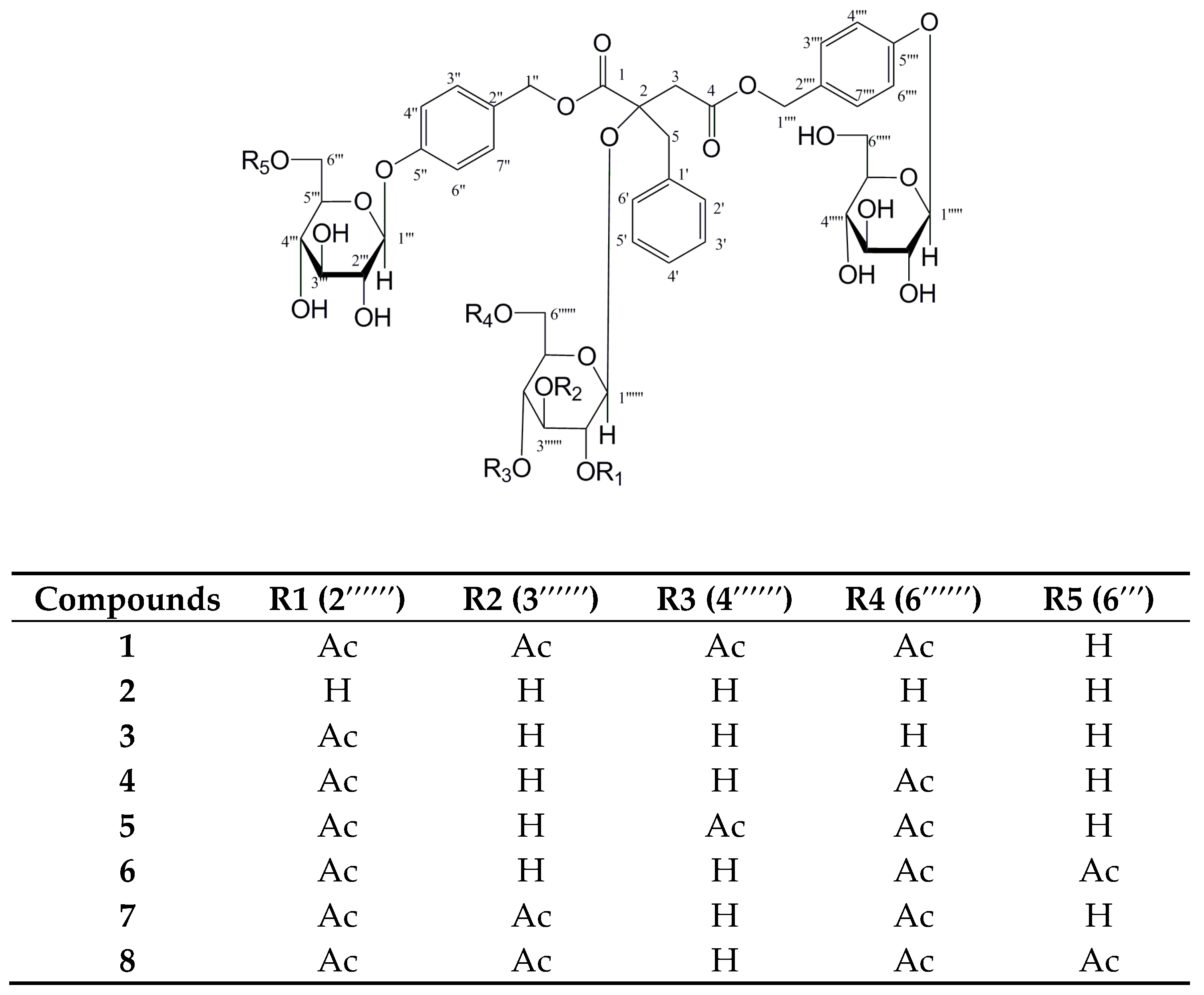
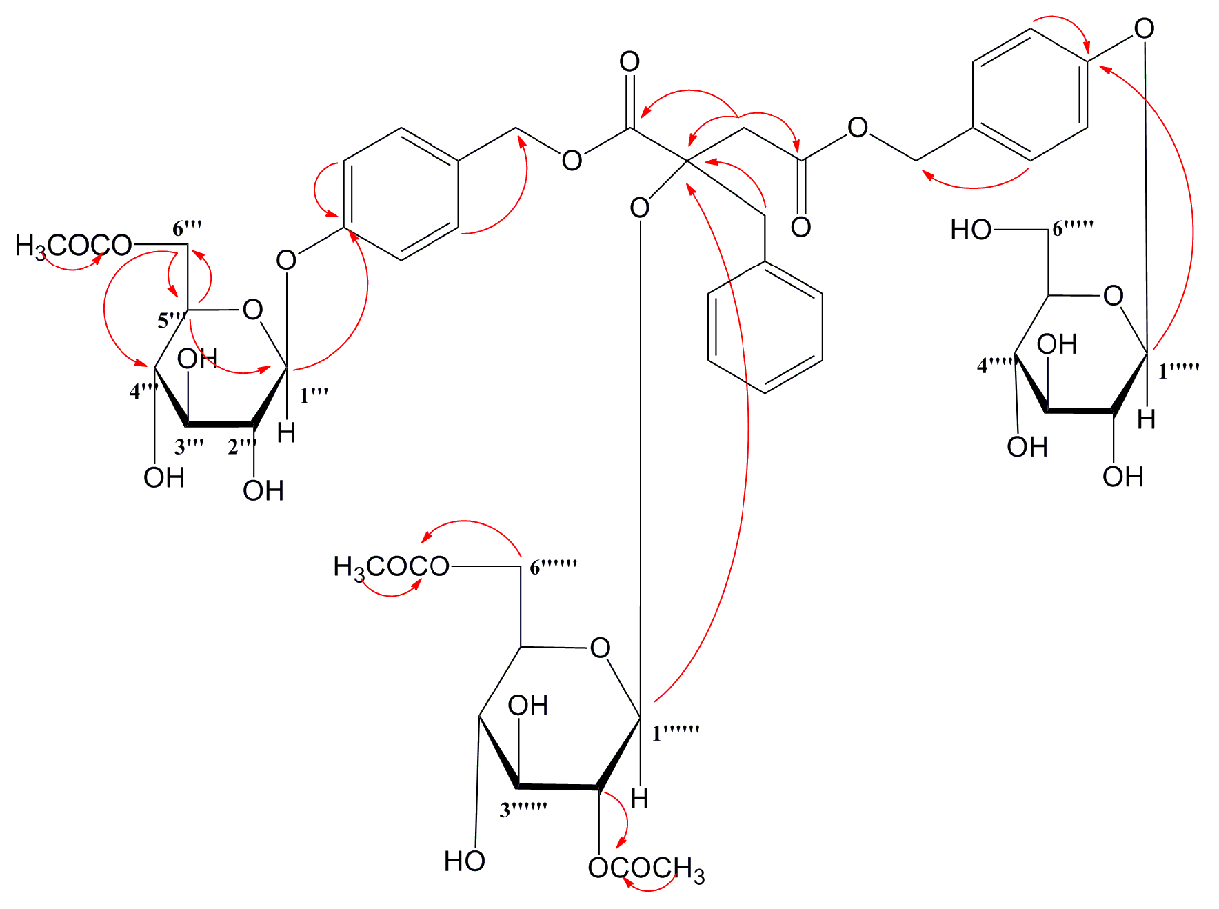
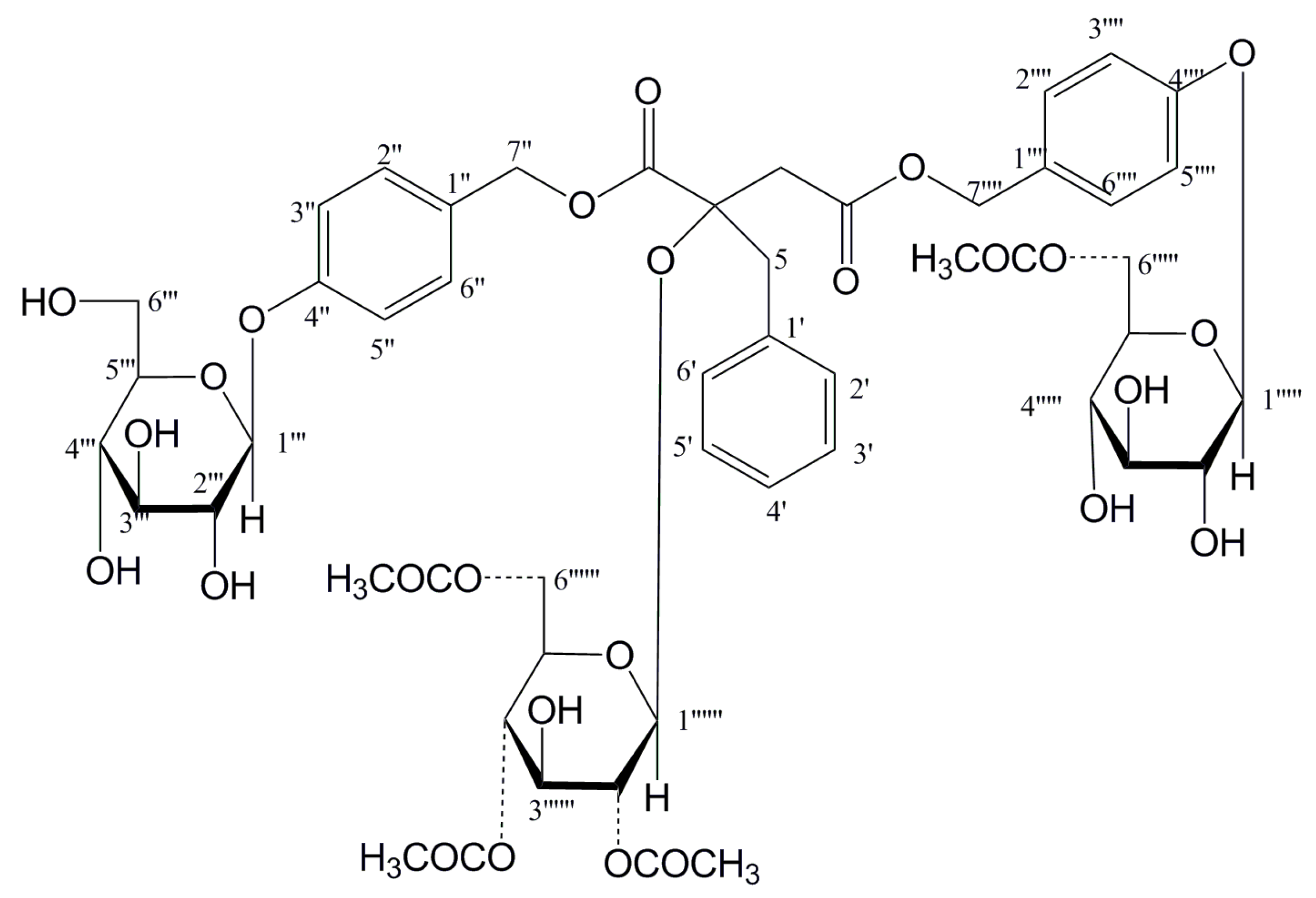
2.1. Structure Elucidation of New Compounds

The HR-ESI-MS showed a [M + NH4]+ ion at m/z 1066.3764, from which the molecular formula of compound 6 was determined to be C49H60O25. The 1H and 13C NMR data (Table 1 and Table 2) showed signals for four methylene groups at δC 41.0 (C-3), δH 2.96 (1H, d, J = 17.8 Hz, H-3), 2.90 (1H, d, J = 17.8 Hz, H-3); δC 43.8 (C-5), δH 3.10 (1H, d, J = 13.8 Hz, H-5), 3.02 (1H, d, J = 14 Hz, H-5); δC 66.6 (C-1″), δH 5.00 (1H, d, J = 12 Hz, H-1″), 4.93 (1H, d, J = 12 Hz, H-1″); δC 66.2 (C-1′′′′), δH 5.00 (1H, d, J = 12 Hz, H-1′′′′), 4.99 (1H, d, J = 12 Hz, H-1′′′′). One quaternary carbon at δC 81.0 (C-2) and two carbonyl groups at δC 170.5 (C-1), and δC 170.1 (C-4) were ascertained by comparing 13C NMR and DEPT spectra, which indicated the basic structure as malic acid [17]. The HMBC correlarions from H2-3 to C-1, C-2 and C-4; H2-5 to C-1 and C-2, combined the comparison of 1D NMR spectra of compound 6 with those of Arundinoside D~F, indicated the presence of 2R-malic acid moiety.
Through the proton signals at δH 7.18 (2H, H-2′/6′), 7.01 (2H, H-3′/5′), 7.17 (1H, H-4′), and the 13C signals at δC 135.5 (C-1′), 128.3 (C-2′/6′), 130.9 (C-3′/5′), 127.1 (C-4′), the benzene group was identified, combining at C-5 based on HMBC correlation between C-1′ and H2-5. Other two benzene groups were identified by the proton signals at δH 7.01 (4H, H-4″/6″/H-4′′′′/6′′′′), 7.23 (2H, H-3′′′′/7′′′′), 7.27 (2H, H-3″/7″), and the carbon signals at δC 157.9 (C-5″), 157.6 (C-5′′′′), 130.3 (C-3″/7″/C-3′′′′/7′′′′), 129.3 (C-2″/2′′′′), 116.7 (C-4″/6″), 116.5 (C-4′′′′/6′′′′). According to HMBC correlations between H2-1″ and C-2″, H2-1″″ and C-2′′′′, the substitution positions of the benzene groups were C-1″ and C-1′′′′, respectively.
The 1H NMR spectrum of compound 6 showed well-resolved signals for three anomeric protons of three glucoses at δH 4.92 (2H, d, J = 7.5 Hz, H-Glc-1′′′/1′′′′′) and 4.86 (1H, d, J = 7.5 Hz, H-Glc-1′′′′′′). The splitting patterns of anomeric proton signals indicated that the sugar units were β-linkage [18]. The long-correlations from H-1′′′ to C-5″, H-1′′′′′ to C-5′′′′, H-1′′′′′′ to C-2 in HMBC experiment ascertained the sugar units combined at C-5″, C-5′′′′ and C-2, respectively. The absolute configuration of the glucoses was d-form by the hydrolysis process [19].
In 1H and 13C NMR spectra, acetyl methyl protons at δH 1.72 (s), 1.92 (s), 1.99 (s) and acetyl carbonyl carbons at δC 169.8 (C), 170.7 (2C) indicated compound 6 possessed three acetyl groups, and the substitution positions were C-6′′′, C-2′′′′′′, C-6′′′′′′ by HMBC correlations from δH 4.27/4.08 (2H, m, H2-6′′′) to 170.7, 4.56 (1H, m, H-2′′′′′′) to δC 169.8, 4.08/4.05 (2H, m, H2-6′′′′′′) to 170.7. The key HMBC correlations of compound 6 were showed in Figure 2. All the protons and carbons were well assigned by NMR analysis. Therefore, compound 6 was determined as 1-(β-d-glucopyranosyloxybenzyl-6′′′-acetyl)-2-(β-d-glucopyranosyl-2′′′′′′,6′′′′′′-diacetyl)-4-(β-d-gluco pyranosyloxybenzyl)-2R-benzylmalate, and named Arundinoside J.
The molecular formula of compound 5 was determined to be C49H60O25 based on the HR-ESI-MS ion [M + NH4]+ at m/z 1066.3766. 1H and 13C NMR data of compound 5 indicated that it was a glucosyloxybenzyl 2R-benzylmalate derivative with three acetyl groups as the same as compound 6, but one group substituted position was different. The structure of compound 5 was further confirmed by HSQC and HMBC experiments. The substituent positions of three acetyl groups were determined at C-2′′′′′′, C-4′′′′′′ and C-6′′′′′′ according to HMBC correlations from δH 4.68 (1H, m, H-2′′′′′′) to δC 169.7, 4.64 (1H, m, H-4′′′′′′) to 169.3, 4.05/3.76 (2H, m, H2-6′′′′′′) to 170.0. Therefore, compound 5 was identified as 1-(β-d-glucopyranosyloxybenzyl)-2-(β-d-glucopyranosyl-2′′′′′′,4′′′′′′,6′′′′′′-triacetyl)-4-(β-d-glucopyranosyloxybenzyl)-2R-benzylmalate, and named Arundinoside I.
The molecular formula of compound 8 was determined to be C51H62O26 based on the HR-ESI-MS ion [M + NH4]+ at m/z 1108.3869. 1H and 13C NMR data indicated the structure of compound 8 was a glucosyloxybenzyl 2R-benzylmalate derivative with four acetyl groups. Further analysis of HMBC correlations from δH 4.08/4.27 (2H, m, H2-6′′′) to 170.7, 4.69 (1H, m, H-2′′′′′′) to 169.6, 4.93 (1H, m, H-3′′′′′′) to 170.3, 4.05/4.08 (2H, m, H-6′′′′′′) to 170.1 suggested that four acetyl groups of compound 8 substituted at C-6′′′, C-2′′′′′′, C-3′′′′′′, C-6′′′′′′, respectively. Therefore, compound 8 was identified as 1-(β-d-glucopyranosyloxybenzyl-6′′′-acetyl)-2-(β-d-glucopyranosyl-2′′′′′′,3′′′′′′,6′′′′′′-triacetyl)-4-(β-d-glucopyranosyloxybenzyl)-2R-benzylmalate, and named Arundinoside K.
The molecular formula of compound 2 was determined to be C43H54O22 based on the HR-ESI-MS ion [M + NH4]+ at m/z 940.3453. 1H and 13C NMR data showed compound 2 was a glucosyloxybenzyl 2-benzylmalate derivative without acetyl group, and its structure was further confirmed by HSQC and HMBC experiments. Therefore, compound 2 was identified as 1-(β-d-glucopyranosyloxybenzyl)-2-(β-d-glucopyranosyl)-4-(β-d-glucopyranosyloxy-benzyl)-2R-benzylmalate, and named Arundinoside H.

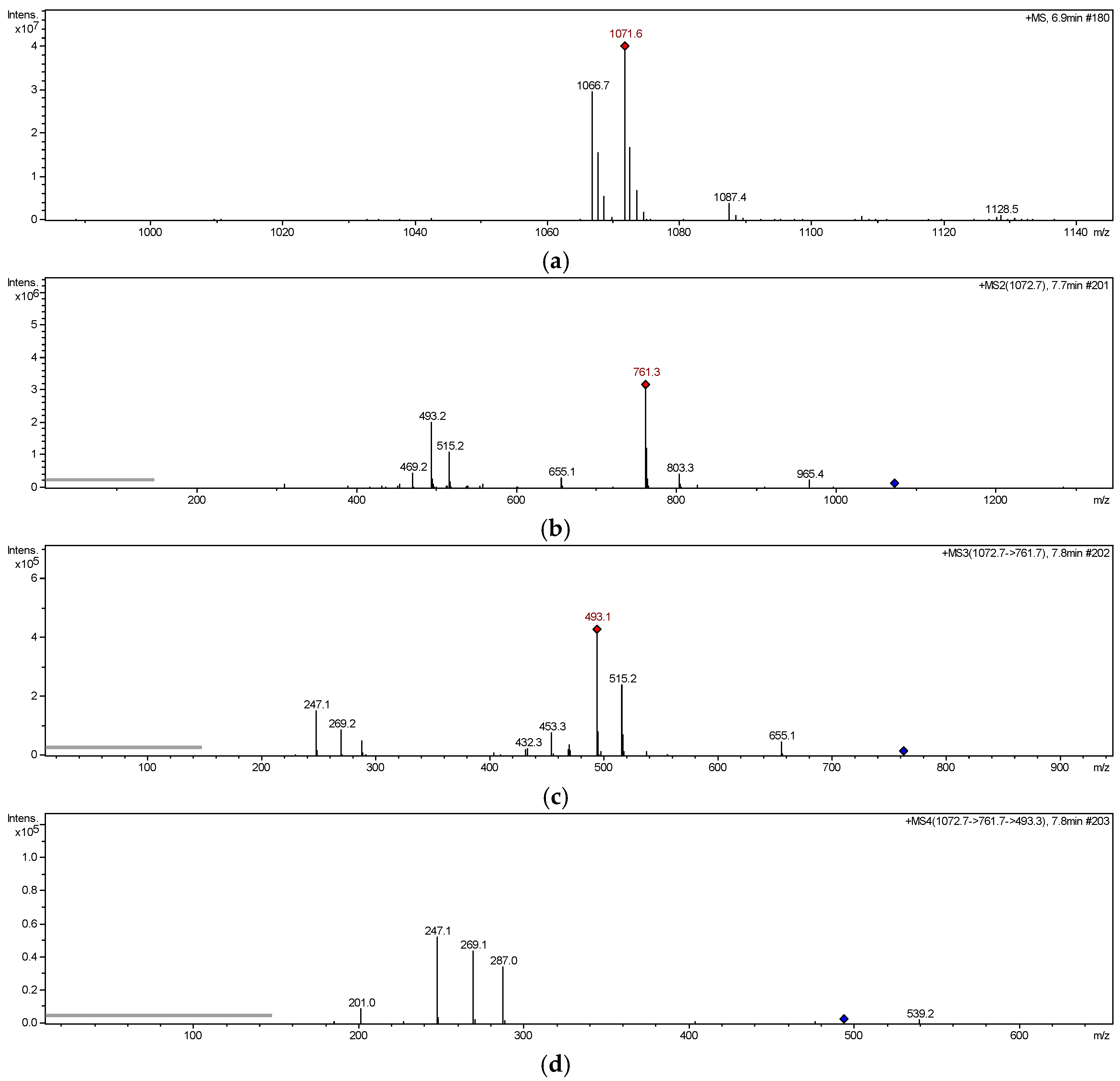
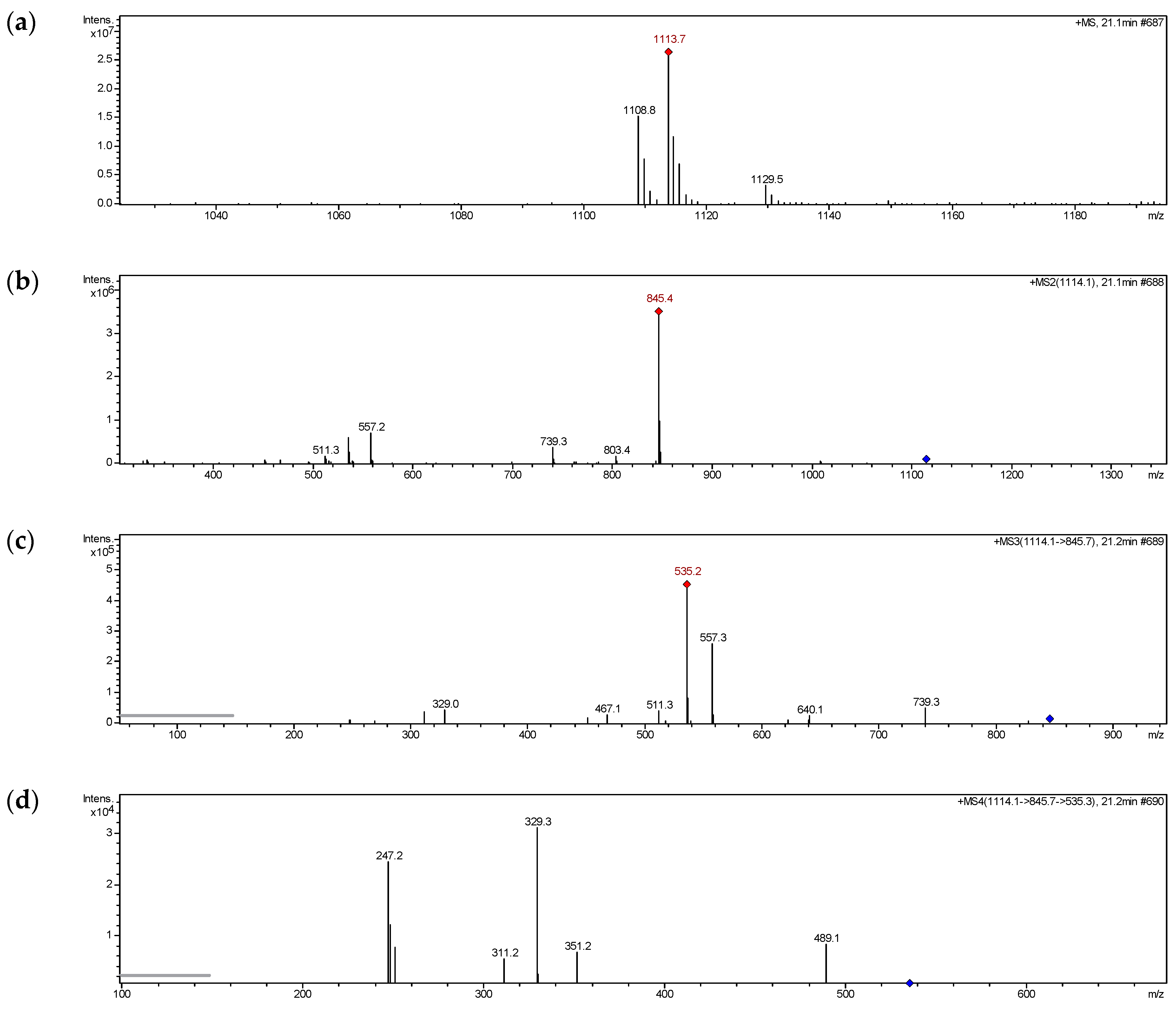
2.2. MS Fragmentation Pattern

HPLC-ESI-MSn experiments were carried out to analysis structural characterization and discuss the fragmentation behaviors of glucosyloxybenzyl 2R-benzylmalates 18 from A. graminifolia. The target glucosyloxybenzyl 2R-benzylmalates recorded at retention times were designed as A1–A6, B1–B6, C1–C3, D1–D6. The positive ion mode was performed on each of these components, and ESI-MSn data were summarized in Table 3 and Table 4.

2.2.1. MS Fragmentation Pathway of Glucosyloxybenzyl 2R-Benzylmalate Derivatives Isolated

In ESI-MS1 spectrum of compound 6 (Figure 3a), significant molecular ion peaks at m/z 1066 [M + NH4]+, 1071 [M + Na]+,1087 [M + K]+ were observed, among which the [M + Na]+ and product ions were sufficient abundance for further analysis. In ESI-MS2 spectrum of compound 6 (Figure 3b), the ion at m/z 761 was produced by loss of 6′′′-acetyl-5″-O-glucosyl-benzyl (CH2-Ph-O-Glc-Ac, 310 Da) from parent ion [M + Na]+. In ESI-MS3 spectrum of compound 6 (Figure 3c), the ions at m/z 515 and 493 were generated by losing 2′′′′′′,6′′′′′′-diacetyl-glucosyl (Glc-2Ac, 246 Da) and 5′′′′-O-glucosyl benzyl (CH2-Ph-O-Glc, 268 Da) from m/z 761, respectively. In ESI-MS4 spectrum of compound 6 (Figure 3d), the fragment at m/z 247, 287, 269 were obviously observed. The ion at m/z 247 could be produced by ions at m/z 493 or 515, which suggested that the basic structure of compound 6 was 2-benzyl-malic acid. The ions at m/z 287 and 269 were obtained by loss of 2-benzyl-malic acid (C11H10O4, 206 Da) and water molecule (H2O, 18 Da) successively from m/z 493. Figure 4 showed the proposed fragmentation pathway of compound 6 [20]. The same rules were found in the MSn analysis of other isolates listed in Table 3.

2.2.2. Structural Prediction of Glucosyloxybenzyl 2R-Benzylmalates Unreported

We also examined unknown glucosyloxybenzyl 2R-benzylmalate derivatives in fractions Ⅰ~Ⅵ of n-BuOH extract with CH3CN-H2O (32:68, v/v) for mass spectrometry analysis. In the MSn spectra, similar fragmentation pathways as described above were observed, and the possible structures of chemical components A1-A6, B1-B6, C1-C3, D1-D6 were inferred (Table 4). Herein, the analytic procedures were explained by peak A8 and A5.
The mass spectra of A8 contained significant ions at m/z 1108 [M + NH4]+, 1113 [M + Na]+, 1129 [M + K]+ (Figure 5a). Neutral loss of 268 Da (CH2-Ph-O-Glc) and 310 Da (CH2-Ph-O-Glc-Ac) were obtained from precursor ion at m/z 1113 to produce fragment ions at m/z 845 and m/z 535 in succession (Figure 5b,c). Then, the ion at m/z 329 obtained by loss of C11H10O4 (206 Da) from m/z 535, combining the ion at m/z 247 (Figure 5d), indicated the presence of 2-benzyl-malic acid moiety in A8. Based on its fragmentation behaviors and previous studies, A8 was inferred to be the structure shown in Figure 6. Moreover, the mass spectra of A5 showed the same molecular formula and similar fragmentation ions with A8, but the retention time on HPLC with the same conditions were different, which indicated A5 was an isomer of A8. The succession of neutral loss of 310 Da and 268 Da obtained from the ion at m/z 845 produced by precursor ion at m/z 1113 suggested one of Ac groups in A5 was located at G2, and not at G1. The same experimental procedures were applied to analyze other molecules list in Table 4, and the main fragments observed in MSn spectra of the [M + Na]+ precursor ions were summarized.

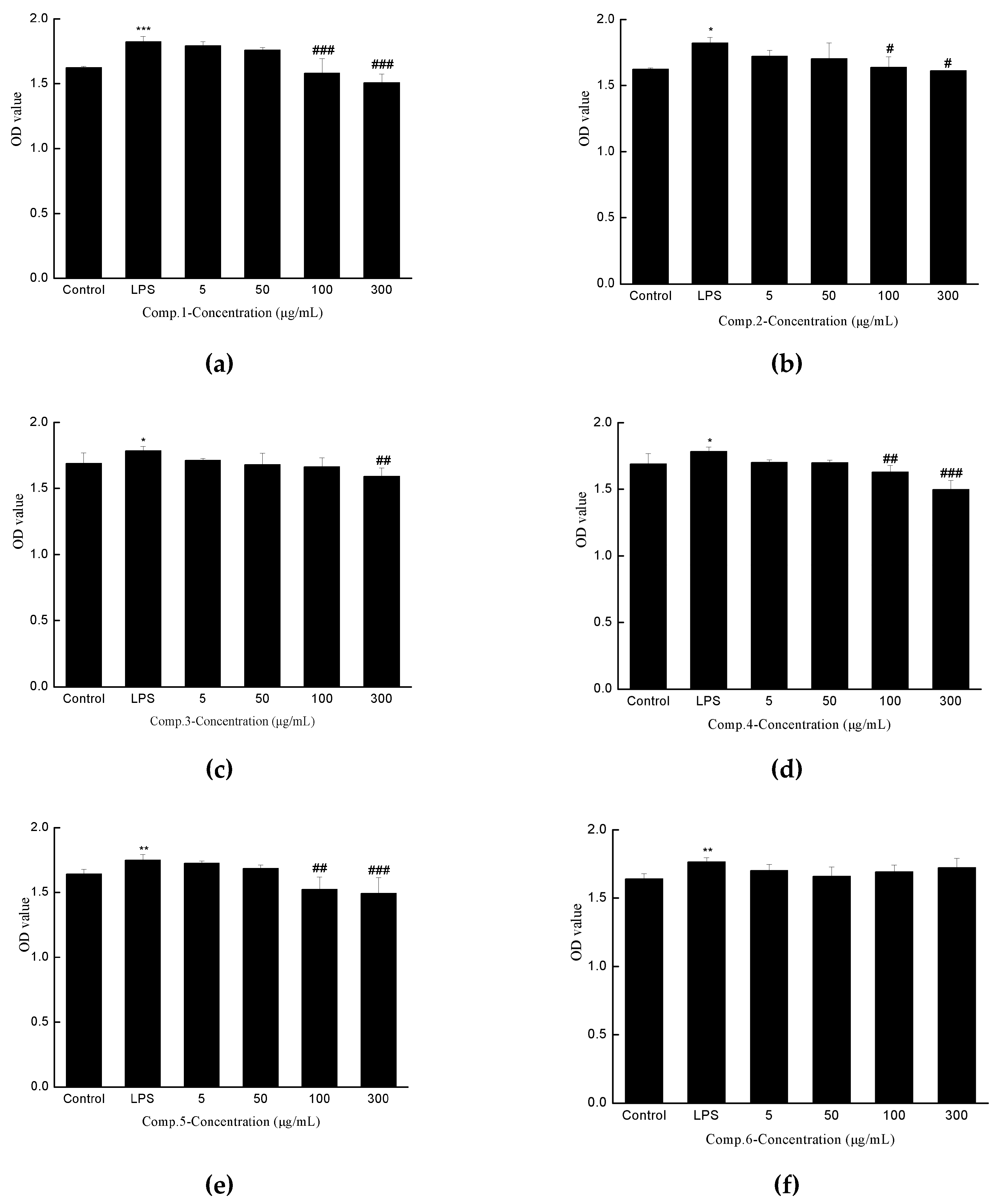
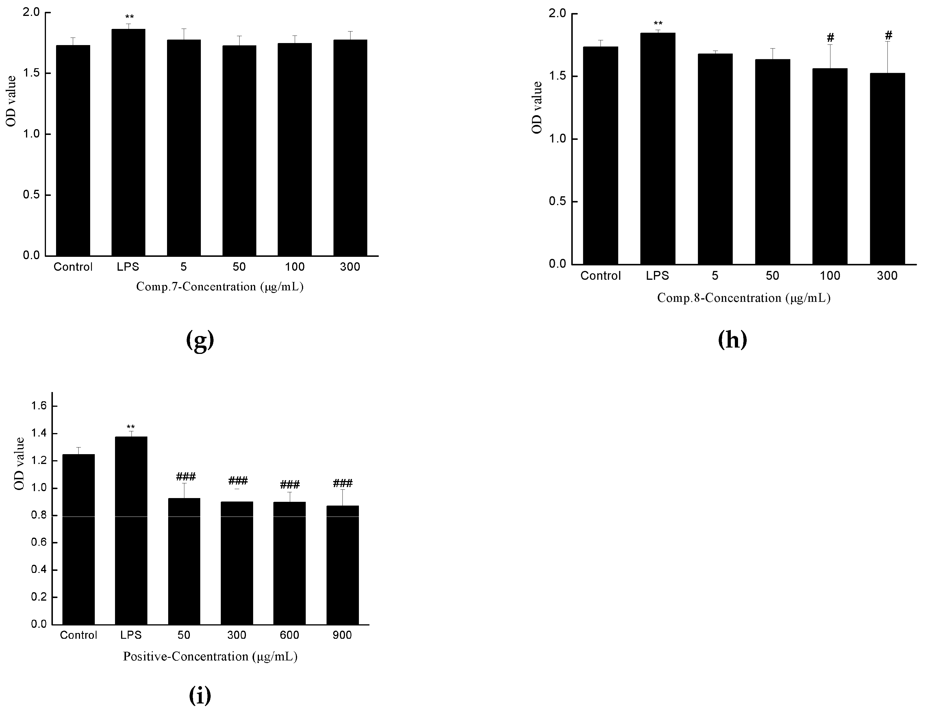
2.3. Anti-hepatic Fibrosis Activity

Emerging studies indicated that HSC in resting state could be induced to activated state by LPS [21], while the inhibition of proliferation of activated HSC, has been considered as an effective target for liver fibrosis [22]. In addition, Considering the bioactive results obtained for the “BaoGan capsule” in our previous work, the anti-hepatic fibrosis activities of the isolates 1–8 were tested on the proliferation of LPS-activated HSC-T6 cells in vitro by MTS method. Legalon (silymarin capsules) was taken as a positive control. As shown in Figure 7, compound 1, 2, 4, 5, 8 were exhibited moderate anti-proliferative activity with significantly different values while the concentration was 100 μg/mL, while positive control showed a significant difference at 50 μg/mL.

3. Materials and Methods

3.1. General Experimental Procedures

HPLC analyses were performed on Shimadzu LC-20AD (Shimadzu, Kyoto, Japan) equipped with a ZORBAX Eclipse XDB-C18 column (250 × 4.6 mm, 5 μm, Agilent, Santa Clara, CA, USA) and a SPD-20A detector. Preparative HPLC separations were conducted on a Shimadzu LC-6AD system with a preparative reversed-phase C18 column (250 × 20 mm, 5 μm, YMC-Pack ODS-A, Tokyo, Japan) and a SPD-6A detector. Mobile phase were purified water and methanol with chromatographic grade, which were bought from Merck. Organic reagents were analytical grade (Beijing Chemical Works, Beijing, China). One dimensional nuclear magnetic resonance (1D NMR: 1H, 13C, DEPT) and two dimensional nuclear magnetic resonance (2D NMR: HSQC, HMBC, 1H-1HCOSY) were measured on Bruker 700MHz AVANCE III spectrometer and Bruker AVANCE DRX-500 spectrometer (Karlsruhe, Germany) in DMSO-d6 (Sigma-Aldrich, St. Louis, MO, USA). Chemical shifts are shown in δ (ppm) with tetramethylsilane (TMS) as an internal standard. High resolution-electrospray ionization-mass spectrometry (HR-ESI-MS) and High-performance liquid chromatography-electrospray ionization- multiple stage mass spectrometry (HPLC-ESI-MSn) data were obtained from a 1100 Agilent Series coupled to an Agilent 6520 Accurate Mass Q-TOF and LC-MSD trap Mass spectrometer (Agilent Technologies, Santa Clara, CA, USA), respectively.

3.2. Plant Material

The whole plant of A. graminifolia was bought from Dai hospital of Xishuanbanna Autonomous Prefecture, Yunnan Province, China. A voucher specimen (batch number: 20111128) was collected in the laboratory.

3.3. Extraction and Isolation

Air dried powder of A. graminifolia (8.0 kg) was decocted with 80% ethanol (3 times, 2 h/time) at room temperature and extracting solution was merged and filtered. The filtrate was evaporated under reduced pressure to acquire crude extraction, which was further extracted with petroleum ether, chloroform, ethyl acetate and n-butanol to obtain the corresponding fractions. The n-BuOH extract was fractioned on a macroporous resin adsorption column eluting with ethanol/water (10:90, 50:50, 100:0, v/v) to yield 3 fractions (A–C). Fraction B (22.2 g) was subjected to Rp-18 silica gel column eluted with acetonitrile/water (10:90→100:0, v/v) to obtain five fractions (B1–B5). Fraction B3 (5.5 g) was then separated by silica gel column eluted with CHCl3/CH3OH (20:1, 2:1, 0:1) to give six fractions (Ⅰ–Ⅵ). Fraction Ⅴ (0.48 g) was submitted to preparative HPLC on a Rp-18 column (250 mm × 20 mm, wavelength 279 nm, flow rate 4 mL/min) with CH3CN-H2O (35:65, v/v) to give compound 1 (26.83 mg, RT = 21.5 min) and peaks D1~D6 eluted by CH3CN-H2O with 5 mM ammonium acetate (32:68, v/v). Fraction Ⅱ (2.64 g) was eluted with CH3CN-H2O (29:71,v/v) to afford compound 2 (1.71 mg, RT = 5 min), compound 3 (1.62 mg, RT = 8.2 min) and compound 4 (3.84 mg, RT = 15.2 min), with CH3CN-H2O (26:74, v/v) to afford compound 5 (2.34 mg, RT = 25 min), compound 6 (3.18 mg, RT = 30 min) and compound 7 (1.85 mg, RT = 41.9 min), with CH3CN-H2O (30:70, v/v) to afford compound 8 (2.01 mg, RT = 42 min). Furthermore, a search of the rest of Fraction Ⅱ was then conducted at 0.2 mL/min for HPLC-ESI-MSn to obtain Peaks A1~A9 eluted by CH3CN-H2O with 5 mM ammonium acetate (30:70, v/v), peaks B1~B6 eluted by CH3CN-H2O with 5 mM ammonium acetate (26:74, v/v); peaks C1~C3 eluted by CH3CN-H2O with 5 mM ammonium acetate (27:73, v/v), and RT values of the peaks were shown in Table 4.

3.4. Cell Proliferation Inhibition Assay

3.4.1. Chemical and Reagents

LPS, RPMI-1640 medium, penicillin-streptomycin, and trypsin were bought from Solarbio, Beijing, China and fetal bovine serum were purchased from Sijiqing, Hangzhou, China. Dimethyl sulfoxide (DMSO), PMS and MTS [3-(4,5-dimethylthiazol-2-yl)-5-(3-carboxymethoxyphenyl)-2-(4- sulfophenyl)-2H-tetrazolium, inner salt] were purchased from Sigma-Aldrich (Steinheim, Germany).

3.4.2. In vitro Evaluation of Anti-Liver Fibrotic Activity

HSC-T6 cells were maintained in RPMI-1640 medium supplemented with 10% fetal bovine serum and 1% penicillin-streptomycin in an incubator with constant temperature at 37 °C and a humidified atmosphere of 5% CO2. The cells were trypsinized and passaged to new plates every two or three days. HSC-T6 cells were seeded in 96-well plates (5 × 10³/100 μL) for 24 h to ensure fully adhesion and good condition. The cell medium in the wells was changed into fresh RPMI-1640 medium supplemented with 5% fetal bovine serum for further incubation. Cells incubated with LPS (1 μg/mL) in the different concentration of compounds 1~8 (0, 5, 50, 100, 300 μg/mL) were cultivated for another 24 h. Each group was provided with 6 independent duplicates. Cell viability was determined using MTS/PMS assay. Absorbance values were read at 490 nm on an ELISA reader. 0.1% DMSO was considered as blank control and legalon (silymarin capsules) as positive control. Cell viability was expressed as a percentage of control cells at 100% viability. Statistical analysis was performed using origin Pro 8.0 (OriginLab Corpration, One Roundhouse Plaza, Northampton, MA, USA).

4. Conclusions

Glucosyloxybenzyl 2R-benzylmalates are a class of naturally occurring substances distributed in Orchidaceae. They were noticed for their novel type of structure and significant activities, while the research of glucosyloxybenzyl 2R-benzylmalates was limited by their higher polarity and less content. In present work, basis on the information acquired from HPLC-ESI-MSn experiment combined with NMR analysis, it was possible not only to identify 8 compounds isolated from A. graminifolia, but also to predict the structures of 24 previously unreported glucosyloxybenzyl 2R-benzylmalates in the extract. The ESI-MSn experiments provide a useful guide for gaining the large structural information of novel compounds, which are important to drug design, although the analytical method cannot confirm the accurate substitution position of Ac groups of these glucosyloxybenzyl 2R-benzylmalates.
In addition, a cell model associated with hepatic fibrosis was established by using LPS to stimulate HSC-T6. The isolates were carried to hepatic fibrosis experiment while compounds 1, 2, 4, 5, 8 showed moderate anti-hepatic fibrosis effects. Nevertheless, the studies on the quantitative structure-antihepatic fibrosis relationship of predicted glucosyloxybenzyl 2R-benzylmalates will be further investigated.

5. Patents

Two patents resulting from the work about new structures, anti-liver fibrotic activity and MS fragment pathway have been submitted to the Chinese Patent Office.

Supplementary Materials

The following are available online: 1H and 13C NMR, DEPT135, HSQC, HMBC, 1H-1H COSY spectra of new compounds 2, 5, 6, and 8; HPLC-ESI-MS spectra of the isolates 18; HPLC-ESI-MSn spectra of peaks A1–A9, B1–B6, C1–C3, and D1–D6.

Author Contributions

Q.L. performed the isolation, MS analyses and structural identification of the compounds under the supervision of F.L. Original draft preparation was wrote by Q.L. and further reviewing and editing was completed by F.L. Q.L. and F.S. were in charge of the biological evaluation. In addition, F.L. provided significant advice regarding the NMR structural elucidation and MS deduction. Y.D. and R.D. contributed to the plant material selection and its resource. This paper was financially supported by F.L., Y.D., R.D.

Funding

This research was financially supported by National Natural Science Foundation of China (Grant No. 81503353) and the National Major Scientific Instruments and Equipment Development Project (Grant No. 2012YQ040140).

Conflicts of Interest

The authors declare no conflict of interest.

References

  1. Jiang Su. New Medical College. Dictionary of Chinese Traditional Medicine; Shanghai Science and Technology Press: Shanghai, China, 1986; pp. 455–456. [Google Scholar]
  2. Li, Y.K.; Zhou, B.; Ye, Y.Q.; Du, G.; Niu, D.Y.; Meng, C.Y.; Gao, X.M.; Hu, Q.F. Two new diphenylethylenes from Arundina graminifolia and their cytotoxicity. Bull. Korean Chem. Soc. 2013, 34, 3257–3260. [Google Scholar] [CrossRef]
  3. Hu, Q.F.; Zhou, B.; Ye, Y.Q.; Jiang, Z.Y.; Huang, X.Z.; Li, Y.K.; Du, G.; Yang, G.Y.; Gao, X.M. Cytotoxic deoxybenzoins and diphenylethylenes from Arundina graminifolia. J. Nat. Product. 2013, 76, 1854–1859. [Google Scholar] [CrossRef] [PubMed]
  4. Meng, C.Y.; Niu, D.Y.; Li, Y.K.; Zhou, B.; Ye, Y.Q.; Du, G.; Hu, Q.F.; Gao, X.M. A new cytotoxic stilbenoid from Arundina graminifolia. Asian J. Chem. 2014, 26, 2411–2413. [Google Scholar] [CrossRef]
  5. Hu, Q.F.; Zhou, B.; Huang, J.M.; Gao, X.M.; Shu, L.D.; Yang, G.Y.; Che, C.T. Antiviral phenolic compounds from Arundina gramnifolia. J. Nat. Prod. 2013, 76, 292–296. [Google Scholar] [CrossRef] [PubMed]
  6. Gao, Z.R.; Xu, S.T.; Wei, J.; Shi, H.L.; Hu, Q.F. Phenolic compounds from Arundina graminifolia and their anti-tobacco mosaic virus activities. Asian J. Chem. 2013, 25, 2747–2749. [Google Scholar] [CrossRef]
  7. Li, L.; Xu, W.X.; Liu, C.B.; Zhang, C.M.; Zhao, W.; Shang, S.Z.; Deng, L.; Guo, Y.D. A new antiviral phenolic compound from Arundina gramnifolia. Asian J. Chem. 2015, 27, 3525–3526. [Google Scholar] [CrossRef]
  8. Li, Y.K.; Yang, L.Y.; Shu, L.D.; Shen, Y.Q.; Hu, Q.F.; Xia, Z.Y. Flavonoid compounds from Arundina graminifolia. Asian J. Chem. 2013, 25, 4922–4924. [Google Scholar] [CrossRef]
  9. Shu, L.D.; Shen, Y.Q.; Yang, L.Y.; Gao, X.M.; Hu, Q.F. Flavonoids derivatives from Arundina graminifolia and their cytotoxicity. Asian J. Chem. 2013, 25, 8358–8360. [Google Scholar] [CrossRef]
  10. Niu, D.Y.; Han, J.M.; Kong, W.S.; Cui, Z.W.; Hu, Q.F.; Gao, X.M. Antiviral fluorenone derivatives from Arundina gramnifolia. Asian J. Chem. 2013, 25, 9514–9516. [Google Scholar] [CrossRef]
  11. Zhu, H.; Song, Q.S. Chemical components of Arundina graminifolia. Nat. Prod. Res. Dev. 2008, 20, 5–7. [Google Scholar]
  12. Meng, W.W.; Nan, G.; Lin, F.K.; Zhao, H.W.; Deng, Y.L.; Dai, R.J.; Chen, Y.; Huang, Y.M.; Zhao, Y.H. Effect of Baogan Yihao on Liver Fibrosis in Rats Induced by CCl4 and High-Fat Feeding. Trans. Beijing Inst. Technol. 2014, 34, 767–770. [Google Scholar]
  13. He, P.P.; Song, C.C.; Meng, W.W.; Deng, Y.L.; Dai, R.J.; Lin, F.K.; Chen, Y.; Zhao, Y.H.; Huang, Y.M. Protective Effect of Baogan Yihao on Acute and Chronic Hepatic Injury. Chin. J. Exp. Tradit. Med. Formul. 2012, 18, 195–198. [Google Scholar]
  14. Dai, R.J.; Su, J.; Wang, F.; Lin, F.K.; Lv, F.; Chen, Y.; Meng, W.W.; Deng, Y.L. The Protective Effects of BGYH on Mice Acute Liver Injury Induced by D-Galn and LPS. Trans. Beijing Inst. Technol. 2015, 35 (Suppl. 1), 67–71. [Google Scholar]
  15. Liu, X.J.; Su, J.; Shi, Y.; Guo, Y.; Suheryani, I.; Zhao, S.C.; Deng, Y.L.; Meng, W.W.; Chen, Y.; Sun, L.L.; Dai, R.J. Herbal Formula, Baogan Yihao (BGYH), Prevented Dimethyl-nitrosamine (DMN)-Induced Liver Injury in Rats. Drug Dev. Res. 2017, 78, 155–163. [Google Scholar] [CrossRef] [PubMed]
  16. Auberon, F.; Olatunji, O.J.; Krisa, S.; Herbette, G.; Antheaume, C.; Bonte, F.; Merillon, J.M.; Lobstein, A. Arundinosides AG, new glucosyloxybenzyl 2R-benzylmalate derivatives from the aerial parts of Arundina graminifolia. Fitoterapia 2018, 125, 199–207. [Google Scholar] [CrossRef] [PubMed]
  17. Morikawa, T.; Xie, H.; Matsuda, H.; Yoshikawa, M. Glucosyloxybenzyl 2-Isobutylmalates from the Tubers of Gymnadenia conopsea. J. Nat. Prod. 2006, 69, 881–886. [Google Scholar] [CrossRef] [PubMed]
  18. Tatsis, C.E.; Boeren, S.; Exarchou, V.; Troganis, A.N.; Vervoort, J.; Gerothanassis, I.P. Identification of the major constituents of Hypericum perforatum by LC/SPE/NMR and/or LC/MS. Phytochemistry 2007, 68, 383–393. [Google Scholar] [CrossRef]
  19. Song, J.; Dai, R.J.; Deng, Y.L.; Lv, F. Rapid structure prediction by HPLC-ESI-MSn of twenty-five polyoxypregnane tetraglycosides from Dregea sinensis with NMR confirmation of eight structures. Phytochemistry 2018, 147, 147–157. [Google Scholar] [CrossRef]
  20. Cai, M.; Zhou, Y.; Gesang, S.L.; Bianba, C.; Ding, L.S. Chemical fingerprint analysis of rhizomes of Gymnadenia conopsea by HPLC–DAD–MSn. J. Chromatogr. B 2006, 844, 301–307. [Google Scholar] [CrossRef] [Green Version]
  21. Kao, Y.H.; Chen, P.H.; Wu, T.Y.; Lin, Y.C.; Tsai, M.S.; Lee, P.H.; Tai, T.S.; Chang, H.R.; Sun, C.K. Lipopolysaccharides induce Smad2 phosphorylation through PI3K/Akt and MAPK cascades in HSC-T6 hepatic stellate cells. Life Sci. 2017, 184, 37–46. [Google Scholar] [CrossRef]
  22. Kang, K.B.; Jin, B.J.; Kim, J.W.; Kim, H.W.; Sung, S.H. Ceanothane- and lupane-type triterpene esters from the roots of Hovenia dulcis, and their antiproliferative activity on HSC-T6 cells. Phytochemistry 2017, 142, 60. [Google Scholar] [CrossRef] [PubMed]
Sample Availability: Samples of the compounds are not available from the authors.
Figure 1. Structures of compounds 1~8.
Molecules 24 00525 g001
Figure 2. Key HMBC correlations of compound 6.
Molecules 24 00525 g002
Figure 3. MSn spectra of compound 6. (a) Full-scan MS1 spectrum, (b) ESI-MS2 spectrum, (c) ESI-MS3 spectrum, (d) ESI-MS4 spectrum.
Molecules 24 00525 g003
Figure 4. Proposed MS fragment pathway of compound 6.
Molecules 24 00525 g004
Figure 5. MSn spectra of A8. (a) Full-scan MS1 spectrum, (b) ESI-MS2 spectrum, (c) ESI-MS3 spectrum, (d) ESI-MS4 spectrum.
Molecules 24 00525 g005
Figure 6. Proposed structure of A8.
Molecules 24 00525 g006
Figure 7. Inhibitory activity of compound 1 (a)–8 (h) and positive control (i) on the proliferation of LPS-activated HSC-T6.
Molecules 24 00525 g007aMolecules 24 00525 g007b
Table 1. 1H nuclear magnetic resonance (NMR) data of four new compounds (700 MHz, DMSO-d6).
Position2568
32.96 (d, 17.7); 2.82 (d, 17.7)2.96 (d, 15); 2.90 (d, 15)2.96 (d, 17.8); 2.90 (d, 17.8)2.96 (d, 17.8); 2.92 (d, 17.8)
53.17 (m); 3.06 (m)3.10 (d, 14); 3.02 (d, 14)3.10(d, 14); 3.02 (d, 14)3.10 (d, 14); 3.02 (d, 14)
2′, 6′7.19 (m)7.18 (m)7.18 (m)7.18 (m)
3′, 5′7.16 (m)7.02 (m)7.01 (d, 8.7)7.01 (d, 8.7)
4′7.19 (m)7.18 (m)7.17 (m)7.17 (m)
1″4.99 (d, 7.9); 4.90 (d, 7.9)5.00 (d, 12); 4.92 (d, 12)5.00 (d, 12); 4.93 (d,12)5.00(d,12); 4.93 (d,12)
3″, 7″7.27 (d, 8.4)7.27 (d, 8.3)7.27 (d, 8.7)7.27 (d, 8.7)
4″, 6″7.01 (d, 8.5)7.02 (m)7.01 (d, 8.7)7.01 (d, 8.7)
Glc-1′′′4.87 (d, 7.5)4.91 (d, 7.9)4.92 (d, 7.5)4.92 (d, 7.7)
Glc-2′′′3.27 (m)3.26 (m)3.27 (m)3.27 (m)
Glc-3′′′3.24 (m)3.23 (m)3.22 (m)3.22 (m)
Glc-4′′′3.16 (m)3.16 (m)3.18 (m)3.18 (m)
Glc-5′′′3.27 (m)3.26 (m)3.27 (m)3.27 (m)
Glc-6′′′3.68 (m); 3.47 (m)3.68 (m); 3.46 (m)4.27 (m); 4.08 (m)4.27 (m); 4.08 (m)
Glc-6′′′-COCH3--1.99 (s)1.99 (s)
1′′′′5.01 (d, 12); 4.90 (d, 12)5.04 (d, 12); 4.91 (d, 12)5.00 (d, 12); 4.99 (d, 12) 5.00 (d,12); 4.99 (d,12)
3′′′′, 7′′′′7.25 (d, 8.4)7.23 (d, 8.3)7.23 (d, 8.7)7.23 (d, 8.7)
4′′′′, 6′′′′7.02 (d, 8.5)7.02 (m)7.02 (d, 8.7)7.02 (d, 8.7)
Glc-1′′′′′4.87 (d, 7.5)4.86 (d, 7.7)4.92 (d, 7.5)4.87 (d, 7.5)
Glc-2′′′′′3.27 (m)3.16 (m)3.30 (m)3.30 (m)
Glc-3′′′′′3.24 (m)3.23 (m)3.22 (m)3.22 (m)
Glc-4′′′′′3.16 (m)3.16 (m)3.18 (m)3.18 (m)
Glc-5′′′′′3.27 (m)3.26 (m)3.27 (m)3.27 (m)
Glc-6′′′′′3.69 (m); 3.47 (m)3.68 (m); 3.46 (m)3.68 (m); 3.46 (m)3.68 (m); 3.46 (m)
Glc-1′′′′′′4.66 (d, 7.8)4.94 (d, 8.0)4.86 (d, 7.6)5.05 (d, 8.0)
Glc-2′′′′′′3.03 (m)4.68 (m)4.56 (m)4.69 (m)
Glc-3′′′′′′3.39 (m)3.46 (m)3.68 (m)4.93 (m)
Glc-4′′′′′′4.36 (m)4.64 (m)3.46 (m)3.46 (m)
Glc-5′′′′′′3.52 (m)3.45 (m)3.61 (m)3.61 (m)
Glc-6′′′′′′3.68 (m); 3.38 (m)4.05 (m); 3.76 (m)4.08 (m); 4.05 (m)4.08 (m); 4.05 (m)
Glc-2′′′′′′-COCH3-1.70 (s)1.72 (s)1.65 (s)
Glc-3′′′′′′-COCH3---1.92 (s)
Glc-4′′′′′′-COCH3-2.01 (s)-
Glc-6′′′′′′-COCH3-1.92 (s)1.92 (s)1.99 (s)
Table 2. 13C NMR data of four new compounds (175MHz, DMSO-d6).
Position2568
1170.9170.1170.5170.1
279.880.781.081.3
340.440.941.041.0
4169.8169.5170.1170.1
542.043.643.843.7
1′135.5135.0135.5135.4
2′, 6′127.9127.9128.3128.3
3′, 5′130.6130.5130.9130.9
4′126.7126.7127.1127.1
1″66.366.266.666.6
2″128.9128.8129.3129.2
3″, 7″129.9129.9130.3130.3
4″, 6″116.2116.2116.7116.7
5″157.4157.4157.9157.9
Glc-1′′′100.3100.3100.5100.5
Glc-2′′′76.876.677.177.1
Glc-3′′′73.273.273.573.6
Glc-4′′′69.669.770.170.2
Glc-5′′′76.676.676.876.8
Glc-6′′′60.760.763.963.8
Glc-6′′′-COCH3--170.7; 21.2170.7; 21.1
1′′′′65.665.766.266.2
2′′′′128.8128.6129.3129.3
3′′′′, 7′′′′129.9129.9130.4130.4
4′′′′, 6′′′′116.1116.2116.5116.5
5′′′′157.4157.4157.6157.6
Glc-1′′′′′100.3100.4100.8100.8
Glc-2′′′′′76.877.077.577.5
Glc-3′′′′′73.273.273.673.4
Glc-4′′′′′69.670.370.170.1
Glc-5′′′′′76.676.676.876.8
Glc-6′′′′′60.760.761.161.1
Glc-1′′′′′′98.396.797.096.7
Glc-2′′′′′′77.070.973.871.2
Glc-3′′′′′′73.770.974.075.1
Glc-4′′′′′′69.772.873.767.9
Glc-5′′′′′′76.770.674.174.1
Glc-6′′′′′′61.261.863.362.9
Glc-2′′′′′′-COCH3-169.7; 20.5169.8; 21.0169.6; 20.7
Glc-3′′′′′′-COCH3--- 170.3; 21.0
Glc-4′′′′′′-COCH3-169.3; 20.8--
Glc-6′′′′′′-COCH3-170; 20.7170.7; 21.1170.1;21.2
Table 3. HR-ESI-MS and key ESI-MSn data of the isolates 18.
CompoundsMolecular FormulaHR-ESI-MS [M + NH4]+ESI-MS1: [M + Na]+ESI-MSn
1C51H62O261108.38611113845, 577, 515, 497, 371, 353, 311, 251, 247
2C43H54O22940.3453945677, 515, 409, 247
3C45H56O23982.3554987719, 515, 451, 247
4C47H58O241024.36651029761, 515, 493, 287, 269, 247, 227
5C49H60O251066.37661071803, 535, 329, 311, 269, 247
6C49H60O251066.37641071761, 515,493, 287, 269, 247
7C49H60O251066.37661071803, 535, 515, 329, 311, 247, 209
8C51H62O261108.38691113803, 535, 515, 329, 311, 247, 209
Table 4. Key ESI-MSn Fragment Ions and structural information of the components predicted.
Molecules 24 00525 i001
PeaksRTMS1 [M + Na]+MSnG1G2G3
A13.9 min10061029, 761, 515, 493, 287, 269, 247 2Ac
A26.4 min10481071, 803, 535, 515, 329, 311 3Ac
A37.3 min10481072, 761, 515, 493, 287, 269, 247Ac 2Ac
A412.7 min10901113, 845, 535, 329, 311 Ac3Ac
A513.9 min10901113, 803, 535, 329, 311, 247Ac 3Ac
A614.0 min10521071, 677, 515, 247Cin
A722.2 min10941117, 849, 645, 247 Cin Ac
A821.1 min10901113, 845, 535, 329, 247 Ac3Ac
A928.0 min10941117, 719, 451, 247Cin Ac
B13.7 min964987, 719, 515, 247 Ac
B24.3 min964987, 719, 515, 247 Ac
B37.0 min760783, 515, 247 OH
B49.0 min10061029, 761, 557, 451, 247 Ac Ac
B511.0 min10061029, 719, 515, 451, 247Ac Ac
B612.6 min10061029, 761, 515, 493, 287, 269, 247 2Ac
C16.4 min10481071, 761, 557, 451, 247AcAc Ac
C28.9 min10481071, 761, 515, 493, 287, 269, 247Ac 2Ac
C311.9 min10481071, 803, 535, 329, 311, 269, 247 3Ac
D116.0 min12521275, 845, 577, 371, 353, 311 4Ac
D28.5 min10521075, 677, 515, 247Cin
D37.0 min10521075, 677, 515, 247Cin
D415.6 min10941117, 719, 515, 451, 247Cin Ac
D518.6 min10941117, 719, 515, 451, 247Cin Ac
D619.3 min10941117, 719, 515, 451, 247Cin Ac

