Abstract
The emerging threat of infections caused by highly drug-resistant bacteria has prompted a resurgence in the use of the lipodecapeptide antibiotics polymyxin B and colistin as last resort therapies. Given the emergence of resistance to these drugs, there has also been a renewed interest in the development of next generation polymyxins with improved therapeutic indices and spectra of action. We report structure-activity studies of 36 polymyxin lipononapeptides structurally characterised by an exocyclic FA-Thr2-Dab3 lipodipeptide motif instead of the native FA-Dab1-Thr2-Dab3 tripeptide motif found in polymyxin B, removing one of the positively charged residues believed to contribute to nephrotoxicity. The compounds were prepared by solid phase synthesis using an on-resin cyclisation approach, varying the fatty acid and the residues at position 2 (P2), P3 and P4, then assessing antimicrobial potency against a panel of Gram-negative bacteria, including polymyxin-resistant strains. Pairwise comparison of N-acyl nonapeptide and decapeptide analogues possessing different fatty acids demonstrated that antimicrobial potency is strongly influenced by the N-terminal L-Dab-1 residue, contingent upon the fatty acid. This study highlights that antimicrobial potency may be retained upon truncation of the N-terminal L-Dab-1 residue of the native exocyclic lipotripeptide motif found in polymyxin B. The strategy may aid in the design of next generation polymyxins.
1. Introduction
The polymyxins (Pmx) are natural product polycationic lipodecapeptides produced by Paenibacillus polymyxa (Figure 1), exemplified by polymyxin B 1 (PmxB) and E 2 (also known as colistin) [1,2,3,4]. First discovered in 1947, with subsequent studies reporting the isolation and characterization of additional Pmx derivatives from natural product sources [5,6,7,8], PmxB 1 and colistin 2 have been part of the clinical antibiotic repertoire for over 50 years, albeit approved for human use in an era with less stringent regulatory requirements compared to contemporary standards. However, toxicity issues, in particular nephrotoxicity [9,10], led to their gradual replacement with safer alternatives. The past decade has seen increasing application of ‘last-resort’ antibiotics due to the ominous rise of infections caused by extended-spectrum β-lactamase- (ESBL) and carbapenemase-producing strains of Acinetobacter baumannii, Pseudomonas aeruginosa and Enterobacteriaceae, with some strains now exhibiting multidrug resistance to practically all known antibiotics [11,12,13,14]. New treatments are urgently needed, but scientific [15] and economic [16] hurdles have slowed progression of the antibiotic clinical pipeline, particularly for Gram-negative therapies [17]. This has prompted a resurgence in the use of PmxB and colistin in spite of their toxicity, as well as renewed interest in the creation of improved analogues, with significant effort focused on optimising dosing strategies [18,19], developing a deeper understanding of structure-toxicity relationships [20,21], and investigating the clinical implications of the increasing prevalence of Pmx-resistance, which has come to the fore in recent years [22].

Figure 1.
Structures of polymyxin B 1 and polymyxin E (colistin) 2.
The polymyxins act initially by binding to lipid A, the membrane-anchoring component of lipopolysaccharide (LPS), which decorates the outer membrane of Gram-negative bacteria. The anionic nature of LPS facilitates electrostatic interaction with the pentacationic polymyxins. This initial interaction then leads to disruption of bacterial outer membrane permeability barrier through destabilisation of the LPS layer, and hydrophobic insertion of the fatty acyl chain of polymyxin into the lipid domain of lipid A. Subsequently, cytoplasmic membrane disruption and potential additional intracellular interactions lead to cell death [23]. In this context, an important structural feature of the polymyxins is the presence of multiple positively charged L-α-γ-diaminobutyric acid (Dab) side chains, which interact with the phosphate groups on lipid A [24]. However, this interaction alone is insufficient to kill bacteria, as polymyxins lacking a fatty acyl tail are poor antibiotics. For example, polymyxin B nonapeptide (PMBN), which contains an N-terminal Thr2-Dab3 dipeptide motif lacking a fatty acyl tail, is not antibacterial [25]. Thus, effective bacterial killing requires the concomitant interaction of LPS and the bacterial outer membrane with the Dab side chains and the lipid tail of polymyxin.
Over the years, several groups have attempted to develop next generation polymyxins with improved safety profiles. New compounds have been reported by Cubist [26], Pfizer [27], Cantab Anti-Infectives [28], MicuRx [29], University of Barcelona [30] and Northern Antibiotics [31]. Most strategies have focused on developing analogues designed to include the native lipodecapeptide structure of Pmx, as both the cyclic heptapeptide ring and the exocyclic lipotripeptide sequence are generally required for optimal antimicrobial activity. In contrast, the team led by Vaara at Northern Antibiotics have demonstrated antimicrobial potency for nonapeptide variants of Pmx in which the exocyclic fatty acyl-diaminobutyryl-threonyl-diaminobutyryl (FA-Dab1-Thr2-Dab3) linear tripeptide segment of Pmx was substituted with a truncated fatty acyl-dipeptide motif (FA-Thr2-XX3, where XX = d-Thr or d-Ser), exemplified by their compound NAB739 [31]. More drastic changes have been reported, including des-fatty acyl derivatives [32,33], but such compounds usually lack intrinsic antimicrobial activity, and instead act as membrane sensitizers that potentiate the activity of other antibiotics. PMBN is an archetypal example [25,34]. Spero Therapeutics have advanced a related analogue SPR-741 (formerly known as NAB741) into Phase I clinical trials; it maintains the PmxB heptapeptide ring, but incorporates an exocyclic fatty acyl-dipeptide motif Ac-Thr2-d-Ser3 [35].
We recently reported a systematic activity-toxicity study of PmxB, with a predominant focus on analogues maintaining the lipodecapeptide structure but with variations at every position [36]. As an extension of this study, we sought to examine the effect of N-terminal Dab-1 truncation, removing one of the positive charges purported to be associated with nephrotoxicity, leading to nonapeptide variants bearing an exocyclic lipodipeptide motif FA-aa2-aa3 (aa = amino acid) instead of the native tripeptide motif FA-Dab1-Thr2-Dab3 of Pmx (Figure 2). Herein we report the synthesis and biological evaluation of 36 unique Pmx nonapeptide analogues, alongside comparative biological data for 10 compounds that have been reported previously [33,36,37,38,39]. In the new nonapeptide series, we examined the effect of altering the fatty acid component, as well as the influence of variations at positions P2, P3 and P4 (numbering based on original Pmx decapeptide scaffold, with P4 the diamino acid residue involved in peptide cyclisation). Collectively the data has enabled a side-by-side comparison of truncated Pmx lipononapeptides versus their lipodecapeptide counterparts, in turn providing insight into the relative contribution of Dab-1 to antimicrobial potency. PmxB 1 and colistin 2 remained the most potent of all the compounds tested, but some nonapeptide analogues possessed similar potency to their decapeptide counterparts, contingent upon the fatty acyl component. Selected analogues also showed moderate activity toward a polymyxin-resistant clinical isolate of P. aeruginosa without appreciable cytotoxicity against human proximal tubular epithelial cells (HK-2).
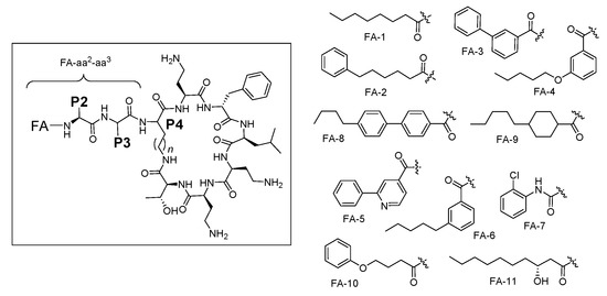
Figure 2.
General structure of polymyxin B nonapeptides and fatty acyl tails used in this study.
2. Results and Discussion
2.1. Chemistry
A total of 36 compounds were synthesized in this study (ten of which have been reported previously [33,36,37,38,39]), with the structures presented in Table 1, Table 2 and Table 3 and Table S1 (Supporting Information). All compounds possessed >95% purity, as determined by LCMS analysis using both ELSD and UV (210 nm) detection. The compounds were prepared by solid phase peptide synthesis (SPPS) (Scheme 1). Several SPPS strategies to construct the polymyxin scaffold have been reported, starting from the C-terminal Leu-7 [27] or Thr-10 [40,41] residue attached to the resin through the carboxyl terminus. Both strategies require a subsequent solution phase cyclisation step. On the other hand, on-resin cyclisation has been reported using Dab-9 as the anchoring point, which was attached to the resin via the Nγ-amino group of the side chain [42]. In the present study, which utilizes an on-resin cyclisation strategy, the polymyxin scaffold was constructed by anchoring the side chain β-hydroxy group of the C-terminal Thr-10 residue onto a dihydropyran DHP HM resin, as previously reported (Scheme 1) [36]. The C-terminal carboxylic acid of Thr-10 and the Nγ-amino group of Dab-4 were masked as allyl ester and carbamate protecting groups, respectively, allowing for orthogonal deprotection in the presence of the remaining Boc-protected Dab side chain amino groups en route to on-resin cyclisation. Thus, the synthesis was initiated using resin-bound Fmoc-L-Thr-CO2Allyl 3 (Scheme 1) [36]. The peptide sequence was constructed using SPPS with sequential Fmoc deprotection (30% piperidine in DMF) followed by Fmoc-amino acid coupling (HCTU, DIPEA in DMF), leading to the intermediate heptapeptide construct 4. On-resin cyclisation was effected by in situ deprotection of both the C-terminus of Thr10 and the Dab-4 side chain amine using Pd(PPh3)4 and PhSiH3, generating 5, which was then treated with DPPA and DIPEA in DMF overnight at room temperature to give the cyclised resin-bound intermediate 6. The synthesis was then completed by removal of the Dab-4 α-amino Fmoc group followed by the sequential addition of the linear exocyclic tail residues Fmoc-L-Dab(Boc)-OH and Fmoc-L-Thr(tBu)-OH to give the penultimate resin-bound precursor 7. Polymyxin B nonapeptide (PMBN) was prepared from 7 by sequential Fmoc removal from Thr-2 followed by treatment with TFA/Et3SiH/H2O (95:1:4), which liberated the peptide from the resin with concomitant side chain deprotection. Analogues 3–10, 12–14, 16–28, 30–44 and 46–47 were also prepared from 7 by sequential Fmoc removal from Thr-2 followed by acylation with the appropriate fatty acid, and cleavage/deprotection using TFA/Et3SiH/H2O (95:1:4). All compounds were purified by rp-HPLC and isolated as their TFA salts.

Table 1.
Summary of antimicrobial activity (MIC, mg/L) and cytotoxicity (CC50, µM) against HK-2 Cells for a series of polymyxin-B nonapeptide (PMBN) derivatives acylated with different fatty acids and varying the P4 residue.
Table 2.
Summary of antimicrobial activity (MIC, mg/L) and cytotoxicity (CC50, µM) against HK-2 cells for a series of polymyxin-B nonapeptide (PMBN) derivatives acylated with different fatty acids and varying the P2 residue.
Table 3.
Summary of antimicrobial activity (MIC, mg/L) and cytotoxicity (CC50, µM) against HK-2 cells for a series of polymyxin-B nonapeptide (PMBN) derivatives acylated with different fatty acids and varying the P3 residue.
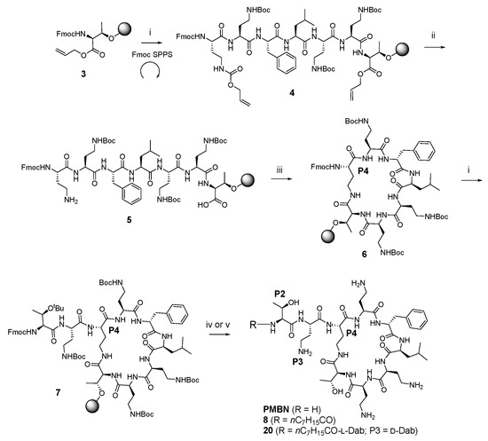
Scheme 1.
General on-resin cyclisation synthetic route to polymyxin nonapeptides, exemplified by the synthesis of PMBN, compound 8 and compound 20 a. a Reagent and conditions: (i) (a) 30% piperidine, DMF; (b) Fmoc-amino acid, HCTU, DIPEA, DMF; (c) repeat; (ii) Pd(PPh3)4, PhSiH3; (iii) DPPA, DIPEA, DMF; (iv) (a) 30% piperidine, DMF; (b) TFA/Et3SiH/H2O (95:1:4); (v) (a) 30% piperidine, DMF; (b) octanoic acid, HCTU, DIPEA, DMF; (c) TFA/Et3SiH/H2O (95:1:4).
2.2. Biological Activity
All compounds synthesised in this study were assessed for their minimum inhibitory concentrations (MIC, mg/L) by broth microdilution assay against five antibiotic-sensitive and resistant ATCC reference strains covering the Gram-negative ESKAPE pathogens (Escherichia coli, Klebsiella pneumoniae, A. baumannii, and P. aeruginosa) (Table 1, Table 2 and Table 3). Staphylococcus aureus was also included as a representative Gram-positive bacterial strain. Antimicrobial profiling was also performed against a subset of polymyxin-resistant MDR clinical isolates of K. pneumoniae, A. baumannii, and P. aeruginosa, but most compounds were inactive (MIC > 32 mg/L), data not shown for K. pneumoniae and A. baumannii isolates. Polymyxin B 1, colistin 2, vancomycin and gentamicin were used as positive inhibitor comparator compounds. Compounds were counter-screened against human proximal tubular epithelial cells (HK-2), using LDH release as a general indicator of cellular toxicity [43,44,45].
Polymyxins without a fatty acyl tail lack antimicrobial potency, exemplified by PMBN [25], which was inactive against all strains except P. aeruginosa ATCC 27853 (MIC 2 mg/L) (Table 1). Interestingly, activity against P. aeruginosa ATCC 27853 was relatively insensitive to structural changes, with most compounds displaying MICs 1–4 mg/L despite a variety of structural modifications (Table 1, Table 2 and Table 3). PMBN potency was restored with incorporation of a C8 tail (octanoic acid, OA), as previously demonstrated by nonapeptide 8, which possessed an exocyclic dipeptide motif OA-Thr2-Dab3 (MIC 1–2 mg/L for most strains) [36,37]. In comparison, PmxB3 [33] incorporating the native exocyclic tripeptide motif FA-Dab1-Thr2-Dab3 of Pmx, was 2- to 4-fold more active than nonapeptide 8 [36,37] against most strains (Table 1). The observation that Pmx nonapeptides produced by truncation of L-Dab1 could retain activity when substituted with an appropriate fatty acid, exemplified by nonapeptide 8, provided impetus to explore this phenomenon further.
In our previous study, we also reported decapeptide 16, possessing a relatively polar 2-chorophenyl urea fatty acyl moiety [36]. This analogue displayed MICs of 1–2 mg/L across most of the tested Gram-negative strains (Table 1). In contrast, nonapeptide 15, the truncated form of decapeptide 16, revealed a more striking difference between the two analogues, with 15 possessing considerably reduced activity during pairwise comparison. This contrasts with the general retention of activity observed between compound 8 [36,37] and PmxB3 [33]. This variation suggests a greater influence of L-Dab1 in the presence of a relatively polar fatty acid, and implies that judicious selection of the fatty acid in nonapeptides lacking the L-Dab1 may compensate for the reduced electrostatic component by additional hydrophobic interactions between the fatty acid and lipid A. This observation prompted further exploration of the fatty acyl component of analogue 8. Additional alterations to the fatty acid tail were generally disfavoured (compare 10–15), with 12 (FA = 4-hexylbenzoic acid) and 13 (FA = 6-phenylhexanoic acid) providing the best activities, albeit with increased cytotoxicity and some activity against S. aureus for 12 (MIC 8 mg/L). Data from this small SAR subset suggests that a simple N-alkyl fatty acyl chain is sufficient for good antimicrobial potency. The activity of 8 was abolished when the L-Thr2-L-Dab3 sequence was reversed (9, L-Dab2-L-Thr3).
The size of the macrocyclic ring was examined by substituting the diamino acid L-Dab4 in 8 with the homologs L-Orn4 17 and L-Lys4 18 (Table 1). Both modifications led to reduced activity, although the L-Orn4 variant was better tolerated, especially against E. coli and P. aeruginosa (MIC 2 mg/L against both). In contrast, reversal of the Dab4 stereochemistry to the D-configuration in 19 was highly detrimental (MIC > 32 mg/L for all strains). The data collectively suggests that optimal antimicrobial potency is highly dependent on the number of atoms forming the heptapeptide ring and the absolute configuration at P4.
The observations of Vaara [38] prompted an examination of the effect of reversing the stereochemistry of L-Dab3 in nonapeptide 8 to D-Dab3 in compound 21 (Table 2). Interestingly, the activity of 21 was somewhat comparable to 8, highlighting the tolerance of P3 to stereochemical inversion. A similar result was observed when the same modification was applied to PmxB3 leading to decapeptide 20, which was made for comparison. Furthermore, the activities of both 20 and 21, possessing exocyclic OA-Dab1-Thr2-D-Dab3 and OA-Thr2-D-Dab3 constructs, respectively, were notably comparable, suggesting the relative contribution of L-Dab1 toward potency was less important in this pairwise series compared to compounds 15 and 16 described earlier. Compound 21 was one of the few analogues examined that was active against a polymyxin-resistant strain of P. aeruginosa FADDI-PA070 (MIC 8 mg/L); cytotoxicity against HK-2 cells was promising (CC50 289 µM). Encouraged by this result, variation of the fatty acid component of 21 was explored, providing compounds 22–27 (Table 2). Analogue 25, containing 6-phenylhexanoic as the fatty acid component, possessed no observable cytotoxicity at the tested concentrations (CC50 > 300 µM) with consistent activity against the ATCC strains (MIC 0.5–4 mg/L), and moderate activity against polymyxin-resistant P. aeruginosa FADDI-PA070 (MIC 8 mg/L). On the other hand, more highly lipophilic fatty acids (e.g., 23, 24) were poorly favoured, instead leading to increased cytotoxicity with accompanying Gram-positive activity (S. aureus MIC 4–16 mg/L), albeit with potent activity against the polymyxin-resistant P. aeruginosa FADDI-PA070 strain (MIC 2–4 mg/L). Compound 21 was also modified to include variants at P2, leading to analogues 28–33 (Table 2). Potency improvements were not observed when L-Thr-2 was replaced with other hydroxylated amino acids (Tyr, Ser, Hse), nor any of the other amino acids examined, suggesting that threonine may be optimal at position 2 in nonapeptides bearing a D-Dab3 substituent. Likewise, in the series 28–33, inversion of the stereochemistry at position 3 (D-Dab3 to L-Dab3) was unproductive, with all compounds possessing MICs ≥ 16 mg/L (data not shown).
Finally, the promising activity observed for 21, containing D-Dab3, prompted further exploration of P3 with both D- and L-amino acids, as well as glycine (Table 3). It was previously demonstrated [36] that Gly3 was poorly tolerated in nonapeptide 49, in contrast to the potent activity observed for its decapeptide counterpart 50, again emphasising the influence of L-Dab1 on potency between the nonapeptide and decapeptide series. Potency could be partially restored in 49 by substitution of the octanoic acid tail with alternative fatty acids (compounds 51 and 52), albeit with variable activity across the Gram-negative panel (Table 3). Substitution of D-Dab3 in 21 with other basic amino acids (His, Lys, Orn, Dap) was generally well tolerated, even with inversion of stereochemistry, with 39 (L-Dap3) possessing the best activity (Table 3). Substitution with histidine was exceptional, with the D-isomer possessing much more potent activity compared to the L-isomer (compare 47 and 48, Table 3). When D-Dab3 in 21 was modified to provide analogues 35 (L-Ser3) or 36 (d-Ser3), activity was significantly reduced (MIC 16–32 mg/L for most strains except E. coli where MIC = 4 mg/L), in contrast to data previously reported by Vaara (35 = NAB743, 36 = NAB739) [31,38]. Of note, the decapeptide 34 [36] possessing an exocyclic OA-Dab1-Thr2-d-Ser3 construct, was significantly more potent than nonapeptide 36 lacking the L-Dab1 residue, again exemplifying the importance of L-Dab1 between nonapeptide and decapeptide variants. Activity across most strains was essentially abolished for analogues containing L-Hse3 (37), L-Asn3 (38), L-Cit3 (44) or L-Trp3 (46).
In the present study, we made a series of fatty N-acyl polymyxin nonapeptides, with alterations of the N-acyl component, and variation of the amino acids at positions P4, P3 and P2. Despite a lack of equipotency with polymyxin B 1 or colistin 2, selected nonapeptide analogues possessed promising antimicrobial activity against the panel of five antibiotic-sensitive and resistant Gram-negative ATCC reference strains tested. Two analogues, 23 and 24, were also active against a polymyxin-resistant strain of P. aeruginosa, but displayed increased cytotoxicity and activity against the Gram-positive S. aureus. Pairwise comparison of nonapeptide and decapeptide analogues (compare 8, 9; 15, 16; 20, 21; 34, 36; and 49, 50) revealed that the influence of the L-Dab1 residue on antimicrobial activity is contingent upon the fatty acyl component; activity was lost with more polar N-acyl components, but was retained with octanoic acid. Nonapeptides with stereochemical inversion at position 3 (i.e., D-Dab3 instead of L-Dab3) retained activity (e.g., 20 and 21), as did analogues possessing alternative positively charged amino acids (e.g., 39–43). On the other hand, activity was lost upon substitution of L-Dab3 with Gly3 in a nonapeptide framework, but could be restored in the decapeptide equivalent due to the presence of L-Dab1 (compare 49 and 50). A similar trend was evident upon substitution of L-Dab3 with a neutral d-Ser3 residue (compare 34 and 36). Collectively the present study demonstrates that polymyxin B nonapeptides may find utility in the design of improved polymxin analogues to fight antibiotic resistant infections.
3. Materials and Methods
3.1. Synthesis
Experimental procedures are described in the Supporting Information. All chemicals were obtained from commercial suppliers and used without further purification. LC-MS analyses were conducted using Agilent Technologies 1200 Series Instrument with a G1316A variable wavelength detector set at λ = 210 nm, 1200 Series ELSD, 6110 quadrupole ESI-MS, using an Agilent Eclipse XDB-Phenyl column (3 × 100 mm, 3.5 μm particle size, flow rate 1 mL/min, the mobile phases 0.05% formic acid in water and 0.05% formic acid in acetonitrile) (Agilent Technologies, Melbourne, Australia). Compound purification was performed using an Agilent 1260 Infinity Preparative HPLC with a G1365D multiple wavelength detector set at λ = 210 nm coupled to an Agilent Eclipse XDB-Phenyl column (21.2 × 100 mm, 5 μm particle size). Identities of final products were confirmed by high resolution mass spectrometry (HRMS), performed on a Bruker Micro TOF mass spectrometer using (+)-ESI calibrated to sodium formate (Bruker Daltonics, Melbourne, Australia). Final purity of more than 95% for all compounds was confirmed by LC-MS analysis using both ELSD and UV (210 nm) detection.
3.2. Minimum Inhibitory Concentration (MIC) Determination by Broth Microdilution Assay
Bacteria were either obtained from American Type Culture Collection (ATCC; Manassas, VA, USA) or independent academic clinical isolate collections, as listed in Table S2. Bacteria were cultured in nutrient broth (NB; Bacto Laboratories, catalog No. 234000) or Mueller Hinton broth (MHB; Bacto Laboratories, catalog No. 211443) at 37 °C overnight with shaking (∼180 rpm). A sample of each culture was then diluted 50-fold in fresh MHB and incubated at 37 °C for 1.5–3 h with shaking (∼180 rpm). Compound stock solutions were prepared as 0.64 or 2.56 mg/mL in water. The compounds, at twice the final desired concentration, were serially diluted 2-fold across the wells of 96-well plates (Polystyrene, Corning, catalogue No. 3370). Mid log phase bacterial cultures (after 1.5–3 h incubation) were diluted to 1 × 10⁶ colony forming units (CFU)/mL, and 50 μL was added to each well giving a final compound concentration range of 32 mg/L to 0.015 mg/L and a final cell density of 5 × 10⁵ CFU/mL. MICs were determined visually after 18 h of incubation at 37 °C, with the MIC defined as the lowest compound concentration at which no bacterial growth was visible.
3.3. Cytotoxicity (Lactate Dehydragenase (LDH) Assay)
Cytotoxicity to human kidney proximal tubular epithelial cell line, HK-2 (ATCC CRL-2190, sourced from ATCC; Manassas, VA, USA) was determined using the LDH assay as previously described [43,44,45]. In brief, HK-2 cells were seeded as 2000 cells/well in black-walled clear bottom 384-well tissue culture treated plates (Corning, catalogue No. 3712) in DMEM/F12 medium (Gibco® 10565-042) containing 10% of Fetal Bovine Serum (FBS, Gibco® 10099-141) and incubated for 24 h at 37 °C, 5% CO2. Compounds were then added into each well with a concentration series from 300 µM to 2.3 µM in 2-fold dilutions. Colistin and polymyxin B were used as controls and tested at a final concentration range of 1 mM to 7.8 µM. The cells were incubated with the compounds for 24 h at 37 °C, 5% CO2. After the incubation, 5 µL of culture medium was added to 45 µL of LDH assay buffer (Biovision, K313-500) and incubated for 30 min at room temperature. The absorbance (ABS) was then read at 450 nm using a Polar Star Omega plate reader. The data was analysed by Prism 6 software (GraphPad Software, La Jolla, CA, USA). Results were calculated using the following equation: cytotoxicity %= (ABSsamples − ABSuntreated/ABS1%Triton X-100 − ABSuntreated) × 100.
Supplementary Materials
The following are available online, Table S1: Compound Structures and HRMS Characterization; Table S2: Bacterial Strains used for Minimum Inhibitory Concentration (MIC) Determinations; Scheme S1: General On-Resin Cyclisation Synthetic Route to Polymyxin Nonapeptides, Exemplified by Synthesis of PMBN, Compound 8 and Compound 20; Figure S1: HR-(+)-ESI-TOF-MS of the [M + 2H]2+ mass ion peak of compound 8.
Author Contributions
Conceptualization: A.G.-G., C.M., B.B., M.S.B., M.A.T.B. and M.A.C.; Data curation: K.A.H.; Formal analysis: K.A.H., A.G.-G., A.G.E. and M.A.T.B.; Funding acquisition: C.M., B.B., M.S.B., M.A.T.B. and M.A.C.; Investigation: K.A.H., A.G.-G., C.M., B.B., A.G.E., J.X.H., R.P. and M.S.B.; Methodology: K.A.H., A.G.-G., C.M., B.B., A.G.E. and J.X.H.; Project administration: K.A.H., A.G.-G., M.A.T.B. and M.A.C.; Writing—original draft: K.A.H.; Writing—review & editing: K.A.H., A.G.E., M.S.B., M.A.T.B. and M.A.C.
Funding
This research was funded by NHMRC grants APP1005350, APP1045326 and APP1139609, and NIH grants R21AI098731/R33AI098731. M.A.T.B., K.A.H., and A.G.E. were supported by Wellcome Trust Seeding Drug Discovery Award (094977/Z/10/Z) and Wellcome Trust Strategic Grant (104797/Z/14/Z). M.A.C. is a NHMRC principle research fellow (APP1059354) and also holds a fractional professorial research fellow appointment at the University of Queensland, with his remaining time as CEO of Inflazome Ltd., a company developing drugs to address clinical unmet needs in inflammatory disease.
Acknowledgments
We thank Soumya Ramu, Angela M. Kavanagh, Emily Furlong, Maite Amado and Ali Hinton for technical support with susceptibility assays.
Conflicts of Interest
The authors declare that there are no conflict of interest.
References
- Benedict, R.G.; Langlykke, A.F. Antibiotic Activity of Bacillus polymyxa. J. Bacteriol. 1947, 54, 24–25. [Google Scholar] [PubMed]
- Stansly, P.G.; Schlosser, M.E. Studies on Polymyxin—Isolation and Identification of Bacillus polymyxa and Differentiation of Polymyxin from Certain Known Antibiotics. J. Bacteriol. 1947, 54, 549–556. [Google Scholar] [PubMed]
- Stansly, P.G.; Brownlee, G. Nomenclature of Polymyxin Antibiotics. Nature 1949, 163, 611. [Google Scholar] [CrossRef] [PubMed]
- Stansly, P.G.; Shepherd, R.G.; White, H.J. Polymyxin—A New Chemotherapeutic Agent. Bull. Johns Hopkins Hosp. 1947, 81, 43–54. [Google Scholar] [PubMed]
- Shoji, J.; Kato, T.; Hinoo, H. Structures of 2 New Polymyxin Group Antibiotics. J. Antibiot. 1977, 30, 427–429. [Google Scholar] [CrossRef] [PubMed]
- Shoji, J.; Hinoo, H.; Wakisaka, Y.; Koizumi, K.; Mayama, M.; Matsuura, S. Isolation of Two New Polymyxin Group Antibiotics (Studies on Antibiotics from Genus Bacillus. XX). J. Antibiot. 1977, 30, 1029–1034. [Google Scholar] [CrossRef]
- Wilkinson, S.; Lowe, L.A. Structures of Polymyxins A and the Question of Identity with the Polymyxins M. Nature 1966, 212, 311. [Google Scholar] [CrossRef]
- Parker, W.L.; Rathnum, M.L.; Dean, L.D.; Nimeck, M.W.; Brown, W.E.; Meyers, E. Polymyxin-F, a New Peptide Antibiotic. J. Antibiot. 1977, 30, 767–769. [Google Scholar] [CrossRef]
- Zavascki, A.P.; Nation, R.L. Nephrotoxicity of Polymyxins: Is There Any Difference between Colistimethate and Polymyxin B? Antimicrob. Agents Chemother. 2017, 61. [Google Scholar] [CrossRef]
- Manchandani, P.; Zhou, J.; Babic, J.T.; Ledesma, K.R.; Truong, L.D.; Tam, V.H. Role of Renal Drug Exposure in Polymyxin B-Induced Nephrotoxicity. Antimicrob. Agents Chemother. 2017, 61. [Google Scholar] [CrossRef]
- Codjoe, F.S.; Donkor, E.S. Carbapenem Resistance: A Review. Med. Sci. 2017, 6, 1. [Google Scholar] [CrossRef] [PubMed]
- Van Loon, K.; Voor In ’t Holt, A.F.; Vos, M.C. A Systematic Review and Meta-analyses of the Clinical Epidemiology of Carbapenem-Resistant Enterobacteriaceae. Antimicrob. Agents Chemother. 2018, 62. [Google Scholar] [CrossRef] [PubMed]
- Bush, K. Past and Present Perspectives on β-Lactamases. Antimicrob. Agents Chemother. 2018, 62. [Google Scholar] [CrossRef] [PubMed]
- Bush, K. Proliferation and significance of clinically relevant β-lactamases. Ann. NY Acad. Sci. 2013, 1277, 84–90. [Google Scholar] [CrossRef] [PubMed]
- PEW. A Scientific Roadmap for Antibiotic Discovery. 2016. Available online: http://www.pewtrusts.org/~/media/assets/2016/05/ascientificroadmapforantibioticdiscovery.pdf (accessed on 31 January 2019).
- Luepke, K.H.; Suda, K.J.; Boucher, H.; Russo, R.L.; Bonney, M.W.; Hunt, T.D.; Mohr, J.F. Past, Present, and Future of Antibacterial Economics: Increasing Bacterial Resistance, Limited Antibiotic Pipeline, and Societal Implications. Pharmacotherapy 2017, 37, 71–84. [Google Scholar] [CrossRef] [PubMed]
- Butler, M.S.; Blaskovich, M.A.T.; Cooper, M.A. Antibiotics in the clinical pipeline at the end of 2015. J. Antibiot. 2017, 70, 3–24. [Google Scholar] [CrossRef] [PubMed]
- Tran, T.B.; Velkov, T.; Nation, R.L.; Forrest, A.; Tsuji, B.T.; Bergen, P.J.; Li, J. Pharmacokinetics/pharmacodynamics of colistin and polymyxin B: Are we there yet? Int. J. Antimicrob. Agent 2016, 48, 592–597. [Google Scholar] [CrossRef] [PubMed]
- Nation, R.L.; Li, J.; Cars, O.; Couet, W.; Dudley, M.N.; Kaye, K.S.; Mouton, J.W.; Paterson, D.L.; Tam, V.H.; Theuretzbacher, U.; et al. Framework for optimisation of the clinical use of colistin and polymyxin B: The Prato polymyxin consensus. Lancet Infect. Dis. 2015, 15, 225–234. [Google Scholar] [CrossRef]
- Roberts, K.D.; Azad, M.A.K.; Wang, J.P.; Horne, A.S.; Thompson, P.E.; Nation, R.L.; Velkov, T.; Li, J. Antimicrobial Activity and Toxicity of the Major Lipopeptide Components of Polymyxin B and Colistin: Last-Line Antibiotics against Multidrug-Resistant Gram-Negative Bacteria. ACS Infect. Dis. 2015, 1, 568–575. [Google Scholar] [CrossRef]
- Rabanal, F.; Cajal, Y. Recent advances and perspectives in the design and development of polymyxins. Nat. Prod. Rep. 2017, 34, 886–908. [Google Scholar] [CrossRef]
- Baron, S.; Hadjadj, L.; Rolain, J.M.; Olaitan, A.O. Molecular mechanisms of polymyxin resistance: Knowns and unknowns. Int. J. Antimicrob. Ag. 2016, 48, 583–591. [Google Scholar] [CrossRef] [PubMed]
- Hancock, R.E.W.; Chapple, D.S. Peptide antibiotics. Antimicrob. Agents Chemother. 1999, 43, 1317–1323. [Google Scholar] [CrossRef] [PubMed]
- Velkov, T.; Thompson, P.E.; Nation, R.L.; Li, J. Structure-Activity Relationships of Polymyxin Antibiotics. J. Med. Chem. 2010, 53, 1898–1916. [Google Scholar] [CrossRef] [PubMed]
- Duwe, A.K.; Rupar, C.A.; Horsman, G.B.; Vas, S.I. In vitro Cytotoxicity and Antibiotic-Activity of Polymyxin-B Nonapeptide. Antimicrob. Agents Chemother. 1986, 30, 340–341. [Google Scholar] [CrossRef] [PubMed]
- Quale, J.; Shah, N.; Kelly, P.; Babu, E.; Backer, M.; Rosas-Garcia, G.; Salamera, J.; George, A.; Bratu, S.; Landman, D. Activity of Polymyxin B and the Novel Polymyxin Analogue CB-182,804 Against Contemporary Gram-Negative Pathogens in New York City. Microb. Drug Resist. 2012, 18, 132–136. [Google Scholar] [CrossRef] [PubMed]
- Magee, T.V.; Brown, M.F.; Starr, J.T.; Ackley, D.C.; Abramite, J.A.; Aubrecht, J.A.; Butler, J.L.; Crandon, F.; Dib-Hajj, M.E.; Flanagan, K.; et al. Discovery of Dap-3 Polymyxin Analogues for the Treatment of Multidrug-Resistant Gram-Negative Nosocomial Infections. J. Med. Chem. 2013, 56, 5079–5093. [Google Scholar] [CrossRef] [PubMed]
- Brown, B.; Boakes, S.; Duperchy, E.; Simonovic, M.; Abdulle, O.; Divall, N.; Stanway, S.J.; Wilson, A.; Moss, S.F.; Dawson, M.J. Synthesis and Structure-Activity Relationships of Polymyxin Nonapeptide Derivatives with N-terminal Aminoacyl Moieties. In Proceedings of the 55th Interscience Conference on Antimicrobial Agents and Chemotherapy, San Diego, CA, USA, 17–21 September 2015. [Google Scholar]
- Gordeev, M.F.; Liu, J.; Wang, X.; Yuan, Z. Antimicrobial Polymyxins for Treatment of Bacterial Infections. WO Patent 2016/100578, 23 June 2016. [Google Scholar]
- Rabanal, F.; Grau-Campistany, A.; Vila-Farres, X.; Gonzalez-Linares, J.; Borras, M.; Vila, J.; Manresa, A.; Cajal, Y. A bioinspired peptide scaffold with high antibiotic activity and low in vivo toxicity. Sci. Rep. 2015, 5, 10558. [Google Scholar] [CrossRef]
- Vaara, M.; Vaara, T.; Tyrrell, J.M. Structure-activity studies on polymyxin derivatives carrying three positive charges only reveal a new class of compounds with strong antibacterial activity. Peptides 2017, 91, 8–12. [Google Scholar] [CrossRef]
- Sato, Y.; Shindo, M.; Sakura, N.; Uchida, Y.; Kato, I. Novel Des-Fatty Acyl-Polymyxin B Derivatives with Pseudomonas aeruginosa-Specific Antimicrobial Activity. Chem. Pharm. Bull. 2011, 59, 597–602. [Google Scholar] [CrossRef]
- Sakura, N.; Itoh, T.; Uchida, Y.; Ohki, K.; Okimura, K.; Chiba, K.; Sato, Y.; Sawanishi, H. The contribution of the N-terminal structure of polymyxin B peptides to antimicrobial and lipopolysaccharide binding activity. Bull. Chem. Soc. Jpn. 2004, 77, 1915–1924. [Google Scholar] [CrossRef]
- Tsubery, H.; Ofek, I.; Cohen, S.; Fridkin, M. Structure-function studies of polymyxin B nonapeptide: Implications to sensitization of gram-negative bacteria. J. Med. Chem. 2000, 43, 3085–3092. [Google Scholar] [CrossRef] [PubMed]
- Corbett, D.; Wise, A.; Langley, T.; Skinner, K.; Trimby, E.; Birchall, S.; Dorali, A.; Sandiford, S.; Williams, J.; Warn, P.; et al. Potentiation of Antibiotic Activity by a Novel Cationic Peptide: Potency and Spectrum of Activity of SPR741. Antimicrob. Agents Chemother. 2017, 61. [Google Scholar] [CrossRef] [PubMed]
- Gallardo-Godoy, A.; Muldoon, C.; Becker, B.; Elliott, A.G.; Lash, L.H.; Huang, J.X.; Butler, M.S.; Pelingon, R.; Kavanagh, A.M.; Ramu, S.; et al. Activity and Predicted Nephrotoxicity of Synthetic Antibiotics Based on Polymyxin B. J. Med. Chem. 2016, 59, 1068–1077. [Google Scholar] [CrossRef] [PubMed]
- O’Dowd, H.; Kim, B.; Margolis, P.; Wang, W.; Wu, C.; Lopez, S.L.; Blais, J. Preparation of tetra-Boc-protected polymyxin B nonapeptide. Tetrahedron Lett. 2007, 48, 2003–2005. [Google Scholar] [CrossRef]
- Vaara, M.; Fox, J.; Loidl, G.; Siikanen, O.; Apajalahti, J.; Hansen, F.; Frimodt-Moller, N.; Nagai, J.; Takano, M.; Vaara, T. Novel polymyxin derivatives carrying only three positive charges are effective antibacterial agents. Antimicrob. Agents Chemother. 2008, 52, 3229–3236. [Google Scholar] [CrossRef] [PubMed]
- Leese, R.A. Antibiotic Compositions for the Treatment of Gram Negative Infections. U.S. Patent 8343912B2, 1 January 2013. [Google Scholar]
- Velkov, T.; Roberts, K.D.; Nation, R.L.; Wang, J.P.; Thompson, P.E.; Li, J. Teaching ‘Old’ Polymyxins New Tricks: New-Generation Lipopeptides Targeting Gram-Negative ‘Superbugs’. ACS Chem. Biol. 2014, 9, 1172–1177. [Google Scholar] [CrossRef] [PubMed]
- Ramesh, S.; Govender, T.; Kruger, H.G.; Albericio, F.; de la Torre, B.G. An improved and efficient strategy for the total synthesis of a colistin-like peptide. Tetrahedron Lett. 2016, 57, 1885–1888. [Google Scholar] [CrossRef]
- Xu, W.L.; Cui, A.L.; Hu, X.X.; You, X.F.; Li, Z.R.; Zheng, J.S. A new strategy for total solid-phase synthesis of polymyxins. Tetrahedron Lett. 2015, 56, 4796–4799. [Google Scholar] [CrossRef]
- Huang, J.X.; Blaskovich, M.A.; Cooper, M.A. Cell- and biomarker-based assays for predicting nephrotoxicity. Expert Opin. Drug Met. 2014, 10, 1621–1635. [Google Scholar] [CrossRef]
- Huang, J.X.; Kaeslin, G.; Ranall, M.V.; Blaskovich, M.A.; Becker, B.; Butler, M.S.; Little, M.H.; Lash, L.H.; Cooper, M.A. Evaluation of biomarkers for in vitro prediction of drug induced nephrotoxicity: Comparison of HK-2, immortalized human proximal tubule epithelial, and primary cultures of human proximal tubular cells. Pharmacol. Res. Perspect. 2015, 3. [Google Scholar] [CrossRef]
- Becker, B.; Butler, M.S.; Hansford, K.A.; Gallardo-Godoy, A.; Elliott, A.G.; Huang, J.X.; Edwards, D.J.; Blaskovich, M.A.T.; Cooper, M.A. Synthesis of octapeptin C4 and biological profiling against NDM-1 and polymyxin-resistant bacteria. Bioorg. Med. Chem. Lett. 2017, 27, 2407–2409. [Google Scholar] [CrossRef] [PubMed]
Sample Availability: Selected compounds are available from the authors. |


© 2019 by the authors. Licensee MDPI, Basel, Switzerland. This article is an open access article distributed under the terms and conditions of the Creative Commons Attribution (CC BY) license (http://creativecommons.org/licenses/by/4.0/).