
The Potential of Novel Chitosan-Based Scaffolds in Pelvic Organ Prolapse (POP) Treatment through Tissue Engineering

 , ,
, ,  and
and
Abstract
:1. Introduction
2. Results and Discussion
2.1. FT–IR Analysis of the Potential Scaffolds
2.2. Porosity and Density Study
2.3. Swelling Capability Study
2.4. Morphology Study
2.5. Antioxidant Activity Study
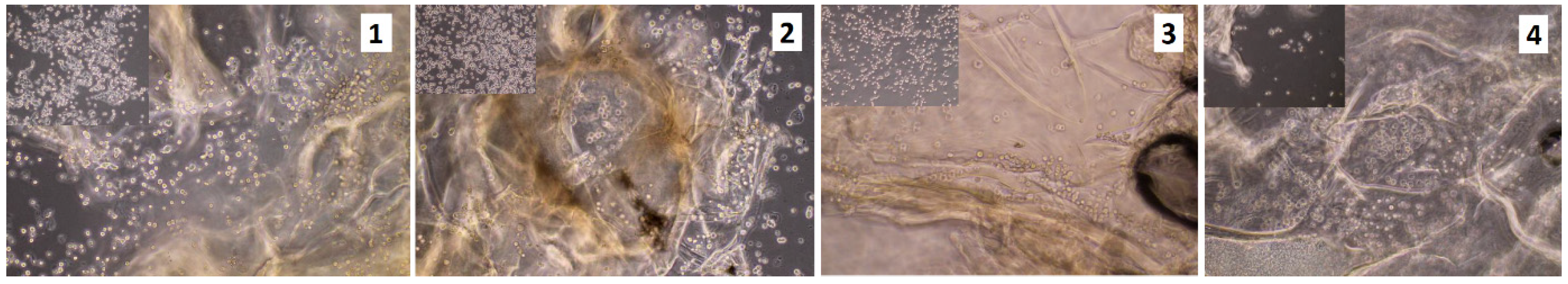
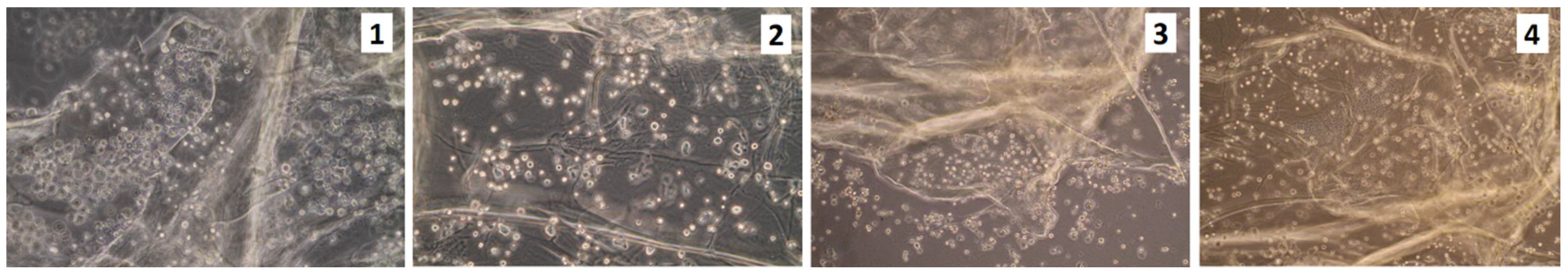
2.6. Cytotoxicity Study
3. Materials and Methods
3.1. Materials
3.2. Methods
3.2.1. Chitosan Scaffolds Synthesis
3.2.2. Chemical Structure Analysis
3.2.3. Porosity and Density Study
3.2.4. Swelling Capability Study
- %SD—swelling degree
- Wt—weight of the investigated sample after time = t, g
- W0—initial weight of the investigated sample, g
3.2.5. Scanning Electron Microscope (SEM) Analysis and X-ray Microanalysis
3.2.6. Antioxidant Activity Study
- %S—the % of the free radicals that were neutralized
- Ac—the absorbance of the DPPH solution without the sample
- As—the absorbance of the DPPH solution containing sample
3.2.7. Cytotoxicity Study
4. Conclusions
Author Contributions
Funding
Conflicts of Interest
References
- Stangel-Wójcikiewicz, K.; Jarocha, D.; Piwowar, M.; Jach, R.; Uhl, T.; Basta, A.; Majka, M. Autologous muscle-derived cells for the treatment of female stress urinary incontinence: A 2-year follow-up of a Polish investigation. Neurourol. Urodyn. 2014, 33, 324–330. [Google Scholar] [CrossRef]
- Wu, J.M.; Vaughan, C.P.; Goode, P.S.; Redden, D.T.; Burgio, K.L.; Richter, H.E.; Markland, A.D. Prevalence and trends of symptomatic pelvic floor disorders in US women. Obs. Gynecol. 2014, 123, 141–148. [Google Scholar] [CrossRef] [Green Version]
- Stangel-Wójcikiewicz, K.; Stec, M.; Nikolavsky, D.; Chancellor, M.B. Cellular therapy for treatment of stress urinary incontinence. Curr. Stem. Cell. Res. 2010, 5, 57–62. [Google Scholar] [CrossRef]
- Stangel-Wójcikiewicz, K.; Piwowar, M.; Jach, R.; Majka, M.; Basta, A. Quality of life assessment in female patients 2 and 4 years after muscle-derived cell transplants for stress urinary incontinence treatment. Ginekol. Pol. 2016, 87, 183–189. [Google Scholar] [CrossRef] [PubMed]
- Pence, J.C.; Clancy, K.B.; Harley, B.A. The induction of pro-angiogenic processes within a collagen scaffold via exogenous estradiol and endometrial epithelial cells. Biotechnol. Bioeng. 2015, 112, 2185–2194. [Google Scholar] [CrossRef] [Green Version]
- Dällenbach, P. To mesh or not to mesh: A review of pelvic organ reconstructive surgery. Int. J. Womens Health 2015, 7, 331–343. [Google Scholar] [CrossRef] [Green Version]
- Barski, D.; Deng, Y. Management of Mesh Complications after SUI and POP Repair: Review and Analysis of the Current Literature. BioMed Res. Int. 2015, 2015, 831285. [Google Scholar] [CrossRef] [PubMed] [Green Version]
- Blaivas, J.G.; Purohit, R.S.; Benedon, M.S.; Mekel, G.; Stern, M.; Billah, M.; Bendavid, R.; Iakovlev, V. Safety considerations for synthetic sling surgery. Nat. Rev. Urol. 2015, 12, 481–509. [Google Scholar] [CrossRef] [PubMed]
- Mangır, N.; Hillary, C.J.; Chapple, C.R.; MacNeil, S. Oestradiol-releasing biodegradable mesh stimulates collagen production and angiogenesis: An approach to improving biomaterial integration in pelvic floor repair. Eur. Urol. Focus. 2019, 5, 280–289. [Google Scholar] [CrossRef] [Green Version]
- Alperin, M. Collagen scaffold: A treatment for large mesh exposure following vaginal prolapse repair. Int. Urogynecol. J. 2014, 25, 1597–1599. [Google Scholar] [CrossRef] [Green Version]
- Mangera, A.; Bullock, A.J.; Roman, S.; Chapple, C.R.; MacNeil, S. Comparison of candidate scaffolds for tissue engineering for stress urinary incontinence and pelvic organ prolapse repair. BJU Int. 2013, 112, 674–685. [Google Scholar] [CrossRef] [PubMed] [Green Version]
- Roman, S.; Mangera, A.; Osman, N.I.; Bullock, A.J.; Chapple, C.R.; MacNeil, S. Developing a tissue engineered repair material for treatment of stress urinary incontinence and pelvic organ prolapse-which cell source? Neurourol. Urodyn. 2014, 33, 531–537. [Google Scholar] [CrossRef] [PubMed]
- Wang, X.; Chen, Y.; Fan, Z.; Hua, K. Comparing different tissue-engineered repair materials for the treatment of pelvic organ prolapse and urinary incontinence: Which material is better? Int. Urogynecol. J. 2018, 29, 131–138. [Google Scholar] [CrossRef] [PubMed]
- Shi, L.B.; Cai, H.X.; Chen, L.K.; Wu, Y.; Zhu, S.A.; Gong, X.N.; Xia, Y.X.; Ouyang, H.W.; Zou, X.H. Tissue engineered bulking agent with adipose-derived stem cells and silk fibroin microspheres for the treatment of intrinsic urethral sphincter deficiency. Biomaterials 2014, 35, 1519–1530. [Google Scholar] [CrossRef] [PubMed]
- Grooms, T.N.; Vuong, H.R.; Tyo, K.M.; Malik, D.A.; Sims, L.B.; Whittington, C.P.; Palmer, K.E.; Matoba, N.; Steinbach-Rankins, J.M. Griffithsin-Modified Electrospun Fibers as a Delivery Scaffold To Prevent HIV Infection. Antimicrob. Agents. Chem. 2016, 11, 6518–6531. [Google Scholar] [CrossRef] [PubMed] [Green Version]
- Younes, I.; Rinaudo, M. Chitin and Chitosan Preparation from Marine Sources. Structure, Properties and Applications. Mar. Drugs 2015, 13, 1133–1174. [Google Scholar] [CrossRef] [Green Version]
- Rinaudo, M. Chitin and chitosan: Properties and applications. Prog. Polym. Sci. 2006, 31, 603–632. [Google Scholar] [CrossRef]
- Rong, H.C.; Hwa, H.D. Effect of molecular weight of chitosan with the same degree of deacetylation on the thermal, mechanical, and permeability properties of the prepared membrane. Carbohydr. Polym. 1996, 29, 353–358. [Google Scholar] [CrossRef]
- Mima, S.; Miya, M.; Iwamoto, R.; Yoshikawa, S. Highly deacetylated chitosan and its properties. J. Appl. Polym. Sci. 1983, 28, 1909–1917. [Google Scholar] [CrossRef]
- Smith, J.; Wood, E.; Dornish, M. Effect of chitosan on epithelial cell tight junctions. Pharm. Res. 2004, 21, 43–49. [Google Scholar] [CrossRef]
- Patil, S.V.; Nanduri, L.S.Y. Interaction of chitin/chitosan with salivary and other epithelial cells—An overview. Int. J. Biol. Macromol. 2017, 104, 1398–1406. [Google Scholar] [CrossRef] [PubMed]
- Choi, J.S.; Yoo, H.S. Pluronic/chitosan hydrogels containing epidermal growth factor with wound-adhesive and photo-crosslinkable properties. J. Biomed. Mater. Res. A 2010, 95, 564–573. [Google Scholar] [CrossRef] [PubMed]
- Wetta, L.A.; Gerten, K.A.; Wheeler, T.L.; Holley, R.L.; Varner, R.E.; Richter, H.E. Synthetic graft use in vaginal prolapse surgery: Objective and subjective outcomes. Int. Urogynecol. J. 2009, 20, 1307–1312. [Google Scholar] [CrossRef] [Green Version]
- Kaczmarek, B.; Sionkowska, A.; Osyczka, A.M. Scaffolds based on chitosan and collagen with glycosaminoglycans crosslinked by tannic acid. Polym. Test. 2018, 65, 163–168. [Google Scholar] [CrossRef]
- Kaya, M.; Baran, T.; Asan-Ozusaglam, M.; Cakmak, Y.S.; Tozak, K.O.; Mol, A.; Mentes, A.; Sezen, G. Extraction and characterization of chitin and chitosan with antimicrobial and antioxidant activities from cosmopolitan Orthoptera species (Insecta). Biotechnol. Bioproc. Eng. 2015, 20, 168–179. [Google Scholar] [CrossRef]
- Piątkowski, M.; Janus, Ł.; Radwan-Pragłowska, J.; Bogdał, D.; Matysek, D. Biodegradable, pH-sensitive chitosan beads obtained under microwave radiation for advanced cell culture. Colloids Surface B 2018, 164, 324–331. [Google Scholar] [CrossRef]
- Radwan-Pragłowska, J.; Piątkowski, M.; Janus, Ł.; Bogdał, D.; Matysek, D. Biodegradable, pH-responsive chitosan aerogels for biomedical applications. RSC Adv. 2017, 7, 32960–32965. [Google Scholar] [CrossRef] [Green Version]
- Piątkowski, M.; Kitala, D.; Radwan-Pragłowska, J.; Janus, Ł.; Klama-Baryła, A.; Łabuś, W.; Tomanek, E.; Glik, J.; Matýsek, D.; Kawecki, M. Chitosan/aminoacid hydrogels with antimicrobial and bioactive properties as new scaffolds for human mesenchymal stem cells culture applicable in wound healing. Express Polym. Lett. 2018, 11, 100–112. [Google Scholar] [CrossRef]
- Piątkowski, M.; Radwan-Pragłowska, J.; Janus, Ł.; Bogdał, D.; Matysek, D.; Cablik, V. Microwave-assisted synthesis and characterization of chitosan aerogels doped with Au-NPs for skin regeneration. Polym. Test. 2019, 73, 366–376. [Google Scholar] [CrossRef]
- Kallak, T.K.; Uvnäs-Moberg, K. Oxytocin stimulates cell proliferation in vaginal cell line Vk2E6E7. Post Reprod. Health 2017, 23, 6–12. [Google Scholar] [CrossRef]
- Fan, F.-Y.; Chiu, C.-C.; Tseng, C.-L.; Lee, H.-S.; Pan, Y.-N.; Yang, K.-C. Glycosaminoglycan/chitosan hydrogel for matrix-associated autologous chondrocyte implantation: An in vitro study. J. Med. Biol. Eng. 2014, 34, 211–217. [Google Scholar] [CrossRef]
Sample Availability: Samples of the compounds are available from the authors on the request. |


| Sample | Crosslinking Agent, g | High Boiling Solvent, mL | Chitosan, g | H2O, mL | Reaction Time, min |
|---|---|---|---|---|---|
| 1 | Adipic, 0.47 Malonic, 0.12 | 10 | 0.5 | 15 | 20 |
| 2 | Adipic, 0.48 l-glutamic, 0.22 | 10 | 0.5 | 15 | 20 |
| 3 | Adipic, 0.16 l-glutamic, 0.16 Malonic, 0.26 | 10 | 0.5 | 20 | 20 |
| 4 | Adipic, 0.5 Levulinic, 0.5 | 10 | 0.5 | 20 | 20 |
© 2020 by the authors. Licensee MDPI, Basel, Switzerland. This article is an open access article distributed under the terms and conditions of the Creative Commons Attribution (CC BY) license (http://creativecommons.org/licenses/by/4.0/).
Share and Cite
Radwan-Pragłowska, J.; Stangel-Wójcikiewicz, K.; Piątkowski, M.; Janus, Ł.; Matýsek, D.; Majka, M.; Amrom, D. The Potential of Novel Chitosan-Based Scaffolds in Pelvic Organ Prolapse (POP) Treatment through Tissue Engineering. Molecules 2020, 25, 4280. https://doi.org/10.3390/molecules25184280
Radwan-Pragłowska J, Stangel-Wójcikiewicz K, Piątkowski M, Janus Ł, Matýsek D, Majka M, Amrom D. The Potential of Novel Chitosan-Based Scaffolds in Pelvic Organ Prolapse (POP) Treatment through Tissue Engineering. Molecules. 2020; 25(18):4280. https://doi.org/10.3390/molecules25184280
Chicago/Turabian StyleRadwan-Pragłowska, Julia, Klaudia Stangel-Wójcikiewicz, Marek Piątkowski, Łukasz Janus, Dalibor Matýsek, Marcin Majka, and Dalia Amrom. 2020. "The Potential of Novel Chitosan-Based Scaffolds in Pelvic Organ Prolapse (POP) Treatment through Tissue Engineering" Molecules 25, no. 18: 4280. https://doi.org/10.3390/molecules25184280