
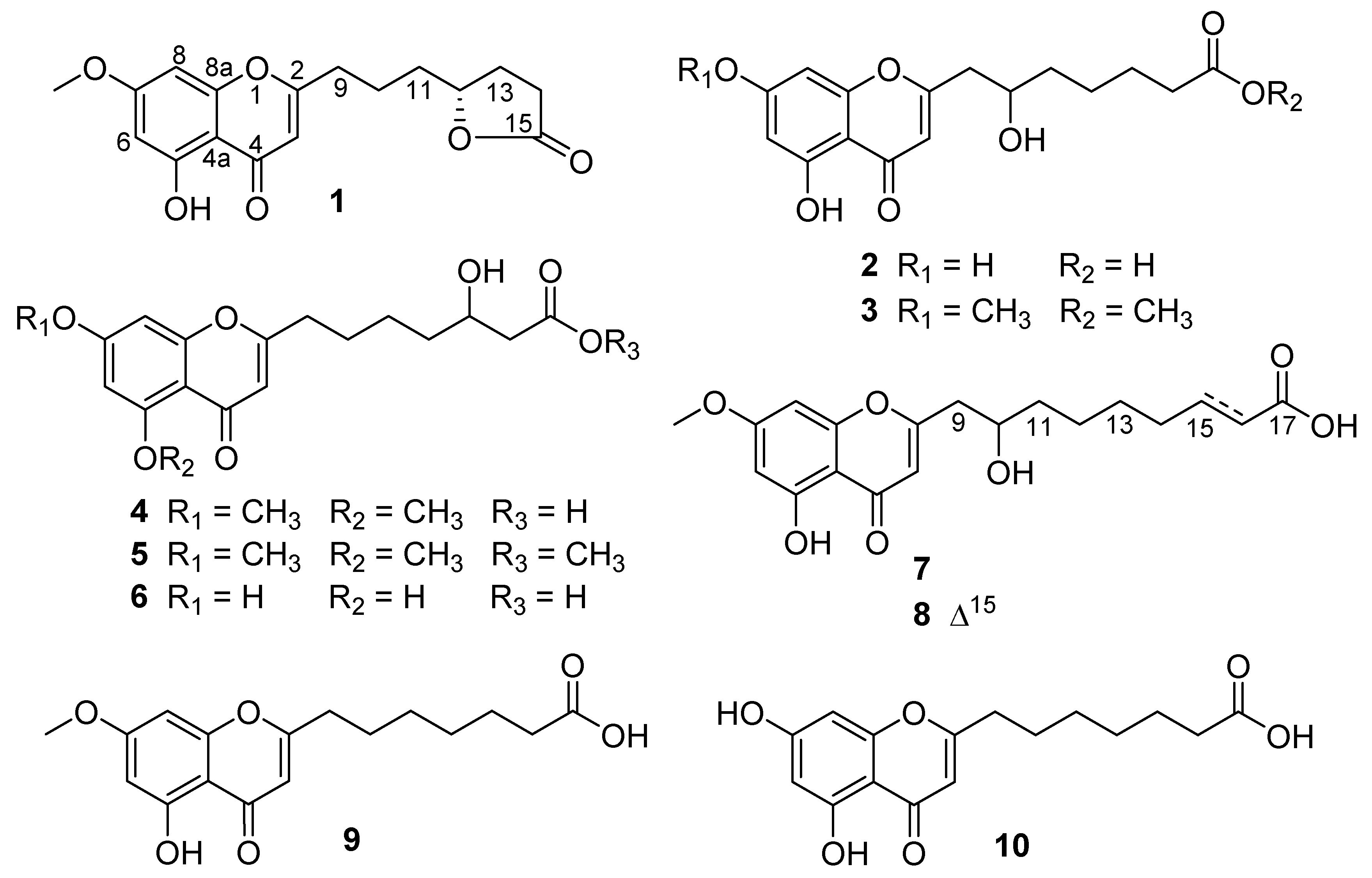
Chromone Derivatives with α-Glucosidase Inhibitory Activity from the Marine Fungus Penicillium thomii Maire

 and
and
Abstract
:
1. Introduction
2. Results
3. Materials and Methods
3.1. General Experimental Procedure
3.2. Fungal Strain and Identification
3.3. Fermentation, Extraction, and Isolation
3.4. α-Glucosidase Assay
3.5. Antioxidant Activity
3.6. Computational Details
4. Conclusions
Supplementary Materials
Author Contributions
Funding
Institutional Review Board Statement
Informed Consent Statement
Data Availability Statement
Conflicts of Interest
Sample Availability
References
- Keri, R.S.; Budagumpi, S.; Pai, R.K.; Balakrishna, R.G. Chromones as a privileged scaffold in drug discovery: A review. Eur. J. Med. Chem. 2014, 78, 340–374. [Google Scholar] [CrossRef] [PubMed]
- Carroll, A.R.; Copp, B.R.; Davis, R.A.; Keyzers, R.A.; Prinsep, M.R. Marine natural products. Nat. Prod. Rep. 2021, 38, 362–413. [Google Scholar] [CrossRef]
- Rateb, M.E.; Ebel, R. Secondary metabolites of fungi from marine habitats. Nat. Prod. Rep. 2011, 28, 290–344. [Google Scholar] [CrossRef] [PubMed]
- Newman, D.J.; Cragg, G.M. Natural products as sources of new drugs over the nearly four decades from 01/1981 to 09/2019. J. Nat. Prod. 2020, 83, 770–803. [Google Scholar] [CrossRef]
- Cardoso, J.; Nakayama, D.G.; Sousa, E.; Pinto, E. Marine-derived compounds and prospects for their antifungal application. Molecules 2020, 25, 5856. [Google Scholar] [CrossRef] [PubMed]
- Alves, A.; Sousa, E.; Kijjoa, A.; Pinto, M. Marine-derived compounds with potential use as cosmeceuticals and nutricosmetics. Molecules 2020, 25, 2536. [Google Scholar] [CrossRef] [PubMed]
- Liang, Z.; Gu, T.; Wang, J.; She, J.; Ye, Y.; Cao, W.; Luo, X.; Xiao, J.; Liu, Y.; Tang, L.; et al. Chromene and chromone derivatives as liver X receptors modulators from a marine-derived Pestalotiopsis neglecta fungus. Bioorg. Chem. 2021, 112, 104927. [Google Scholar] [CrossRef] [PubMed]
- Luo, X.; Yang, J.; Chen, F.; Lin, X.; Chen, C.; Zhou, X.; Liu, S.; Liu, Y. Structurally diverse polyketides from the mangrove-derived fungus Diaporthe sp. SCSIO 41011 with their anti-influenza A virus activities. Front. Chem. 2018, 6, 286. [Google Scholar] [CrossRef] [PubMed] [Green Version]
- Bao, J.; Luo, J.F.; Qin, X.C.; Xu, X.Y.; Zhang, X.Y.; Tu, Z.C.; Qi, S.H. Dihydrothiophene-condensed chromones from a marine-derived fungus Penicillium oxalicum and their structure–bioactivity relationship. Bioorg. Med. Chem. Lett. 2014, 24, 2433–2436. [Google Scholar] [CrossRef] [PubMed]
- Cheng, Z.; Li, Y.; Xu, W.; Liu, W.; Liu, L.; Zhu, D.; Kang, Y.; Luo, Z.; Li, Q. Three new cyclopiane-type diterpenes from a deep-sea derived fungus Penicillium sp. YPGA11 and their effects against human esophageal carcinoma cells. Bioorg. Chem. 2019, 91, 103129. [Google Scholar] [CrossRef] [PubMed]
- Li, Y.L.; Liu, W.; Xu, W.; Zeng, X.; Cheng, Z.B.; Li, Q. Aspterrics A and B, new sesquiterpenes from deep sea-derived fungus Aspergillus terreus YPGA10. Rec. Nat. Prod. 2020, 14, 18–22. [Google Scholar] [CrossRef]
- Li, Q.; Xu, W.; Fan, R.; Zhang, J.; Li, Y.; Wang, X.; Han, S.; Liu, W.; Pan, M.; Cheng, Z. Penithoketone and penithochromones A-L, polyketides from the deep-sea-derived fungus Penicillium thomii YPGA3. J. Nat. Prod. 2020, 83, 2679–2685. [Google Scholar] [CrossRef] [PubMed]
- Cheng, Z.; Xu, W.; Wang, Y.; Bai, S.; Liu, L.; Luo, Z.; Yuan, W.; Li, Q. Two new meroterpenoids and two new monoterpenoids from the deep sea-derived fungus Penicillium sp. YPGA11. Fitoterapia 2019, 133, 120–124. [Google Scholar] [CrossRef] [PubMed]
- Cheng, Z.; Zhao, J.; Liu, D.; Proksch, P.; Zhao, Z.; Lin, W. Eremophilane-type sesquiterpenoids from an Acremonium sp. fungus isolated from deep-sea sediments. J. Nat. Prod. 2016, 79, 1035–1047. [Google Scholar] [CrossRef] [PubMed]
- Yuan, W.H.; Wei, Z.W.; Dai, P.; Wu, H.; Zhao, Y.X.; Zhang, M.M.; Jiang, N.; Zheng, W.F. Halogenated metabolites isolated from Penicillium citreonigrum. Chem. Biodivers. 2014, 11, 1078–1087. [Google Scholar] [CrossRef] [PubMed]
- Dhameja, M.; Gupta, P. Synthetic heterocyclic candidates as promising α-glucosidase inhibitors: An overview. Eur. J. Med. Chem. 2019, 176, 343–377. [Google Scholar] [CrossRef] [PubMed]
- Wu, Z.; Wang, Y.; Liu, D.; Proksch, P.; Yu, S.; Lin, W. Antioxidative phenolic compounds from a marine-derived fungus Aspergillus versicolor. Tetrahedron 2016, 72, 50–57. [Google Scholar] [CrossRef]
- Li, Y.; Liu, W.; Han, S.; Zhang, J.; Xu, W.; Li, Q.; Cheng, Z. Penitholabene, a rare 19-nor labdane-type diterpenoid from the deep-sea-derived fungus Penicillium thomii YPGA3. Fitoterapia 2020, 146, 104691. [Google Scholar] [CrossRef] [PubMed]
- Liu, W.; Liu, Y.; Yang, F.; Han, S.; Zhang, J.; Yang, H.; Cheng, Z.; Li, Q. Asperflaloids A and B from Aspergillus flavipes DZ-3, an endophytic fungus of Eucommia ulmoides Oliver. Molecules 2021, 26, 3514. [Google Scholar] [CrossRef] [PubMed]
- Sybyl Software, Version X 2.0; Tripos Associates Inc.: St. Louis, MO, USA, 2013.
- Frisch, M.J.L.; Trucks, G.W.; Schlegel, H.B.; Scuseria, G.E.; Robb, M.A.; Cheeseman, J.R.; Scalmani, G.; Barone, V.; Mennucci, B.; Petersson, G.A.; et al. Gaussian 09, Rev. C 01; Gaussian Inc.: Wallingford, CT, USA, 2009. [Google Scholar]
 ) and HMBC (
  ) correlations of 1–6.
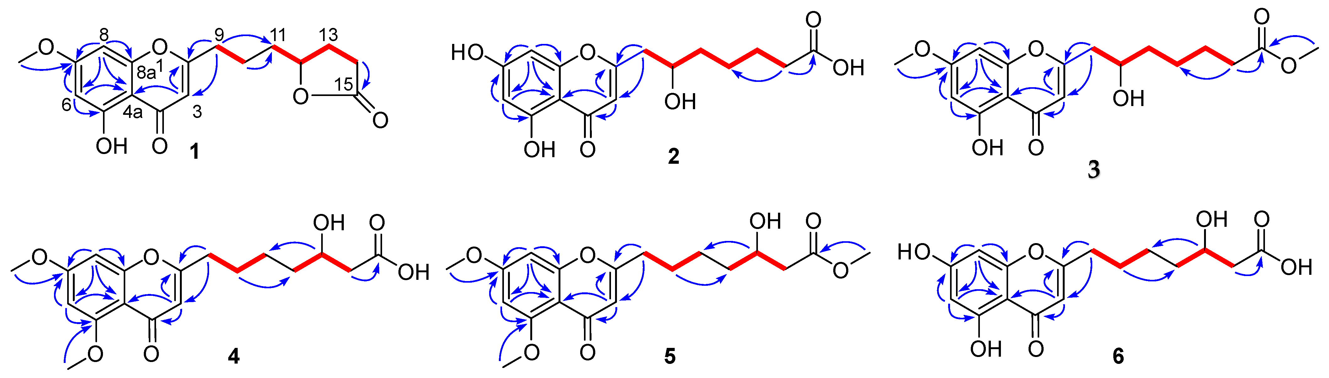
 ) and HMBC (
  ) correlations of 7 and 8.
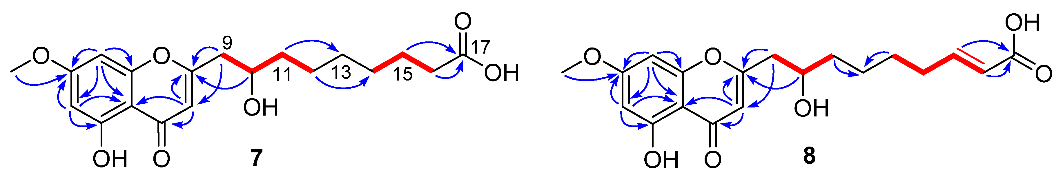

| Position | 1 a | 2 b | 3 b | |||
|---|---|---|---|---|---|---|
| δH | δC | δH | δC | δH | δC | |
| 2 | 170.6 | 170.0 | 170.4 | |||
| 3 | 6.25, s | 107.8 | 6.10, s | 110.1 | 6.15, s | 110.4 |
| 4 | 182.0 | 183.9 | 184.0 | |||
| 4a | 104.6 | 105.3 | 106.2 | |||
| 5 | 161.3 | 163.2 | 163.0 | |||
| 6 | 6.36, d (1.9) | 98.0 | 6.18, d (1.9) | 100.0 | 6.33, d (2.0) | 99.2 |
| 7 | 165.2 | 165.9 | 167.4 | |||
| 8 | 6.61, d (1.9) | 92.5 | 6.33, d (1.9) | 95.0 | 6.54, d (2.0) | 93.5 |
| 8a | 157.8 | 159.9 | 159.8 | |||
| 9 | 2.69, t (6.5) | 32.9 | 2.81, dd (14.5, 3.9) 2.63, dd (14.5, 8.8) | 43.2 | 2.82, dd (14.3, 3.9) 2.66, dd (14.3, 8.7) | 43.2 |
| 10 | 1.77, m | 22.3 | 4.0, m | 70.0 | 4.0, m | 69.9 |
| 11 | 1.69, m | 34.0 | 1.57, m | 38.1 | 1.56, m | 38.0 |
| 12 | 4.52, m | 79.9 | 1.45, m 1.56, m | 26.3 | 1.43, m 1.54, m | 26.2 |
| 13 | 2.27, m 1.80, m | 27.3 | 1.65, m | 26.0 | 1.65, m | 25.9 |
| 14 | 2.49, m | 28.3 | 2.32, t (7.2) | 35.0 | 2.35, t (7.3) | 34.7 |
| 15 | 177.1 | 177.7 | 176.0 | |||
| 5-OCH3 | ||||||
| 7-OCH3 | 3.84, s | 56.1 | 3.87, s | 56.5 | ||
| 15-OCH3 | 3.65, s | 52.0 | ||||
| Position | 4 | 5 | 6 | |||
|---|---|---|---|---|---|---|
| δH | δC | δH | δC | δH | δC | |
| 2 | 169.7 | 169.6 | 172.4 | |||
| 3 | 6.04, s | 111.3 | 6.05, s | 111.3 | 6.05, s | 108.4 |
| 4 | 180.1 | 180.1 | 184.0 | |||
| 4a | 109.1 | 109.2 | 105.2 | |||
| 5 | 162.1 | 162.1 | 163.2 | |||
| 6 | 6.49, d (2.1) | 97.3 | 6.50, d (2.1) | 97.2 | 6.17, d (2.0) | 100.0 |
| 7 | 166.3 | 166.3 | 165.9 | |||
| 8 | 6.62, d (2.1) | 94.0 | 6.64, d (2.1) | 94.0 | 6.31, d (2.0) | 94.9 |
| 8a | 161.8 | 161.8 | 159.9 | |||
| 9 | 2.63, t (7.5) | 34.4 | 2.63, t (7.7) | 34.3 | 2.64, t (7.6) | 34.9 |
| 10 | 1.76, m | 27.8 | 1.76, m | 27.7 | 1.75, m | 27.8 |
| 11 | 1.52, m | 26.1 | 1.52, m | 26.0 | 1.52, m | 26.0 |
| 12 | 1.52, m | 37.6 | 1.54, m | 37.6 | 1.53, m | 37.6 |
| 13 | 3.99, m | 69.3 | 4.0, m | 69.1 | 3.99, m | 69.1 |
| 14 | 2.41, m | 43.6 | 2.41, dd (15.1, 8.1) 2.50, dd (15.1, 4.9) | 43.2 | 2.39, dd (15.3, 7.9) 2.46, dd (15.3, 5.1) | 43.3 |
| 15 | 174.5 | 174.0 | 175.7 | |||
| 5-OCH3 | 3.89, s | 56.5 | 3.89, s | 56.5 | ||
| 7-OCH3 | 3.90, s | 52.0 | 3.91, s | 56.5 | ||
| 15-OCH3 | 3.67, s | 52.0 | ||||
| Position | 7 | 8 | ||
|---|---|---|---|---|
| δH | δC | δH | δC | |
| 2 | 170.4 | 170.4 | ||
| 3 | 6.15, s | 110.4 | 6.15, s | 110.4 |
| 4 | 184.0 | 184.0 | ||
| 4a | 106.2 | 106.2 | ||
| 5 | 163.0 | 163.0 | ||
| 6 | 6.32, d (1.9) | 99.1 | 6.32, d (2.0) | 99.1 |
| 7 | 167.3 | 167.3 | ||
| 8 | 6.52, d (1.9) | 93.5 | 6.53, d (2.0) | 93.4 |
| 8a | 159.8 | 159.8 | ||
| 9 | 2.82, dd (14.3, 3.7) 2.65, dd (14.3, 8.4) | 43.3 | 2.83, dd (14.3, 3.5) 2.66, dd (14.3, 8.8) | 43.3 |
| 10 | 4.0, m | 70.1 | 4.0, m | 70.0 |
| 11 | 1.55, m | 38.4 | 1.57, m | 38.1 |
| 12 | 1.41, m | 26.5 | 1.48, m 1.57, m | 26.3 |
| 13 | 1.37, m | 30.3 | 1.54, m | 29.1 |
| 14 | 1.37, m | 30.2 | 2.25, m | 33.1 |
| 15 | 1.62, m | 26.1 | 6.95, dd (15.7, 7.0) | 150.6 |
| 16 | 2.28, t (7.4) | 35.1 | 5.81, d (15.7) | 123.0 |
| 17 | 177.9 | 170.5 | ||
| 5-OCH3 | ||||
| 7-OCH3 | 3.86, s | 56.5 | 3.87, s | 56.5 |
| Compounds | α-Glucosidase Inhibitory | Antioxidant | ||
|---|---|---|---|---|
| % Inhibition (667 μM) | IC50 (μM) | % Inhibition (1000 μM) | IC50 (μM) | |
| 1 | −3.43 | 7.24 | ||
| 2 | 15.10 | 35.7 | 1970 ± 156 | |
| 3 | 23.78 | 20.4 | ||
| 4 | −9.718 | 12.2 | ||
| 5 | 1.41 | 11.2 | ||
| 6 | 27.3 | 842 ± 11 | 33.8 | |
| 7 | 25.2 | 917 ± 8 | 23.6 | |
| 8 | 41.7 | 1017 ± 22 | 19.3 | |
| 9 | 40.18 | 688 ± 24 | 18.1 | |
| 10 | 74.66 | 268 ± 25 | 21.8 | |
| Acarbose a | 27.7 | 1330 | ||
| Vitamin C a | 96.4 | 26.7 | ||
Publisher’s Note: MDPI stays neutral with regard to jurisdictional claims in published maps and institutional affiliations. |
© 2021 by the authors. Licensee MDPI, Basel, Switzerland. This article is an open access article distributed under the terms and conditions of the Creative Commons Attribution (CC BY) license (https://creativecommons.org/licenses/by/4.0/).
Share and Cite
Han, S.; Liu, Y.; Liu, W.; Yang, F.; Zhang, J.; Liu, R.; Zhao, F.; Xu, W.; Cheng, Z. Chromone Derivatives with α-Glucosidase Inhibitory Activity from the Marine Fungus Penicillium thomii Maire. Molecules 2021, 26, 5273. https://doi.org/10.3390/molecules26175273
Han S, Liu Y, Liu W, Yang F, Zhang J, Liu R, Zhao F, Xu W, Cheng Z. Chromone Derivatives with α-Glucosidase Inhibitory Activity from the Marine Fungus Penicillium thomii Maire. Molecules. 2021; 26(17):5273. https://doi.org/10.3390/molecules26175273
Chicago/Turabian StyleHan, Shouye, Yu Liu, Wan Liu, Fan Yang, Jia Zhang, Ruifeng Liu, Fenqin Zhao, Wei Xu, and Zhongbin Cheng. 2021. "Chromone Derivatives with α-Glucosidase Inhibitory Activity from the Marine Fungus Penicillium thomii Maire" Molecules 26, no. 17: 5273. https://doi.org/10.3390/molecules26175273
APA StyleHan, S., Liu, Y., Liu, W., Yang, F., Zhang, J., Liu, R., Zhao, F., Xu, W., & Cheng, Z. (2021). Chromone Derivatives with α-Glucosidase Inhibitory Activity from the Marine Fungus Penicillium thomii Maire. Molecules, 26(17), 5273. https://doi.org/10.3390/molecules26175273