Article

The Antifungal Action Mode of N-Phenacyldibromobenzimidazoles

1
Centre for Advanced Materials and Technologies CEZAMAT, Warsaw University of Technology, Poleczki 19, 02-822 Warsaw, Poland
2
Department of Virology, National Institute of Public Health-National Institute of Hygiene, Chocimska 24, 00-791 Warsaw, Poland
3
Clinical Science, Targovax Oy, Lars Sonckin Kaari 14, Espoo Stella Luna Business Park, 02600 Espoo, Finland
4
Faculty of Chemistry, Warsaw University of Technology, Noakowskiego St 3, 00-664 Warsaw, Poland
*
Authors to whom correspondence should be addressed.
Molecules 2021, 26(18), 5463; https://doi.org/10.3390/molecules26185463
Submission received: 1 August 2021 / Revised: 25 August 2021 / Accepted: 2 September 2021 / Published: 8 September 2021
(This article belongs to the Special Issue Design, Synthesis, and Biological Evaluation of Novel Antifungals)

Abstract

:
Our study aimed to characterise the action mode of N-phenacyldibromobenzimidazoles against C. albicans and C. neoformans. Firstly, we selected the non-cytotoxic most active benzimidazoles based on the structure–activity relationships showing that the group of 5,6-dibromobenzimidazole derivatives are less active against C. albicans vs. 4,6-dibromobenzimidazole analogues (5ef and 5h). The substitution of chlorine atoms to the benzene ring of the N-phenacyl substituent extended the anti-C. albicans action (5e with 2,4-Cl2 or 5f with 3,4-Cl2). The excellent results for N-phenacyldibromobenzimidazole 5h against the C. albicans reference and clinical isolate showed IC50 = 8 µg/mL and %I = 100 ± 3, respectively. Compound 5h was fungicidal against the C. neoformans isolate. Compound 5h at 160–4 µg/mL caused irreversible damage of the fungal cell membrane and accidental cell death (ACD). We reported on chitinolytic activity of 5h, in accordance with the patterns observed for the following substrates: 4-nitrophenyl-N-acetyl-β-d-glucosaminide and 4-nitrophenyl-β-d-N,N′,N″-triacetylchitothiose. Derivative 5h at 16 µg/mL: (1) it affected cell wall by inducing β-d-glucanase, (2) it caused morphological distortions and (3) osmotic instability in the C. albicans biofilm-treated. Compound 5h exerted Candida-dependent inhibition of virulence factors.

1. Introduction

Studies conducted during the past two decades have documented changes in the causative agents of nosocomial blood stream infections, and emphasized an increase of very critical fungal infections, particularly due to Candida spp. and Cryptococcus spp. [1]. The emergence of antifungal resistance required more concern to find out effective antimycotics with novel modes of action. Thus, introduction of N-phenacyldibromobenzimidazoles as another antimycotics destroying the fungal cell wall and membrane may be a milestone in the development of antifungal therapies. Moreover, treatment with anti-filamentation compound benefits the host by modulating immune responses [1]. An inhibition of morphological switch may provide an alternative approach to finding compounds with a potential to control the Candida albicans infections [2]. Morphogenesis is critical for biofilm formation, thus compounds able to inhibit sessile growth are needed [3].
Azoles are easy-to-use scaffolds in antifungal drug discovery [4]. Moreover, azoles are often functionalized with phenacyl group as a result of N-alkylation to gain excellent antifungal activity. There are known as biologically active N-phenacyl imidazoles [5,6,7,8,9,10], benzimidazoles [11,12,13,14], triazoles [5,7,15] or pyrazoles [16,17]. N-phenacyl azoles are often used as substrates for further synthesis of antifungal active agents [7,9,10,11,13,14,15,18,19,20]. In this study, we focused on dibromobenzimidazole synthesis due to promising antifungal activity rarely undertaken by scientists in worldwide studies on drug discovery, probably due to tedious synthesis [21,22,23,24].
We evaluated the toxicity of various N-phenacyldibromobenzimidazoles towards a mammalian cell line as well as the fungistatic and fungicidal effect against the C. albicans and Cryptococcus neoformans reference and clinical isolates resistant to azoles and echinocandins. The experiments with N-phenacyldibromobenzimidazoles have been encouraging in the current study because of the following action modes need to be assessed: 1. Phosphatidylserine externalization affecting subsequently the chitin content; 2. Cell wall stress induced by N-phenacyldibromobenzimidazoles resulted in the decreased/ increased ROS; 3. Lysosomotropic N-phenacyldibromobenzimidazoles exerted direct membrane lyses and caused osmotic pressure; 4. The concept regarding extensity of accidental cell death (ACD) under N-phenacyldibromobenzimidazoles. Since the primary targets of commercially available antimycotics are β-1.3-glucan and ergosterol, respectively, we underwent study if any compensatory mechanism in the cell wall and membrane occurs after the N-phenacyldibromobenzimidazole treatment. In our study, morphological changes enabled N-phenacyldibromobenzimidazoles gaining access to intracellular targets by facilitating membrane transience.

2. Results

2.1. Synthesis of N-Phenacyldibromobenzimidazoles

As it is shown in Table 1 and Scheme 1, compounds 45 were synthesized by N-alkylation of 5,6-dibromobenzimidazole 1 or 4,6-dibromobenzimidazole 2 with phenacyl chlorides or bromides 3 in the presence of K2CO3 in MeCN. The time of the reactions as well as the yields depended on the structure of the phenacyl derivative 3 used. In the case of unsubstituted phenacyl bromide 3a and monofunctionalized derivatives 3bd, the respective products 4ad and 5ad were isolated in 72–94%. Meanwhile, in reactions with alkylating agents, 3ej possessing two or three halogen atoms in the benzene ring, afforded complicated mixture of products, so the target compound 4ej and 5ej were isolated in 13–24% [25]. All N-phenacylbenzimidazoles 45 were purified by column chromatography, followed by crystallization.

2.2. The Antifungal Effect of Dibromobenzimidazole Derivatives

As it is shown in Table 2 and Figures S1–S12 (in Supplementary file), in our initial screening of twenty dibromobenzimidazole derivatives we assessed the percentage of cell growth inhibition (%I). At the inhibitory concentration of 50% (IC50), the concentration of benzimidazoles that reduces the cell growth of C. albicans SC5314 by ≥50% was determined. Secondly, randomly selected (5f) and the most effective inhibitors (5e and 5h) were tested against the C. albicans SPZ176 isolate resistant to Flu and Itr (Table 2). Further, 5e displayed IC50 at 4–16 µg/mL (Table 2) and the mode of fungicidal action against SC5314 at 8–16 µg/mL (lg R ≤ 1.19 in Table 3). 5f showed lg R = 1 at 8 µg/mL (Table 3). Contrariwise, 5h displayed no candidacidal action (lg R ≤ 0.43 in Table 3). Moreover, a paradoxical growth phenomenon of the reference strain SC5314 [26] was noted for the following derivatives: 4f, 4h, 5a, and 5gi (Figures S5, S7, S9, S11 and S12) as well as 5b, 5ef, 5h, 5j (Table 2). Briefly, we noted a slow decrease in the viable cell growth at higher concentrations (e.g., %I = 53 ± 8 at 16 µg/mL for 5b) vs. the lowest concentrations at which the cell growth was substantially inhibited (e.g., %I = 95 ± 8 at 8 µg/mL for 5b).
We determined the effectiveness of dibromobenzimidazole derivatives against the fungal isolates using colony forming unites (cfu) assay (Table 4). The exhaustive data clearly demonstrated that cfu were recovered after treatment with the tested dibromobenzimidazoles (Table 4). The most effective 5h at 16 µg/mL totally inhibited recovery of cfu of both clinical isolates. In the case of C. neoformans, there was no cfu recovery after treatment with 5h at the concentration range of 8–16 µg/mL. Thus, C. neoformans was more sensitive to 5h than C. albicans. We identified the leading fungicidal compound 5h to be used in a series of follow-up analyses to establish its action mode in vitro.

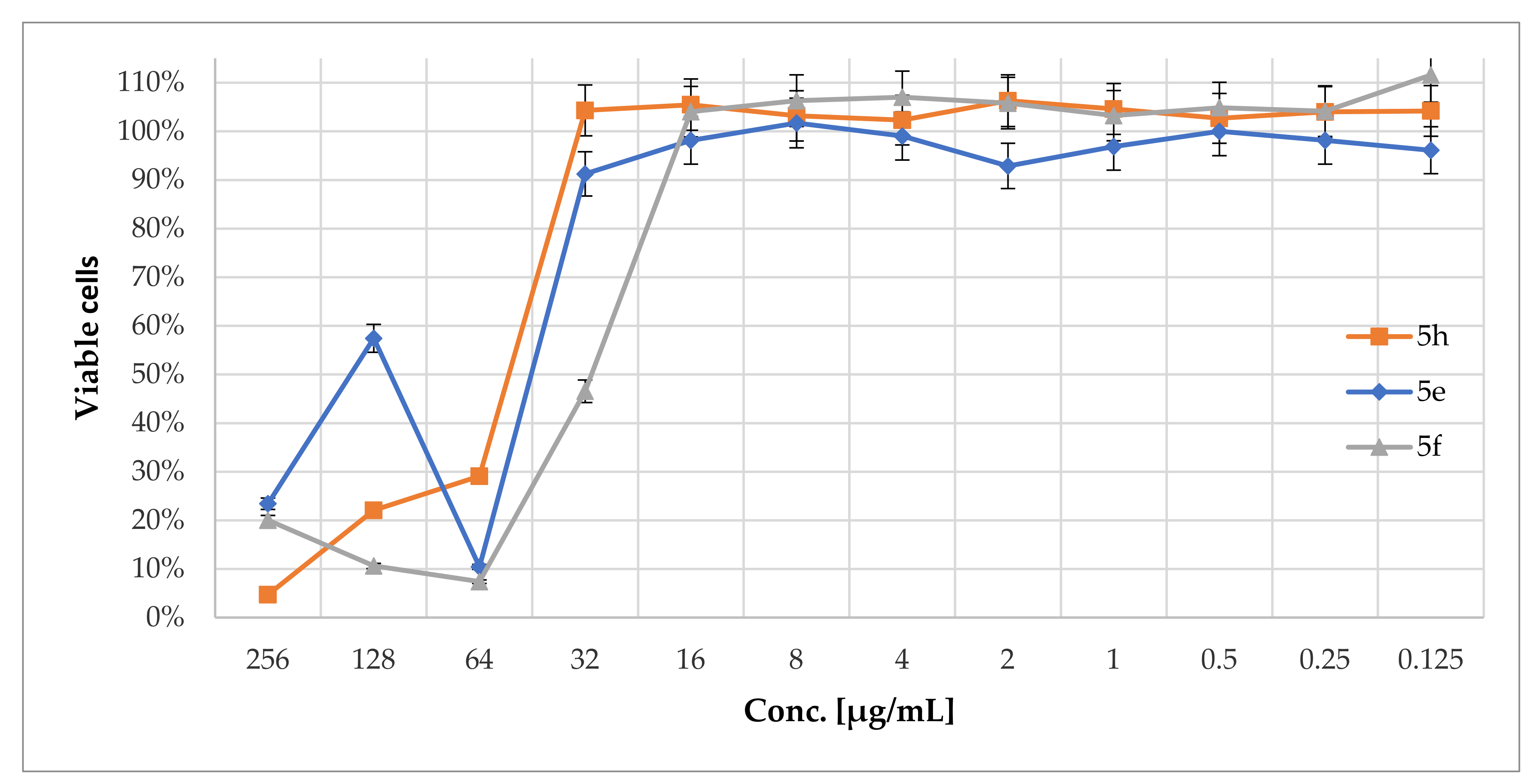
2.3. Cytotoxicity of N-Phenacyldibromobenzimidazole Derivatives

As it was shown in Figure 1, the Vero cell viability or cytotoxicity generated by the most active compounds (fungicidal) was assessed using the MTS method. Figure 1 indicates CC50 = 32–64 µg/mL and CC90 = 64–256 µg/mL for 5e and 5f. Moreover, 5h displayed CC50 = 32–64 µg/mL and CC90 = 256 µg/mL. Thus, all compounds did not inhibit the NAD(P)H dehydrogenase (quinone) activity and disturb cell membrane permeability.

2.4. Antifungal Activity of 5h in Combination with Osmoprotectant

The %I values of 5h were changed in the presence of sorbitol, and it suggests influence of 5h on the cell wall structure of the C. albicans clinical isolate (Table 5). In details, 5h displayed lack or weak (%I = 8 ± 18) cell growth inhibition at 16 µg/mL in the presence of 0.8 M sorbitol as an osmotic protectant in the medium vs. one without sorbitol (%I = 100 ± 3 in Table 2). For the C. neoformans isolate, the antifungal activity of 5h was as follows: (1) 5h at 4 µg/mL causes no cell growth inhibition in medium with sorbitol added vs. 1 × 10⁵ cfu/mL recovered in medium without sorbitol (Table 4); (2) 5h displays no growth recovery at 8–16 µg/mL in medium without sorbitol vs. %I = 79–95 at the same range of concentrations in medium with sorbitol added.

2.5. Chitinolytic Activity of 5h

As it was shown in Table 6, the detailed studies on chitinolytic activity showed affinity of 5h to the following substrates: 4-nitrophenyl-N-acetyl-β-d-glucosaminide and 4-nitrophenyl-N,N′-diacetyl-β-d-chitobioside. Contrariwise, 5h displayed no affinity to 4-nitrophenyl-β-d-N,N′,N″-triacetylchitothiose.

2.6. Efflux Disorder under 5h

Rho123 was not able to leave the mitochondrion due to the membrane potential decreased (efflux decreased) as a results of cell death. For the C. albicans ref. strain and C. neoformans isolate, efflux decreased in line with increased conc. of 5h (Table 7). Contrariwise, in the case of C. albicans clinical isolate, efflux was noted for the 5h-treated cells at 16 μg/mL (Table 7).

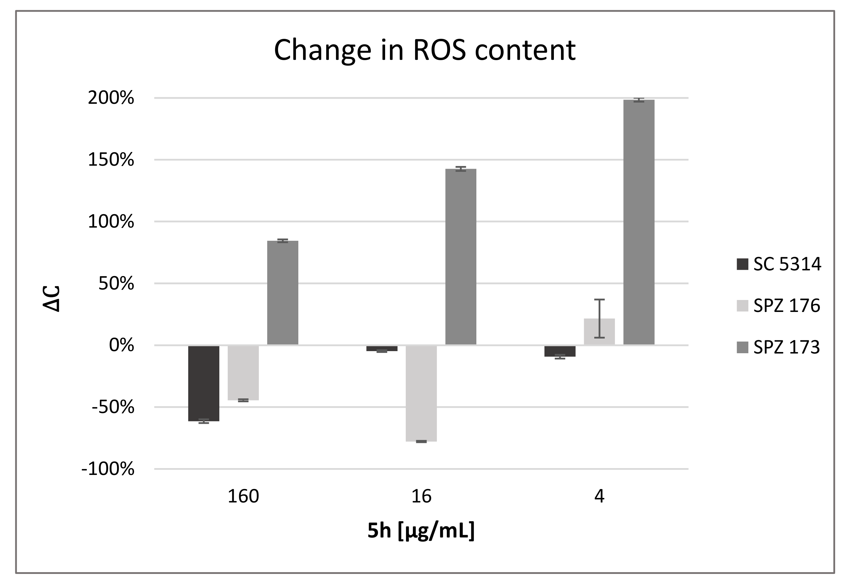
2.7. Compound 5h Induces ROS Generation

Treatment of the fungal cells with low concentration of 5h led to the ROS production at high level (198%In Figure 2). Generally, in the case of C. neoformans, the level of ROS production increased in line with decreasing concentrations of 5h. Remaining strains showed ROS under detectable level, with exception of C. albicans SPZ176 generating ROS at 22% under treatment with 5h at 4 µg/mL.

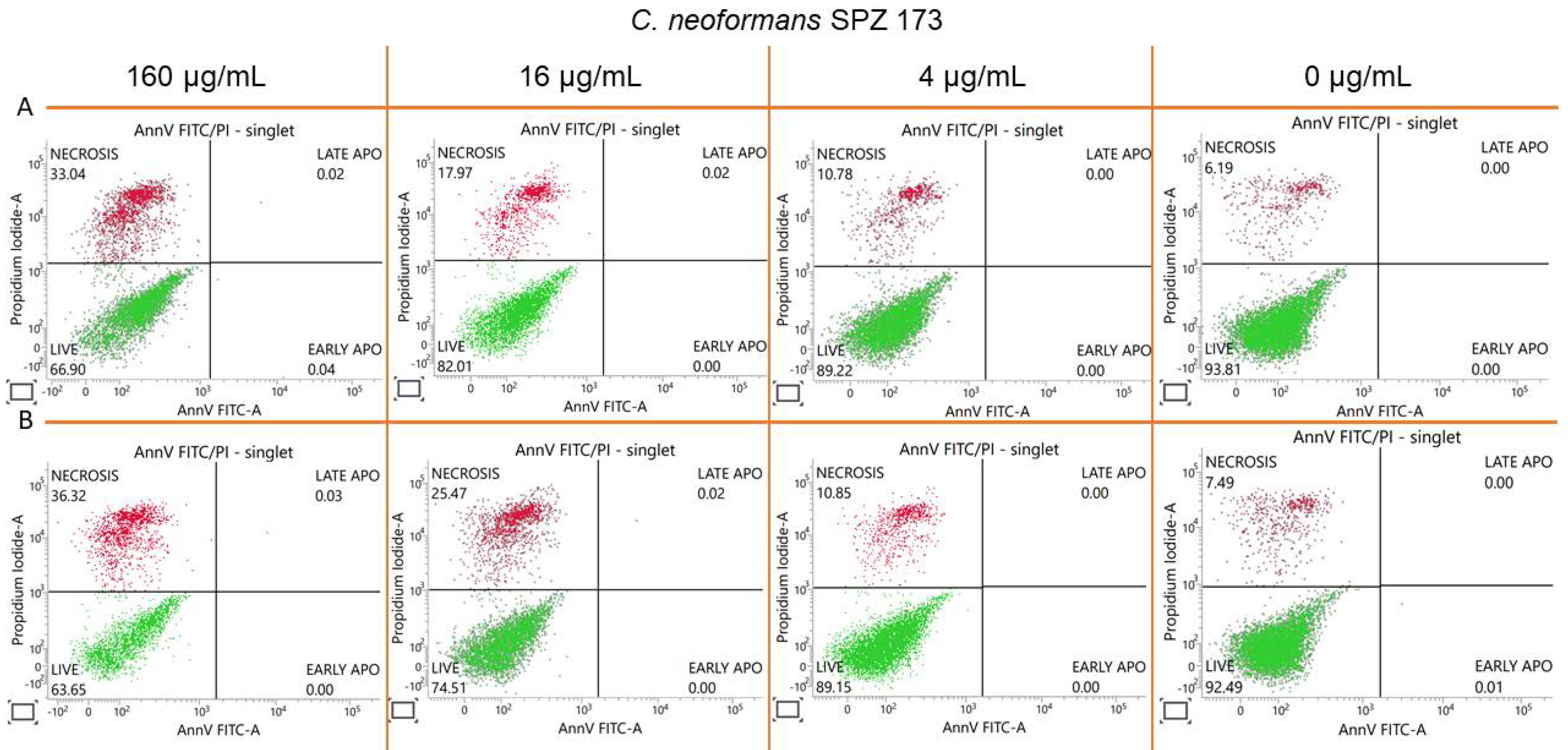
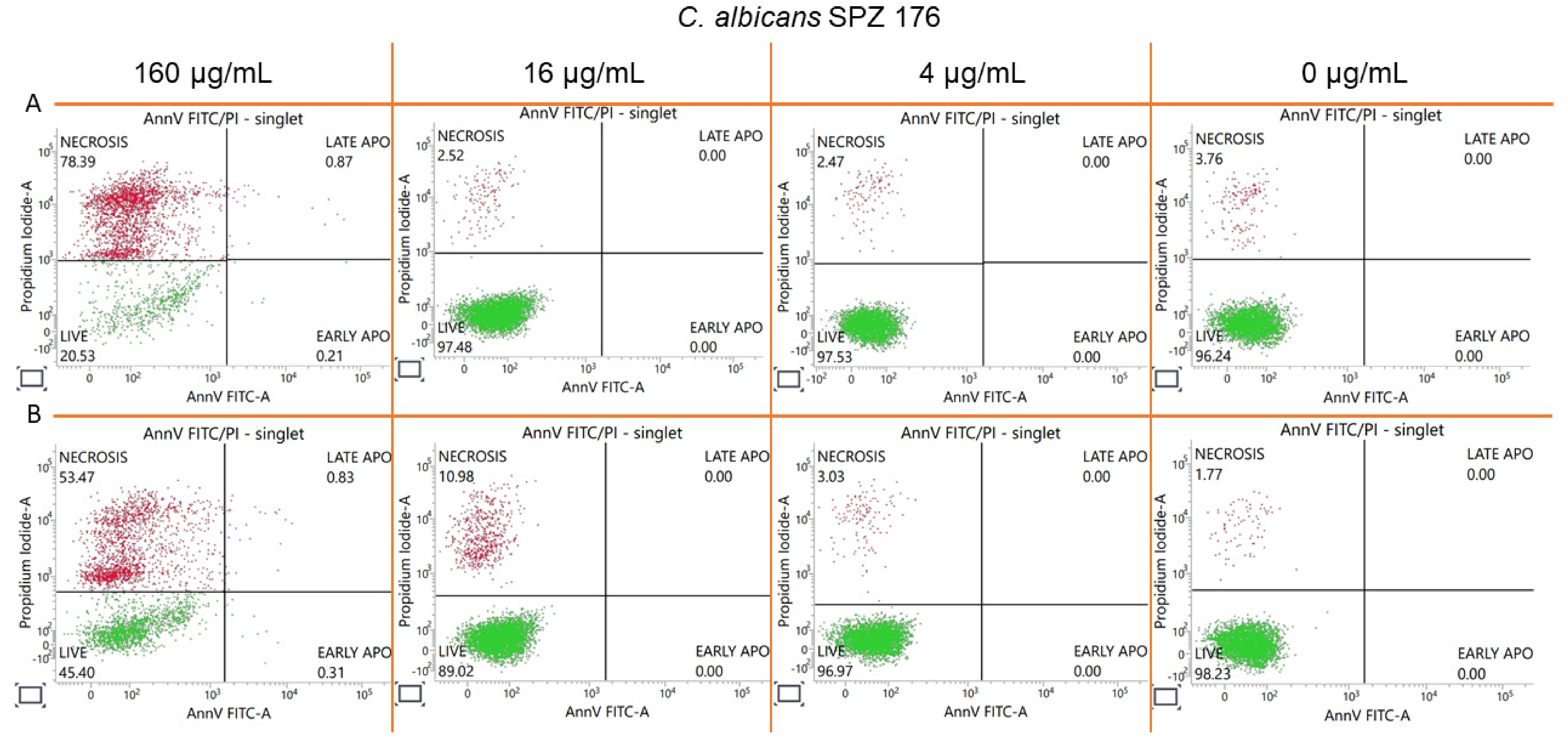
2.8. Estimation of Accidental Cell Death in the 5h-Treated Fungi

As shown in Figure 3 and Figure 4, 5h at the concentrations ranging from 4 to 160 µg/mL generated necrosis (accidental cell death ACD) in the fungal cells and protoplasts. Apoptosis early or late was induced approx. at 0.31% or 0.83% in the C. albicans protoplasts under treatment with 5h at 160 µg/mL. In the case of C. neoformans, apoptosis was generated approx. at 0.04% (early) and 0.02% (late) by 5h at 160 µg/mL. In the case of the C. neoformans protoplasts, late apoptosis was noted approx. at 0.03% or 0.02%, respectively, for 160 or 16 µg/mL.

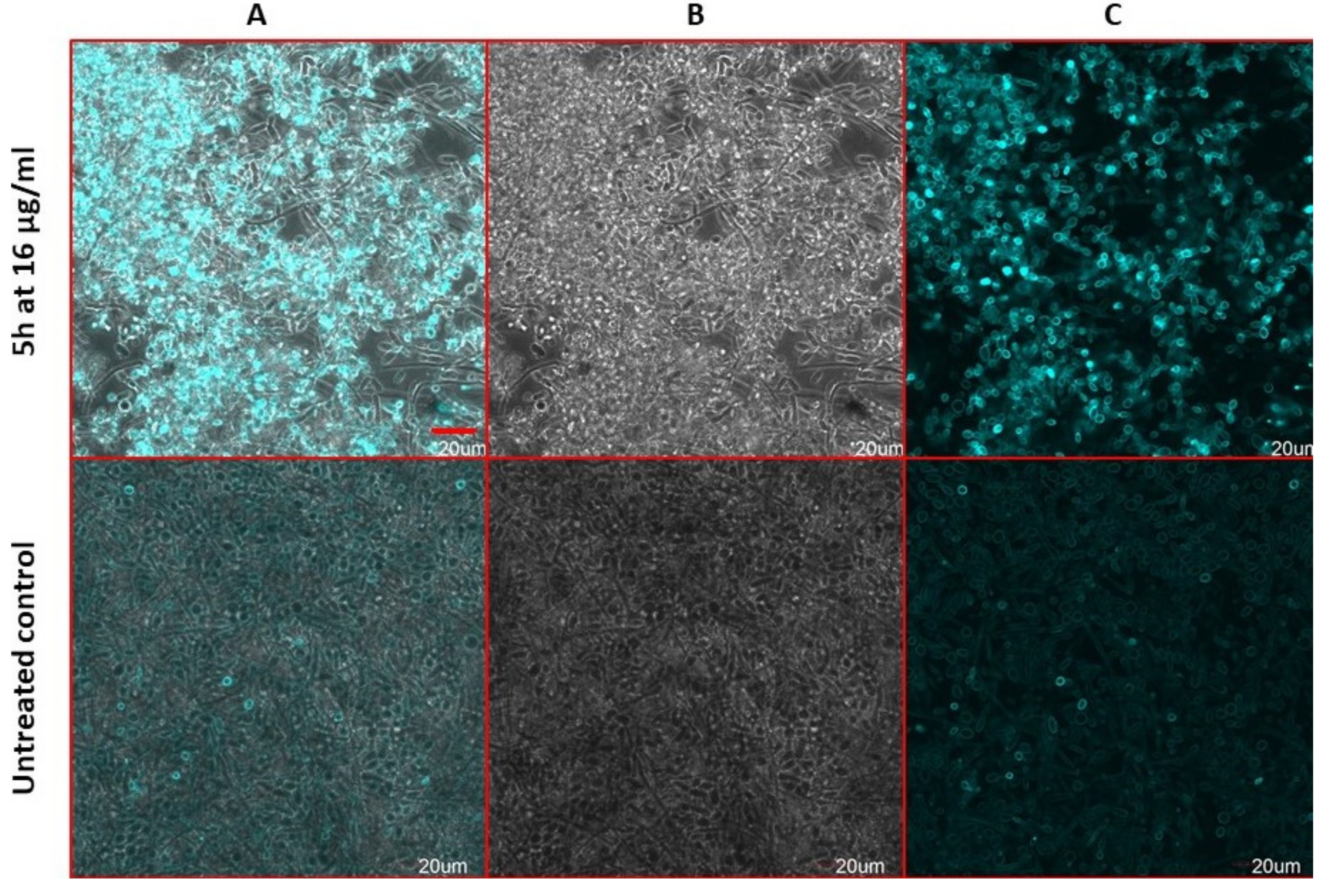
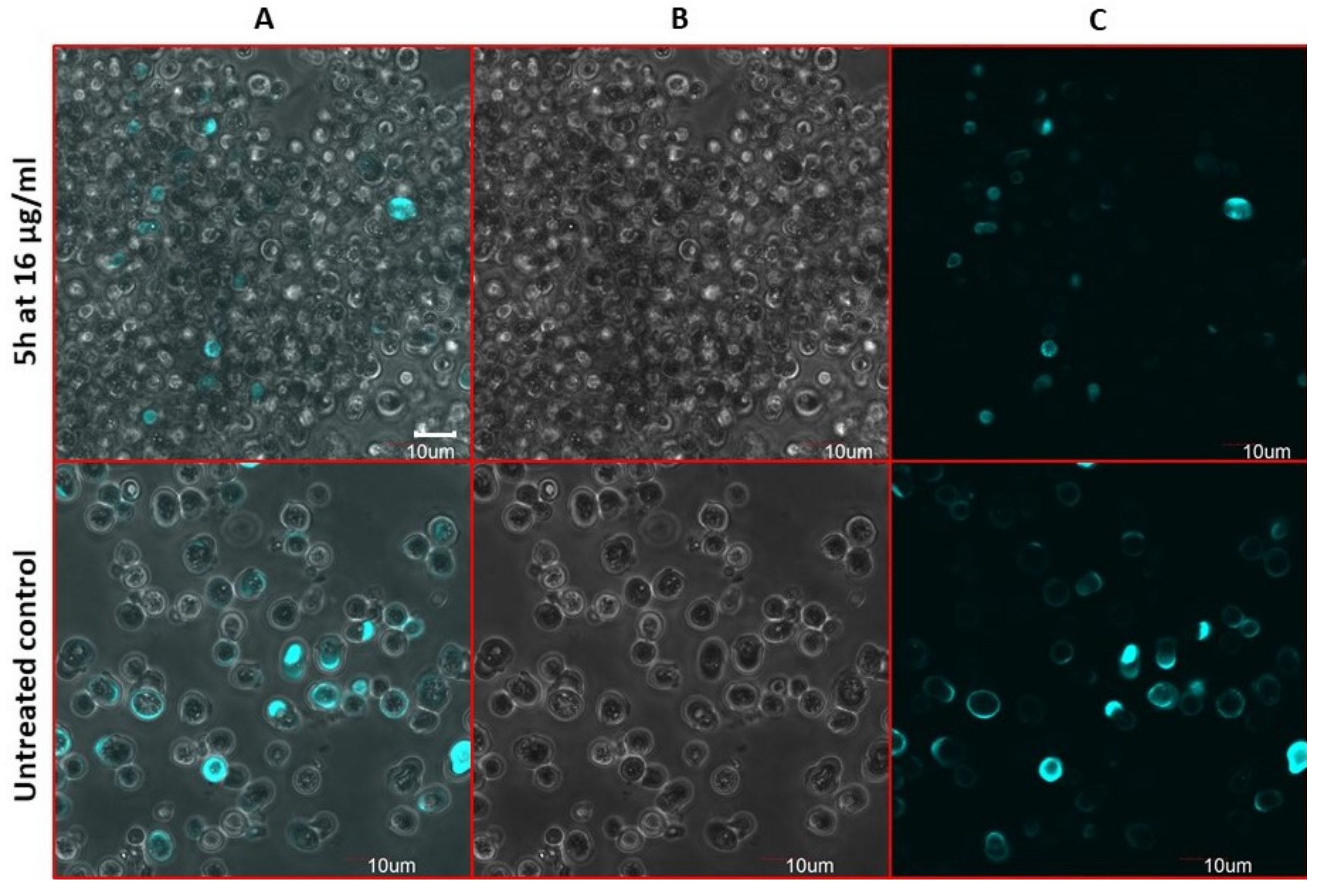
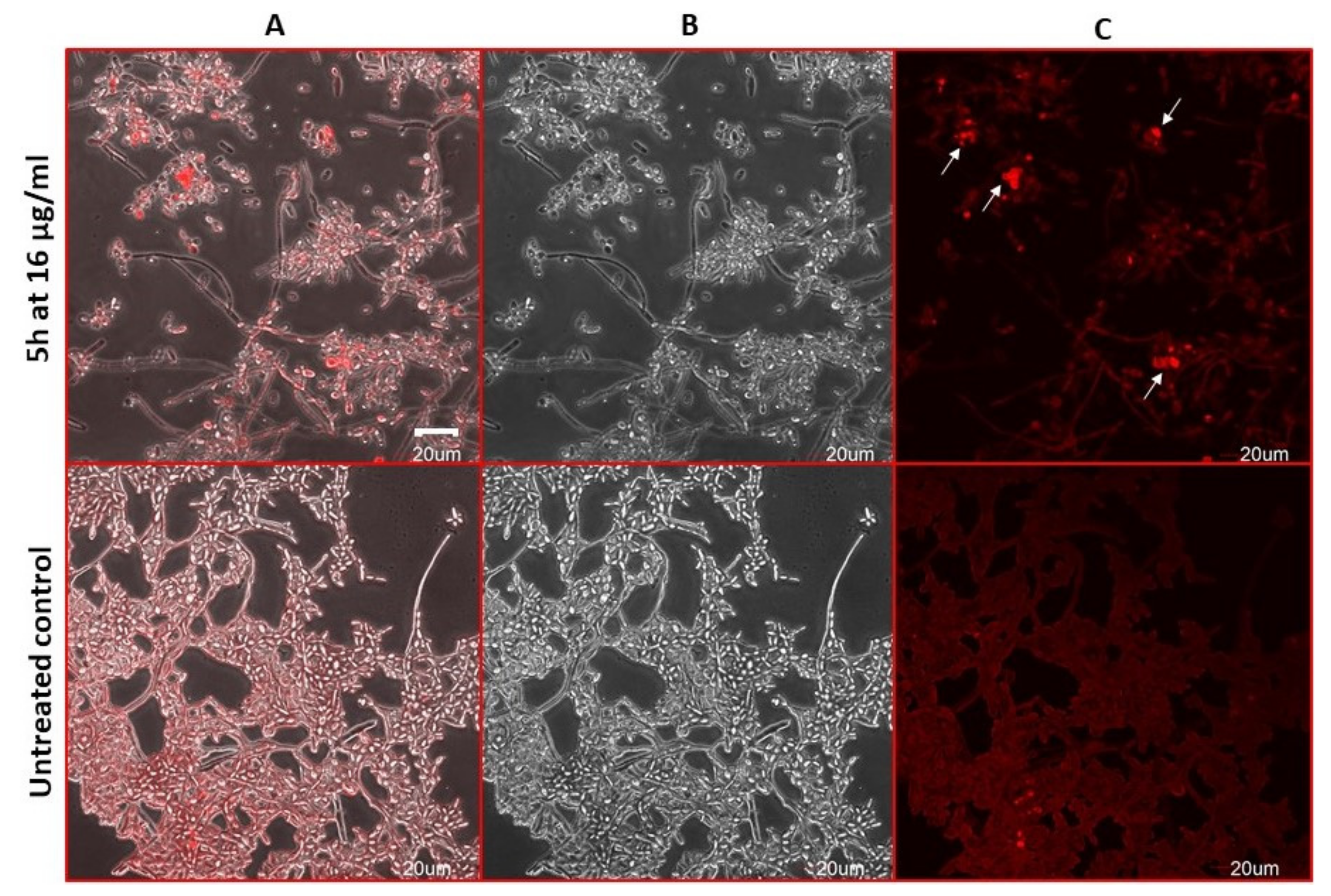
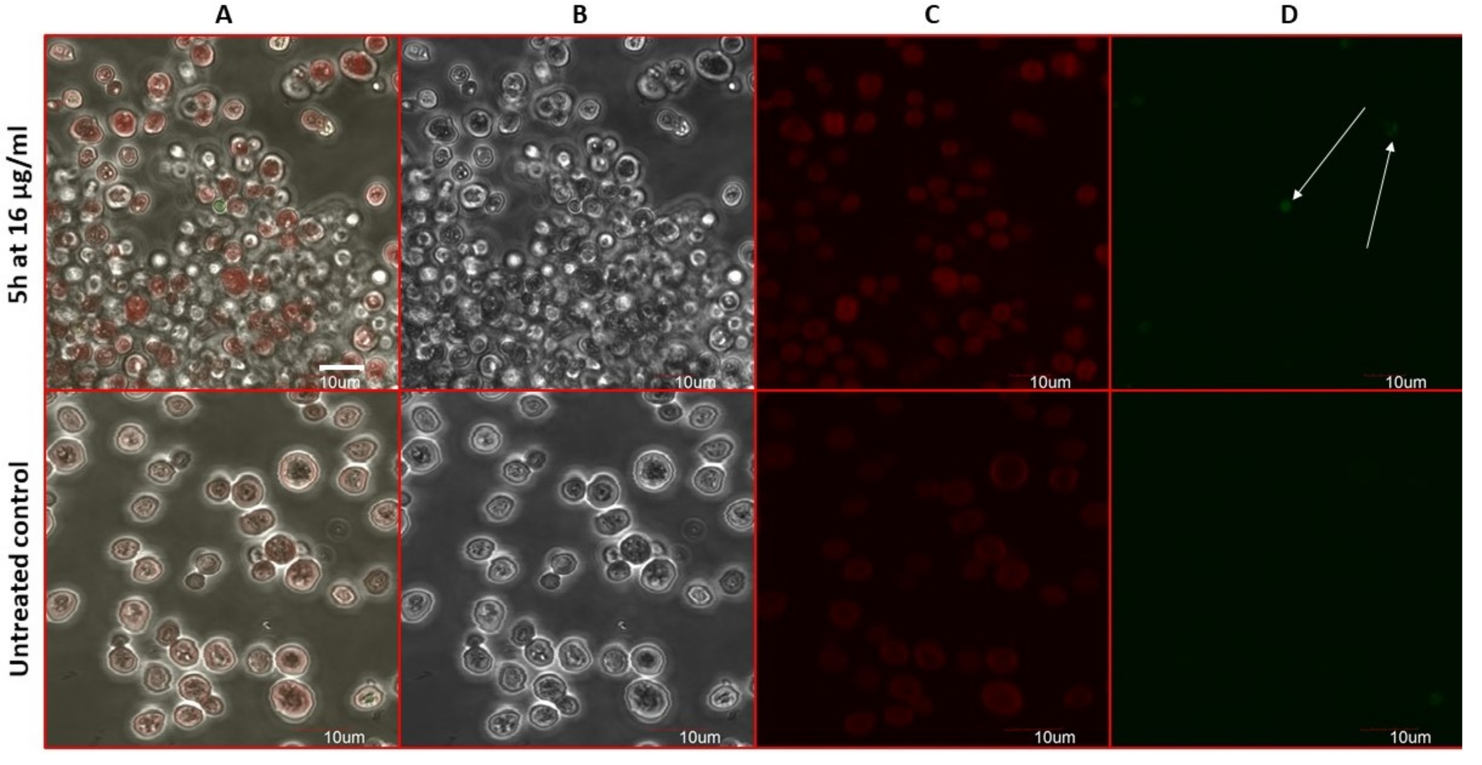
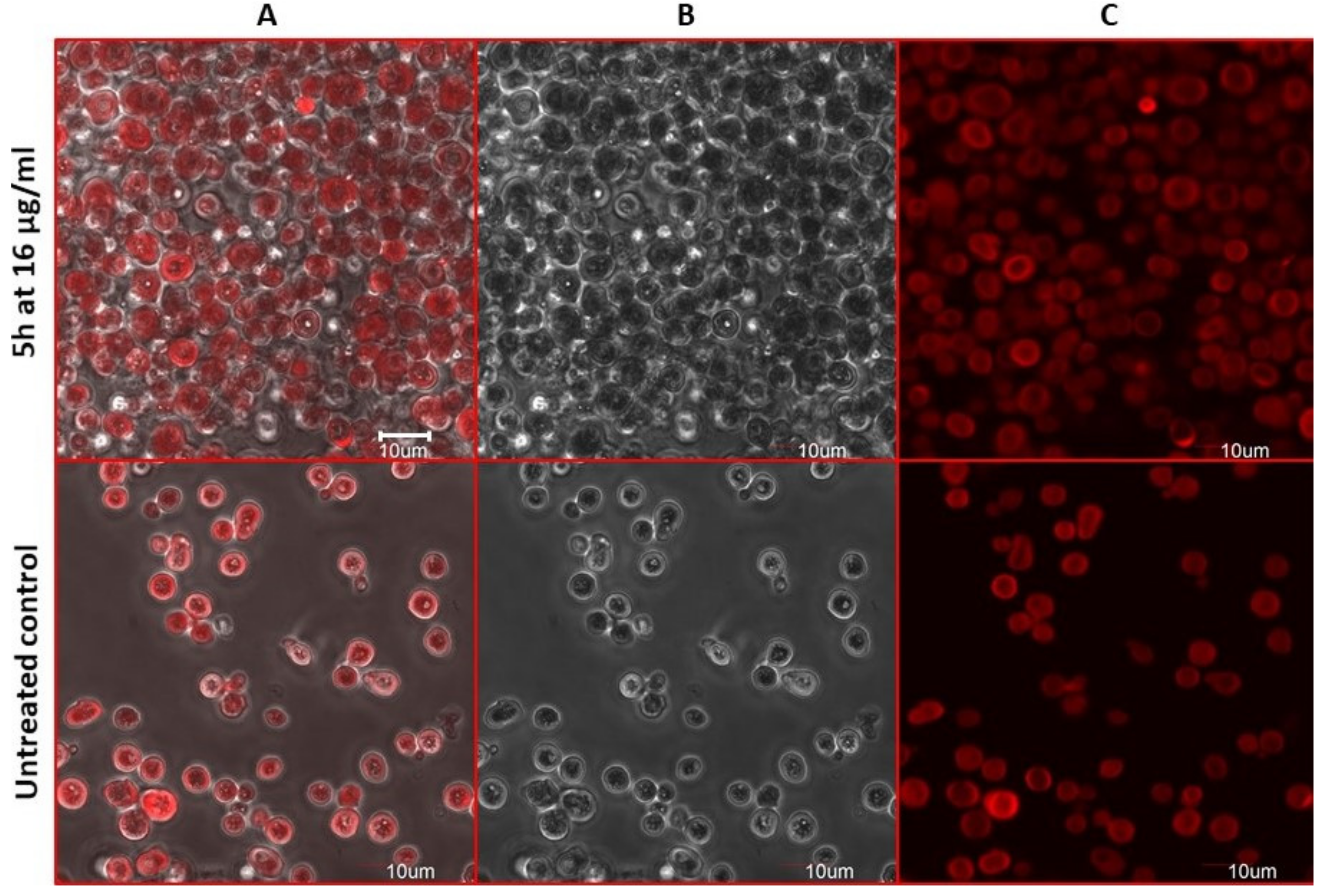
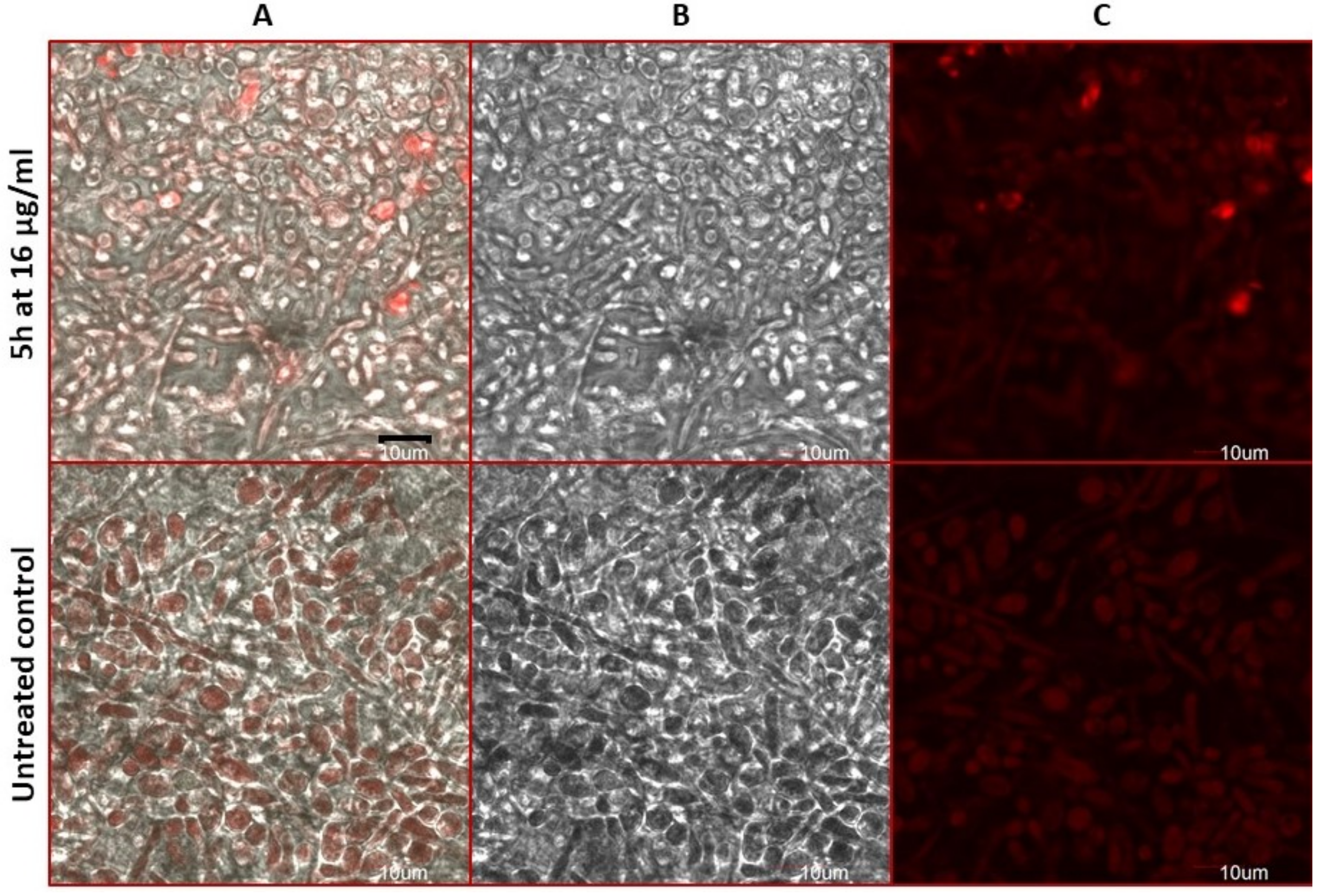
2.9. Antifungal Action and Accidental Cell Death by Fluorescent Structural Staining Techniques

The resulting cell wall damage and cell viability were assessed using Confocal laser scanning microscopy (CLSM) after treatment with 5h (twelve images were assessed for each treatment/staining). As it was shown using CFW staining (Figure 5), 5h at 16 µg/mL induced the cell wall rearrangement of the C. albicans sessile conglomerate. Biofilm’s chitin content was redistributed and elevated under treatment with 5h (vivid blue fluorescence of elevated chitin in Figure 5). Contrariwise, action of 5h against the C. neoformans sessile growth was not significant (Figure 6). In Figure 6, very few cells were totally stained with CFW in conglomerate vs. the untreated control showing several cells with bright blue fluorescence. Thus 5h did not reorganize the cell wall chitin content of C. neoformans.
Congo red (CR) interacts with β-d-glucan of the 5h-treated C. albicans sessile cells (Figure 7). Thus, the cells exposed to 5h at 16 µg/mL exhibit increased frequencies of the cell wall damage (arrows in Figure 7). Contrariwise, the biofilm of C. neoformans treated with 5h was found CR sensitive in comparable level to the untreated sessile cells (Figure 8). Thus 5h did not disturb the glucan content of C. neoformans.
Compound 5h altered plasma membrane permeability, which is indicated by intensive red fluorescence of the 5h-treated sessile cells (Figure 9), compounds induced necrosis-like cell death (bright red fluorescence of ethidium bromide EB inside the damaged sessile cells in Figure 9). Contrariwise, C. neoformans was resistant to 5h (arrows indicate weak green fluorescence of acridine orange AO inside the viable cells in Figure 10).

3. Discussion

Antifungal structure–activity relationships showed that the group of 5,6-dibromobenzimidazole derivatives are less active against C. albicans vs. 4,6-dibromobenzimidazole analogues (Table 2 and Figures S1–S12 (in Supplementary file)). Moreover, the substitution of chlorine atoms to the benzene ring of the N-phenacyl substituent extended anti-C. albicans activity (5e with 2,4-Cl2 or 5f with 3,4-Cl2 in Table 1, Table 2, Table 3 and Table 4 and Table S1 in Supplementary file). Contrariwise, the substitution of bromine or fluorine atoms in the same positions influences weak activity against Candida spp. The findings described above are in line with Vargas-Oviedo et al. [14]. It is worth to mention that 5h substituted with fluorine atoms at C2 and C4 of the benzene ring of the N-phenacyl group exhibited excellent fungicidal activity against the C. albicans reference and clinical strain as well as the C. neoformans isolate (Table 2, Table 3 and Table 4). In our study, the leading compound 5h (Table 4) was <16-times less active than AmB with minimal fungicidal concentration MFC90 = 1 µg/mL [31] and MFC = 0.5 [32] against the C. albicans isolates and SC5314, respectively. Structure–activity relationships provide opportunities for synthesis of dibromobenzimidazole analogues with improved antifungal action. Moreover, the most active antifungals (5ef, 5h) at the concentration range of 32–0.125 µg/mL were developed to generate viable and vital eukaryotic cells (Figure 1 and Figure S13; Tables S2 and S3). Thus, the tested dibromobenzimidazole were proved to be less cytotoxic against the Vero cells compared to AmB (toxic at 15–20 µg/mL after 24 h) [33].
In line with the results obtained in the presence of the osmo-protectant in the growth medium [34], we showed that 5h is the C. albicans cell wall inhibitor, displaying reverse effect in the presence of sorbitol (Table 5). The effect is characterized by decreasing in %I (Table 5) as observed in the medium with sorbitol vs. medium without protectant (Table 2). Our studies demonstrated that osmotic protector reduces anti-Candida activity of 5h. In alignment with Górska-Nieć et al. [35], we proved that enhanced biomass production leads to loss of antifungal activity of 5h at concentrations ranging from 4 to 16 µg/mL. Moreover, the activity of 5h did not correspond with AmB affecting cell wall due to activity accompanied by an increase concentration in medium with sorbitol [36].
Moreover, the micromorphological evaluation of the C. albicans-treated with 5h revealed the lack of structures indicating fungal mycelium typical for biofilm. Thus 5h was able to inhibit the biofilm formation (Figure 5, Figure 6, Figure 7, Figure 8, Figure 9 and Figure 10). Since the yeast-hyphae morphological transition is relevant for C. albicans virulence [37] we indicated that 5h represents promising therapeutic.
We showed that 5h acts by the cell wall chitin lysis originated from the comparison studies with chitinase (Table 6). Chitin is a polymer of β-1,4-linked N-acetyl-d-glucosamine (GlcNAc), which is an integral component of the fungal cell wall [38]. 5h was able to hydrolase 4-nitrophenyl-N-acetyl-β-d-glucosaminide and 4-nitrophenyl-β-d-N,N′,N″-triacetylchitothiose without activity against triacetylchitothiose (Table 6). Based on our results and in line with Nielsen and Sörensen [39], we hypothesized that 5h displays comparable activity to chitinase (EC 3.2.1.14). Since the ability of Congo red CR fluorescent tracker to visualize the fungal cell wall elements was described previously [40,41], we used CR as a diazo compound pertaining to its high affinity to polysaccharides in the 5h-treated C. albicans [40,41]. In line with Shalmy et al. [40] we found good staining result of CR in the 5h-treated C. albicans vs. 5h-treated capsule of C. neoformans which was poorly stained (Figure 7 vs. Figure 8). We showed that 5h displayed Candida spp. dependent activity.
Furthermore, 5h induced the phosphatidylserine PE translocation and membrane permeability [41], these were shown using the Annexin V and propidium iodide PI staining assay (Figure 3 and Figure 4). We hypothesized that PE externalization affect subsequently the elevated chitin content (Figure 5) and activity of 5h. Moreover, this polymer play an essential role in the sensitivity (or resistance) of C. albicans to AmB [42,43,44]. Since ROS play a crucial role in intracellular signalling [44], C. neoformans treated with 5h displayed elevated ROS (Figure 2 and Table S4) regarded as a cell death phenotype in connection with plasma membrane disintegration (at 16–160 µg/mL in Figure 3 and Figure 4 and Table S5) and loss of clonogenicity (at 8–16 µg/mL in Table 4). In details, the high levels of ROS at the 5h-treated cells at 4 µg/mL activate apoptosis pathway capable of inducing ACD (Figure 2). We hypothesized that the adaptive response of C. neoformans showing elevated ROS production promotes stress resistance to 5h. Contrariwise, decrease in the ROS level by incubation with 5h can induce the lethal process adequately monitored by cytometric analysis (Figure 3 and Figure 4 and Table S5). Based on the latter findings, 5h can act such as anti-oxidant. Moreover, elevated ROS under 5h correlated with fungicidal effect typical for AmB [44].
We used Rho123 as a membrane-potential-sensitive cationic fluorophore [45] to show that it was not able to leave the mitochondrion due to decreased membrane potential as a result of the 5h-treated cell death (Figure 2, Figure 3 and Figure 4, Figure 9 and Figure 10 as well as Tables S4 and S5). We concluded that 5h can be mitochondrial inhibitor of C. albicans ref and C. neoformans. Contrariwise, the Rho123 efflux simply increased pump activity in the 5h-treated C. albicans isolate resistant to azoles. The compelling evidence for reduced filamentation and ACD (progenitors of mycoses) are targets for dibromobenzimidazole. Finally, our findings suggested a general strategy for antimycotics development that might be useful in limiting the emergence of fungal resistance. We selected 5h as the most compound with significant response against the fungal virulence factors. We propose that 5h acts synergistically to disrupt the C. albicans cell wall/membrane. These structures establish an excellent target for specific inhibition of pathogenic fungi.

4. Materials and Methods

4.1. General Remarks of the N-Phenacyl Dibromobenzimidazole Derivatives Synthesis

Commercially available reagents from Sigma Aldrich (Darmstadt, Germany), Fluka (Charlotte, NC, USA) and Avantor (Gliwice, Poland) were used as supplied. The measured melting points were not corrected. The column chromatography was performed using Silica gel 60 (Merck) of 40–63 μm. Thin-layer chromatography was carried out on TLC aluminium plates with silica gel Kieselgel 60 F254 (Merck, Darmstadt, Germany) (0.2 mm thickness film). The 1H and 13C NMR spectra were measured with a Varian 500 spectrometer operating at 500 MHz for 1H and 125 MHz for 13C nuclei. Chemical shifts (δ) are given in parts per million (ppm); signal multiplicity assignment: s, singlet; d, doublet; dd, doublet of doublets; m, multiplet; coupling constant (J) are given in hertz (Hz). High resolution mass spectrometry (HRMS) was carried out on Q Exactive Hybrid Quadrupole-Orbitrap Mass Spectrometer (Bremen, Germany), ESI (electrospray) with spray voltage 4.00 kV at Institute of Biochemistry and Biophysics Polish Academy of Science (IBB PAS, Warsaw, Poland. The most intensive signals are reported.

4.1.1. Synthesis of 4ad and 5ad

To a stirred suspension of 5,6-dibromobenzimidazole 1 or 4,6-dibromobenzimidazole 2 (1 mmol, 0.276 g) in MeCN (20 mL) K2CO3 (4 mmol, 0.553 g) followed by 3ad (1 mmol) was added. The reaction was carried out at room temperature (20–22 °C) for 24 h. After this time the solid products were filtered, washed out with MeCN (25 mL), evaporated. The residue was purified by column chromatography (silica gel/CHCl3, eluent CHCl3). Analytical sample was crystallized (EtOH).

4.1.2. Synthesis of 4ei and 5ei

To a stirred suspension of 5,6-dibromobenzimidazole 1 or 4,6-dibromobenzimidazole 2 (1 mmol, 0.276 g) in MeCN (20 mL) K2CO3 (8 mmol, 1.106 g) followed by 3ej (2 mmol) was added. The reaction was carried out at room temperature (20–22 °C) for 3h for 3e-i and 96 h for 3j. After this time the solid products were filtered, washed out with MeCN (25 mL), evaporated. The residue was purified twice by column chromatography (silica gel/CHCl3, eluent CHCl3 followed by silica gel/toluene, eluent toluene/EtOAc gradient, 50:0 to 50:15). Analytical sample was crystallized (EtOH).

4.2. Biological Studies

4.2.1. Yeast Cultures

Antifungal activity of new N-phenacyldibromobenzimidazole derivatives was carried out against two C. albicans strains: reference C. albicans SC5314 from American Type Culture Collection (ATCC) and clinical SPZ176 strain (resistant to antifungal drugs: fluconazole Flu and itraconazole Itr) and clinical C. neoformans SPZ173 strain (naturally resistant to echinocandins). Fungal strains were stored at −80 °C in Microbank system (ProLab Diagnostics, Richmond Hill, ON, Canada) and cultured for 24 h at 30 °C with shaking at 100 rpm prior to each examination in liquid medium: YEPD (Yeast Extract Peptone Dextrose) or YNB (Yeast Nitrogen Base 0.67% w/v, glucose 2% w/v, CSM-URA 0.077% w/v, sterile water). After centrifugation at 3000 rpm at 4 °C for 5 min, cells were washed twice with sterile water and resuspended to prepare suspensions for experiments (ranging from 1.9 × 10⁷ to 2.0 × 10¹¹ cfu/mL; where cfu/mL = (number of colonies) × (inverse dilution of coefficient plated) × 10.

4.2.2. Broth Microdilution Assay: MIC and MFC Determination

Stock solutions of 1600 µg/mL were prepared by dissolving the following compounds: 4a, 4j, 5b, 5d, 5e, 5f, 5h, and 5j in 96% DMSO. Concentrations of 800, 400 and 200 µg/mL were later prepared form stock solutions and stored at −20 °C. Antifungal susceptibility testing was performed by broth microdilution assay according to the method M27-A3 by CLSI (Clinical and Laboratory Standards Institute) [27]. The microtiter plates were prepared containing compound test wells (CTW), sterility control wells (STW) and growth control wells (GCW) in triplicate in YEPD or YNB liquid medium. Compounds were added to proper wells (CTW and STW) to final concentration of 16, 8 and 4 µg/mL. Initial yeast suspensions (prepared as described above) were diluted 105-fold in sterile water and 20-fold in liquid medium before examination and then added to wells (CTW and GCW). To obtain the same concentration of DMSO in each well, DMSO was also added to growth control wells. Microtiter plates were incubated for 48 h at 30 °C. After 48 h visual assessments and absorbance measurements at 405 nm were performed using Synergy H4 Hybrid Reader (BioTek Instruments, Winooski, VT, USA). Antifungal activity was calculated as the percentage of cell growth inhibition using formula: % of inhibition = 100 × (1 − (ODCTW − ODSCW)/(ODGCW − ODSCW), were OD means absorbance of each well. CTWs containing each concentration of tested compounds were mixed and diluted 104-fold in sterile water. Then, 100 µL of each suspension was spread on the plates containing solid YEPD or YNB medium and incubated at 30 °C for 48 h. After 48 h, visual assessments were performed and Colony Forming Unit per 1 mL (cfu/mL) was calculated. Logarithmic cfu growth reduction factor (R) was calculated by formula: R = log (cfu/mL GCW) − log (cfu/mL CTW). Minimum Fungicidal Concentration (MFC) was determined as the concentration which resulted in ≥99.9% CFU/mL reduction (R > 3).

4.2.3. Determination of 5ef and 5h Cytotoxicity

Cytotoxicity evaluation was performed using MTS reagent (3-(4,5-dimethylthiazol-2-yl)-5-(3-carboxymethoxyphenyl)-2-(4-sulfophenyl)-2H-tetrazolium, MTS, Promega, USA) against mammal Vero cell line (ATCC CCL-81, LGC Standards, Lomianki, Poland). Vero cell line was cultured in vitro at 37 °C and 5% CO2 in EMEM medium (Eagle’s Minimum Essential Medium, Sigma-Aldrich, St. Louis, MO, USA) supplemented with 10% FBS (foetal bovine serum, Gibco, Waltham, MA, USA) and 1% antibiotics. Cells were passaged several times and eventually transferred to microtiter plate (final density of 400,000 cells per mL) and incubated for 24 h prior to examination [46]. Resulting cell monolayer was maintained in EMEM medium supplemented with 10% FBS. Stock solutions of each comp. were prepared (conc. of 512 μg/mL) and added in triplicate in 2-fold dilutions to the plate until final conc. of 0.125 μg/mL. Positive control with cells and without tested comp. and negative control without cells were also prepared. After 24 h of incubation, 10 μL of MTS reagent was added to each well and the plates were incubated for 3 h in darkness [46]. Finally, the absorbance at 490 and 660 nm was measured with Synergy H4 Hybrid Reader (BioTek Instruments, Winooski, VT, USA) and specific absorbance (SA) was calculated as follows: SA = A490 − A660. Viability of Vero cells was calculated using formula: % viability = (SA Test − SA Blank) / (SA Positive control − SA Blank) × 100, and the cytotoxicity of the compounds: % cytotoxicity = (SA Positive control − SA Test)/(SA Positive control − SA Blank) × 100 [41].

4.2.4. Broth Microdilution Assay: Activity of 5h Accompanied by Osmotic Protector

The evaluation of antifungal activity of 5h against clinical C. albicans SPZ176 and C. neoformans SPZ173 strains was performed by the CLSI M27-A3 method described above with modifications. Compound test wells (CTW), sterility control wells (STW) and growth control wells (GCW) were prepared as previously mentioned in liquid medium consisting of YNB and 0.8 M sorbitol (Sigma-Aldrich, USA) as an osmotic protector [34]. Plates were incubated for 120 h at 30 °C. Absorbance was measured at 405 nm after 96 and 120 h of incubation using Synergy H4 Hybrid Reader (BioTek Instruments, USA). Antifungal activity was calculated as the percentage of cell growth inhibition using formula presented above.

4.2.5. Examination of Chitinolytic Activity of 5h

Test was preformed using Chitinase Assay Kit (CS0980, Sigma-Aldrich, USA). Procedure was based on technical bulletin obtained from producer [30]. Four groups of samples were prepared on microtiter plate: (1) Blanc—40 μL of substrate A, B or C; (2) Standard—120 μL of standard solution (included in Assay Kit); (3) Test—36 μL of substrate A, B or C with 5h to final concentration of 16 μg/mL (4 μL of 5h at 160 mg/mL); (4) Control—36 μL of substrate A, B or C with 4 μL of 0.2 mg/mL chitinase. Plate was incubated for 30 min at 37 °C and then the reaction was stopped with stop solution form the Assay Kit. Absorbance at 405 nm was measured using Synergy H4 Hybrid Reader (BioTek Instruments, USA) [27]. Chitinolytic activity was calculated using formula:
ACT = A T A B   × 0.05   ×   0.07   ×   D F A S   × t   ×   V PR
where: ACT —chitinolytic activity [U/mL]; A T —absorbance of test sample at 405 nm [-]; A B —absorbance of blank at 405 nm [-]; 0.05 p-nitrofenol concentration in standard solution [μmol/mL]; 0.07 —final volume of samples in each test well (after addition of stop solution) [mL]; D F —enzyme dilution factor (here equal to 1—enzyme was not diluted); A S —absorbance of standard [-]; t —reaction duration time [min]; V PR —volume of 5h or chitinase [mL] (here 0.004 mL).

4.2.6. Determination of the Rhodamine 123 Efflux from the Cells Treated with 5h

C. albicans SC5314 ref. strain’s, C. albicans SPZ176 clinical strains and C. neoformans SPZ173 clinical strain’s culture were prepared as previously described. Test samples were prepared by adding of 100 μL of 105-fold diluted cells suspensions to 900 μL YNB medium with 5h at conc. of 160, 16 or 4 μg/mL. Control was obtained by adding 105-fold diluted cells suspensions to 900 μL YNB without 5h. All samples were incubated at 30 °C with shaking at 120 rpm for 18 h. Suspensions were then centrifuged at 9500 rpm for 2 min and cells were washed with PBS. Following, 100 μL of the washed cells were added to 900 μL of PBS with glucose (5 mM) and rhodamine B (7.18 mg/mL) (Sigma-Aldrich, Darmstadt, Germany). After 30 min of incubation at 37 °C, suspensions were centrifuged at 9500 rpm for 2 min and the cells were washed with PBS. Then, the cells were resuspended in PBS with glucose (1 mM) and incubated at 37 °C with shaking at 120 rpm for 18 h. Then, the post growth medium was separated from the cells by centrifugation at 9500 rpm for 2 min and 20 μL of supernatant was added to microtiter plate. To prepare 10-fold diluted samples, 180 μL of sterile water was added to each well. Fluorescence was measured with excitation at 521 nm and emission at 627 nm using Synergy H4 Hybrid Reader (BioTek Instruments, Winooski, VT USA). Concentration of Rho123 was calculated using formula: C = (E − Blank − 1151.2) × 10/556.91; where: E—emission; Blank—emission of PBS/glucose medium; 1151.2 and 556,91—coefficients of rhodamine standard curve; 10—dilution coefficient. Decrease of the Rho123 content was determined using the formula: ΔC% = [C(Test) − C(Control)]/C(Control) × 100; where: C(Test)—concentration of rhodamine in tested samples; C(Control)—concentration of rhodamine in control samples [45].

4.2.7. Determination of Reactive Oxygen Species (ROS) Concentration after Incubation with 5h

Examination was preformed using DCFDA/H2DCFDA kit (Thermo Fisher Scientific, Waltham, MA, USA) [45]. C. albicans SC5314, C. albicans SPZ 176 clinical isolate and C. neoformans SPZ 173 clinical isolate were prepared as previously described. Test samples were prepared as described in Rho123 assay (see Section 4.2.6). Positive control treated with hydrogen peroxide at conc. of 3% and untreated negative control were used. Test and control tubes were incubated for at 30 °C with shaking at 120 rpm for 18 h. Suspensions were then centrifuged at 5000 rpm for 5 min and cells were washed with PBS. Then, 999.5 μL of the cell suspension was transferred to new test tube and 0.5 μL of fluorescein solution (10 mM) in DMSO (96%) was added. All samples were incubated for at 30 °C with shaking at 120 rpm for 40 min. Test samples (without positive control) were centrifuged at 5000 rpm for 5 min and cells were resuspended in YNB medium. Then, all samples were incubated at 30 °C with shaking at 120 rpm for 18 h. The positive control was transferred on a microtiter plate as well as 10-fold dilution of test samples. Fluorescence was measured with extinction at 485 nm and emission at 530 nm using Synergy H4 Hybrid Reader (BioTek Instruments, Winooski, VT, USA). Change in ROS concentration was calculated using formula: ΔC = [E(Test) − E(Control)] × 100%/E(Control); where ΔC—change in ROS concentration; E(Test)—fluorescence of test samples; E(Control)—fluorescence of negative control [45].

4.2.8. Cytometric Analysis of Cell Death Type

To determine the type of cell death induced by the action of 5h, flow cytometry analysis was performed using the protoplasts and C. albicans SPZ176 and C. neoformans SPZ173 cells. Protoplasts were obtained according to the method previously described [41]. Cells and protoplasts were then incubated with 160, 16 or 4 μg/mL of 5h at 30 °C with shaking at 120 rpm for 24 h. Compound-free growth controls were also prepared. After harvesting by centrifugation at 3000 rpm at 4 °C for 5 min; cells were washed and resuspended with sterile water. Determination of the cell death type was conducted by staining using annexin V and propidium iodide (FITC Annexin V/Dead Cell Apoptosis Kit with FITC annexin V and PI, for Flow Cytometry, (Invitrogen, Waltham, MA, USA) [42]. Suspensions were diluted by 10-fold with the proper buffer from the kit and then incubated for 10–15 min with 1 μL of annexin. After centrifugation at 3000 rpm at 4 °C for 5 min cells and protoplasts were resuspended in the buffer and incubated in ice for 5–15 min with 1 μL of propidium iodide (PI). Fluorescence was analysed by flow cytometry using BD FACSLyrics 2L6C with FACSuite Software 1.4 RUO (BD Biosciences, Mississauga, ON, Canada).

4.2.9. Confocal Laser Scanning Microscopy (CLSM) Analyses of the C. albicans and C. neoformans Biofilms Treated with 5h

C. albicans SPZ 176 and clinical C. neoformans SPZ 173 cultures were prepared, as previously described [41]. Suspensions were centrifuged at 5000 rpm at 4 °C for 5 min and resuspended in 2 mL of the YNB medium. Then, 500 μL of suspensions were placed on coverslips on the bottom of a 24 well plate (two wells were prepared for each strain). Plate was then incubated at 37 °C for 24 h without shaking. Then, the plate was washed twice with PBS. To the tested well, 500 μL of 5h solution in PBS (final conc. of 16 μg/mL) was added (test sample), to the control one 500 μL PBS was added. Plate was incubated at 37 °C for 18 h. Biofilms were then washed twice with PBS and then 495 μL of PBS and 5 μL of staining solution was added. The following staining solutions were used: CR (Congored, Sigma-Aldrich) at stock conc. of 200 µg/mL; CFW (Calcofluor White, Sigma-Aldrich) at stock conc. of 250 µg/mL; AO (Acridine Orange, Roche Diagnostics GmbH, Mannheim, Germany) at stock conc. of 100 µg/mL; EB (Ethidium Bromide, Roche Diagnostics GmbH) at stock conc. of 100 µg/mL [41]. Final staining solutions were diluted by 100-fold. The plate was incubated at 37 °C for 18 h. Microscope observations were carried out using confocal laser scanning microscopy (CLSM) with Olympus FLUOREVIEW FV1000 (Olympus, Osaka, Japan).

Supplementary Materials

The following are available online. Analytical data of compounds 4a, 4j, 5b, 5d, 5e, 5f, 5h, 5j; 1H and 13C NMR of compounds 4a, 4j, 5b, 5d, 5e, 5f, 5h, 5j; HRMS of compounds 4a, 4j, 5b, 5d, 5e, 5f, 5h, 5j; Figures S1–S12. Cell growth inhibition under 4b, 4c, 4d, 4e, 4f, 4g, 4h, 4i, 5a, 5c, 5g, 5i; Figure S13. Cytotoxicity of phenacyl dibromide derivatives; Table S1. Characteristics of 4,6-dibromidebenzimidazol N-phenacyl derivates; Table S2. Viability of Vero cells treated with phenacyl dibromide derivatives; Table S3. Cytotoxicity of phenacyl dibromide derivatives; Table S4. Change in ROS content [ΔC ± RSD] in post growth medium of C. albicans SC5314, C. albicans SPZ176 or C. neoformans SPZ173 treated with 5h compared to untreated control; Table S5. Fractions of live, necrotic, early, and late apoptosis C. albicans SPZ176 and C. neoformans SPZ173 cells and protoplasts (P), treated with different concentration of 5h.

Author Contributions

Conceptualization, M.S. and A.K.; methodology, M.S. and A.K.; software, J.B.; validation, M.S., A.K., and Ł.K.; formal analysis, M.S.; investigation, A.G. and J.K.; resources, A.G.; data curation, A.G.; writing—original draft preparation, A.G., M.S. and J.K.; writing—review and editing, M.S.; visualization, M.R. and A.G; supervision, M.S. and Ł.K.; project administration, M.S.; funding acquisition, M.S. All authors have read and agreed to the published version of the manuscript.

Funding

M.S. was financed by the Centre for Advanced Materials and Technologies CEZAMAT, Warsaw University of Technology, Poland. Ł.K. was supported by the grant (BW-3/2021 and 1BWBW/2021) funded by the National Institute of Public Health (NIH)-National Research Institute (NIPH NIH-NRI), Poland. M.R., A.G., J.K. and J.B. were supported by the Faculty of Chemistry, Warsaw University of Technology, Poland. A.K. was supported by the Faculty of Chemistry, Warsaw University of Technology, Poland.

Institutional Review Board Statement

Not applicable.

Informed Consent Statement

Not applicable.

Data Availability Statement

Not available.

Conflicts of Interest

The authors declare no conflict of interest.

Sample Availability

Not available.

References

  1. Pierce, C.G.; Lopez-Ribot, J.L. Candidiasis drug discovery and development: New approaches targeting virulence for discovering and identifying new drugs. Expert Opin. Drug Discov. 2013, 8, 1117–1126. [Google Scholar] [CrossRef] [Green Version]
  2. Lu, Y.; Su, C.; Liu, H. Candida albicans hyphal initiation and elongation. Trends Microbiol. 2014, 22, 707–714. [Google Scholar] [CrossRef] [Green Version]
  3. Leite, M.C.; Bezerra, A.P.; de Sousa, J.P.; Guerra, F.Q.; Lima Ede, O. Evaluation of Antifungal Activity and Mechanism of Action of Citral against Candida albicans. Evid. Based Complement. Altern. Med. 2014, 2014, 378280. [Google Scholar] [CrossRef] [PubMed] [Green Version]
  4. Shafiei, M.; Peyton, L.; Hashemzadeh, M.; Foroumadi, A. History of the development of antifungal azoles: A review on structures, SAR, and mechanism of action. Bioorg. Chem. 2020, 104, 104240. [Google Scholar] [CrossRef] [PubMed]
  5. Emami, S.; Foroumadi, A.; Falahati, M.; Lotfali, E.; Rajabalian, S.; Ebrahimi, S.A.; Farahyar, S.; Shafiee, A. 2-Hydroxyphenacyl azoles and related azolium derivatives as antifungal agents. Bioorg. Med. Chem. Lett. 2008, 18, 141–146. [Google Scholar] [CrossRef] [PubMed]
  6. Olender, D.; Żwawiak, J.; Lukianchuk, V.; Lesyk, R.; Kropacz, A.; Fojutowski, A.; Zaprutko, L. Synthesis of some N-substituted nitroimidazole derivatives as potential antioxidant and antifungal agents. Eur. J. Med. Chem. 2009, 44, 645–652. [Google Scholar] [CrossRef]
  7. Nelson, R.; Kesternich, V.; Pérez-Fehrmann, M.; Salazar, F.; Marcourt, L.; Christen, P.; Godoy, P. Synthesis and Antifungal activity of phenacyl azoles. J. Chem. Res. 2014, 38, 549–552. [Google Scholar] [CrossRef]
  8. Olender, D.; Zaprutko, L.; Mertas, A.; Szliszka, E.; Wyrozumski, D.; Król, W. Anti-Candida Activity of 4-Morpholino-5-Nitro- and 4,5-Dinitro-Imidazole Derivatives. Pharm. Chem. J. 2018, 51, 1063–1067. [Google Scholar] [CrossRef]
  9. Elejalde, N.R.; Macías, M.; Castillo, J.C.; Sortino, M.; Svetaz, L.; Zacchino, S.; Portilla, J. Synthesis and in vitro Antifungal Evaluation of Novel N-Substituted 4-Aryl-2-methylimidazoles. Chem. Sel. 2018, 3, 5220–5227. [Google Scholar] [CrossRef]
  10. Sari, S.; Kart, D.; Öztürk, N.; Kaynak, F.B.; Gencel, M.; Taşkor, G.; Karakurt, A.; Saraç, S.; Eşsiz, Ş.; Dalkara, S. Discovery of new azoles with potent activity against Candida spp. and Candida albicans biofilms through virtual screening. Eur. J. Med. Chem. 2019, 179, 634–648. [Google Scholar] [CrossRef]
  11. Shaker, Y.M.; Omar, M.A.; Mahmoud, K.; Elhallouty, S.M.; El-Senousy, W.M.; Ali, M.M.; Mahmoud, A.E.; Abdel-Halim, A.H.; Soliman, S.M.; El Diwani, H.I. Synthesis, in vitro and in vivo antitumor and antiviral activity of novel 1-substituted ben-zimidazole derivatives. J. Enzym. Inhib. Med. Chem. 2015, 30, 826–845. [Google Scholar] [CrossRef] [Green Version]
  12. Kamil, A.; Akhter, S.; Ahmed, M.; Rizwani, G.H.; Hassan, S.; Naeem, S.; Jahan, S.; Khursheed, R.; Zahid, H. Antimalarial and insecticidal activities of newly synthesized derivatives of Benzimidazole. Pak. J. Pharm. Sci. 2015, 28, 2179–2184. [Google Scholar]
  13. Panchal, S.N.; Vekariya, R.H.; Patel, K.D.; Prajapati, S.M.; Rajani, D.P.; Rajani, S.D.; Patel, H.D. An efficient synthesis of novel carbohydrate and thiosemicarbazone hybrid benzimidazole derivatives and their antimicrobial evaluation. Indian J. Chem. 2016, 55B, 604–612. [Google Scholar]
  14. Vargas-Oviedo, D.; Butassi, E.; Zacchino, S.; Portilla, J. Eco friendly synthesis and antifungal evaluation of N substituted benzimidazoles. Mon. Chem.-Chem. Mon. 2020, 151, 575–588. [Google Scholar] [CrossRef]
  15. Zhang, H.; Lin, J.; Rasheed, S.; Zhou, C. Design, synthesis, and biological evaluation of novel benzimidazole derivatives and their interaction with calf thymus DNA and synergistic effects with clinical drugs. Bioorg. Med. Chem. Lett. 2012, 22, 5363–5366. [Google Scholar] [CrossRef]
  16. Kumar, V.; Kaur, K.; Karelia, D.N.; Beniwal, V.; Gupta, G.K.; Sharma, A.K.; Gupta, A.K. Synthesis and biological evaluation of some 2-(3,5-dimethyl-1H-pyrazol-1-yl)-1-arylethanones: Antibacterial, DNA photocleavage, and anticancer activities. Eur. J. Med. Chem. 2014, 81, 267–276. [Google Scholar] [CrossRef]
  17. Jacob, K.S.; Ganguly, S. Synthesis, antimicrobial screening and cytotoxic studies of some novel pyrazole analogs. J. Appl. Pharm. Sci. 2016, 6, 135–141. [Google Scholar] [CrossRef] [Green Version]
  18. Karki, R.G.; Gokhale, V.M.; Kharkar, P.S.; Kulkarni, V.M. Azole compounds designed by molecular modelling show antifungal activity as predicted. Indian J. Chem. 2003, 42B, 372–381. [Google Scholar] [CrossRef]
  19. Gaikwad, N.D.; Patil, S.V.; Bobade, V.D. Hybrids of ravuconazole: Synthesis and biological evaluation. Eur. J. Med. Chem. 2012, 54, 295–302. [Google Scholar] [CrossRef]
  20. Gaikwad, N.D.; Patil, S.V.; Bobade, V.D. Synthesis and biological evaluation of some novel thiazole substituted benzotriazole derivatives. Bioorg. Med. Chem. Lett. 2012, 22, 3449–3454. [Google Scholar] [CrossRef]
  21. Meggio, F.; Shugar, D.; Pinna, L.A. Ribofuranosyl-benzimidazole derivatives as inhibitors of casein kinase-2 and casein kinase-1. Eur. J. Biochem. 1990, 187, 89–94. [Google Scholar] [CrossRef]
  22. Genieser, H.G.; Winkler, E.; Butt, E.; Zorn, M.; Schulz, S.; Iwitzki, F.; Störmann, R.; Jastorff, B.; Døskeland, S.O.; Øgreid, D.; et al. Derivatives of 1-β-d-ribofuranosylbenzimidazole 3′,5′-phosphate that mimic the actions of adenosine 3′,5′-phosphate (cAMP) and guanosine 3′,5′-phosphate (cGMP). Carbohydr. Res. 1992, 234, 217–235. [Google Scholar] [CrossRef]
  23. Zou, R.; Drach, J.C.; Townsend, L.B. Interaction of the putative human cytomegalovirus portal protein pUL104 with the large terminase subunit pUL56 and its inhibition by benzimidazole-D-ribonucleosides. J. Med. Chem. 1997, 40, 811–818. [Google Scholar] [CrossRef]
  24. Mancebo, H.S.Y.; Lee, G.; Flygare, J.; Tomassini, J.; Luu, P.; Zhu, Y.; Peng, J.; Blau, C.; Hazuda, D.; Price, D.; et al. P-TEFb kinase is required for HIV Tat transcriptional activation in vivo and in vitro. Gene. Dev. 1997, 11, 2633–2644. [Google Scholar] [CrossRef] [Green Version]
  25. Kowalkowska, A.; Chojnacki, K.; Wińska, P.; Mierzejewska, J.; Lewiński, R. Optimization of N-phenacyldibromobenzimidazole synthesis. in preparation.
  26. Stover, K.R.; Cleary, J.D. The Eagle-Like Effect of the Echinocandins: Is It Relevant for Clinical Decisions? Curr. Fungal Infect. Rep. 2015, 9, 88–93. [Google Scholar] [CrossRef]
  27. Rex, J.H.; Alexander, B.D.; Andes, D.; Arthington-Skaggs, B.; Brown, S.D.; Chaturvedi, V.; Ghannoum, M.A.; Espinel-Ingroff, A.; Knapp, C.C.; Ostrosky-Zeichner, L.; et al. Reference Method for Broth Dilution Antifungal Susceptibility Testing of Yeast, 3rd ed.; Approved Standard M27-A3; Clinical and Laboratory Standards Institute: Wayne, PA, USA, 2008. [Google Scholar]
  28. Carmona-Gutierrez, D.; Bauer, M.A.; Zimmermann, A.; Aguilera, A.; Austriaco, N.; Ayscough, K.; Balzan, R.; Bar-Nun, S.; Barrientos, A.; Belenky, P.; et al. Guidelines and recommendations on yeast cell death nomenclature. Microb. Cell 2018, 5, 4–31. [Google Scholar] [CrossRef] [Green Version]
  29. Borowiecki, P.; Wińska, P.; Bretner, M.; Gizińska, M.; Koronkiewicz, M.; Staniszewska, M. Synthesis of Novel Proxyphylline Derivatives with Dual Anti-Candida albicans and Anticancer Activity. Eur. J. Med. Chem. 2018, 150, 307–333. [Google Scholar] [CrossRef]
  30. Technical Bulletin: Chitinase Assay Kit (CS0980); Sigma-Aldrich: St. Louis, MO, USA, 2017; Available online: https://www.sigmaaldrich.com/deepweb/assets/sigmaaldrich/product/documents/105/220/cs0980bul.pdf (accessed on 20 June 2021).
  31. Cantón, E.; Pemán, J.; Viudes, A.; Quindós, G.; Gobernado, M.; Espinel-Ingroff, A. Minimum fungicidal concentrations of amphotericin B for bloodstream Candida species. Diagn. Microbiol. Infect. Dis. 2003, 45, 203–206. [Google Scholar] [CrossRef]
  32. Staniszewska, M.; Bondaryk, M.; Kazek, M.; Gliniewicz, A.; Braunsdorf, C.; Schaller, M.; Mora-Montes, H.M.; Ochal, Z. Effect of serine protease KEX2 on Candida albicans virulence under halogenated methyl sulfones. Future Microbiol. 2017, 12, 285–306. [Google Scholar] [CrossRef] [PubMed]
  33. França, F.; Tagliati, C.; Ferreira, A.; Chaves, M.M. Amphotericin B nephrotoxicity in vitro: Differential profile of PKC signaling in VERO and MDCK cell lines. Curr. Top. Toxicol. 2014, 9, 15–19. [Google Scholar]
  34. Pereira, J.V.; Freires, I.A.; Castilho, A.R.; da Cunha, M.G.; Alves Had, S.; Rosalen, P.L. Antifungal potential of Sideroxylon obtusifolium and Syzygium cumini and their mode of action against Candida albicans. Pharm. Biol. 2016, 54, 2312–2319. [Google Scholar] [CrossRef] [Green Version]
  35. Górka-Nieć, W.; Perlińska-Lenart, U.; Zembek, P.; Palamarczyk, G.; Kruszewska, J.S. Influence of sorbitol on protein production and glycosylation and cell wall formation in Trichoderma reesei. Fungal Biol. 2010, 114, 855–862. [Google Scholar] [CrossRef]
  36. Makarasen, A.; Reukngam, N.; Khlaychan, P.; Chuysinuan, P.; Isobe, M.; Techasakul, S. Mode of action and synergistic effect of valinomycin and cereulide with amphotericin B against Candida albicans and Cryptococcus albidus. J. Mycol. Méd. 2018, 28, 112–121. [Google Scholar] [CrossRef]
  37. Brand, A. Hyphal growth in human fungal pathogens and its role in virulence. Int. J. Microbiol. 2012, 2012, 517529. [Google Scholar] [CrossRef] [Green Version]
  38. Tabata, E.; Wakita, S.; Kashimura, A.; Sugahara, Y.; Matoska, V.; Bauer, P.O.; Oyama, F. Residues of acidic chitinase cause chitinolytic activity degrading chitosan in porcine pepsin preparations. Sci. Rep. 2019, 9, 15609. [Google Scholar] [CrossRef]
  39. Nielsen, M.N.; Sørensen, J. Chitinolytic activity of Pseudomonas fluorescens isolates from barley and sugar beet rhizosphere. FEMS Microbiol. Ecol. 1999, 30, 217–227. [Google Scholar] [CrossRef]
  40. Shamly, V.; Kali, A.; Srirangaraj, S.; Umadevi, S. Comparison of Microscopic Morphology of Fungi Using Lactophenol Cotton Blue (LPCB), Iodine Glycerol and Congo Red Formaldehyde Staining. J. Clin. Diagn. Res. JCDR 2014, 8, DL01–DL02. [Google Scholar] [CrossRef] [PubMed]
  41. Staniszewska, M.; Sobiepanek, A.; Gizińska, M.; Peña-Cabrera, E.; Arroyo-Córdoba, I.J.; Kazek, M.; Kuryk, Ł.; Wieczorek, M.; Koronkiewicz, M.; Kobiela, T.; et al. Sulfone derivatives enter the cytoplasm of Candida albicans sessile cells. Eur. J. Med. Chem. 2020, 191, 1121139. [Google Scholar] [CrossRef]
  42. Eisenberg, T.; Carmona-Gutierrez, D.; Büttner, S.; Tavernarakis, N.; Madeo, F. Necrosis in yeast. Apoptosis 2010, 15, 257–268. [Google Scholar] [CrossRef]
  43. Bahmed, K.; Bonaly, R.; Coulon, J. Relation between cell wall chitin content and susceptibility to amphotericin B in Kluyveromyces, Candida and Schizosaccharomyces species. Res. Microbiol. 2003, 154, 215–222. [Google Scholar] [CrossRef]
  44. Mesa-Arango, A.C.; Trevijano-Contador, N.; Román, E.; Sánchez-Fresneda, R.; Casas, C.; Herrero, E.; Argüelles, J.C.; Pla, J.; Cuenca-Estrella, M.; Zaragoza, O. The production of reactive oxygen species is a universal action mechanism of Amphotericin B against pathogenic yeasts and contributes to the fungicidal effect of this drug. Antimicrob. Agents Chemother. 2014, 58, 6627–6638. [Google Scholar] [CrossRef] [PubMed] [Green Version]
  45. Saibabu, V.; Fatima, Z.; Khan, L.A.; Hameed, S. Vanillin Confers Antifungal Drug Synergism in Candida albicans by Impeding CaCdr2p Driven Efflux. J. Mycol. Med. 2020, 30, 100921. [Google Scholar] [CrossRef] [PubMed]
  46. Product Information Sheet: Reactive Oxygen Species (ROS) Detection Reagents; Thermo Fisher Scientific: Waltham, MA, USA, 2005; Available online: https://www.thermofisher.com/order/catalog/product/C369?SID=srch-srp-C369?query=DCFDA#/C369?SID=srch-srp-C369%3Fqueryassets.thermofisher.com (accessed on 20 June 2021).
Scheme 1. N-phenacyldibromobenzimidazoles 45.
Molecules 26 05463 sch001
Figure 1. Viability of the Vero cells treated with N-phenacyldibromobenzimidazole derivatives. Legend: Range of conc. of 5ef and 5h where CC50 and CC90 were noted against the Vero cells: CC50 = 32–64 µg/mL and CC90 = 64–256 µg/mL for 5ef; CC50 = 32–64 µg/mL and CC90 = 256 µg/mL for 5h. Values are presented as means ± standard deviation. Data represent mean ± SD, n = 3.
Molecules 26 05463 g001
Figure 2. Change in ROS content in fungal strains treated with 5h. Legend: ΔC = [E(Test) − E(Control)] × 100%/E(Control); where ΔC − change in the ROS content; E(Test) − fluorescence of test samples; E(Control) − fluorescence of negative control [29,30]. C. albicans SC5314, C. albicans SPZ176 and C. neoformans SPZ173 treated with 5h compared to the untreated control. Values are presented as means ± relative standard deviation. Data represent mean ± SD, n = 3.
Molecules 26 05463 g002
Figure 3. Flow cytometric analysis of the cell death type of C. albicans SPZ176 treated with 5h. Legend: (A) cells; (B) protoplasts; EARLY APO means early apoptosis; LATE APO means late apoptosis. Data represent mean ± SD, n = 3.
Molecules 26 05463 g003
Figure 4. Flow cytometric analysis of the cell death type of C. neoformans SPZ173 treated with 5h. Legend: (A) cells; (B) protoplasts; EARLY APO means early apoptosis; LATE APO means late apoptosis. Data represent mean ± SD, n = 3.
Molecules 26 05463 g004
Figure 5. The fluorescent staining of the C. albicans SPZ176 biofilm with calcofluor white (CFW). Legend: Confocal laser scanning microscope (CLSM) analyses: (A) phase contrast and fluorescence merged; (B) phase contrast and (C) fluorescence images. (AC) Upper raw depicts sessile cells treated with 5h at 16 µg/mL; lower raw included the untreated controls. Sessile cells stained with calcofluor white (CFW) at final concentration of 2.5 µg/mL showed vivid blue fluorescence of elevated chitin in the 5h-treated morphotypes (upper raw). Reduced fluorescence of chitin in the intact untreated control sessile cells was noted (lower raw).
Molecules 26 05463 g005
Figure 6. The fluorescent staining of the C. neoformans SPZ173 biofilm with calcofluor white (CFW). Legend: Confocal laser scanning microscope (CLSM) analyses: (A) phase contrast and fluorescence merged; (B) phase contrast and (C) fluorescence images. (AC) Upper raw depicts sessile cells treated with 5h at 16 µg/mL. Sessile cells-treated with 5h showed weak CFW staining. Lower raw included the untreated controls showing several cells with bright blue fluorescence.
Molecules 26 05463 g006
Figure 7. Congo red (CR)-stained C. albicans SPZ176 sessile growth. Legend: Confocal laser scanning microscope (CLSM) analyses: (A) phase contrast and fluorescence merged; (B) phase contrast and (C) fluorescence images. (AC) Upper raw depicts elevated beta-glucan CR-stained (arrows) in several cells in the conglomerate of sessile growth treated with 5h at 16 µg/mL. Lower raw included the untreated controls showing lack of CR staining.
Molecules 26 05463 g007
Figure 8. The C. neoformans SPZ173 sessile growth stained with Congo red (CR). Legend: Confocal laser scanning microscope (CLSM) analyses: (A) phase contrast and fluorescence merged; (B) phase contrast and (C) fluorescence images. (AC) Upper raw depicts beta-glucan CR-stained in the conglomerate of sessile cells treated with 5h at 16 µg/mL. Lower raw included the untreated controls showing CR staining.
Molecules 26 05463 g008
Figure 9. The C. albicans SPZ 176 sessile cell-death assessment. Legend: Confocal laser scanning microscope (CLSM) analyses of acridine orange (AO)- and ethidium bromide (EB)-stained biofilm cells. (A) phase contrast and fluorescence merged; (B) phase contrast and (C) fluorescence images. (AC) Upper raw shows necrotic cells treated with 5h at 16 µg/mL. Lower raw displays viable untreated control cells without uptaking EB.
Molecules 26 05463 g009
Figure 10. The C. neoformans SPZ173 biofilm-death assessment. Legend: Confocal laser scanning microscope (CLSM) analyses of acridine orange (AO)- and ethidium bromide (EB)-stained biofilm cells. (A) phase contrast and fluorescence merged; (B) phase contrast and (C) fluorescence images. (AC) Upper raw shows alive cells treated with 5h at 16 µg/mL. (D) Viable cells appear green with intact nuclei (arrows). Lower raw displays viable untreated control cells.
Molecules 26 05463 g010
Table 1. N-phenacyldibromobenzimidazoles 45.
Lp.3, Ar COCH2, X3/1 or 3/2
[mol/mol]
Time of the
Reaction [h]
4 [%]5 [%]
13a, Ph, Br1/1244a, 945a, 74
23b, 4-FC6H4, Cl1/1244b, 815b, 79
33c, 4-ClC6H4, Br1/1244c, 835c, 72
43d, 4-BrC6H4, Cl 1/1244d, 795d, 73
53e, 2,4-Cl2C6H3, Cl2/134e, 175e, 21
63f, 3,4-Cl2C6H3, Cl2/134f, 135f, 15
73g, 2,4,6-Cl3C6H2, Cl2/1964g, 145g, 15
83h, 2,4-F2C6H3, Cl2/134h, 215h, 22
93i, 2,5-F2C6H3, Cl2/134i, 165i, 17
103j, 2,4,6-F3C6H2, Cl2/134j, 235j, 24
Table 2. In vitro antifungal activity of dibromobenzimidazole derivatives against C. albicans ATCC SC5314 and the randomly selected potential antifungals against C. albicans SPZ176 after 48 h.
Candida albicansComp.
[µg/mL]
Cell Growth Inhibition (%I)
4816
ATCC SC5314 4a0 ± 00 ± 018 ± 1
4j0 ± 120 ± 285 ± 8 a
5b0 ± 295 ± 8 a53 ± 8 a
5d0 ± 100 ± 176 ± 1 a
5e71 ± 1 a58 ± 1 a51 ± 1 a
5f0 ± 146 ± 237 ± 1
5h0 ± 581 ± 4 a75 ± 11 a
5j0 ± 383 ± 1 a0 ± 3
AmB100 ± 1100 ± 2100 ± 1
Isolate SPZ1765e0 ± 0.017 ± 780 ± 8 a
5f0 ± 100 ± 494 ± 5 a
5h0 ± 70 ± 2100 ± 3 a
AmB100 ± 2100 ± 1100 ± 1
Legend: a inhibitory concentration of IC50 (concentration resulting in the cell growth inhibition by ≥50%) at 405 nm using spectrophotometric measurement SPE (SPARK Tecan Group. Austria) [27]; Amphotericin B (AmB) as positive control; Ref. C. albicans SC5314 from American Type Culture Collection (ATCC) and clinical strain SPZ176 (resistant to fluconazole Flu and itraconazole Itr) were tested. Data represent mean ± SD, n = 3.
Table 3. Logarithmic cell growth reduction factor (R) of C. albicans ATCC SC5314.
DerivativesConc. [µg/mL]a Dibromobenzimidazole Treated Cells
[cfu × 10−7]
b Growth Control Cells
[cfu × 10−7]
Logarithm Reduction R
4a164.553.59−0.10
84.01−0.05
44.97−0.14
4j163.250.04
82.650.13
44.51−0.10
5b165.70−0.20
81.130.50
43.470.01
5d161.390.41
83.82−0.03
42.060.24
5e163.100.06
80.231.19
40.301.08
5f163.97−0.04
80.361.00
40.950.58
5j167.65−0.33
81.340.43
43.450.02
5h161.320.43
82.210.21
47.50−0.32
Legend: a cfu of C. albicans after treatment with dibromobenzimidazole at a proper concentration, b cfu of untreated inoculum of C. albicans. Decrease is expressed as decimal log reduction using formula lgR = lg cfu/mL control cells − lg cfu/mL benzoxazole treated cells. Dibromobenzimidazole reducing C. albicans cells at least for lg R ≥ 1 was defined as fungicidal. Data represent mean ± SD, n = 2.
Table 4. Recovery of the colony forming units of the fungal isolates after 48 h-treatment with N-phenacyldibromobenzimidazole.
Comp.Conc.
[µg/mL]
C. albicans
[cfu/mL]
C. neoformans
[cfu/mL]
5e4105105
87 × 102
16105
5f4104
8104
16104
5h4104
80
160
AmB40
80
160
Legend: the clinically derived strains: C. albicans SPZ176 and C. neoformans SPZ173 (naturally resistant to echinocandins). Amphotericin B (AmB) as positive control. 0 means no cfu recovery [27,28]. Data represent mean ± SD, n = 2.
Table 5. Antifungal activity of 5h against the clinical isolates in sorbitol as osmotic protector.
StrainsConc.
[μg/mL]
Growth Inhibition [% ± SD]
96 h120 h
C. neoformans SPZ173400
893 ± 1579 ± 9
1695 ± 2686 ± 15
C. albicans SPZ176400
800
1608 ± 18
Legend: data presented as mean% ± RSD%, cells were incubated with 5h for 96 and 120 h; 0 means no cell growth inhibition. Data represent mean ± SD, n = 3.
Table 6. Chitinolytic activity [U/mL] of 5h.
SubstrateU/mL × 105
ABC
5h21021
Chitinase314035603360
Legend: Three substrates were used: A (4-nitrophenyl-N-acetyl-β-d-glucosaminide), B (4-nitrophenyl-β-d-N,N′,N″-triacetylchitothiose), C (4-nitrophenyl-N,N′-diacetyl-β-d-chitobioside). Lack of chitinolytic activity was concluded if the equation yielded a value lower than one U/mL. The equation was used as Equation (1). Data represent mean ± SD, n = 3.
Table 7. Decrease of rhodamine content [ΔC% ± RSD%].
5h [μg/mL]C. albicans SC5314C. albicans SPZ176C. neoformans SPZ173
1604 ± 90 ± 015 ± 2
166 ± 26 ± 114 ± 4
40 ± 10 ± 518 ± 8
Legend: Concentration of Rho123 in the postgrowth medium of the 5h-treated cells vs. the untreated control. ΔC% = [C(Test) − C(Control)]/C(Control) × 100; where: C(Test) − concentration of Rho123 in the tested samples; C(Control) − concentration of Rho123 in the untreated control. Data represent mean ± SD, n = 3.
Publisher’s Note: MDPI stays neutral with regard to jurisdictional claims in published maps and institutional affiliations.

