Review

Recent Advances in the Development of Exogenous dsRNA for the Induction of RNA Interference in Cancer Therapy

by
Tatiana S. Golubeva
1,2,*,
Viktoria A. Cherenko
1,2 and
Konstantin E. Orishchenko
1,2
1
Department of Genetic Technologies, Novosibirsk State University, Novosibirsk 630090, Russia
2
Federal Research Center Institute of Cytology and Genetics, Siberian Branch of the Russian Academy of Sciences, Novosibirsk 630090, Russia
*
Author to whom correspondence should be addressed.
Molecules 2021, 26(3), 701; https://doi.org/10.3390/molecules26030701
Submission received: 30 December 2020 / Revised: 21 January 2021 / Accepted: 24 January 2021 / Published: 29 January 2021
(This article belongs to the Special Issue Advances in Therapeutic RNA Design and Delivery)

Abstract

:
Selective regulation of gene expression by means of RNA interference has revolutionized molecular biology. This approach is not only used in fundamental studies on the roles of particular genes in the functioning of various organisms, but also possesses practical applications. A variety of methods are being developed based on gene silencing using dsRNA—for protecting agricultural plants from various pathogens, controlling insect reproduction, and therapeutic techniques related to the oncological disease treatment. One of the main problems in this research area is the successful delivery of exogenous dsRNA into cells, as this can be greatly affected by the localization or origin of tumor. This overview is dedicated to describing the latest advances in the development of various transport agents for the delivery of dsRNA fragments for gene silencing, with an emphasis on cancer treatment.

1. Introduction

RNA interference is a natural mechanism for gene silencing. It is achieved by cleaving a large double-stranded RNA (dsRNA) precursor into small fragments (21–25 base pairs in length) that act as end effectors through their complementarity to mRNA. The resulting complex is degraded with endonucleases, leading to a reduction in the target mRNA level and a reduction in a synthesis of the corresponding protein. Endogenous siRNAs have not been found in mammals including humans. However, they could be derived from precursor dsRNA (~500 base pairs in length) and small hairpin RNAs (shRNAs) after a cleavage with Dicer or RNAse III (Figure 1). The exploration of this mechanism has made a revolution in the biomedical field. Currently, this approach is used to selectively regulate the activity of specific genes in animals, plants, and humans; as of the beginning of 2020, at least 10 oligonucleotide-based medications have received FDA approval for the treatment of various diseases, including Duchenne muscular dystrophy, spinal muscular atrophy, and cytomegalovirus retinitis [1]. Moreover, gene therapy has other advantages over the conventional treatment. Firstly, it can be administered locally, thereby providing local delivery of a high therapeutic dose without the risk of systemic side effects. Secondly, as most gene therapies are applied on a one-off basis, it can be cost-effective in the long term [2].
RNA interference is applied in many areas of fundamental and practical science including tumor biology. Using a selective knockdown of specific genes (for example, vascular endothelial growth factor (VEGF), c-myc, c-fos), researchers have been able to study the roles of each of them in oncogenesis, and to reveal new factors, which promote or suppress oncogenic cell transformation. The treatment of tumor diseases by RNA interference-mediated therapies also seems to be a very promising approach, as it can be used to selectively knock down almost any gene, thus enabling treatments that account for the patient’s genetic characteristics. Moreover, owing to Watson–Crick pair formation, siRNAs have a significant advantage over the use of monoclonal antibodies or artificially synthesized macromolecules aimed at the recognition of the complex spatial structure of proteins. The latter type of interaction imposes a significant limitation on the use of medications based on antibodies or small molecules, as it is often impossible to identify the target molecule with high activity, affinity, and specificity [1,3]. However, the siRNA method also has its shortcomings: small RNAs degrade quickly under in vivo conditions owing to the abundance of nucleases and phosphatases, a situation that current efforts are endeavoring to solve with chemical modification of oligonucleotides and using various delivery systems based on viral particles, lipids, peptides, exosomes, and inorganic nanoparticles [4,5,6,7,8,9,10,11,12].
This overview describes the main approaches to the delivery of small dsRNA into cancer cells, and discusses the advantages, disadvantages, and prospects of these methodologies in clinical practice (Table 1). Unfortunately, despite a great potential of RNAi application for the therapy of cancer, currently, there are no universal pipelines for a targeted delivery of exogenously synthetized RNA into cells. All of the approaches have multiple limitations such as delivery technique (local vs. systemic), tumor type (solid vs. ascites), and tumor origin (for some tumors, such as hepatocarcinoma, rather effective delivery techniques have been developed).
Currently, a large variety of delivery methods and their modifications have been developed, and there are also a considerable number of target genes to knock down via RNAi. In our review, we attempted to elucidate several common features in already published works within the field of cancer treatment using RNA with a similar object/subject, while it is extremely important in terms of knowledge systematization.

2. Viral Particles

2.1. Lentiviruses

Lentiviral vectors have the highest transfection efficiency among all viral systems [13]. Being mammalian viruses, lentiviruses can be effectively used to transfer genes into cells [14]. Compared with traditional non-viral delivery vectors, they have many advantages such as high delivery efficiency, speed, and low cost [15]. To date, many viral vectors have been developed for ex vivo and in vivo cell transfection.
In studies on gene silencing in cancer cells, there are many targets for lentiviral, and it is worth mentioning the studies on VEGF (vascular endothelial growth factor) knockdown. Several of them have demonstrated that the inactivation of this gene decreases the rate of cancer cell division in culture, as well as significantly reduces tumor development, angiogenesis, and in vivo invasion in nude mouse xenograft models [16,17,18,19,20].
Diverse microRNA signaling molecules may also be a promising target for inhibition via RNAi by viral delivery systems. MicroRNAs play an important role in the modulation of gene expression as endogenous regulators. Thus, the researchers were able to enhance the apoptosis significantly [21], reduce angiogenesis in tumors [22], diminish the rate of metastasis formation [23], and accelerate differentiation [24].
Also noteworthy is the lentiviral system for efficient overexpression of mutant-template human telomerase RNA [25]. This intervention leads to a significant lengthening of telomeres in cancer cells, which leads to suppression of growth and induction of apoptosis in cancer and precancerous cells. Unfortunately, this method is promising for telomerase-positive cells; no similar effects were observed in telomerase-negative cells.

2.2. Adenoviruses

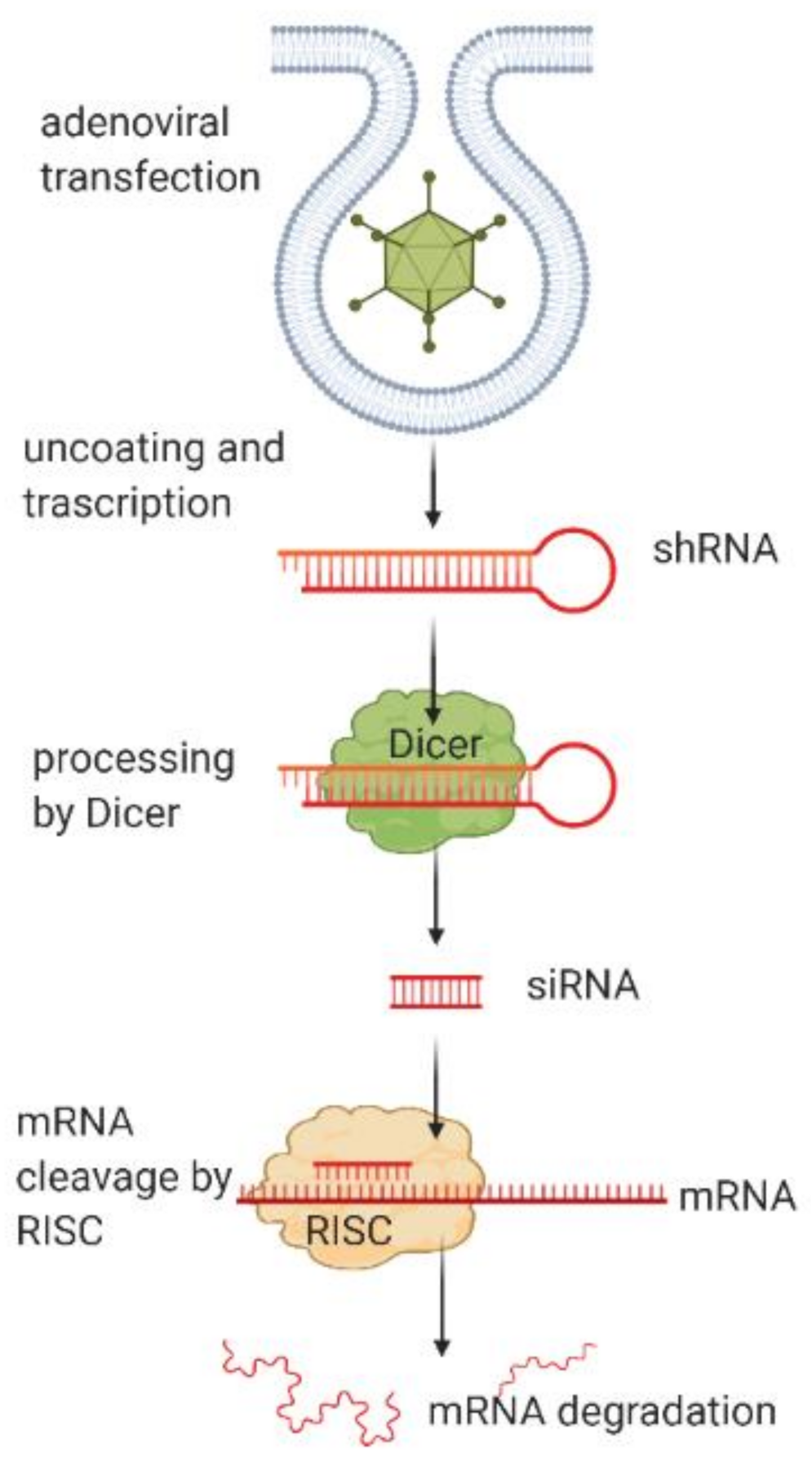
Another system for the delivery of target fragments for dsRNA synthesis is the use of adenoviruses (Figure 2), and these have a number of advantages as compared with lentiviruses.
First, the majority of cells in the human body express primary adenovirus receptors and secondary integrin receptors, thus making the adenovirus one of the most effective vehicles for in vivo gene delivery. Secondly, adenoviruses do not integrate their DNA into the host cell genome. Thirdly, despite the safety concerns, adenovirus-based genetic vectors have now been widely used in clinical practice, and currently, safe dosages and injection techniques are already well established. Fourth, adenovirus-based vectors represent a universal platform for the virus capsid modification for the optimization of targeting specificity and other therapeutic characteristics [26].
There has been a concern that their use could be very limited owing to the frequent contact of humans with adenoviruses, resulting in the presence of antibodies that are highly likely to destroy the viral particle before it reaches the target cells. However, there are clinical studies in which oncolytic adenoviruses have avoided destruction by the immune system and successfully reached tumors [27,28]. There are also efforts aimed at the protection of the adenoviral constructs from antibodies and degradation through the use of silicon coating [29]. Such encapsulation of the virus particles significantly improves their distribution and effects in tumor inhibition.
As with lentivirus, search queries reveal a great number of studies covering a wide range of gene targets that were silenced by means of RNA interference. For example, the growth of hepatocellular carcinomas has been significantly inhibited using adenoviral constructs containing fragments able to form short hairpin RNA and miRNA, resulting in a subsequent suppression of the corresponding genes, with the inhibition of cancer cell growth being demonstrated in both cell cultures and in vivo models [30,31,32,33].
There is still a concern about the safety of viruses as vectors for the gene therapy of human diseases. Primarily, the genome regions responsible for the replication of the viral particles are obligatorily deleted from the constructs for safety reasons. However, lentiviruses, for example, typically insert themselves into the genome of the host cell as proviruses, and this can pose a risk of the cell transitioning into a cancerous form. However, these concerns have not been confirmed in any studies to date [34,35]. A hepatotoxic effect has been demonstrated for adenoviruses in the case of intravenous administration, owing to sequestration of the viral particles by the liver, followed by transaminitis and vascular disruption [36,37]. However, no serious adverse effects have been reported in the case of oral adenovirus administration [38]. In general, it should be noted that the local use of viral vectors in gene therapy is preferable to systemic administration, being both a safer and a more effective technique [39].

3. Nanoparticles

3.1. Lipid-Based Nanoparticles

Lipid-based nanoparticles are used as delivery agents in the first FDA-approved human medication, patisiran (Onpattro®), which is used to treat amyloid polyneuropathy. Its biologically active substance is a small fragment of modified dsRNA that works via RNA interference. In addition to amyloid polyneuropathy, this delivery method has also been approved for cancer treatment, including combined treatments using dsRNA and chemotherapy [40]. Compared with the viral vectors, this method of siRNA delivery can be used for the systemic administration of medications owing to the high biocompatibility of the lipid-based nanoparticles [41], thus it can be applied for the treatment of both solid and diffuse tissues. This type of nanoparticles usually consists of phospholipids with inlaid by conjugation lipids, which can be modified with various ligands.
It should be noted that lipid-based nanoparticles bear the advantage of being the least toxic for in vivo applications, and significant progress has been made in the area of RNA delivery using lipid-based nanoassemblies. However, this approach has a significant disadvantage of the low tissue selectivity of drug delivery and the low transfection of cancer cells.
First, it is very important to note that glycotargeting, the main method of hepatocyte-targeting, was developed using lipid-based nanoparticles. Targeting unique markers of hepatocytes, asialoglycoprotein receptors, allows to achieve very high results of transfection and inhibition of growth rate in hepatocarcinoma cell lines [42,43]. Moreover, one of the main modifications of nanoparticles is aimed specifically at solving these problems. For example, in hepatocarcinoma therapy, cationic lipid-based nanoparticles can successfully deliver the shNUPR plasmid to suppress the corresponding NUPR1 gene (which is involved in hepatocellular carcinoma growth and chemoresistance), by protecting the plasmid from DNase I action [40], while building an apolipoprotein crown onto the particle significantly increases its transfection and selectivity to hepatocytes [41]. The same study noted the key roles of both the length of the PEG-conjugated (polyethylene glycol) lipid chain and the amount of PEG in the nanoparticle needed for successful RNA interference therapy of the solid tumors.
Specific modifications in the structure of antibodies have also been used to increase the selectivity of lipid-based nanoparticles for certain types of cells. For example, this approach has worked well with delivering siRNA to lymphocytes, which are normally particularly difficult to transfect with RNA, because they are resistant to traditional transfection reagents and are distributed throughout the body, hindering successful delivery by a systemic administration [44]. In addition to this, a high concentration of reactive oxygen species (ROS) is used as a marker of cancer cells. It facilitates the selective decomposition of the lipid-based carrier to nanoparticles in the cancerous cells [45].
Combined therapy, which includes both a standard cytostatic agent and dsRNA, for silencing a specific gene, within lipid-based nanoparticles, is also a promising approach to the treatment of resistant tumors. An example is the combined delivery of cisplatin together with siRNA, targeting the endonuclease xeroderma pigmentosum group F (XPF), a key of nucleotide excision repair component in mammals. The lipid-based nanoparticles can efficiently encapsulate both cytostatic agents and molecules of siRNA in a specified ratio. Both components are effectively transported into the cells and released therein. As a result, the cisplatin damages the DNA, while the siRNA specifically suppresses the levels of both mRNA and the corresponding XPF protein to enhance the action of the cisplatin, thus leading to increased levels of expression of apoptosis markers and increased cytotoxicity in both cisplatin-sensitive and -resistant cells [46].

3.2. Gold Nanoparticles

Gold nanoparticles are also used for stable and safe delivery of various medications, including siRNA. They can be synthesized in a wide range of sizes with diverse surface functionalities. Tunability in size and surface characteristics makes them promising candidates as drug delivery vehicles. Moreover, there is an opportunity for a very precise control over the size, shape, and surface properties of such gold nanoparticles and of their functionalization using various biomolecules [47]. For example, oligopeptides have been used to develop siRNA-delivery systems for the treatment of glioblastomas and breast cancer [48,49]. Unmodified gold nanoparticles possess low transfection efficiency as siRNA delivery agents. However, in vitro experiments have demonstrated a positive role for the incorporation of cations onto the delivery agent surface in order to enhance the uptake of exogenous RNA by cells, for example, functionalizing them with arginine Fe3O4 [50,51,52,53]. Indeed, for some modifications, the transfection frequency was higher than that seen with the commercial reagent Lipofectamine 2000. Despite this, in vivo experimental results are not so optimistic; that is, systemic administration of cationic delivery systems without biological stabilizing fragments results in their non-specific binding to negatively charged serum proteins, leading to the aggregation and opsonization of the particles. Therefore, PEGylated nanoparticles, having a practically neutral charge, seem more promising for cancer disease therapy [54].

3.3. Polymeric Nanoparticles

Polymeric nanoparticles are probably the most diverse category of delivery agents owing to the availability of various polymeric materials. Unlike the abovementioned nanoparticles, the possibilities for their chemical composition and modification are practically unlimited. Moreover, modern materials permit the creation of a 3D nanoparticle structure with the siRNA embedded in it, either throughout the particle or comprising part of its layers. Some materials (especially chitosan-based) are highly promising as oral delivery agents for targeted RNAs. Unlike lipid-based nanoparticles, these delivery agents have no adverse effects on the liver [55].
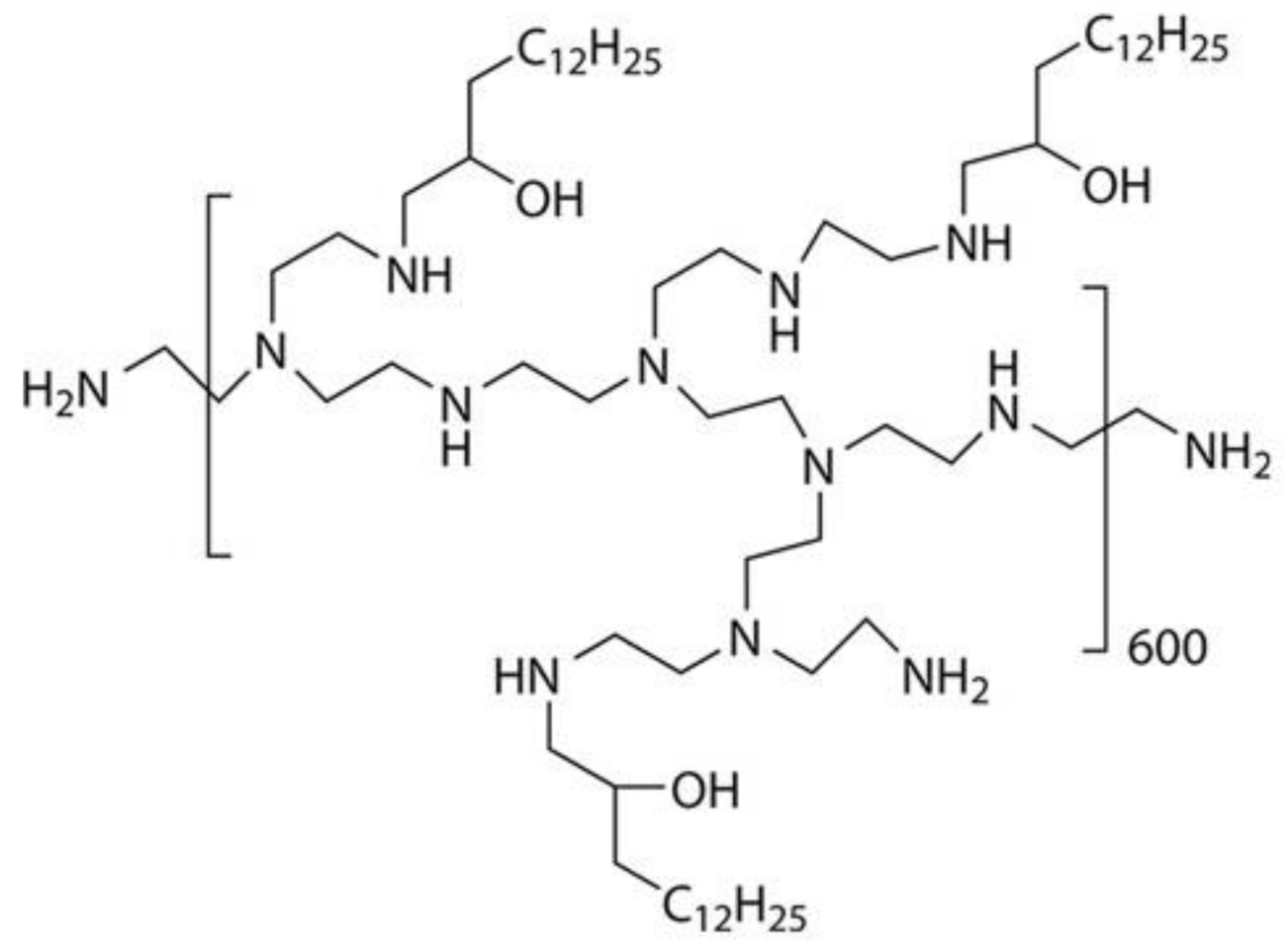
Various polymeric materials (gelatin-based, poly(lactic-co-glycolic) (PLGA-modified), cationic amphiphilic) were developed in a series of studies and proved themselves effective as siRNA delivery agents in in vitro systems directed against breast cancer, hepatoma, and myeloid leukemia [56,57,58,59,60]. Despite the fundamentally different nature of the polymer material for the nanoparticles in these studies, they all have the common feature of being positively charged at their surfaces owing to various functional groups aimed at increasing transfection effectiveness. As mentioned above for gold nanoparticles, a positive charge can be a significant disadvantage in the case of systemic administration because of the interaction with serum proteins. In 2018, the first study was published that demonstrated the effect of systemically administered siRNA on nonhuman primates in combination with a 7C1 polymer (Figure 3), low-molecular weight, ionizable polymer that forms nanoparticles [61]. The study provided an extensive histochemical analysis of tissues, showing there were no toxic effects, while the control of cytokines in the blood serum suggested that there were no inflammatory effects in the body. Biochemical blood tests and analysis of the liver function before and after treatment confirmed that the concentrations of proteins and enzymes remained within their normal ranges. The obtained data indicate that the 7C1 complex is a promising siRNA delivery system for systemic administration.
A chitosan-based galactose-modified polymer has also been developed for the oral delivery of siRNA medications [62]. The level of VEGF gene silencing was investigated in mice hepatoma cells, characterized by increased galactose uptake, and thus expected to accumulate an increased concentration of the anti-VEGF siRNA. As a result of the study, significant suppression of the corresponding gene expression, an increase of apoptosis, and an inhibition of angiogenesis have been demonstrated.
Both polymeric nanoparticles and gold nanoparticles are used for combined cancer therapy; for example, when using photocontrolled toxicity to fight cancer cells [63]. In one of the studies, the system of photosensitive polymer nanoparticles included the Pt(IV) prodrug and si (c-fos), thus the release and activation of these components were irradiation-dependent. During exposure to blue light (430 nm), the material was stimulated to release the active components, resulting in cell death. Selective phototherapeutic agents are the basis of an emerging and rapidly developing industry and the use of such medications seems very promising within anti-cancer and anti-bacterial treatment schemes; however, there is a concern related to their photoactivation inside the body because of the low penetrating ability of the radiation used.

3.4. Silicon Nanoparticles

Silicon nanoparticles provide an alternative approach to address the maintenance of siRNA integrity while delivering it in the quantities required. The authors previously mentioned the silicon encapsulation of viruses to protect these from the immune system, yet using silicon as an independent delivery agent is much less popular. The main characteristic of silicon that allows it to be considered as a potentially effective siRNA delivery agent is its porosity—in this case, such encapsulation of the RNA molecules protects them from degradation in the body. As with other materials used in nanoparticles, silicon allows for a variety of modifications to enhance uptake selectivity, specifically by cancer cells. For example, it provides for the use of additional peptide or lipid coatings [64]. It has been demonstrated that the amount of siRNA that can be loaded into silicon nanoparticles is significantly affected by the concentration of salts and urea in solution, so this must be taken into account in vivo [65].

4. Exosomes and Exosome-Mimetic Nanovesicles

Exosomes (also called “extracellular vesicles”) are natural, nanoscale vesicles that can interact with cell membranes owing to the presence of various adhesive proteins on their surfaces, thus exosomes are considered promising delivery vehicles, also because they are highly biocompatible. This feature provoked many attempts to apply these lipid structures in medicine, and especially in gene therapy for siRNA transport [66]. For example, exosomes were used for silencing of the RAD1 gene, which is one of the main therapeutic targets in cancer treatment [67]. The application of exosomes also enabled to reduce premature ovarian failure, an irreversible effect that women can face after chemotherapy, where anti-apoptotic miRNAs are essential for the restoration of granulosa cells in the follicles. Amniotic fluid can be used as a source of the corresponding exosomes [68].
Taking into account that the cells of multicellular organisms secrete enormous quantities of exosomes, their targeted delivery for gene silencing in cancer tumors is a critical issue. Viral modification of exosomes, as a targeting method, has been approved for RNA delivery in vivo [69]. The main obstacle in the therapeutic application of exosomes is their relatively low yield in any cell culture system and currently complicated purification processes [70].
An alternative to natural exosomes can be artificial exosomes; for example, cell-derived mimetic nanovesicles are a potentially promising alternative to exosomes for clinical applications, demonstrating higher yield without incumbent production and isolation issues [71]. Mimetic nanovesicles could be derived from any cell type. They possess comparable characteristics to exosomes and could be used instead of them. The main source of mimetic nanovesicles is mesenchymal stem cells [72,73]. Firstly, the issues with using mesenchymal stem cells directly are due to their poor engraftment rate, and secondly, there are certain safety problems with their use in humans. Therefore, as an alternative devoid of these shortcomings, mimetic nanovesicles based on exosomes began to be utilized. Artificially synthesized analogs of exosomes have another advantage—their membrane can be modified synthetically in order to obtain optimal physical and chemical properties for purification and release of the contents.
The literature describes the preparation of mimetic nanovesicles using macrophages or macrophages fused with mesenchymal stem cells [72,73,74,75]. Using this technique, it was possible to significantly reduce the proliferation rate of cancer cells in the case of RNAi of the c-Myc gene, one of the key regulators of cell proliferation [70,76,77]. In addition to mimetic nanovesicles, the study also used Lipofectamine 2000 and native RNA non-associated with any transport agent as controls. It was shown that the capture of the target RNA occurs equally efficiently with both Lipofectamine 2000 and mimetic nanovesicles, which indicates that they are promising as RNA deliveries for RNAi. It is also worth noting that, when the native RNA was introduced without any delivery system, no RNA interference was detected, suggesting that transport systems are absolutely necessary to protect RNA from degradation.
The complications of the techniques for obtaining nanovesicles are similar to those for exosomes, which are effective delivery vehicles of dsRNA for RNAi. In this regard, it is worth mentioning another study, where researchers proposed to make exosome analogs without the use of cells and to synthesize mimetic nanovesicles completely artificially, in vitro [78]. Using such a methodology, mimetic nanovesicles were obtained based on chitosan nanoparticles covered with a lipid layer that mimics exosomes. Owing to electrostatic interaction, RNA molecules adhered to chitosan particles, while the bilipid layer provided interaction with cells for successful delivery of dsRNA and its protection from degradation. The low toxicity of the developed delivery method is another great advantage, in addition to the possibility of obtaining a large number of mimetic nanovesicles Notably, the toxicity is more than four times lower compared with Lipofectamine 2000. Low transfection efficiency is among the disadvantages of the developed system (lower than lipofectamine). Despite this, the mimetic nanovesicle-mediated delivery system can be very promising for gene therapy thanks to its safety. However, additional research is required for its improvement.

5. Peptides

Peptides as siRNA delivery agents may be another promising platform in gene therapy for cancer. They possess flexibility in design, simple compositions and formulations, and diverse physicochemical functions [79]. This delivery system also has drawbacks—peptide agents are very sensitive to proteases, which imposes restrictions on the use of this methodology when peptides are administered systemically. Local administration is preferred for peptide delivery vehicles, however, it is not optimal in the treatment of solid tumors.
First, it is worth noting dendrimers—tree-like polypeptides with a large number of branches. Their branchy structure allows solving several problems at once—part of the molecule is responsible for protecting siRNA from enzymatic degradation, the other part can be functionalized for targeted delivery of RNA molecules to a specific cell type (for example, using antibodies), and additional modifications can be introduced to improve transfection effectiveness. Amphiphilic phospholipid peptide dendrimers successfully delivered siRNA into castration-resistant prostate cancer PC-3 cells [80]. The Hsp27 gene (heat shock protein 27), one of the main therapeutic targets for the treatment of castration-resistant prostate cancer, was selected to be knocked down. The delivery system had a hydrophobic part based on natural lipids and responsible for interaction with the cell membrane and capture of the vector, and the hydrophilic part consisted of dendritic l-lysin, capable of compacting siRNA into nanoparticles to protect it from enzymatic degradation. In this study, a balance between the hydrophobic and hydrophilic parts of the vector has been achieved, which is reflected in a sufficiently high level of transfection of target cells by the siRNA. In another equally outstanding work, the dendrimer was based on a flexible triethanolamine-core with a polyamidoamine dendritic structure [81]. Here, the researchers applied dual targeting by modifying the dendrimer with additional proteins, interacting with integrin and neuropilin-1 receptors, which led to improved cell penetration, gene silencing, and anticancer activity for the prostate cancer model.
To protect peptides from proteases, an approach using D-isomer amino acids was proposed [82]. The amphipathic peptides created with this methodology demonstrated not only high resistance to proteases, but a capability of self-assembly with siRNA molecules. The researchers note the retention of the basic biophysical characteristics of the retro-inverse form of the protein in comparison with its L-parent homologue. Treatment of cells with the developed complex also produced an effective knockdown of the target gene through RNAi.
Nanocarrier based on aminated poly (α) glutamate was chosen as another promising agent for the systemic use of polypeptides as siRNA delivery technology against solid tumors [83]. The RNA molecules interacted electrostatically with the carrier, leading to the formation of a complex extremely stable in plasma/blood. This approach was approved for systemic administration in vivo against solid tumors—ovarian cancer and lung carcinoma. As a result, a reduction in the expression of the target gene Rac1 was achieved by 33 and 38%, respectively. The tumor size decreased by 73% and 87%, which indicates the high efficiency and future potential of this approach for the treatment of solid tumors.

6. Conjugates

Among the conjugates for targeted delivery of oligonucleotides, givosiran is currently the standard. It is the second drug after patisiran approved by the FDA for the treatment of acute hepatic porphyria [84]. Givosiran is a small interfering RNA (siRNA) directed toward the 5-aminolevulinic acid synthase, an important enzyme in the production of heme part in hemoglobin. It is covalently bound to a ligand containing three N-acetylgalactosamine residues that facilitate uptake into hepatocytes via asialoglycoprotein receptors, which are highly expressed on the cell surface of hepatocytes and are selective for glycoproteins containing N-acetylgalactosamine residues [85]. It is worth highlighting N-acetylgalactosamine as a very promising molecule for the delivery of siRNA to hepatocytes; in addition to the already approved givosiran, there are another seven conjugates in registrational review or phase 3 trials and at least another 21 conjugates at earlier stages of clinical development [86]. Additional enhancements, such as hexopyranose chemical modification altriol nucleic acid within siRNA, significantly enhanced the protection of the oligonucleotide against 5-exonuclease degradation [87]. Studies on the targeted delivery of siRNA through modifications using N-acetylgalactosamine residues are sufficient for a separate full review; in this review, only a small part of them is considered.
An example of a successful approach can be the conjugation of siRNA with docosanoic acid to target myostatin, a key determinant of muscle loss and cachexia in cancer, in skeletal and cardiac muscle cells [88,89]. The siRNAs delivered in this way provided more than 55% of gene silencing in muscle cells and about 80% in cardiac cells, increasing tissue volume by more than half.
Conjugation of siRNA with cholesterol allows to neutralize the negative charge of RNA and, consequentially, the impermeability of the cell membrane for it. In one of the studies, 356 cholesterol-conjugated siRNA molecules with various additional modifications were tested, and an algorithm that effectively predicts the activity of miRNA was developed based on linear regression approach [90]. As a result, conjugates were obtained that effectively transfect cancer cells, the chemical modifications of which were predicted by the algorithm. This approach eliminates the need for stochastic screening and optimizes the development of delivery systems.

7. Conclusions

This overview is an attempt to summarize the main trends in siRNA delivery in the field of cancer disease therapy. Taking into account the rapid development of gene therapy in general, and the possibilities for individual gene regulation using RNA interference, the authors identified a great number of studies from which it was extremely difficult to draw general conclusions to produce a coherent overview.
Summarizing the work performed in this field, the authors can declare that RNA interference has a promising future in cancer treatment, and that the number of approaches to the targeted delivery of siRNA will only increase. The limitations of the existing approaches will eventually be overcome, enabling the selection of optimal delivery systems for exceptionally effective gene therapy of oncological diseases.

Author Contributions

Conceptualization, T.S.G.; writing—original draft preparation, V.A.C.; writing—review and editing, T.S.G. and K.E.O.; funding acquisition, K.E.O. All authors have read and agreed to the published version of the manuscript.

Funding

This research was funded by Ministry of Education and Science of Russian Federation, grant #2019-0546 (FSUS-2020-0040).

Institutional Review Board Statement

Not applicable.

Informed Consent Statement

Not applicable.

Acknowledgments

The authors would like to thank Vladimir V. Muzyka for critical review of the text.

Conflicts of Interest

The authors declare no conflict of interest.

Sample Availability

Not available.

Abbreviations

dsRNAdouble-stranded RNA
siRNAsmall interfering RNA
RNAiRNA interference
shRNAshort hairpin RNA

References

  1. Roberts, T.C.; Langer, R.; Wood, M.J.A. Advances in oligonucleotide drug delivery. Nat. Rev. Drug Discov. 2020, 19, 673–694. [Google Scholar] [CrossRef] [PubMed]
  2. Wirth, T.; Ylä-Herttuala, S. Gene Therapy Used in Cancer Treatment. Biomedicines 2014, 2, 149–162. [Google Scholar] [CrossRef] [PubMed]
  3. Hu, B.; Zhong, L.; Weng, Y.; Peng, L.; Huang, Y.; Zhao, Y.; Liang, X.J. Therapeutic siRNA: State of the art. Signal Transduct. Target. Ther. 2020, 5, 101. [Google Scholar] [CrossRef] [PubMed]
  4. Weng, Y.; Xiao, H.; Zhang, J.; Liang, X.J.; Huang, Y. RNAi therapeutic and its innovative biotechnological evolution. Biotechnol Adv. 2019, 37, 801–825. [Google Scholar] [CrossRef] [PubMed]
  5. Guo, D.; Ji, X.; Peng, F.; Zhong, Y.; Chu, B.; Su, Y.; He, Y. Photostable and Biocompatible Fluorescent Silicon Nanoparticles for Imaging-Guided Co-Delivery of siRNA and Doxorubicin to Drug-Resistant Cancer Cells. Nano-Micro Lett. 2019, 11, 27. [Google Scholar] [CrossRef] [Green Version]
  6. Liu, J.; Guo, N.; Gao, C.; Liu, N.; Zheng, X.; Tan, Y.; Lei, J.; Hao, Y.; Chen, L.; Zhang, X. Effective Gene Silencing Mediated by Polypeptide Nanoparticles LAH4-L1-siMDR1 in Multi-Drug Resistant Human Breast Cancer. J. Biomed. Nanotechnol. 2019, 15, 531–543. [Google Scholar] [CrossRef] [PubMed]
  7. Kim, M.; Kim, G.; Hwang, D.; Lee, M. Delivery of high mobility group Box-1 siRNA using brain-targeting Exosomes for ischemic stroke therapy. J. Biomed. Nanotechnol. 2019, 15, 2401–2412. [Google Scholar] [CrossRef]
  8. Wang, Y.; Li, C.; Du, L.; Liu, Y. A reactive oxygen species-responsive dendrimer with low cytotoxicity for efficient and targeted gene delivery. Chin. Chem. Lett. 2020, 31, 275–280. [Google Scholar] [CrossRef]
  9. Chen, Y.; Li, B.; Chen, X.; Wu, M.; Ji, Y.; Tang, G.; Ping, Y. A supramolecular co-delivery strategy for combined breast cancer treatment and metastasis prevention. Chin. Chem. Lett. 2020, 31, 1153–1158. [Google Scholar] [CrossRef]
  10. Nasehi, L.; Ghahremani, M.H.; Yavari, K.; Hadjati, J.; Fallah, A.; Abdolhossein Zadeh, B.; Saltanatpour, Z. Stable silencing of IGF1R using Lentiviral-mediated shRNA in HEK293T cells. Cell. Mol. Biol. 2017, 63, 62–66. [Google Scholar] [CrossRef]
  11. Qiu, J.; Zhao, J.; Zuo, A.; Liu, L.; Liu, Q.; Pan, H.; Yuan, X. Lentiviral RNA interference-mediated downregulation of forkhead box M1 expression suppresses growth of oral squamous cell carcinoma in vitro. Oncol. Lett. 2019, 17, 525–531. [Google Scholar] [CrossRef] [PubMed] [Green Version]
  12. Xu, X.; Lai, Y.; Zhou, W.; Hua, Z. Lentiviral delivery of a shRNA sequence analogous to miR-4319/miR-125-5p induces apoptosis in NSCLC cells by arresting G2/M phase. J. Cell. Biochem. 2019, 120, 14017–14027. [Google Scholar] [CrossRef] [PubMed]
  13. Liao, Z.; Wang, X.; Lin, D.; Zou, Q. Construction and Identification of the RNAi Recombinant Lentiviral Vector Targeting Human DEPDC7 Gene. Interdiscip. Sci. Comput. Life Sci. 2017, 9, 350–356. [Google Scholar]
  14. Osten, P.; Grinevich, V.; Cetin, A. Viral Vectors: A Wide Range of Choices and High Levels of Service. In Conditional Mutagenesis: An Approach to Disease Models; Springer: Berlin/Heidelberg, Germany, 2007; pp. 177–202. [Google Scholar]
  15. Liu, Y.P.; Westerink, J.T.; Ter Brake, O.; Berkhout, B. RNAi-Inducing lentiviral vectors for anti-hiv-1 gene therapy. Methods Mol. Biol. 2011, 721, 293–311. [Google Scholar] [PubMed]
  16. Qiu, J.F.; Zhang, Z.Q.; Wang, Y.; You, J. Lentivirus-mediated RNAi knockdown of VEGFA in RKO colorectal cancer cells decreases tumor formation and growth in vitro and in vivo. Int. J. Clin. Exp. Pathol. 2012, 5, 290–298. [Google Scholar]
  17. Sun, P.; Yu, H.; Zhang, W.Q.; Hu, M.; Lv, R. Lentivirus-mediated siRNA targeting VEGF inhibits gastric cancer growth in vivo. Oncol. Rep. 2012, 28, 1687–1692. [Google Scholar] [CrossRef] [Green Version]
  18. Zhao, X.; Zhu, D.M.; Gan, W.J.; Li, Z.; Zhang, J.L.; Zhao, H.; Zhou, J.; Li, D.C. Lentivirus-mediated shRNA interference targeting vascular endothelial growth factor inhibits angiogenesis and progression of human pancreatic carcinoma. Oncol. Rep. 2013, 29, 1019–1026. [Google Scholar] [CrossRef] [Green Version]
  19. Lin, J.; Pang, H.; Guo, X.; Ding, Y.; Geng, J.; Zhang, J.; Min, J. Lentivirus-Mediated RNAi Silencing of VEGF Inhibits Angiogenesis and Growth of Renal Cell Carcinoma in a Nude Mouse Xenograft Model. DNA Cell Biol. 2015, 34, 717–727. [Google Scholar] [CrossRef]
  20. Su, S.F.; Chang, Y.W.; Andreu-Vieyra, C.; Fang, J.Y.; Yang, Z.; Han, B.; Lee, A.S.; Liang, G. miR-30d, miR-181a and miR-199a-5p cooperatively suppress the endoplasmic reticulum chaperone and signaling regulator GRP78 in cancer. Oncogene 2013, 32, 4694–4701. [Google Scholar] [CrossRef] [Green Version]
  21. Emeagi, P.U.; Maenhout, S.; Dang, N.; Heirman, C.; Thielemans, K.; Breckpot, K. Downregulation of Stat3 in melanoma: Reprogramming the immune microenvironment as an anticancer therapeutic strategy. Gene Ther. 2013, 20, 1085–1092. [Google Scholar] [CrossRef]
  22. Tang, X.R.; Wen, X.; He, Q.M.; Li, Y.Q.; Ren, X.Y.; Yang, X.J.; Zhang, J.; Wang, Y.Q.; Ma, J.; Liu, N. MicroRNA-101 inhibits invasion and angiogenesis through targeting ITGA3 and its systemic delivery inhibits lung metastasis in nasopharyngeal carcinoma. Cell Death Dis. 2017, 8, e2566. [Google Scholar] [CrossRef] [PubMed] [Green Version]
  23. Jian, P.; Li, Z.W.; Fang, T.Y.; Jian, W.; Zhuan, Z.; Mei, L.X.; Yan, W.S.; Jian, N. Retinoic acid induces HL-60 cell differentiation via the upregulation of miR-663. J. Hematol. Oncol. 2011, 4, 20. [Google Scholar] [CrossRef] [PubMed] [Green Version]
  24. Li, S.; Rosenberg, J.E.; Donjacour, A.A.; Botchkina, I.L.; Hom, Y.K.; Cunha, G.R.; Blackburn, E.H. Rapid inhibition of cancer cell growth induced by lentiviral delivery and expression of mutant-template telomerase RNA and anti-telomerase short-interfering RNA. Cancer Res. 2004, 64, 4833–4840. [Google Scholar] [CrossRef] [PubMed] [Green Version]
  25. Zafar, S.; Quixabeira, D.C.A.; Kudling, T.V.; Cervera-Carrascon, V.; Santos, J.M.; Grönberg-Vähä-Koskela, S.; Zhao, F.; Aronen, P.; Heiniö, C.; Havunen, R.; et al. Ad5/3 is able to avoid neutralization by binding to erythrocytes and lymphocytes. Cancer Gene Ther. 2020. [Google Scholar] [CrossRef] [PubMed]
  26. Lee, C.S.; Bishop, E.S.; Zhang, R.; Yu, X.; Farina, E.M.; Yan, S.; Zhao, C.; Zheng, Z.; Shu, Y.; Wu, X.; et al. Adenovirus-Mediated Gene Delivery: Potential Applications for Gene and Cell-Based Therapies in the New Era of Personalized Medicine. Genes Dis. 2017, 2, 43–63. [Google Scholar] [CrossRef]
  27. Zhang, Y.; Wu, J.; Zhang, H.; Wei, J.; Wu, J. Extracellular Vesicles-Mimetic Encapsulation Improves Oncolytic Viro-Immunotherapy in Tumors with Low Coxsackie and Adenovirus Receptor. Front. Bioeng. Biotechnol. 2020, 8. [Google Scholar] [CrossRef]
  28. Kong, H.; Zhao, R.; Zhang, Q.; Iqbal, M.Z.; Lu, J.; Zhao, Q.; Luo, D.; Feng, C.; Zhang, K.; Liu, X.; et al. Biosilicified oncolytic adenovirus for cancer viral gene therapy. Biomater. Sci. 2020, 8, 5317–5328. [Google Scholar] [CrossRef]
  29. Li, X.; Su, Y.; Sun, B.; Ji, W.; Peng, Z.; Xu, Y.; Wu, M.; Su, C. An artificially designed interfering lncRNA expressed by oncolytic adenovirus competitively consumes OncomiRs to exert antitumor efficacy in hepatocellular carcinoma. Mol. Cancer Ther. 2016, 15, 1436–1451. [Google Scholar] [CrossRef] [Green Version]
  30. Huang, M.; Li, G.; Pan, T.; Cheng, Y.; Ren, W.; Jia, W.; Ma, J.; Xu, G. A Novel multi-target RNAi adenovirus inhibits hepatoma cell proliferation, migration, and induction of angiogenesis. Oncotarget 2016, 7, 57705–57713. [Google Scholar] [CrossRef] [Green Version]
  31. Wang, M.; Liu, J.; Xi, D.; Luo, X.; Ning, Q. Adenovirus-mediated artificial microRNA against human fibrinogen like protein 2 inhibits hepatocellular carcinoma growth. J. Gene Med. 2016, 18, 102–111. [Google Scholar] [CrossRef]
  32. Chen, T.; Xiong, J.; Yang, C.; Shan, L.; Tan, G.; Yu, L.; Tan, Y. Silencing of FOXM1 transcription factor expression by adenovirus-mediated RNA interference inhibits human hepatocellular carcinoma growth. Cancer Gene Ther. 2014, 21, 133–138. [Google Scholar] [CrossRef] [PubMed]
  33. Kalesnykas, G.; Kokki, E.; Alasaarela, L.; Lesch, H.P.; Tuulos, T.; Kinnunen, K.; Uusitalo, H.; Airenne, K.; Yla-Herttuala, S. Comparative Study of Adeno-associated Virus, Adenovirus, Bacu lovirus and Lentivirus Vectors for Gene Therapy of the Eyes. Curr. Gene Ther. 2017, 17. [Google Scholar] [CrossRef] [PubMed]
  34. Montini, E.; Cesana, D.; Schmidt, M.; Sanvito, F.; Ponzoni, M.; Bartholomae, C.; Sergi, L.S.; Benedicenti, F.; Ambrosi, A.; Di Serio, C.; et al. Hematopoietic stem cell gene transfer in a tumor-prone mouse model uncovers low genotoxicity of lentiviral vector integration. Nat. Biotechnol. 2006, 24, 687–696. [Google Scholar] [CrossRef] [PubMed]
  35. Shayakhmetov, D.M.; Li, Z.-Y.; Ni, S.; Lieber, A. Analysis of Adenovirus Sequestration in the Liver, Transduction of Hepatic Cells, and Innate Toxicity after Injection of Fiber-Modified Vectors. J. Virol. 2004, 78, 5368–5381. [Google Scholar] [CrossRef] [Green Version]
  36. Shayakhmetov, D.M.; Gaggar, A.; Ni, S.; Li, Z.-Y.; Lieber, A. Adenovirus Binding to Blood Factors Results in Liver Cell Infection and Hepatotoxicity. J. Virol. 2005, 79, 7478–7491. [Google Scholar] [CrossRef] [Green Version]
  37. Stephenson, K.E.; Keefer, M.C.; Bunce, C.A.; Frances, D.; Abbink, P.; Maxfield, L.F.; Neubauer, G.H.; Nkolola, J.; Peter, L.; Lane, C.; et al. First-in-human randomized controlled trial of an oral, replicating adenovirus 26 vector vaccine for HIV-1. PLoS ONE 2018, 13. [Google Scholar] [CrossRef]
  38. Merentie, M.; Lottonen-Raikaslehto, L.; Parviainen, V.; Huusko, J.; Pikkarainen, S.; Mendel, M.; Laham-Karam, N.; Kärjä, V.; Rissanen, R.; Hedman, M.; et al. Efficacy and safety of myocardial gene transfer of adenovirus, adeno-associated virus and lentivirus vectors in the mouse heart. Gene Ther. 2016, 23, 296–305. [Google Scholar] [CrossRef]
  39. He, W.; Turkeshi, A.; Li, X.; Zhang, H. Progress in systemic co-delivery of microRNAs and chemotherapeutics for cancer treatment by using lipid-based nanoparticles. Ther. Deliv. 2020, 11, 591–603. [Google Scholar] [CrossRef]
  40. Botto, C.; Augello, G.; Amore, E.; Emma, M.R.; Azzolina, A.; Cavallaro, G.; Cervello, M.; Bondì, M.L. Cationic solid lipid nanoparticles as non viral vectors for the inhibition of hepatocellular carcinoma growth by RNA interference. J. Biomed. Nanotechnol. 2018, 14, 1009–1016. [Google Scholar] [CrossRef]
  41. Chen, D.; Parayath, N.; Ganesh, S.; Wang, W.; Amiji, M. The role of apolipoprotein- and vitronectin-enriched protein corona on lipid nanoparticles for- And vivo targeted delivery and transfection of oligonucleotides in murine tumor models. Nanoscale 2019, 11, 18806–18824. [Google Scholar] [CrossRef]
  42. Huang, X.; Leroux, J.C.; Castagner, B. Well-Defined Multivalent Ligands for Hepatocytes Targeting via Asialoglycoprotein Receptor. Bioconjug. Chem. 2017, 28, 283–295. [Google Scholar] [CrossRef] [PubMed]
  43. Mkhwanazi, N.K.; de Koning, C.B.; van Otterlo, W.A.L.; Ariatti, M.; Singh, M. PEGylation potentiates hepatoma cell targeted liposome-mediated in vitro gene delivery via the asialoglycoprotein receptor. Z. Naturforsch C. J. Biosci. 2017, 72, 293–301. [Google Scholar] [CrossRef] [PubMed]
  44. Hazan-Halevy, I.; Rosenblum, D.; Ramishetti, S.; Peer, D. Systemic modulation of lymphocyte subsets using siRNAs delivered via targeted lipid nanoparticles. In Methods in Molecular Biology; Humana Press: New York, NY, USA, 2019; pp. 151–159. [Google Scholar]
  45. Liang, C.; Chang, J.; Jiang, Y.; Liu, J.; Mao, L.; Wang, M. Selective RNA interference and gene silencing using reactive oxygen species-responsive lipid nanoparticles. Chem. Commun. 2019, 55, 8170–8173. [Google Scholar] [CrossRef] [PubMed]
  46. Li, C.; Li, T.; Huang, L.; Yang, M.; Zhu, G. Self-assembled Lipid Nanoparticles for Ratiometric Codelivery of Cisplatin and siRNA Targeting XPF to Combat Drug Resistance in Lung Cancer. Chem. Asian J. 2019, 14, 1570–1576. [Google Scholar] [CrossRef]
  47. Ferreira, D.; Fontinha, D.; Martins, C.; Pires, D.; Fernandes, A.R.; Baptista, P.V. Gold nanoparticles for vectorization of nucleic acids for cancer therapeutics. Molecules 2020, 25, 3489. [Google Scholar] [CrossRef]
  48. Ahwazi, R.P.; Kiani, M.; Dinarvand, M.; Assali, A.; Tekie, F.S.M.; Dinarvand, R.; Fatemeh, A. Immobilization of HIV-1 TAT peptide on gold nanoparticles: A feasible approach for siRNA delivery. J. Cell. Physiol. 2020, 235, 2049–2059. [Google Scholar] [CrossRef]
  49. Kong, L.; Qiu, J.; Sun, W.; Yang, J.; Shen, M.; Wang, L.; Shi, X. Multifunctional PEI-entrapped gold nanoparticles enable efficient delivery of therapeutic siRNA into glioblastoma cells. Biomater. Sci. 2017, 5, 258–266. [Google Scholar] [CrossRef]
  50. Zhao, E.; Zhao, Z.; Wang, J.; Yang, C.; Chen, C.; Gao, L.; Feng, Q.; Hou, W.; Gao, M.; Zhang, Q. Surface engineering of gold nanoparticles for in vitro siRNA delivery. Nanoscale 2012, 4, 5102–5109. [Google Scholar] [CrossRef]
  51. Kong, W.H.; Bae, K.H.; Jo, S.D.; Kim, J.S.; Park, T.G. Cationic lipid-coated gold nanoparticles as efficient and non-cytotoxic intracellular siRNA delivery vehicles. Pharm. Res. 2012, 29, 362–374. [Google Scholar] [CrossRef]
  52. Chen, Y.; Xu, M.; Guo, Y.; Tu, K.; Wu, W.; Wang, J.; Tong, X.; Wu, W.; Qi, L.; Shi, D. Targeted chimera delivery to ovarian cancer cells by heterogeneous gold magnetic nanoparticle. Nanotechnology 2017, 28, 025101. [Google Scholar] [CrossRef]
  53. Jiang, Y.; Tang, R.; Duncan, B.; Jiang, Z.; Yan, B.; Mout, R.; Rotello, V.M. Direct cytosolic delivery of siRNA using nanoparticle-stabilized nanocapsules. Angew. Chem. Int. Ed. Engl. 2015, 54, 506–510. [Google Scholar] [CrossRef] [PubMed] [Green Version]
  54. Rahme, K.; Guo, J.; Holmes, J.D.; O’Driscoll, C.M. Evaluation of the physicochemical properties and the biocompatibility of polyethylene glycol-conjugated gold nanoparticles: A formulation strategy for siRNA delivery. Colloids Surfaces B Biointerfaces 2015, 135, 604–612. [Google Scholar] [CrossRef] [PubMed]
  55. Dahlman, J.E.; Barnes, C.; Khan, O.F.; Thiriot, A.; Jhunjunwala, S.; Shaw, T.E.; Xing, Y.; Sager, H.B.; Sahay, G.; Speciner, L.; et al. In vivo endothelial siRNA delivery using polymeric nanoparticles with low molecular weight. Nat. Nanotechnol. 2014, 9, 648–655. [Google Scholar] [CrossRef] [PubMed]
  56. Zhang, W.; Han, B.; Lai, X.; Xiao, C.; Xu, S.; Meng, X.; Li, Z.; Meng, J.; Wen, T.; Yang, X.; et al. Stiffness of cationized gelatin nanoparticles is a key factor determining RNAi efficiency in myeloid leukemia cells. Chem. Commun. 2020, 56, 1255–1258. [Google Scholar] [CrossRef] [PubMed]
  57. Huang, W.; Lv, M.; Gao, Z.G.; Jin, M.J.; Xu, Y.J.; Yu, X.D.; Jin, Z.H.; Yin, X.Z. Preparation and characterization of polymeric nanoparticles for siRNA delivery to down-regulate the expressions of exogenous and endogenous target genes. Pharmazie 2012, 67, 676–680. [Google Scholar] [PubMed]
  58. Kapadia, C.H.; Ioele, S.A.; Day, E.S. Layer-by-layer assembled PLGA nanoparticles carrying miR-34a cargo inhibit the proliferation and cell cycle progression of triple-negative breast cancer cells. J. Biomed. Mater. Res. Part A 2020, 108, 601–613. [Google Scholar] [CrossRef]
  59. Halman, J.R.; Kim, K.T.; Gwak, S.J.; Pace, R.; Johnson, M.B.; Chandler, M.R.; Rackley, L.; Viard, M.; Marriott, I.; Lee, J.S.; et al. A cationic amphiphilic co-polymer as a carrier of nucleic acid nanoparticles (Nanps) for controlled gene silencing, immunostimulation, and biodistribution. Nanomedicine 2020, 23, 102094. [Google Scholar] [CrossRef]
  60. Li, H.; Yu, S.S.; Miteva, M.; Nelson, C.E.; Werfel, T.; Giorgio, T.D.; Duvall, C.L. Matrix metalloproteinase responsive, proximity-activated polymeric nanoparticles for siRNA delivery. Adv. Funct. Mater. 2013, 23, 3040–3052. [Google Scholar] [CrossRef] [Green Version]
  61. Khan, O.F.; Kowalski, P.S.; Doloff, J.C.; Tsosie, J.K.; Bakthavatchalu, V.; Winn, C.B.; Haupt, J.; Jamiel, M.; Langer, R.; Anderson, D.G. Endothelial siRNA delivery in nonhuman primates using ionizable low–molecular weight polymeric nanoparticles. Sci. Adv. 2018, 4, 8409–8436. [Google Scholar] [CrossRef] [Green Version]
  62. Han, L.; Tang, C.; Yin, C. Oral delivery of shRNA and siRNA via multifunctional polymeric nanoparticles for synergistic cancer therapy. Biomaterials 2014, 35, 4589–4600. [Google Scholar] [CrossRef]
  63. Zhang, Q.; Kuang, G.; He, S.; Lu, H.; Cheng, Y.; Zhou, D.; Huang, Y. Photoactivatable Prodrug-Backboned Polymeric Nanoparticles for Efficient Light-Controlled Gene Delivery and Synergistic Treatment of Platinum-Resistant Ovarian Cancer. Nano Lett. 2020, 20, 3039–3049. [Google Scholar] [CrossRef] [PubMed]
  64. Kim, B.; Sun, S.; Varner, J.A.; Howell, S.B.; Ruoslahti, E.; Sailor, M.J. Securing the Payload, Finding the Cell, and Avoiding the Endosome: Peptide-Targeted, Fusogenic Porous Silicon Nanoparticles for Delivery of siRNA. Adv. Mater. 2019, 31. [Google Scholar] [CrossRef] [PubMed]
  65. Tieu, T.; Dhawan, S.; Haridas, V.; Butler, L.M.; Thissen, H.; Cifuentes-Rius, A.; Voelcker, N.H. Maximizing RNA Loading for Gene Silencing Using Porous Silicon Nanoparticles. ACS Appl. Mater. Interfaces 2019, 11, 22993–23005. [Google Scholar] [CrossRef] [PubMed]
  66. Darband, S.G.; Mirza-Aghazadeh-Attari, M.; Kaviani, M.; Mihanfar, A.; Sadighparvar, S.; Yousefi, B.; Majidinia, M. Exosomes: Natural nanoparticles as bio shuttles for RNAi delivery. J. Control. Release 2018, 289, 158–170. [Google Scholar] [CrossRef] [PubMed]
  67. Shtam, T.A.; Kovalev, R.A.; Varfolomeeva, E.Y.; Makarov, E.M.; Kil, Y.V.; Filatov, M.V. Exosomes are natural carriers of exogenous siRNA to human cells in vitro. Cell Commun. Signal. 2013, 11, 88. [Google Scholar] [CrossRef] [Green Version]
  68. Xiao, G.Y.; Cheng, C.C.; Chiang, Y.S.; Cheng, W.T.K.; Liu, I.H.; Wu, S.C. Exosomal miR-10a derived from amniotic fluid stem cells preserves ovarian follicles after chemotherapy. Sci. Rep. 2016, 6, 23120. [Google Scholar] [CrossRef] [Green Version]
  69. Koppers-Lalic, D.; Hogenboom, M.M.; Middeldorp, J.M.; Pegtel, D.M. Virus-modified exosomes for targeted RNA delivery; A new approach in nanomedicine. Adv. Drug Deliv. Rev. 2013, 65, 348–356. [Google Scholar] [CrossRef]
  70. Lunavat, T.R.; Jang, S.C.; Nilsson, L.; Park, H.T.; Repiska, G.; Lässer, C.; Nilsson, J.A.; Gho, Y.S.; Lötvall, J. RNAi delivery by exosome-mimetic nanovesicles—Implications for targeting c-Myc in cancer. Biomaterials 2016, 102, 231–238. [Google Scholar] [CrossRef]
  71. Nasiri Kenari, A.; Kastaniegaard, K.; Greening, D.W.; Shambrook, M.; Stensballe, A.; Cheng, L.; Hill, A.F. Proteomic and Post-Translational Modification Profiling of Exosome-Mimetic Nanovesicles Compared to Exosomes. Proteomics 2019, 19, e1800161. [Google Scholar] [CrossRef] [Green Version]
  72. Lee, J.R.; Kyung, J.W.; Kumar, H.; Kwon, S.P.; Song, S.Y.; Han, I.B.; Kim, B.S. Targeted Delivery of Mesenchymal Stem Cell-Derived Nanovesicles for Spinal Cord Injury Treatment. Int. J. Mol. Sci. 2020, 21, 4185. [Google Scholar] [CrossRef]
  73. Ko, K.W.; Yoo, Y.I.; Kim, J.Y.; Choi, B.; Park, S.B.; Park, W.; Rhim, W.K.; Han, D.K. Attenuation of Tumor Necrosis Factor-α Induced Inflammation by Umbilical Cord-Mesenchymal Stem Cell Derived Exosome-Mimetic Nanovesicles in Endothelial Cells. Tissue Eng. Regen. Med. 2020, 17, 155–163. [Google Scholar] [CrossRef] [PubMed]
  74. Lee, J.R.; Park, B.W.; Kim, J.; Choo, Y.W.; Kim, H.Y.; Yoon, J.K.; Kim, H.; Hwang, J.W.; Kang, M.; Kwon, S.P.; et al. Nanovesicles derived from iron oxide nanoparticles-incorporated mesenchymal stem cells for cardiac repair. Sci. Adv. 2020, 6, 0952. [Google Scholar] [CrossRef] [PubMed]
  75. Choo, Y.W.; Kang, M.; Kim, H.Y.; Han, J.; Kang, S.; Lee, J.R.; Jeong, G.J.; Kwon, S.P.; Song, S.Y.; Go, S.; et al. M1 Macrophage-Derived Nanovesicles Potentiate the Anticancer Efficacy of Immune Checkpoint Inhibitors. ACS Nano 2018, 129, 8977–8993. [Google Scholar] [CrossRef] [PubMed]
  76. Habib, S.; Ariatti, M.; Singh, M. Anti-c-myc RNAi-Based Onconanotherapeutics. Biomedicines 2020, 8, 612. [Google Scholar] [CrossRef]
  77. Daniels, A.N.; Singh, M. Sterically stabilized siRNA:gold nanocomplexes enhance c-MYC silencing in a breast cancer cell model. Nanomedicine 2019, 14, 1387–1401. [Google Scholar] [CrossRef]
  78. Lu, M.; Zhao, X.; Xing, H.; Liu, H.; Lang, L.; Yang, T.; Xun, Z.; Wang, D.; Ding, P. Cell-free synthesis of connexin 43-integrated exosome-mimetic nanoparticles for siRNA delivery. Acta Biomater. 2019, 96, 517–536. [Google Scholar] [CrossRef]
  79. Cummings, J.C.; Zhang, H.; Jakymiw, A. Peptide carriers to the rescue: Overcoming the barriers to siRNA delivery for cancer treatment. Transl. Res. 2019, 214, 92–104. [Google Scholar] [CrossRef]
  80. Dong, Y.; Chen, Y.; Zhu, D.; Shi, K.; Ma, C.; Zhang, W.; Rocchi, P.; Jiang, L.; Liu, X. Self-assembly of amphiphilic phospholipid peptide dendrimer-based nanovectors for effective delivery of siRNA therapeutics in prostate cancer therapy. J. Control. Release 2020, 322, 416–425. [Google Scholar] [CrossRef]
  81. Liu, X.; Liu, C.; Chen, C.; Bentobji, M.; Cheillan, F.A.; Piana, J.T.; Qu, F.; Rocchi, P.; Peng, L. Targeted delivery of Dicer-substrate siRNAs using a dual targeting peptide decorated dendrimer delivery system. Nanomedicine 2014, 10, 1627–1636. [Google Scholar] [CrossRef]
  82. Vaissière, A.; Aldrian, G.; Konate, K.; Lindberg, M.F.; Jourdan, C.; Telmar, A.; Seisel, Q.; Fernandez, F.; Viguier, V.; Genevois, C.; et al. A retro-inverso cell-penetrating peptide for siRNA delivery. J. Nanobiotechnol. 2017, 15, 34. [Google Scholar] [CrossRef]
  83. Polyak, D.; Krivitsky, A.; Scomparin, A.; Eliyahu, S.; Kalinski, H.; Avkin-Nachum, S.; Satchi-Fainaro, R. Systemic delivery of siRNA by aminated poly(α)glutamate for the treatment of solid tumors. J. Control. Release 2017, 257, 132–143. [Google Scholar] [CrossRef] [PubMed]
  84. Second RNAi drug approved. Nat. Biotechnol. 2020, 38, 385. [CrossRef] [PubMed] [Green Version]
  85. Sardh, E.; Harper, P.; Balwani, M.; Stein, P.; Rees, D.; Bissell, D.M.; Desnick, R.; Parker, C.; Phillips, J.; Bonkovsky, H.L.; et al. Phase 1 Trial of an RNA Interference Therapy for Acute Intermittent Porphyria. N. Engl. J. Med. 2019, 380, 549–558. [Google Scholar] [CrossRef] [PubMed]
  86. Debacker, A.J.; Voutila, J.; Catley, M.; Blakey, D.; Habib, N. Delivery of Oligonucleotides to the Liver with GalNAc: From Research to Registered Therapeutic Drug. Mol. Ther. 2020, 28, 1759–1771. [Google Scholar] [CrossRef]
  87. Kumar, P.; Degaonkar, R.; Guenther, D.C.; Abramov, M.; Schepers, G.; Capobianco, M.; Jiang, Y.; Harp, J.; Kaittanis, C.; Janas, M.M.; et al. Chimeric siRNAs with chemically modified pentofuranose and hexopyranose nucleotides: Altritol-nucleotide (ANA) containing GalNAc-siRNA conjugates: In vitro and in vivo RNAi activity and resistance to 5’-exonuclease. Nucleic Acids Res. 2020, 48, 4028–4040. [Google Scholar] [CrossRef]
  88. Biscans, A.; Caiazzi, J.; McHugh, N.; Hariharan, V.; Muhuri, M.; Khvorova, A. Docosanoic acid conjugation to siRNA enables functional and safe delivery to skeletal and cardiac muscles. Mol. Ther. 2020, 18, 23. [Google Scholar] [CrossRef]
  89. Han, Y.Q.; Ming, S.L.; Wu, H.T.; Zeng, L.; Ba, G.; Li, J.; Lu, W.F.; Han, J.; Du, Q.J.; Sun, M.M.; et al. Myostatin knockout induces apoptosis in human cervical cancer cells via elevated reactive oxygen species generation. Redox Biol. 2018, 19, 412–428. [Google Scholar] [CrossRef]
  90. Shmushkovich, T.; Monopoli, K.R.; Homsy, D.; Leyfer, D.; Betancur-Boissel, M.; Khvorova, A.; Wolfson, A.D. Functional features defining the efficacy of cholesterol-conjugated, self-deliverable, chemically modified siRNAs. Nucleic Acids Res. 2018, 46, 10905–10916. [Google Scholar] [CrossRef] [Green Version]
Figure 1. Short scheme of RNAi in cells.
Molecules 26 00701 g001
Figure 2. Mechanism of action of adenoviral particle transfection.
Molecules 26 00701 g002
Figure 3. Chemical structure of a 7C1 repeat unit.
Molecules 26 00701 g003
Table 1. Short summary of methods for exogenous RNA delivery.
Type of DeliveryAdvantagesDisadvantages
1. Viral particles
- lentivirusesHigh delivery efficiency, speed, and low costDNA integrating into the host cell genome
- adenovirusesAdenoviruses do not integrate their DNA into the host cell genomeLow transfection efficiency, the presence of antibodies that are highly likely to destroy the viral particle before it reaches the target cells
2. Nanoparticles
- lipid-based nanoparticlesCan be used for the systemic administration of medications owing to the high biocompatibility, can be applied for the treatment of both solid and diffuse tissuesLow tissue selectivity of drug delivery and the low transfection of cancer cells
- gold nanoparticlesVery precise control over the size, shape and surface propertiesLow transfection efficiency as siRNA delivery agents
- polymeric nanoparticlesPossibilities for their chemical composition and modification are practically unlimited Low tissue selectivity of drug delivery and relatively low transfection of cancer cells
- silicon nanoparticles Silicon encapsulation of dsRNA protects them from the degradation The amount of siRNA that can be loaded into silicon nanoparticles is significantly affected by the concentration of salts and urea
3. Exosomes and exosome-mimetic nanovesicles High biocompatibility Relatively low yield in any cell culture system and currently complicated purification processes
4. Peptides Flexibility in design, simple compositions and formulations, diverse physicochemical functions Peptide agents are very sensitive to proteases, which imposes restrictions on the use of this methodology when peptides are administered systemically
5. ConjugatesHigh biocompatibility and low toxicity Low tissue selectivity of drug delivery and the low transfection of cancer cells
Publisher’s Note: MDPI stays neutral with regard to jurisdictional claims in published maps and institutional affiliations.