1. Introduction
Urolithiasis, or kidney stones, is a major health concern with increasing prevalence rates worldwide [
1]. It results from free or attached mineral crystallizations in the renal calyces [
2]. The crystalline and organic components are formed when the urine becomes supersaturated with minerals, then they grow, and aggregate before being retained [
3]. About 80% of calculi are composed of calcium phosphate (CaP) mixed with calcium oxalate (CaOx) [
4]. Multiple factors can cause urolithiasis, including a sedentary lifestyle, unhealthy diet, irregular food habits and obesity [
5]. the treatment of kidney stones involves the administration of symptomatic drugs (diuretics, anti-inflammatory drugs), percutaneous, nephrolithotomy and lithotripsy [
6]. However, these treatments are frequently associated with complications such as hemorrhage, hypertension, and tubular necrosis followed by subsequent fibrosis [
7]. In addition, they are very expensive and to date, there is no promising drug for the treatment and prevention of recurrence [
8].
Herbal remedies could be an alternative to anti-urolithiasic drugs due to their many active compounds that can act synergistically and have—most the time—minimal side effects [
9]. It was suggested that plants with anti-urolithiasic properties induce their effect via antioxidant capacities that mitigate the toxicity caused by free radicals involved in the initiation and development of urolithiasis [
9]. Many experimental studies support this hypothesis. Indeed, phenols and flavonoids have been shown to be effective in attenuating the process of calculus formation, both in animal models and in humans [
10]. Polyphenols from grape seeds prevented renal papilla from calcium monohydrate oxalate, calculi formation and lesions induced by oxidant cytotoxic substances [
11]. Furthermore, polyphenols-rich extract from
Quercus gilva Blume showed an anti-urolithiasic effect associated with its antioxidant and anti-inflammatory properties [
12].
Rubia tinctorum L. (madder root) (RT) is a plant belonging to the Rubiaceae family whose root is used as a folk medicine to cure various ailments, including kidney stones and bladder diseases in several countries in Asia, Russia and Europe [
9,
13]. The therapeutic properties of RT such as anti-inflammatory, antioxidant, hepatoprotective and antibacterial activities were confirmed in vivo and in vitro by experimental data [
14]. A local survey in Morocco has previously revealed the frequent use of
Rubia tinctorum L. (RT) for the treatment of kidney stones [
15]. However, to our knowledge, no experimental study reported an anti-urolithiasic effect of RT. Therefore, the present study was conducted on an ethylene glycol (EG)/ammonium chloride (AC) experimental model of urolithiasis in rats. The aim of this study was to evaluate the potential protective effect of both ethanolic and ethyl acetate extracts of
Rubia tinctorum L. (E-RT and EA-RT, respectively) in this model. Then, the objective was to assess the RT extract’s antioxidant activity and to identify the polyphenols contained in E-RT and EA-RT extracts which could be linked to these effects.
3. Discussion
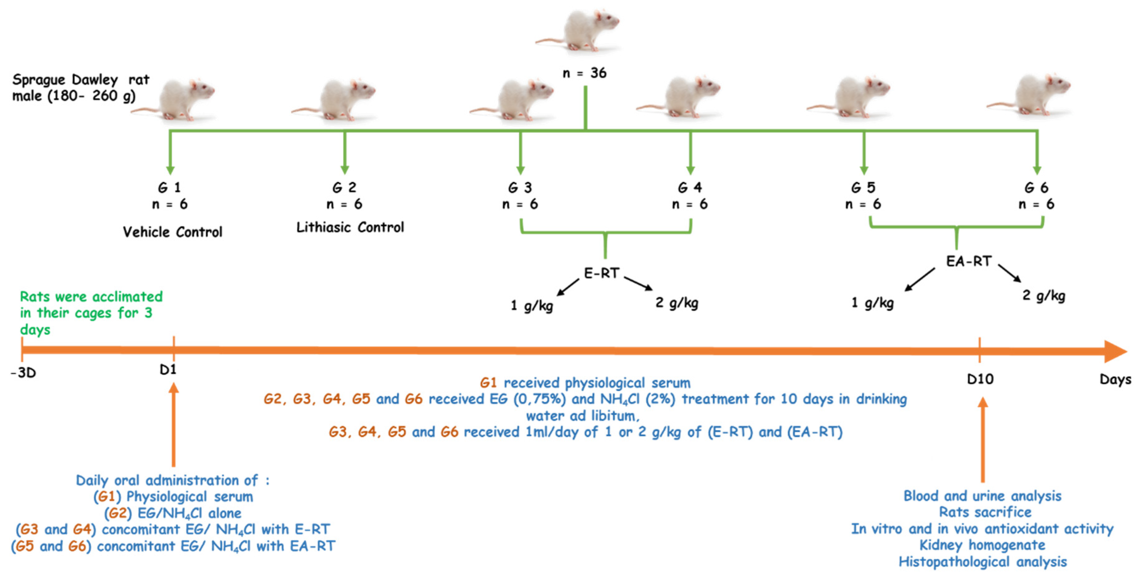
The results of the present study show that in response to 0.75% ethylene glycol (EG)/2% ammonium chloride (AC) oral administration over a 10-day period, young male rats developed kidney stones (or urolithiasis) mainly composed of calcium oxalate (CaOx). As shown in the
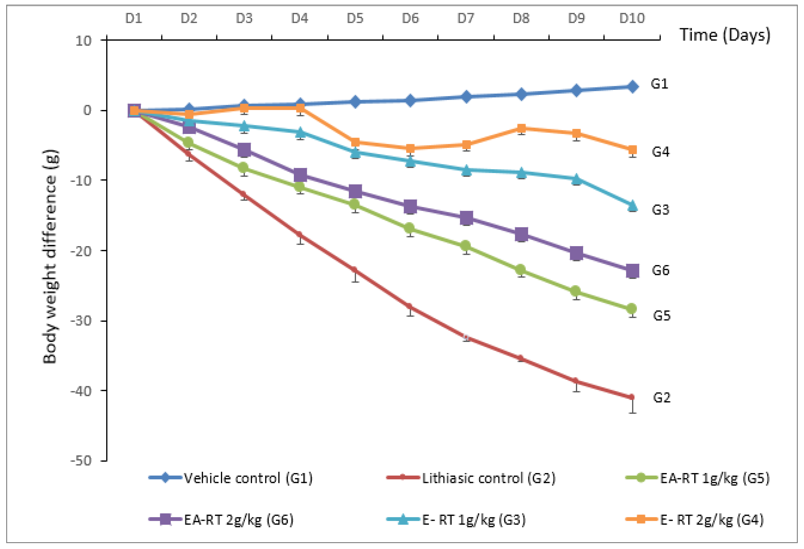
Figure 1, treated rats (urolithiasis control group or G2) lost body weight because they drank less water and almost stopped eating. The EG/AC model used in the present this study is similar to the previously described experimental models of urolithiasis by Ravindra and colleagues [
16]. The pathophysiological mechanisms responsible for the alterations elicited in this model could be related to an increase in the urinary oxalate (Ox) concentration. Indeed, EG is easily absorbed along the intestine and metabolized in the liver to Ox, leading to hyperoxaluria. Ox precipitates in the urine as CaOx because of its low solubility. High levels of Ox and CaOx crystals, particularly in the epithelial cells of the nephron, induce heterogeneous nucleation followed by crystal aggregation [
17]. AC potentiates the action of EG and accelerates the phenomenon of urolithiasis [
18].
Microscopic examination clearly showed that the treatment inducing urolithiasis led to the appearance of characteristic crystals of CaOx (i.e., with a bipyramid form) in the urine, while the urine of the untreated control group (G1) was free of these crystals. This result is similar to previously published data [
16] and may be associated with a decreased urinary output, an elevated pH, hyperoxaluria, and hypercalciuria [
19]. The biochemical analysis of the urine confirmed these results as lithiasic rats presented an increase in the excretion of phosphorus and calcium. A high concentration of phosphorus in the renal tubules could potentiate the Ox-induced lithiasis [
20], whereas calcium would act as an important factor in the nucleation and precipitation of Ox in the form of CaOx [
17] and in the resulting crystal growth [
20]. An increase in urinary protein excretion has also been recorded indicating proximal tubular dysfunction [
21]. Protein excretion could be related to severe lesions of the glomeruli and to tubular dilatation [
22]. Another factor contributing to protein urea could be an interstitial inflammation attested by mononucleated cells’ infiltration (
Table 3).
Serum levels of urea, creatinine, and uric acid were significantly increased in the urolithiasic group (G2) compared to the untreated control group (G1), indicating renal damage (
Table 1). These results are consistent with those of a previous study and indicate that the accumulation of nitrogenous substances in the serum may be a consequence of a decreased glomerular filtration rate (GFR) due to lithiasic obstruction [
23]. Uric acid binds to CaOx and modulates its crystallization and solubility and also reduces the inhibitory activity of glycosaminoglycans [
20]. Na
+, K
+, and Cl
− plasma concentrations were significantly increased in the lithiasic group (G2). Electrolyte imbalance disturbs the metabolism of the renal cells leading to the development of cell structure alterations [
24].
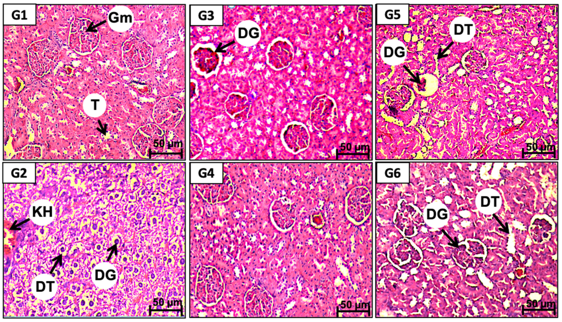
The EG/AC treatment caused tubular dilation, glomeruli lesions, and mononucleocyte infiltration. These renal damages observed in the lithiasic group (G2) could be attributed to a peroxidative action on the renal epithelium resulting from the elevated rate of urinary Ox and its deposition in the tubules and glomeruli. Indeed, CaOx deposition was shown to induce oxidative stress, which could be responsible for papillary tissue lesions [
2].
In the present study, we investigated the antilithiasic activities of ethanol and ethyl acetate extract of
Rubia tinctorum L. (RT) on EG/AC-induced renal lithiasis in rats. RT is one of the several medicinal plants that are widely used in traditional medicine systems to cure various ailments. The plant has been extensively studied for its biological activities and therapeutic potentials such as anti-inflammatory, anti-aggregant, antioxidant and antibacterial properties [
14,
25,
26]. In traditional medicine, RT dried roots are used for treatment of cardiovascular diseases including high blood pressure [
15,
27] liver pain, anemia and diarrhea [
28,
29]. Moreover, several studies have described the nephro-protective effects of plants [
9]. However, to our knowledge, no previous studies have demonstrated an anti-urolithiasic activity of RT.
A previous toxicological experiment, conducted in our laboratory, demonstrated that up to 5 g/kg of the RT extracts, administrated orally, triggered no major side effects [
26]. Therefore, the selected doses of 1 g/kg and 2 g/kg were free of any toxicological effect.
Treatment with E-RT and EA-RT prevented the alterations induced by EG/AC to values close to the untreated control group (G1). This preventive effect concerned the formation of crystals in the urine and the biochemical parameters of the serum and urine. RT extracts also prevented the body weight loss induced by the lithiasic treatment in a dose-dependent manner (
Figure 1). This finding is similar to that obtained with a standardized extract of fenugreek seed [
30] that was linked to an improvement in diuresis which resulted in the dissolution of the formed calculus and an interruption in the process of aggregation and deposition of the additional crystals. E-RT and the higher dose of EA-RT prevented the increase in serum urea, creatinine, and uric acid levels, probably by preserving a normal GFR. Electrolyte (calcium, phosphorus, K
+, Na
+, and Cl
−) concentration enhancements were also inhibited by the RT extracts. Maintaining electrolytic balance may, therefore, result in the preservation of cell metabolism.
E-RT (2 g/kg) was the most effective in decreasing levels of urinary proteins and creatinine and restored EG/AC-induced low diuresis by improving the GFR. RT extracts also significantly reduced the levels of phosphorus, sodium, potassium, calcium, and uric acid. EA-RT (2 g/kg) had a lower beneficial effect and its action was limited to recovery of the GFR and the inhibition of stone formation. Of note, the lower dose (1 g/kg) of both EA-RT and E-RT was less effective than the higher dose 2 g/kg in inhibiting EG/AC-induced lithiasic effects as documented by biochemical changes in
Table 1 and
Table 2.
Histopathological analyses showed concordant results with biochemical changes. RT extracts’ dose consistently prevented the degenerative changes in kidney tissues that could be induced by EG/AC. Interestingly, RT extracts’ preventive effects depended on the type of extract. E-RT (2 g/kg) was the most effective in protecting from EG/AC-induced disorganization in kidney architecture even though no differences were found between EA-RT and E-RT at 2 g/kg regarding their protective effect on calcium oxalate urolithiasis formation. Results obtained with RT extracts were similar to those previously obtained with
Acorus calamus ethanolic extract [
22],
Peucedanum grande hydroalcoholic extract [
31], and cystone [
32]. Biochemical and histopathological improvements in lithiasic animals observed after treatment with several plant extracts were proposed to be directly related to their antioxidant capacity. Antioxidants could have an important action in preventing the formation of the intrapapillary calcifications induced by oxidative stress that lead to papillary calculi formation [
11].
To verify the antioxidant preventing effect hypothesis, the antioxidant properties of RT extracts were evaluated both in vitro and in vivo.
The in vitro assays showed a promising antioxidant activity of both E-RT and EA-RT extracts. As documented in the DPPH scavenging, reducing power and ß-carotene assays, the E-RT extract has a better antioxidant activity compared to EA-RT extract. However, this effect is less important than reference antioxidant agents (
Table 5).
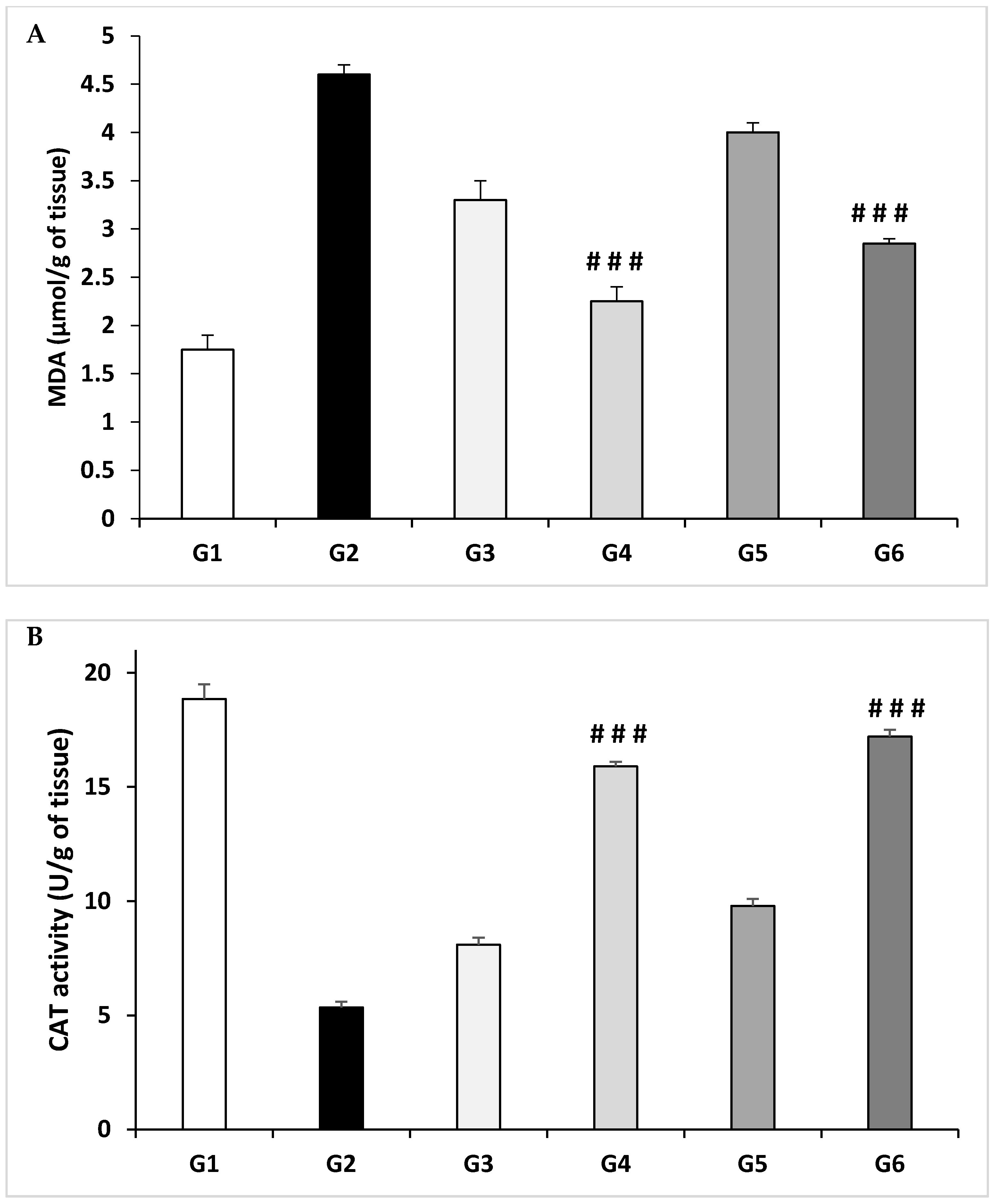
In vivo, EG treatment increased the level of MDA and significantly decreased the activity of the catalase enzyme in kidneys compared to the untreated control group (G1) (
Figure 3). Indeed, elevated free radical production, as observed by an increase in MDA level due to EG ingestion in the formation of nephrolithiasis, confirms that kidney tissue is under oxidative stress. This hypothesis is strengthened by the report that patients with kidney stones have less activity of antioxidant enzymes with increased lipid peroxidation [
33].
Oxidative damages, as reflected by higher lipid peroxidation (MDA) and lower antioxidant enzymes activity such as catalase, deteriorate kidney structure and functions as observed in calculi-induced rats. Antioxidant and reactive oxygen scavengers have been shown to be effective for protecting the kidney in animals [
33]. Here, treatment with E-RT and EA-RT showed an increase in catalase enzyme activity and a decrease in MDA levels in a dose-dependent manner in kidney homogenates.
Overall, our results present evidence that Rubia tinctorum L. extracts exhibited a marked protective effect against oxidative stress both in vitro and in vivo.
Rubia tinctorum L. extract contain large amounts of antioxidants which can play an important role in adsorbing and neutralizing free radicals, quenching oxygen, or decomposing peroxides. This antioxidant activity may be due, in a large part, to the specific polyphenolic composition identified by the HPLC analysis (
Table 6). Indeed, a positive correlation exists between total phenolic contents and the antioxidant capacity [
34]. This correlation is confirmed here, as qualitative and quantitative analysis of the RT extracts’ composition showed that E-RT, which contained more various polyphenols at higher concentrations, has the most powerful antioxidant effect. The antioxidant potential of E-RT could be considered quite significant compared to the powerful phenol, gallic acid (this study) but it is important compared to extracts from other plants [
35]. Analysis of the specific composition of each RT extract lead to the conclusion that E-RT antioxidant activity may be due to vanillin, rosmarinic acid, quercetin, catechin, syringic acid and cinnamic acid; while EA-RT antioxidant activity may be due to quercetin, cinnamic acid, vanillin and rutin. Several polyphenols with an antioxidant capacity were found to possess an antiurolithiasic preventing effect. As a matter of fact, quercetin antioxidant capacity was linked to a protective effect against oxidative stress associated with renal failure in the EG/AC model [
29,
36], to decreased oxidation of DNA bases [
37] and to lead-induced DNA damage prevention and apoptosis [
36]. There is also evidence that catechin has a preventive effect on renal calcium crystallization in vitro and in the EG model [
38]. Vanillin and cinnamic acid may also contribute to an additional preventive effect as both compounds have antioxidant potential [
39]. Therefore, it could be suggested that the superiority of E-RT over EA-RT related to their antioxidant action and prevention from urolithiasis is due to their richer polyphenolic constitution and their synergism.
These results are consistent with other studies that revealed that another species of the
Rubiaceae family,
Rubia cordifolia L., showed a great protective potential against different kidney and urinary disorders [
21,
40,
41]. However, to our knowledge, for
Rubia tinctorum L. this is the first time that a study reports such a preventive effective in urolithiasis.


 ,
, 


