Article

Andrographolide Inhibits Biofilm and Virulence in Listeria monocytogenes as a Quorum-Sensing Inhibitor

1
School of Life Sciences & Basic Medicine, Xinxiang University, Xinxiang 453000, China
2
Key Laboratory of Biomedicine and Health Risk Warning of Xinxiang City, Xinxiang University, Xinxiang 453000, China
3
Henan Engineering Laboratory for Bioconversion Technology of Functional Microbes, College of Life Sciences, Henan Normal University, Xinxiang 453007, China
*
Author to whom correspondence should be addressed.
Molecules 2022, 27(10), 3234; https://doi.org/10.3390/molecules27103234
Submission received: 26 April 2022 / Revised: 14 May 2022 / Accepted: 17 May 2022 / Published: 18 May 2022
(This article belongs to the Special Issue Functional and Bioactive Properties of Foods and Natural Products)

Abstract

:
Listeria monocytogenes is a major foodborne pathogen that can cause listeriosis in humans and animals. Andrographolide is known as a natural antibiotic and exhibits good antibacterial activity. We aimed to investigate the effect of andrographolide on two quorum-sensing (QS) systems, LuxS/AI-2 and Agr/AIP of L. monocytogenes, as well as QS-controlled phenotypes in this study. Our results showed that neither luxS expression nor AI-2 production was affected by andrographolide. Nevertheless, andrographolide significantly reduced the expression levels of the agr genes and the activity of the agr promoter P2. Results from the crystal violet staining method, confocal laser scanning microscopy (CLSM), and field emission scanning electron microscopy (FE-SEM) demonstrated that andrographolide remarkably inhibited the biofilm-forming ability of L. monocytogenes 10403S. The preformed biofilms were eradicated when exposed to andrographolide, and reduced surviving cells were also observed in treated biofilms. L. monocytogenes treated with andrographolide exhibited decreased ability to secrete LLO and adhere to and invade Caco-2 cells. Therefore, andrographolide is a potential QS inhibitor by targeting the Agr QS system to reduce biofilm formation and virulence of L. monocytogenes.

1. Introduction

Listeria monocytogenes, a Gram-positive bacterium, is recognized as an important foodborne pathogen that can cause listeriosis, a severe invasion infection in humans and animals [1]. L. monocytogenes appears to be widely distributed in nature and can easily enter food processing environments. The ability of L. monocytogenes to persist for long periods in equipment and environments of the food industry increases the risk of contaminated food and outbreaks of listeriosis. The typical clinical symptoms of listeriosis include septicemia, meningitis, abortion, and neonatal death [2]. Listeriosis is a rare but serious disease with high case-fatality rates [3]. Food contaminated by L. monocytogenes is recognized as the main vehicle for acquiring listerial infections [4]. Therefore, controlling L. monocytogenes in the food industry is an important issue for food safety and public health.
Quorum sensing (QS) is a cell-to-cell communication process that allows bacteria to behave coordinately through the exchange of extracellular signaling molecules called autoinducers [5]. Once the concentration of autoinducer reaches a certain threshold, QS is activated and regulates the expression of a specific set of genes [5]. Therefore, QS is also an important regulatory mechanism in bacteria, and it controls a variety of physiological activities such as bioluminescence, biofilm formation, and virulence [6,7,8].
In L. monocytogenes, two QS systems, LuxS/autoinducer 2 (AI-2) and Agr/autoinducing peptide (AIP), have been identified. The LuxS/AI-2 appears in many species of Gram-negative and Gram-positive bacteria [9]. LuxS is the key enzyme in the AI-2 biosynthesis pathway, and it is highly conserved in bacteria [10]. Thus, AI-2 is considered a QS signaling molecule for interspecies communication. In L. monocytogenes, LuxS is involved in the repression of biofilm formation [11,12]. The Agr/AIP consists of the four-gene operon agrBDCA, whose expression is driven by the P2 promoter upstream of agrB [13]. Many studies have suggested that the Agr system contributes to biofilm formation and virulence in L. monocytogenes [13,14,15]. As QS is vital in the regulation of many important phenotypes of bacteria, interfering with the QS networks is considered to be a potential way to prevent food contamination and human infection. Although several synthetic and natural compounds, such as 4-hydroxy-2,5-dimethyl-3(2H)-furanone, ambuic acid, and lactocin AL705, have been found to be capable of inhibiting the QS systems in L. monocytogenes [16,17,18], it is still urgent to search for more novel QS inhibitors.
Andrographis paniculata (Burm. F) Nees is a well-known traditional medicinal and edible plant in China. Andrographolide (its chemical structure is presented in Figure 1A), a diterpene lactone, is the major bioactive component of A. paniculata. Andrographolide has attracted much attention because of its multiple medicinal properties, such as antibacterial and anti-inflammatory activities [19]. Recently, several studies have demonstrated that andrographolide can inhibit the virulence and pathogenicity of Pseudomonas aeruginasa and Escherichia coli by interfering with the bacterial QS system [20,21]. However, its effect on the QS systems of L. monocytogenes is still unknown.
Therefore, the aim of this study was to investigate the QS inhibitory activity of andrographolide against L. monocytogenes. The effects of andrographolide on biofilm formation and mature biofilms were evaluated by identifying changes in biofilm biomass, morphology, and architecture. Swimming and swarming motilities of L. monocytogenes were measured in the presence of andrographolide. In addition, the effects of andrographolide treatment on the secretion of hemolysin, as well as on the ability to adhere to and invade Caco-2 cells, were also investigated to determine its anti-infective activity.

2. Results

2.1. Determination of Minimum Inhibitory Concentration (MIC) of Andrographolide

In this study, the antimicrobial susceptibility of L. monocytogenes to andrographolide was assessed, and the concentration range of andrographolide was 0.25–3 mg/mL with 0.5 mg/mL steps. The MIC of andrographolide against L. monocytogenes 10403S was 2 mg/mL.

2.2. Growth in the Presence of Andrographolide

Growth curves of L. monocytogenes 10403S in the presence of sub-MICs of andrographolide are shown in Figure 1B. Our results showed that andrographolide at concentrations of 1 mg/mL or below had no inhibitory effect on bacterial growth compared with the control.

2.3. Effect of Andrographolide on luxS Expression and AI-2 Production

As shown in Figure 2A, the presence of andrographolide did not result in a significant change in the expression level of luxS. Results from the bioluminescence assay confirmed that L. monocytogenes 10403S had the ability to produce AI-2 (Figure 2B). However, the addition of sub-MICs of andrographolide did not significantly affect AI-2 production (Figure 2B).

2.4. Effect of Andrographolide on agr Expression and the agr Promoter (P2) Activity

The expression levels of agrBD, agrC, and agrA genes were significantly decreased (p < 0.05) in L. monocytogenes 10403S after exposure to andrographolide (Figure 2A). The P2 activity was determined by the β-galactosidase assay in this study. As shown in Figure 2C, andrographolide treatment at 0.125, 0.25, 0.5, and 1 mg/mL reduced the P2 activity by approximately 26%, 29%, 36%, and 46%, compared with the control. These data demonstrated that andrographolide possesses anti-QS capacity by inhibiting the Agr/AIP system in L. monocytogenes.

2.5. Inhibition of Biofilm Formation by Andrographolide

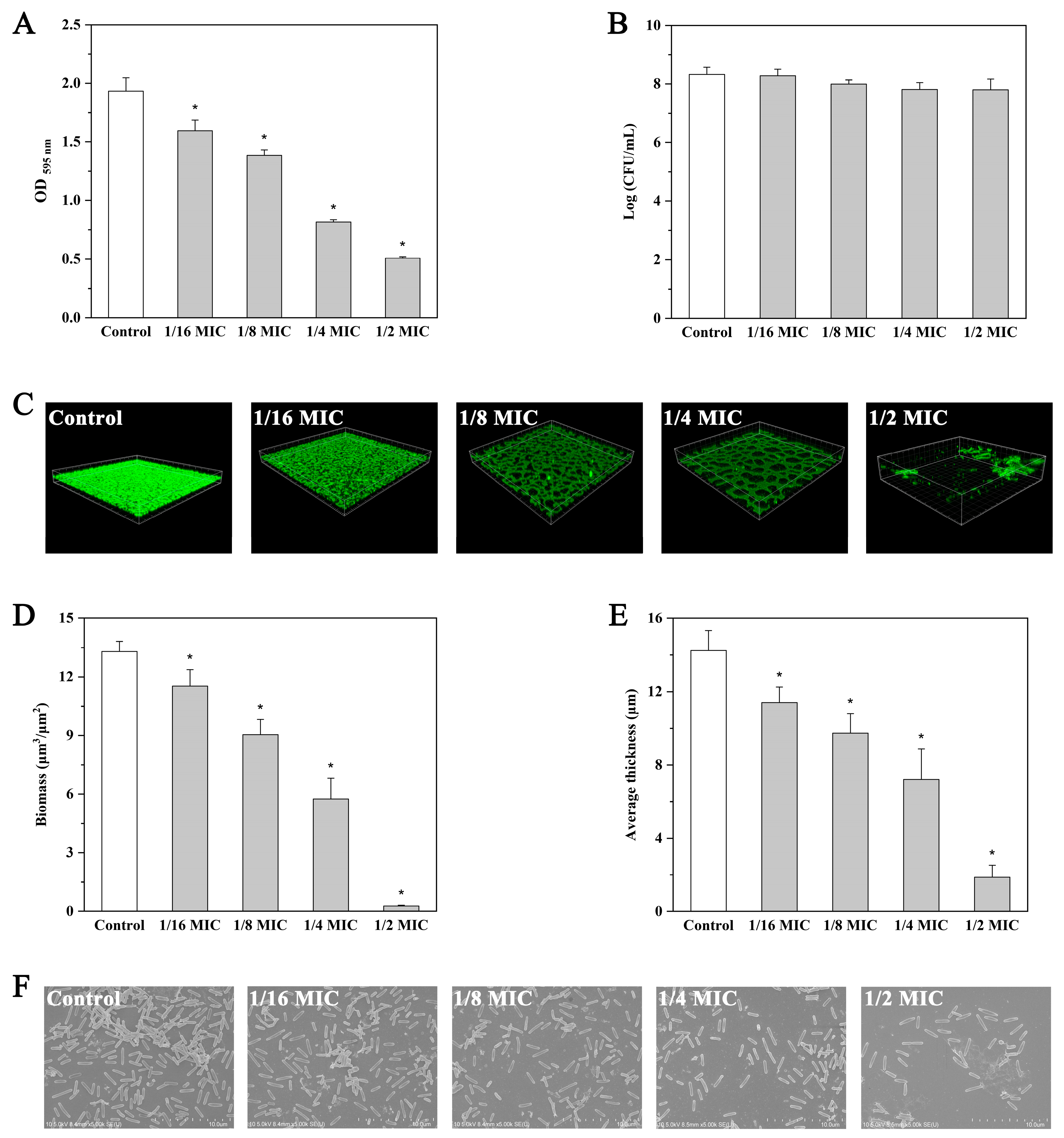
Treatment with andrographolide at concentrations of 0.125, 0.25, 0.5, and 1 mg/mL significantly reduced (p < 0.05) the biofilm biomass by 17.5%, 28.3%, 57.7%, and 73.7%, respectively (Figure 3A). The difference in biofilm-forming ability is not due to a difference in growth because the addition of andrographolide had no effect on the viability of planktonic cells compared with that of the control (Figure 3B).
Confocal laser scanning microscopy (CLSM) and field emission scanning electron microscopy (FE-SEM) were applied to observe the morphology of L. monocytogenes biofilms. CLSM images showed a compact biofilm structure in the control (Figure 3C–E). In contrast, reduced thickness of biofilms was observed following andrographolide treatment (Figure 3C–E). Similar results were also obtained with the FE-SEM images, in which biofilms showed a more scattered appearance in the presence of andrographolide (Figure 3F).

2.6. Effect of Andrographolide on Preformed Biofilms

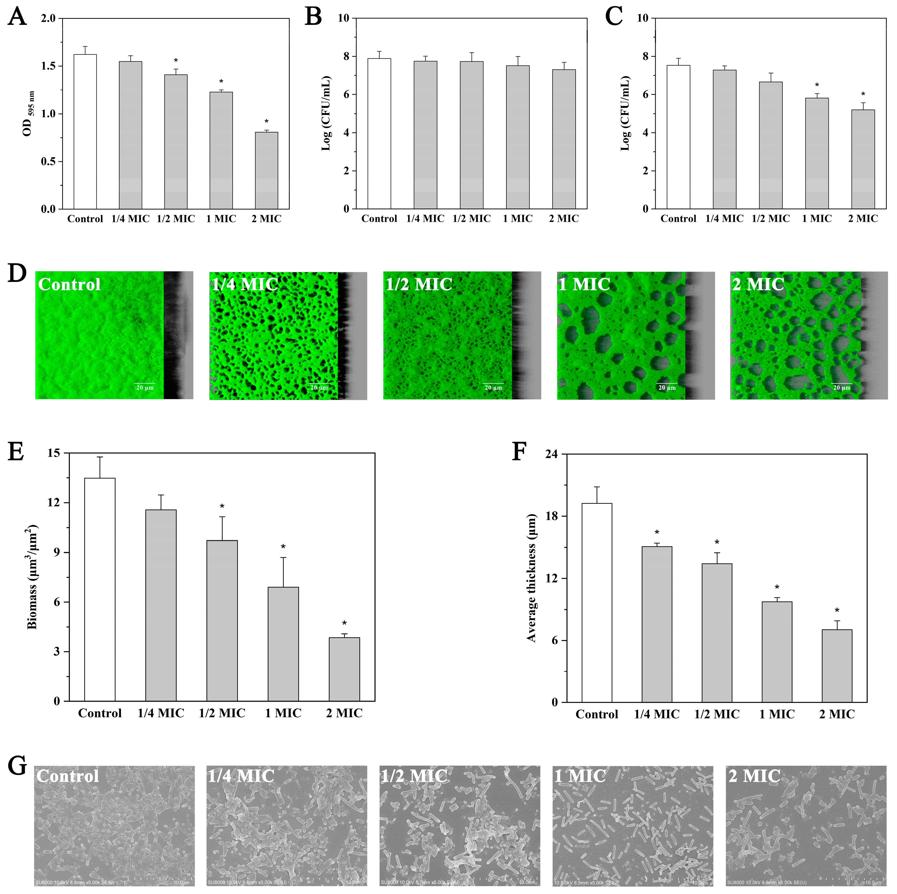
In the present study, andrographolide at sub-MICs (0.5 and 1 mg/mL) and inhibitory concentrations (2 and 4 mg/mL) was used to investigate the effect of andrographolide on mature biofilms. As shown in Figure 4A, andrographolide at 0.5 mg/mL (1/4MIC) resulted in only a minor reduction (4.6%) in the preformed biofilms (p > 0.05). When treated with andrographolide at 1 (1/2MIC), 2 (MIC), and 4 mg/mL (2MIC), mature biofilms were eradicated by 13.2%, 24.3%, and 50.2%, respectively (p < 0.05). The counting results of surviving cells in treated biofilm (Figure 4B) further confirmed the eradication effect of andrographolide at concentrations above 1/2MIC on preformed biofilms of L. monocytogenes 10403S. Our results also showed that the viability of planktonic cells in the culture supernatant was not influenced by andrographolide (0.5 to 4 mg/mL; Figure 4C).
In addition to quantitative analysis, treated biofilms were also visualized using CLSM and FE-SEM. CLSM images showed complete and uniformly distributed biofilm in the control experiment, whereas andrographolide treatment, especially at 2 and 4 mg/mL, significantly reduced the number of attached cells (Figure 4D–F). The FE-SEM images also indicated decreased adherence in the andrographolide-treated biofilms (Figure 4G).

2.7. Effect of Andrographolide on Motility

In this study, swimming and swarming motilities of L. monocytogenes 10403S grown with andrographolide (0.125, 0.25, 0.5, and 1 mg/mL) were evaluated. After incubation at 25 °C, no significant difference (p > 0.05) in the mean diameter of the swim and swarm ring was observed between the andrographolide treatment group and the control (Figures S1 and S2). Expression levels of the flagella gene (flaA) and motility-related genes (motA and motB) were not affected by andrographolide (Figure S3). These data suggest that andrographolide has no effect on swimming and swarming motilities of L. monocytogenes.

2.8. Effect of Andrographolide on Virulence Factors

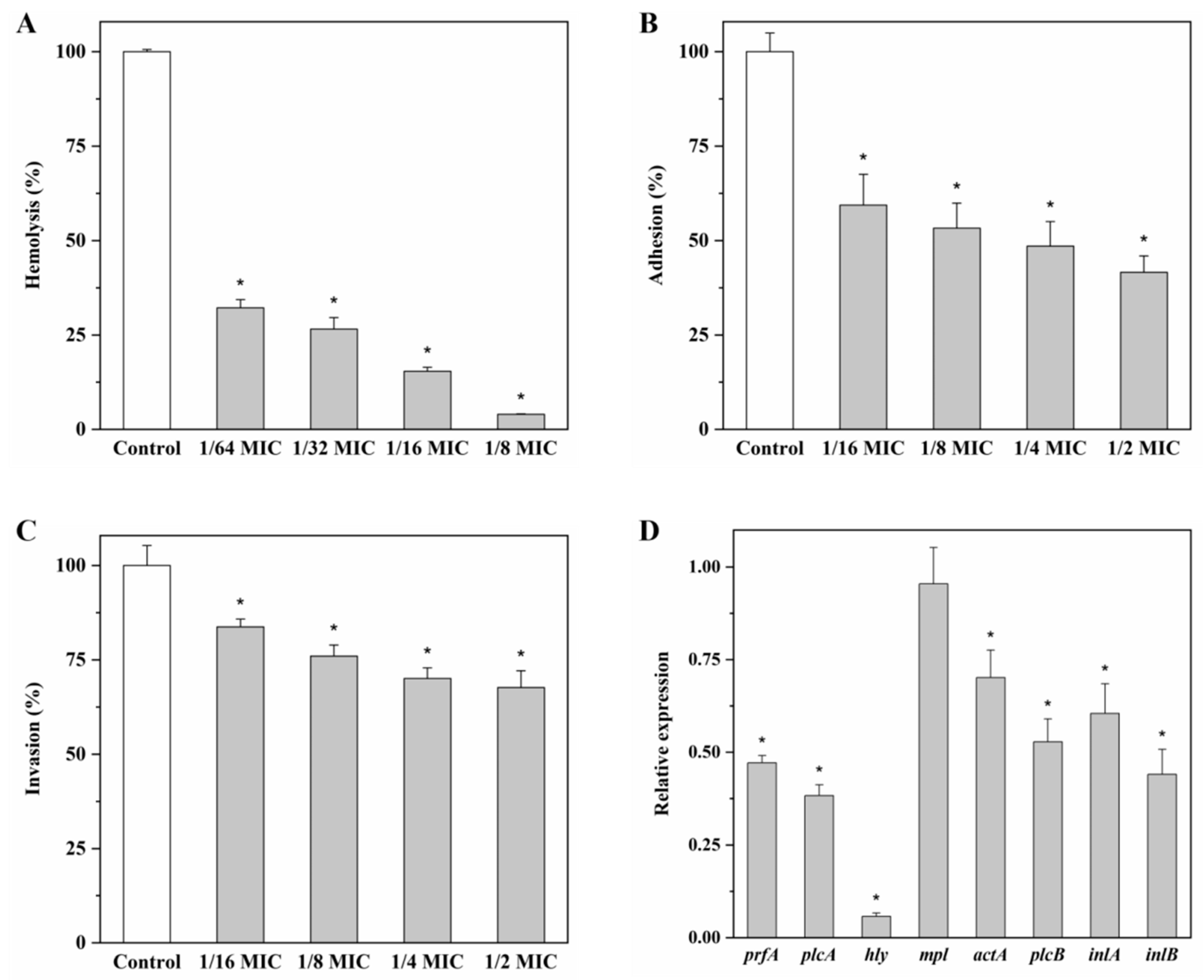
As shown in Figure 5A, the hemolytic activity of L. monocytogenes 10403S was significantly reduced (p < 0.05) by the addition of andrographolide. When treated with andrographolide at 0.03125, 0.0625, 0.125, and 0.25 mg/mL, the hemolysis rate of 10403S was 32.2%, 26.6%, 15.4%, and 4.0%, respectively.
The capacity of L. monocytogens 10403S to attach and invade Caco-2 cells was also determined in the presence or absence of andrographolide. When treated with andrographolide at 0.125, 0.25, 0.5, and 1 mg/mL, adhesion of 10403S was decreased to 59.4%, 53.3%, 48.5%, and 41.6%, respectively (Figure 5B); the invasion rate was reduced to 83.8%, 76.0%, 70.1%, and 67.7%, respectively (Figure 5C).
Expression levels of virulence-associated genes (prfA, plcA, hly, mpl, actA, plcB, inlA, and inlB) were investigated in this study. All virulence genes tested, except mpl, were down-regulated (p < 0.05) after treatment of andrographolide (Figure 5D).

3. Discussion

QS regulates many important physiological processes in several pathogenic bacteria, such as biofilm formation and the production of virulence factors. L. monocytogenes has been considered an important foodborne pathogen all over the world, and it has the ability to form biofilms in food-processing environments [22,23]. L. monocytogenes growing in biofilms is significantly more resistant to disinfection than its free-living counterparts, leading to the increased risk of food contamination by this bacterium. Consumption of L. monocytogenes-contaminated food may cause listeriosis, which threatens public health. Therefore, controlling biofilm formation and virulence of L. monocytogenes is one of the most important issues in the food industry. Application of QS inhibitor to decrease biofilm formation and virulence by blocking QS is considered to be an effective risk-reducing way of food contamination by L. monocytogenes.
Andrographolide exhibits not only excellent antibacterial activity but also inhibitory effects on QS. Guo et al. (2014) have reported that andrographolide interferes with the LuxS/AI-2 QS system in avian pathogenic E. coli and inhibits the AI-2 activity [21]. P. aeruginosa has two acylated homoserine lactone (AHL)-based QS systems, LasI/LasR and RhlI/RhlR. It has been reported that 14-alpha-lipoyl andrographolide, a derivative of andrographolide, represses the expression levels of QS genes and AHL production, and consequently, biofilm formation regulated by QS systems is inhibited in P. aeruginosa PAO1 [20].
In the present study, the effects of andrographolide on two QS systems were investigated in Gram-positive bacterium L. monocytogenes. Unlike E. coli, the LuxS/AI-2 system of L. monocytogenes was not affected by andrographolide. Neither luxS expression nor AI-2 production changed after exposure to this agent. In E. coli, LsrB, the receptor for signaling molecule AI-2, has been identified; however, a similar receptor has not been reported in L. monocytogenes [24]. Some scholars support the view that AI-2 plays the role of metabolite rather than a QS signaling molecule in L. monocytogenes [11,12]. Thus, it is not surprising that andrographolide exhibited different effects on the LuxS/AI-2 systems in E. coli and L. monocytogenes. Interestingly, the presence of andrographolide significantly reduced the expression levels of the agr genes and the activity of the agr promoter P2, indicating that andrographolide is a potential QS inhibitor targeting the Agr/AIP system in L. monocytogenes. To our knowledge, the Agr/AIP system is a typical QS system in Gram-positive bacteria. In L. monocytogenes, AgrD, a precursor peptide, is processed into an active signaling molecule AIP by AgrB, and then, AIP is released outside the cells. When the concentration of AIP in the extracellular space reaches a certain threshold, it activates the two-component system consisting of AgrC (histidine kinase) and AgrA (response regulator) by binding specifically to AgrC. AgrA not only regulates the expression of target genes but also autoregulates the agr operon via driving the P2 promoter upstream of agrB. So how does andrographolide interfere with the Agr/AIP system? Is it by inhibiting AIP biosynthesis, by disrupting the interaction between AIP and AgrC, or by other means? Further studies are needed to clarify the mechanism of andrographolide in the Agr QS system of L. monocytogenes.
We tried to investigate the effect of andrographolide on AIP production by quantifying actual AIP concentrations in bacterial culture supernatants using high-performance liquid chromatography (HPLC) but failed. The interference of the complex components and the high levels of peptides in the BHI medium may be one of the reasons [25]. It is predicted that peptide-signaling molecules are more metabolically costly, and their concentrations in bacterial culture were much lower than other types of signaling molecules [26]. Therefore, there is also the possibility that L. monocytogenes produce a very small amount of AIP, which also makes it difficult to quantify AIP by HPLC. Since andrographolide can block the Agr system, the phenotype controlled by this system may also be affected by andrographolide. Previous studies have confirmed positive regulation by the Agr system on biofilm formation in L. monocytogenes [14]. Results from the crystal violet staining method, CLSM, and FE-SEM demonstrated that the addition of andrographolide remarkably inhibited the biofilm-forming ability of L. monocytogenes 10403S. The mature biofilms formed by L. monocytogenes are difficult to remove even after routine cleaning and disinfection procedures. Therefore, the effects of andrographolide on preformed biofilms were also evaluated in this study. After exposure to andrographolide, decreased biofilm biomass and surviving cells were observed in treated biofilms. Andrographolide could not only destroy the structure of mature biofilms but also reduce the number of bacterial cells in biofilms.
Flagellum-mediated motilities, swimming motility, and swarming motility play a significant role in the first stages of biofilm formation [27]. It has been reported that these flagellar motility genes (flaA, motA, and motB) play important roles in the biofilm formation of L. monocytogenes [28,29]. In the current study, swimming and swarming motilities and expression of flagellar motility genes were not influenced by andrographolide.
L. monocytogenes is an intracellular pathogen that can cause severe invasive infections in humans. In fact, the process of L. monocytogenes infection of host cells is regulated by virulence factors [30]. Given that the Agr system is associated with virulence and invasion of L. monocytogenes, effects of andrographolide on some key virulence factors were also investigated in the current study. Listeriolysin O (LLO) encoded by the hly gene is a Listeria-specific hemolysin [31]. Our results showed that andrographolide significantly reduced the hemolytic activity of LLO. Notably, the LLO activity was almost completely inhibited in the presence of andrographolide at 0.25 mg/mL (1/8MIC), suggesting the excellent inhibitory effect of andrographolide at low concentration on the LLO activity. As a result, concentrations below 0.25 mg/mL were selected to evaluate the inhibitory effect of andrographolide on the LLO activity. In addition, the ability of L. monocytogenes to adhere to and invade Caco-2 cells was also suppressed when exposed to andrographolide at concentrations ranging from 0.125 to 1 mg/mL. Consistently, most virulence genes tested in our study were down-regulated after treatment of andrographolide.
Andrographolide is considered a potential QS inhibitor for some Gram-negative bacteria. The effect of andrographolide on QS systems of L. monocytogenes was investigated in our study. Andrographolide interfered with the Agr QS system of L. monocytogenes and consequently inhibited bacterial behaviors regulated by this QS system. L. monocytogenes treated with andrographolide exhibited decreased ability to form biofilms, secrete LLO, and adhere to and invade Caco-2 cells. Therefore, andrographolide has the potential to control L. monocytogenes contamination in the food industry as a QS inhibitor.

4. Materials and Methods

4.1. Bacterial Strains and Growth Conditions

Wild-type L. monocytogenes 10403S was obtained as a gift from Prof. Qin Luo and was grown in brain heart infusion (BHI; Oxoid Ltd., Basingstoke, UK) broth at 37 °C. E. coli DH5α was purchased from Bomaide Gene Technology Co., Ltd. (Beijing, China) and was grown in Luria-Bertani (Huankai Ltd., Guangzhou, China) broth. Vibrio harveyi BB170, an AI-2 reporter strain, was kindly provided by Xiangan Han (Shanghai Veterinary Research Institute) and was grown in AB medium at 30 °C.

4.2. Determination of MIC of Andrographolide

Andrographolide (purity > 98%, CAS 5508-58-7) was purchased from Shanghai Macklin Biochemical Co., Ltd. (Shanghai, China). Stock solutions were prepared by dissolving andrographolide in dimethyl sulfoxide (DMSO, Macklin, Shanghai, China). The final concentration of DMSO in all samples was 0.1%, which had no significant effect on the growth of L. monocytogenes 10403S (data not shown). The MIC of andrographolide against L. monocytogenes 10403S was determined using the agar dilution method, as described previously [32]. Bacterial cultures were diluted using sterilized saline solution to approximately 107 CFU/mL. One microliter of suspension was inoculated on Mueller–Hinton agar (MHA; Huankai, Guangzhou, China). MHA containing DMSO was used as the blank control. MHA containing DMSO inoculated with bacterial culture was used as the negative control. The MIC was defined as the lowest concentration of andrographolide that prevented the visible growth of L. monocytogenes.

4.3. Growth Curve Analysis

For growth measurement, L. monocytogenes 10403S was inoculated into BHI broth in the absence or presence of andrographolide (0.125, 0.25, 0.5, and 1 mg/mL). BHI with DMSO served as the negative control. The samples were incubated in a Bioscreen C microbiology reader (Growth Curves, Helsinki, Finland) at 37 °C for 24 h, with OD600nm measurements collected hourly and used to generate bacterial growth curves.

4.4. Gene Expression Analyses

Quantitative real-time PCR (qRT-PCR) was performed to measure the relative expression levels of the target genes using the LightCycler 96 real-Time PCR system (Roche, Basel, Switzerland), as described previously [33]. Primers for eight virulence genes are listed in Table S1, and other primers have been reported in our previous study [33]. To assess the effect of andrographolide on gene expression, L. monocytogenes 10403S was grown in BHI supplemented with or without andrographolide (1 mg/mL). 16S rRNA was used as a reference gene, and the fold changes of the target genes were calculated using the 2−ΔΔCt method.

4.5. Detection of AI-2

The bioluminescence assay was used to detect AI-2 as described previously [34] using an EnSpire Multimode Plate Reader (PerkinElmer, Waltham, MA, USA). To investigate the effect of andrographolide on AI-2 production in L. monocytogenes 10403S, bacteria were incubated in BHI broth with different concentrations of andrographolide (0.125, 0.25, 0.5, and 1 mg/mL), and the medium containing DMSO was used as the blank control. Results were normalized to the DMSO control.

4.6. Determination of the agr Promoter P2 Activity

The activity of the agr promoter P2 was determined in this study. The agr promoter (P2)-lacZ fusion was constructed as described previously [35,36]. β-galactosidase activity assay was performed based on the method by Miller [37].

4.7. Effect of Andrographolide on Biofilm Formation by L. monocytogenes

Biofilms of L. monocytogenes were measured using the microplate assay with crystal violet staining in 96-well polystyrene plates (Costar 3599; Corning Inc., Kennebunk, ME, USA), as described previously [38]. Briefly, overnight cultures of L. monocytogenes were diluted at 1:100 in fresh BHI broth, and andrographolide was added to the cultures to achieve final concentrations of 0, 0.125, 0.25, 0.5, and 1 mg/mL. BHI with DMSO served as the blank control. Two hundred microliters of bacterial cultures were transferred into wells of the microtiter plate. The plates were incubated at 37 °C for 48 h without agitation. To determine the growth of planktonic cells, 100 μL of cultures were centrifuged, and the pellets were resuspended in 1 mL of sterile saline. The bacterial cultures were 10-fold serially diluted, and 100 μL volumes were taken for colony counting. To assess biofilm formation, the medium containing planktonic cells was removed after incubation. Then, biofilms were stained with 1% crystal violet solution for 45 min and then decolorized with 95% ethanol. The absorbance at OD595nm was measured to determine biofilm production.

4.8. Effect of Andrographolide on Preformed Biofilms by L. monocytogenes

L. monocytogenes biofilms were incubated as described above. After 48 h of incubation at 37 °C, the medium was removed, and wells were washed three times with sterile water. Then, fresh BHI broth supplemented with different concentrations of andrographolide was added to the wells, with DMSO serving as the control. Plates were incubated at 37 °C for 4 h. Planktonic cells were quantified as described above. To quantify biofilm biomass, biofilms were stained with crystal violet after removing the medium and then decolorized by 95% ethanol. The absorbance at OD595nm was measured. To quantify biofilm bacteria, the wells were washed several times with sterile water. Then, 200 μL of sterile saline was added to each well, and its surface was thoroughly scratched with a plastic pipette tip. Recovered biomasses were vortexed, serially diluted, and plated onto BHI agar. These plates were incubated at 37 °C for 24 h prior to enumeration by counting colonies.

4.9. CLSM Biofilm Formation Assay

Biofilm assay by CLSM was performed as described previously [33]. Biofilms were stained using the Live/Dead BacLight Bacterial viability kit (Molecular Probes, Eugene, OR, USA) and then observed by a Leica TCS-SP8 Confocal Laser Scanning Microscope (Leica-Microsystems, Wetzlar, Germany). To construct the three-dimensional projections of biofilms, the IMARIS 7.1 software (Bitplane, Zürich, Switzerland) was applied to process the CLSM acquisitions. Biofilm biomass and thickness were quantified by the COMSTAT software [39].

4.10. FE-SEM Analysis

FE-SEM (Hitachi SU8010; Hitachi, Tokyo, Japan) was applied to observe biofilms of L. monocytogenes as described previously [40]. After 48 h of incubation at 37 °C, L. monocytogenes biofilms were fixed in 4% glutaraldehyde and then washed with phosphate-buffered saline (PBS; pH 7.4). Samples were progressively dehydrated by passage through a graded series of ethanol solutions from 30% to 100%. The samples were coated with gold in an ion sputter coater (SBC-12; KYKY Technology Co. Ltd., Beijing, China) and observed with FE-SEM.

4.11. Motility

Swimming and swarming motilities of L. monocytogenes were tested in our study. Briefly, 1 μL of overnight L. monocytogenes 10403S culture was inoculated at the center of 0.3% and 0.5% tryptic soy broth (TSB) agar plate with different concentrations of andrographolide, respectively. Plates were cultivated at 25 °C, and the diameter of the bacterial swim and swarm was measured 48 h later.

4.12. Effect of Andrographolide on L. monocytogenes Key Virulence Factors

The hemolytic activity of L. monocytogenes was assayed as described previously [41]. The bacterial cultures were centrifuged (5500× g, 4 °C, 10 min), and 250 μL of supernatant was mixed with 900 μL of hemolysin buffer and 100 μL of sheep red blood cells. BHI broth and the sheep red blood cells treated with 1% Triton X-100 served as the negative control and positive control, respectively. The samples were determined by measuring the absorbance at OD543nm. The relative hemolysis was presented as a percentage of the positive control.
Adhesion and invasion assays were carried out as previously described [14]. Briefly, bacterial cultures of L. monocytogenes were mixed with Caco-2 cells at a multiplicity of infection (MOI) of 100 and then incubated at 37 °C for 1 h. For adhesion assay, cells were washed with pre-warmed PBS and then lysed with ice-cold distilled water. For invasion assay, cells were incubated in DMEM medium with 10 μg/mL gentamicin after washing once with pre-warmed PBS. The cells were washed and lysed according to the steps described above. The lysed cells were plated on BHI agar and incubated at 37 °C for 24 h.

4.13. Statistical Analysis

Statistical analysis was performed using the SPSS version 20.0 (SPSS Inc., Chicago, IL, USA). Differences were defined as significant at p < 0.05.

5. Conclusions

In summary, andrographolide reduced the expression levels of the agr genes and the activity of the agr promoter P2, suggesting the inhibitory effect of andrographolide on the Agr QS system. Andrographolide could not only inhibit biofilm formation but also remove mature biofilms of L. monocytogenes, indicating the anti-biofilm activity of andrographolide against L. monocytogenes. Additionally, the hemolytic activity and the ability to adhere to and invade Caco-2 cells of L. monocytogenes were suppressed by andrographolide. In L. monocytogenes, the Agr QS system is involved in biofilm formation and virulence. Thus, andrographolide is a potential QS inhibitor by targeting the Agr QS system to reduce biofilms and virulence of L. monocytogenes.

Supplementary Materials

The following supporting information can be downloaded at: https://www.mdpi.com/article/10.3390/molecules27103234/s1, Table S1: Primers for qRT-PCR used in this study. Figure S1: Effect of andrographolide on swimming motility of L. monocytogenes 10403S incubated at 25 °C. (a) control; (b–e) andrographolide (0.125, 0.25, 05 and 1 mg/mL). Figure S2: Effect of andrographolide on swarming motility of L. monocytogenes 10403S incubated at 25 °C. (a) control; (b–e) andrographolide (0.125, 0.25, 05 and 1 mg/mL). Figure S3: Relative expression levels of flagella gene and motility-related genes in L. monocytogenes. Results are presented as fold changes relative to the expression level of the target gene in L. monocytogenes 10403S in BHI. Error bars represent the standard deviation of triplicate experiments (n = 3). The asterisk indicates a value statistically different from that of the control, with p < 0.05.

Author Contributions

Conceptualization, T.Y. and X.J. (Xiaobing Jiang); methodology, X.X.; software, C.J.; validation, C.J. and R.K.; writing—original draft preparation, T.Y. and X.J. (Xiaojie Jiang); writing—review and editing, X.J. (Xiaobing Jiang); funding acquisition, T.Y., X.X., and X.J. (Xiaobing Jiang). All authors have read and agreed to the published version of the manuscript.

Funding

This research was funded by the Cultivation Fund of the National Scientific Research Project of Henan Normal University (20200148), the Natural Science Foundation of Henan Province (222300420470, 212300410220, and 202300410018), the Key Project of Natural Science of the Education Department of Henan Province (22A180003), and Henan Province Young Backbone Teacher Project (2021GGJS162).

Institutional Review Board Statement

Not applicable.

Informed Consent Statement

Not applicable.

Data Availability Statement

Not applicable.

Acknowledgments

We are grateful to Xiangan Han, Shanghai Veterinary Research Institute, for kindly providing V. harveyi BB170. We also thank Douwe van Sinderen for providing the pPTPL plasmid and Jean-Francois Collet for providing E. coli MC1000 (cloning host for pPTPL).

Conflicts of Interest

The authors declare no conflict of interest.

Sample Availability

Samples of the compounds are not available from the authors.

References

  1. Drevets, D.A.; Bronze, M.S. Listeria monocytogenes: Epidemiology, human disease, and mechanisms of brain invasion. FEMS Immunol. Med. Microbiol. 2008, 53, 151–165. [Google Scholar] [CrossRef] [PubMed] [Green Version]
  2. McLauchlin, J.; Mitchell, R.T.; Smerdon, W.J.; Jewell, K. Listeria monocytogenes and listeriosis: A review of hazard characterisation for use in microbiological risk assessment of foods. Int. J. Food Microbiol. 2004, 92, 15–33. [Google Scholar] [CrossRef]
  3. Clark, C.G.; Farber, J.; Pagotto, F.; Ciampa, N.; Dor, K.; Bernard, K.; Ng, L.K. Surveillance for Listeria monocytogenes and listeriosis, 1995–2004. Epidemiol. Infect. 2010, 138, 559–572. [Google Scholar] [CrossRef] [PubMed] [Green Version]
  4. Todd, E.C.D.; Notermans, S. Surveillance of listeriosis and its causative pathogen, Listeria monocytogenes. Food Control 2011, 22, 1484–1490. [Google Scholar] [CrossRef]
  5. Waters, C.M.; Bassler, B.L. Quorum sensing: Cell-to-cell communication in bacteria. Annu. Rev. Cell Dev. Biol. 2005, 21, 319–346. [Google Scholar] [CrossRef] [Green Version]
  6. Chong, G.; Kimyon, Ö.; Manefield, M. Quorum sensing signal synthesis may represent a selective advantage independent of its role in regulation of bioluminescence in Vibrio fischeri. PLoS ONE 2013, 8, e67443. [Google Scholar] [CrossRef]
  7. Hammer, B.K.; Bassler, B.L. Quorum sensing controls biofilm formation in Vibrio cholerae. Mol. Microbiol. 2003, 50, 101–104. [Google Scholar] [CrossRef]
  8. Chong, Y.M.; How, K.Y.; Yin, W.F.; Yin, W.F.; Chan, K.G. The effects of chinese herbal medicines on the quorum sensing-regulated virulence in Pseudomonas aeruginosa PAO1. Molecules 2018, 23, 972. [Google Scholar] [CrossRef] [Green Version]
  9. Schauder, S.; Shokat, K.; Surette, M.G.; Bassler, B.L. The LuxS family of bacterial autoinducers: Biosynthesis of a novel quorum-sensing signal molecule. Mol. Microbiol. 2001, 41, 463–476. [Google Scholar] [CrossRef]
  10. Pei, D.; Zhu, J. Mechanism of action of S-ribosylhomocysteinase (LuxS). Curr. Opin. Chem. Biol. 2004, 8, 492–497. [Google Scholar] [CrossRef]
  11. Belval, S.C.; Gal, L.; Margiewes, S.; Garmyn, D.; Piveteau, P.; Guzzo, J. Assessment of the roles of LuxS, S-ribosyl homocysteine, and autoinducer 2 in cell attachment during biofilm formation by Listeria monocytogenes EGD-e. Appl. Environ. Microbiol. 2006, 72, 2644–2650. [Google Scholar] [CrossRef] [PubMed] [Green Version]
  12. Sela, S.; Frank, S.; Belausov, E.; Pinto, R. A Mutation in the luxS gene influences Listeria monocytogenes biofilm formation. Appl. Environ. Microbiol. 2006, 72, 5653–5658. [Google Scholar] [CrossRef] [PubMed] [Green Version]
  13. Autret, N.; Raynaud, C.; Dubail, I.; Berche, P.; Charbit, A. Identification of the agr locus of Listeria monocytogenes: Role in bacterial virulence. Infect. Immun. 2003, 71, 4463–4471. [Google Scholar] [CrossRef] [PubMed] [Green Version]
  14. Riedel, C.U.; Monk, I.R.; Casey, P.G.; Waidmann, M.S.; Gahan, C.G.M.; Hill, C. AgrD-dependent quorum sensing affects biofilm formation, invasion, virulence and global gene expression profiles in Listeria monocytogenes. Mol. Microbiol. 2009, 71, 1177–1189. [Google Scholar] [CrossRef] [PubMed]
  15. Rieu, A.; Weidmann, S.; Garmyn, D.; Piveteau, P.; Guzzo, J. agr system of Listeria monocytogenes EGD-e: Role in adherence and differential expression pattern. Appl. Environ. Microbiol. 2007, 73, 6125–6133. [Google Scholar] [CrossRef] [Green Version]
  16. Wu, Y.; Liu, Y.; Zhang, Y.; Fang, Z.; Sun, L. Interferce effect of 4-hydroxy-2,5-dimethyl-3(2H)-furanone on AI-2 quorum sensing of Listeria monocytogenes. Sci. Technol. Food Ind. 2018, 39, 87–92. (In Chinese) [Google Scholar] [CrossRef]
  17. Todd, D.A.; Parlet, C.P.; Crosby, H.A.; Malone, C.L.; Heilmann, K.P.; Horswill, A.R.; Cech, N.B. Signal biosynthesis inhibition with ambuic acid as a strategy to target antibiotic-resistant infections. Antimicrob. Agents Chemother. 2017, 61, e00263-17. [Google Scholar] [CrossRef] [Green Version]
  18. Melian, C.; Segli, F.; Gonzalez, R.; Vignolo, G.; Castellano, P. Lactocin AL705 as quorum sensing inhibitor to control Listeria monocytogenes biofilm formation. J. Appl. Microbiol. 2019, 127, 911–920. [Google Scholar] [CrossRef]
  19. Chao, C.Y.; Lii, C.K.; Tsai, I.T.; Li, C.C.; Liu, K.L.; Tsai, C.W.; Chen, H.W. Andrographolide inhibits ICAM-1 expression and NF-κB activation in TNF-α-treated EA.hy926 cells. J. Agric. Food Chem. 2011, 59, 5263–5271. [Google Scholar] [CrossRef]
  20. Ma, L.; Liu, X.; Liang, H.; Che, Y.; Chen, C.; Dai, H.; Yu, K.; Liu, M.; Ma, L.; Yang, C.H.; et al. Effects of 14-alpha-lipoyl andrographolide on quorum sensing in Pseudomonas aeruginosa. Antimicrob. Agents Chemother. 2012, 56, 6088–6094. [Google Scholar] [CrossRef] [Green Version]
  21. Guo, X.; Zhang, L.Y.; Wu, S.C.; Xia, F.; Fu, Y.X.; Wu, Y.L.; Leng, C.Q.; Yi, P.F.; Shen, H.Q.; Wei, X.B.; et al. Andrographolide interferes quorum sensing to reduce cell damage caused by avian pathogenic Escherichia coli. Vet. Microbiol. 2014, 174, 496–503. [Google Scholar] [CrossRef] [PubMed]
  22. Di Bonaventura, G.; Piccolomini, R.; Paludi, D.; D’Orio, V.; Vergara, A.; Conter, M.; Ianieri, A. Influence of temperature on biofilm formation by Listeria monocytogenes on various food-contact surfaces: Relationship with motility and cell surface hydrophobicity. J. Appl. Microbiol. 2008, 104, 1552–1561. [Google Scholar] [CrossRef] [PubMed]
  23. In Lee, S.H.; Barancelli, G.V.; de Camargo, T.M.; Corassin, C.H.; Rosim, R.E.; da Cruz, A.G.; Cappato, L.P.; de Oliveira, C.A.F. Biofilm-producing ability of Listeria monocytogenes isolates from Brazilian cheese processing plants. Food Res. Int. 2017, 91, 88–91. [Google Scholar] [CrossRef]
  24. Rezzonico, F.; Duffy, B. Lack of genomic evidence of AI-2 receptors suggests a non-quorum sensing role for luxS in most bacteria. BMC Microbiol. 2008, 8, 154. [Google Scholar] [CrossRef] [PubMed] [Green Version]
  25. Zetzmann, M.; Sánchez-Kopper, A.; Waidmann, M.S.; Blombach, B.; Riedel, C.U. Identification of the agr peptide of Listeria monocytogenes. Front. Microbiol. 2016, 7, 989. [Google Scholar] [CrossRef] [PubMed]
  26. Dogsa, I.; Spacapan, M.; Dragoš, A.; Danevčič, T.; Pandur, Ž.; Mandic-Mulec, I. Peptide signaling without feedback in signal production operates as a true quorum sensing communication system in Bacillus subtilis. Commun. Biol. 2021, 4, 58. [Google Scholar] [CrossRef]
  27. Guttenplan, S.B.; Kearns, D.B. Regulation of flagellar motility during biofilm formation. FEMS Microbiol. Rev. 2013, 37, 849–871. [Google Scholar] [CrossRef] [Green Version]
  28. Lemon, K.P.; Higgins, D.E.; Kolter, R. Flagellar motility is critical for Listeria monocytogenes biofilm formation. J. Bacteriol. 2007, 189, 4418–4424. [Google Scholar] [CrossRef] [Green Version]
  29. Todhanakasem, T.; Young, G.M. Loss of flagellum-based motility by Listeria monocytogenes results in formation of hyperbiofilms. J. Bacteriol. 2008, 190, 6030–6034. [Google Scholar] [CrossRef] [Green Version]
  30. Camejo, A.; Carvalho, F.; Reis, O.; Leitão, E.; Sousa, S.; Cabanes, D. The arsenal of virulence factors deployed by Listeria monocytogenes to promote its cell infection cycle. Virulence 2011, 2, 379–394. [Google Scholar] [CrossRef] [Green Version]
  31. Sheehan, B.; Klarsfeld, A.; Msadek, T.; Cossart, P. Differential activation of virulence gene expression by PrfA, the Listeria monocytogenes virulence regulator. J. Bacteriol. 1995, 177, 6469–6476. [Google Scholar] [CrossRef] [PubMed] [Green Version]
  32. Jiang, X.; Yu, T.; Xu, Y.; Wang, H.; Korkeala, H.; Shi, L. MdrL, a major facilitator superfamily efflux pump of Listeria monocytogenes involved in tolerance to benzalkonium chloride. Appl. Microbiol. Biotechnol. 2019, 103, 1339–1350. [Google Scholar] [CrossRef] [PubMed]
  33. Jiang, X.; Ren, S.; Geng, Y.; Jiang, C.; Liu, G.; Wang, H.; Yu, T.; Liang, Y. Role of the VirSR-VirAB system in biofilm formation of Listeria monocytogenes EGD-e. Food Res. Int. 2021, 145, 110394. [Google Scholar] [CrossRef] [PubMed]
  34. Taga, M.E.; Xavier, K.B. Methods for analysis of bacterial autoinducer-2 production. Curr. Protoc. Microbiol. 2011, 1, 1–15. [Google Scholar] [CrossRef]
  35. Collins, B.; Guinane, C.M.; Cotter, P.D.; Hill, C.; Paul Ross, R. Assessing the contributions of the lias histidine kinase to the innate resistance of Listeria monocytogenes to nisin, cephalosporins, and disinfectants. Appl. Environ. Microbiol. 2012, 78, 2923–2929. [Google Scholar] [CrossRef] [Green Version]
  36. O’Driscoll, J.; Glynn, F.; Cahalane, O.; O’Connell-Motherway, M.; Fitzgerald, G.F.; Van Sinderen, D. Lactococcal plasmid pNP40 encodes a novel, temperature-sensitive restriction-modification system. Appl. Environ. Microbiol. 2004, 70, 5546–5556. [Google Scholar] [CrossRef] [Green Version]
  37. Deng, Z.; Shan, Y.; Pan, Q.; Gao, X.; Yan, A. Anaerobic expression of the gadE-mdtEF multidrug efflux operon is primarily regulated by the two-component system ArcBA through antagonizing the H-NS mediated repression. Front. Microbiol. 2013, 4, 194. [Google Scholar] [CrossRef] [Green Version]
  38. Djordjevic, D.; Wiedmann, M.; McLandsborough, L.A. Microtiter plate assay for assessment of Listeria monocytogenes biofilm formation. Appl. Environ. Microbiol. 2002, 68, 2950–2958. [Google Scholar] [CrossRef] [Green Version]
  39. Heydorn, A.; Nielsen, A.T.; Hentzer, M.; Sternberg, C.; Givskov, M.; Ersboll, B.K.; Molin, S. Quantification of biofilm structures by the novel computer program COMSTAT. Microbiology 2000, 146, 2395–2407. [Google Scholar] [CrossRef] [Green Version]
  40. Guilbaud, M.; Piveteau, P.; Desvaux, M.; Brisse, S.; Briandet, R. Exploring the diversity of Listeria monocytogenes biofilm architecture by high-throughput confocal laser scanning microscopy and the predominance of the honeycomb-like morphotype. Appl. Environ. Microbiol. 2015, 81, 1813–1819. [Google Scholar] [CrossRef] [Green Version]
  41. Liu, Z.; Meng, R.; Zhao, X.; Shi, C.; Zhang, X.; Zhang, Y.; Guo, N. Inhibition effect of tea tree oil on Listeria monocytogenes growth and exotoxin proteins listeriolysin O and p60 secretion. Lett. Appl. Microbiol. 2016, 63, 450–457. [Google Scholar] [CrossRef] [PubMed]
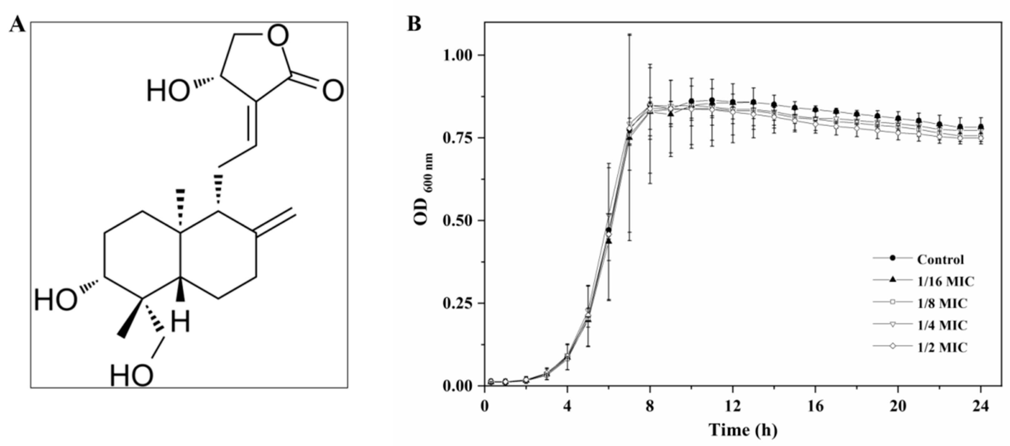
Figure 1. (A) Chemical structure of andrographolide and (B) growth curves of L. monocytogenes in BHI broth with different concentrations of andrographolide.
Molecules 27 03234 g001
Figure 2. Effect of andrographolide on two QS systems in L. monocytogenes. (A) Relative expression levels of QS-related genes in L. monocytogenes 10403S grown in BHI with or without andrographolide (1 mg/mL). Results are presented as fold changes relative to the expression level of the target gene in L. monocytogenes 10403S in BHI. (B) The production of AI-2. Vibrio harveyi BB170 and Escherichia coli DH5α were used as the positive control and the negative control, respectively. The relative activity of AI-2 was presented as a percentage of the positive control. (C) The agr promoter (P2) activity. Error bars represent the standard deviation of triplicate experiments (n = 3). The asterisk indicates a value statistically different from that of the control, with p < 0.05.
Molecules 27 03234 g002
Figure 3. Effect of andrographolide on biofilm formation. (A) Biofilm assay by microtiter plater with crystal violet staining. (B) Surviving planktonic cells in the bacterial culture. (C) CLSM images. (D) The biofilm biomass of CLSM analysis. (E) Mean thickness of biofilm by CLSM analysis. (F) SEM images. Error bars represent the standard deviation of triplicate experiments (n = 3). The asterisk indicates a value statistically different from that of the control, with p < 0.05.
Molecules 27 03234 g003
Figure 4. Effect of andrographolide on preformed biofilms. (A) Biofilm assay by microtiter plater with crystal violet staining. (B) Surviving cells in biofilms. (C) Surviving planktonic cells in the bacterial culture. (D) CLSM images. (E) The biofilm biomass of CLSM analysis. (F) Mean thickness of biofilm by CLSM analysis. (G) SEM images. Error bars represent the standard deviation of triplicate experiments (n = 3). The asterisk indicates a value statistically different from that of the control, with p < 0.05.
Molecules 27 03234 g004
Figure 5. Effect of andrographolide on (A) the hemolytic activity, (B) the capacity to attach Caco-2 cells, (C) the capacity to invade Caco-2 cells, and (D) the expression levels of virulence genes, and the results from qRT-PCR are presented as fold changes relative to the expression level of the target gene in L. monocytogenes 10403S in BHI. Error bars represent the standard deviation of triplicate experiments (n = 3). The asterisk indicates a value statistically different from that of the control, with p < 0.05.
Molecules 27 03234 g005
Publisher’s Note: MDPI stays neutral with regard to jurisdictional claims in published maps and institutional affiliations.