Article

In Vitro and In Silico Investigation of Diterpenoid Alkaloids Isolated from Delphinium chitralense

1
Department of Pharmacy, Shaheed Benazir Bhutto University, Sheringal Dir (Upper) 18000, Khyber Pakhtunkhwa, Pakistan
2
Department of Chemistry, University of Malakand, Chakdara Dir (Lower) 18550, Khyber Pakhtunkhwa, Pakistan
3
Department of Clinical Laboratory Sciences, College of Applied Medical Sciences, Taif University, P.O. Box 11099, Taif 21944, Saudi Arabia
4
Faculty of Pharmacy, Universiti Teknologi MARA Cawangan Selangor Kampus Puncak Alam, Bandar Puncak Alam 42300, Selangor, Malaysia
5
Atta-Ur-Rahman Institute for Natural Products Discovery (AuRIns), Universiti Teknologi MARA Cawangan Selangor Kampus Puncak Alam, Bandar Puncak Alam 42300, Selangor, Malaysia
6
Department of Chemistry, Shaheed Benazir Bhutto University, Sheringal Dir (Upper) 18000, Khyber Pakhtunkhwa, Pakistan
7
Department of Agriculture, Shaheed Benazir Bhutto University, Sheringal Dir (Upper) 18000, Khyber Pakhtunkhwa, Pakistan
8
Department of Biochemistry, Abdul Wali Khan University Mardan, Mardan 23200, Khyber Pakhtunkhwa, Pakistan
9
Department of Medical Laboratory Technology, College of Applied Sciences, Jazan University, Jazan 45142, Saudi Arabia
*
Authors to whom correspondence should be addressed.
Molecules 2022, 27(14), 4348; https://doi.org/10.3390/molecules27144348
Submission received: 15 May 2022 / Revised: 2 July 2022 / Accepted: 5 July 2022 / Published: 7 July 2022
(This article belongs to the Special Issue In Silico Activity Profiling of Natural Products)

Abstract

:
This study reports the isolation of three new C20 diterpenoid alkaloids, Chitralinine A–C (13) from the aerial parts of Delphinium chitralense. Their structures were established on the basis of latest spectral techniques and single crystal X-rays crystallographic studies of chitralinine A described basic skeleton of these compounds. All the isolated Compounds (13) showed strong, competitive type inhibition against acetylcholinesterase (AChE) and butyrylcholinesterase (BChE) in comparison to standard allanzanthane and galanthamine however, chitralinine-C remained the most potent with IC50 value of 11.64 ± 0.08 μM against AChE, and 24.31 ± 0.33 μM against BChE, respectively. The molecular docking reflected a binding free energy of −16.400 K Cal-mol−1 for chitralinine-C, having strong interactions with active site residues, TYR334, ASP72, SER122, and SER200. The overall findings suggest that these new diterpenoid alkaloids could serve as lead drugs against dementia-related diseases including Alzheimer’s disease.

Graphical Abstract

1. Introduction

Delphinium chitralense is a high altitude (1520 to 1830 m) annual herb, belonging to the family Ranunculaceae. The roots of D. denudatum have been found to possess anticonvulsant properties are commonly utilized in Pakistan in the Unani system of medicine [1]. Phytochemically, the genus Delphinium is reported to be a rich source of pharmacologically active diterpenoid alkaloids that pronounce potent antipyretic and analgesic activities. The nor-diterpenoid alkaloids have been found to show ten times more toxic effects as compared to any of the tested alkaloids [2]. A large number of natural products acting as cholinesterase inhibitors, especially diterpenoids and norditerpenoids alkaloids, have been investigated and isolated from different plant species as described extensively in recent studies [3,4,5,6]. In the past decade, a large number of alkaloids possessing C20 and C19 diterpenoid skeletons (Figure 1) have been isolated from different species of Delphinium [7]. Some of delphinium alkaloids are strong inhibitors of cell death, caused by oxidative stress in H9C2 cells [8]. A C-20 diterpenoid alkaloid, deoxylappaconitine, showed very strong analgesic activities, higher than the standard drug, lappaconitine [9]. Moreover, these alkaloids have been under investigation for their cardiac, relaxant, and anti-inflammatory properties, as well as antiproliferative activities against numerous cancer cell lines [7]. Some of the effects have been well documented using the structural activity relationship as it has been observed that the OH groups at positions 1, 8, and 14, as well as N-CH3 or N-H are necessary for their various therapeutic cardiac effects [10]. In the early 1990s, methyllyconitine, a major constituent, was found to be an effective ligand for neuronal nicotinic acetylcholine receptor which prompted the scientists to find suitable natural cholinesterase inhibiters to treat cerebral dementia as well as Alzheimer’s diseases (AD) [7].
Alzheimer’s disease (AD) is considered to be one of the most closely related forms of dementia to neurodegeneration disorders. The main cause of AD is the presence of the AChE enzyme which hydrolyzes acetylcholine and is present in at the neuromuscular junction of brain synapses [11]. The abnormal decrease in brain activity with regards to cholinergic function can cause memory defacement in senile dementia disease [12]. In AD, the decline of cognition is connected to the immediate loss of cholinergic neurons and shortage of ACh, which is enhanced by the neuronal ACh. The AChE generally regulates the quantity of ACh in the brain. Consequently, the deficiency of ACh in cells is redressed by the inhibition of the AChE enzyme to improve cognitive abilities. In severe cases of AD, the AChE level is decreased by up to 90% in comparison to a normal healthy brain [12], leading to uncontrollable alterations in the last stages of AD. Most of the literature reports show that a sufficient quantity of BChE is stored in Alzheimer’s plaques in comparison to the quantity of plaques present in normal healthy brains. A number of synthetic compounds such as donepezil, tacrine, and rivastigmine have been applied for the treatment of memory impairment and cognitive dysfunction [13], but these compounds were found to have antagonistic effects, including gastrointestinal complications and problems related to bioavailability [14]. Cholinesterase inhibitors obtained from plants such as jadwarine-A, jadwarine-B, 1β-hydroxy,14β-acetyl condelphine [15], swatinine-C, hohenackerine, aconorine, and lappaconitine [16] are found to be potent and demonstrate competitive and non-competitive enzyme inhibition. The investigation of natural cholinesterase inhibitors is a big task in the area of drug development, particularly for the treatment of Alzheimer’s and other related diseases [17,18].
In the current work, we describe the isolation, structure elucidation, and in vitro and in silico anticholinesterase inhibitory potential of three new C20 diterpenoids alkaloids isolated for the first time from D. chitralense. The crystal structure determination and DFT calculations of Compound 1 have also been discussed.

2. Results and Discussion

2.1. Structure Elucidation and Identification

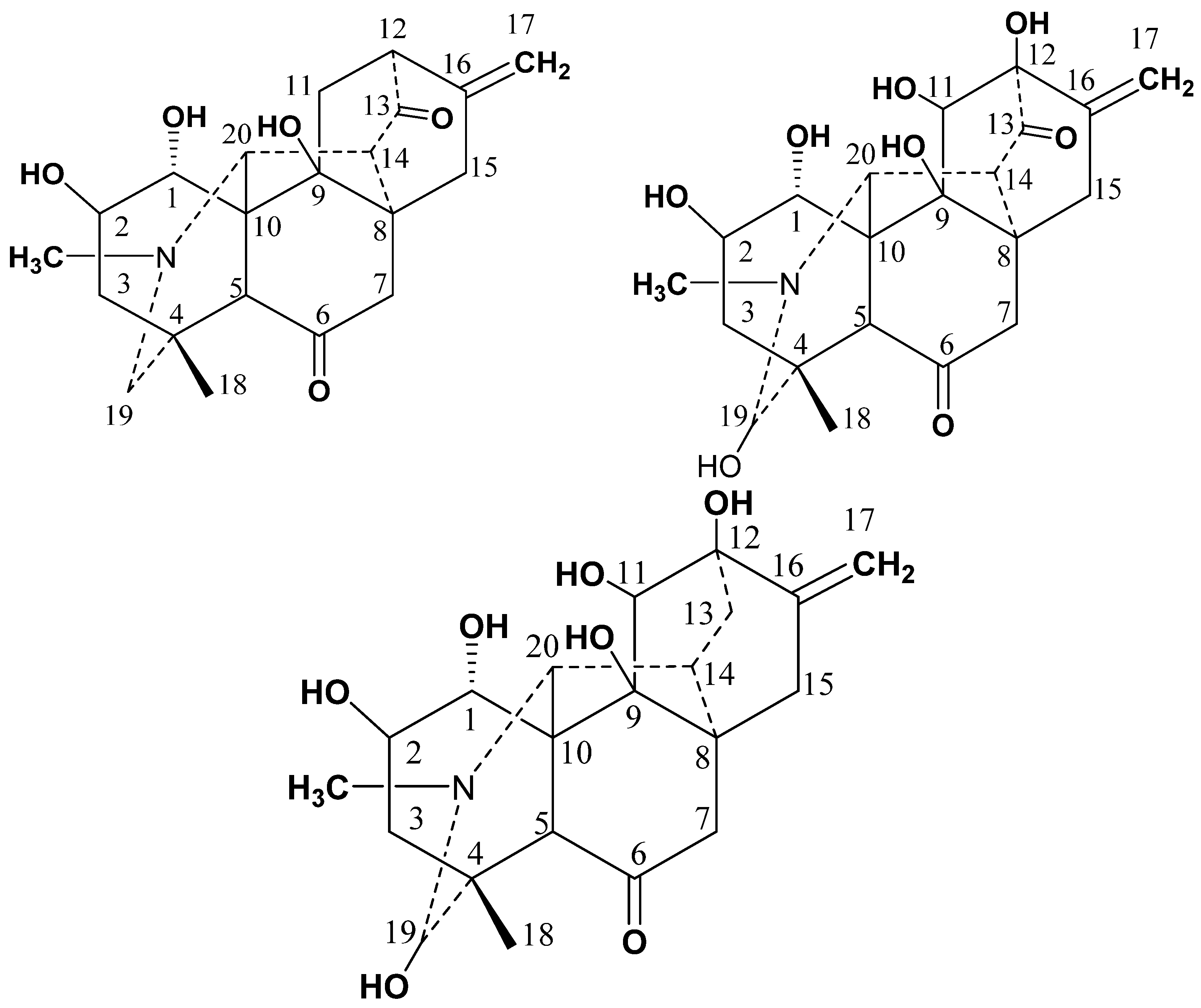
Three new Compounds (13) were isolated by the procedures (see material and methods section) from the aerial parts of D. chitralense (Figure 2).
The molecular formula for Compound 1 (C21H27NO5) was established on the basis of its molecular peak in HR-EIMS [M+] at m/z 373.4405 (calcd. 373.4417) and NMR spectral data including single X-ray crystallography.
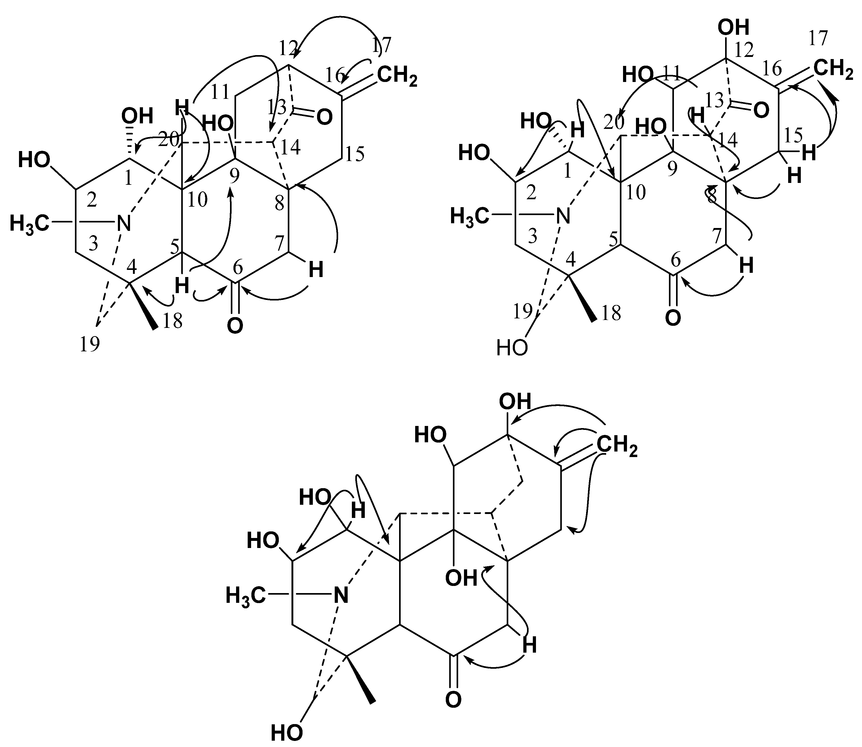
The NMR spectral data of Compound 1 displayed signals of N-methyl at δH 2.25 (3H, s, CH3-N); δC 48; methyl group at δH 1.17 (3H, s, H-18); δC 27.3 (C18); a terminal methylene proton at δH 5.07 (H-17a) and 4.80 (H-17b), δC 110.3 (C-17), two oxygenated methines at δH 4.22 (H-1); δC 78.4 (C-1), and δH 4.67 (H-2); δC 75.9 (C-2) along with additional signals of five methylene, four methines, and seven quaternary carbons. These structural features were suggestive of the structure of C20 diterpenoid alkaloid. Long-range 1H-13C correlation of 1 (Figure 3) was obtained through the heteronuclear multiple bond correlations (HMBC) experiment which suggested that H-15 (δH 2.59) signal interacted with C-9 (δC 78.6), C-10 (δC 39.7) and C-1 (δC 78.4), as well as signal of H-5 (δH 2.44) correlated to C-4 (δC 36.7) and C-6 (δC 209.4) (Table 1). The chemical shifts of C-6 and C-13 suggested two ketonic carbonyls in the molecules.

2.1.1. Crystal Structure Determination

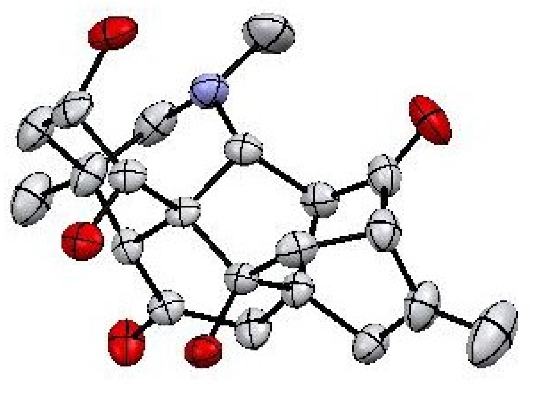
Finally, the structure and relative stereochemistry of Compound 1 was established by the study of X-ray diffraction technique (Figure 4).
Compound 1 was crystallized as monoclinic unit of crystal system with C2 space group. The crystal determination and refinement data of isolated natural product (1) are tabulated in Table 2 and Supplementary Materials.
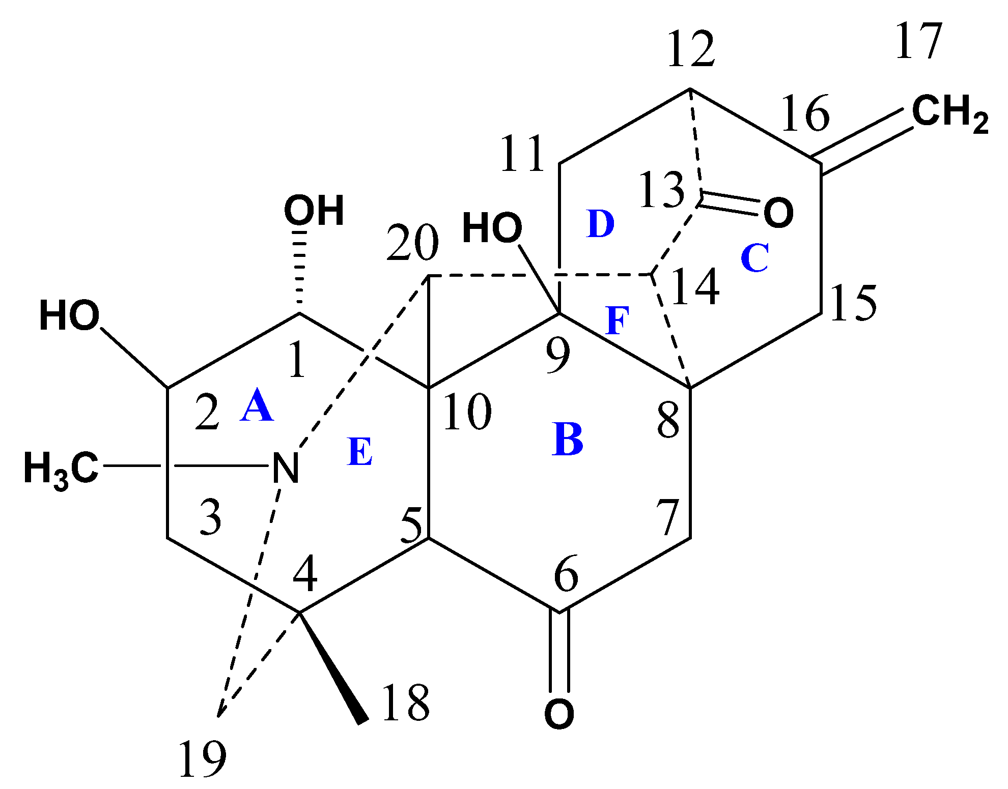
Compound 1 contains six main cyclic rings (A–F) (Figure 5). The rings A–E are six-membered rings whereas ring F is five-membered ring. Analyzing the basic/core skeleton, the junction of ring A/E [C-5—C-10—C-20 = 113.4 (4)0 ] and B/C [C-8—C-9—C-11 =106.8 (4)0 ] are trans fused while rings A/B [C-5—C-10—C-1 =107.2 (4)0] and rings E/F [C-5—C-10—C-9 = 110.6 (4)0] are cis-fused. The bond lengths and angles were in observed in predicted ranges [19]. The two-hydroxyl group at C-2 and C-9 are β-oriented while the other hydroxyl groups at C-1 was noticed to be α-oriented. In observing stereochemistry, all the rings showed chair, boat, and half boat conformations. The absolute configuration of Compound 1 cannot be constituted by Mo-Kα diffraction data, although it can be assumed to be equivalent as reported for other isolated C20-diterpenoid alkaloids [19]. Keeping in view, the above mentioned spectral and crystal data, the structure of Compound 1 was deduced as 9β-hydroxy hetidine (named Chitralinine-A).

2.1.2. DFT Calculation of Compound 1

The DFT simulations are necessary to gain deeper insights into the molecular structure and electronic properties as it was recently reported in many examples in the literature that DFT simulations were used along with experimental study to obtain the electronic properties which are harder to obtain through experimental analysis. Therefore, we performed the DFT calculations and studied different properties for the studied compound such as HOMO-LUMO analysis, reactivity, global hardness, and optimized structure etc. DFT calculations were generally accomplished on a single unit cell of the molecule [20]. The geometry of Compound 1 with appropriate orientation and spatial arrangement was optimized by using B3LYP-631G (p) and 6-311 + G(d,p)/wB97XD basis sets [21]. The optimized geometry and structure of the compound under investigation is shown in Figure 6. The electronic properties, calculated energy and other relevant parameters are given in Table 3.
HOMO-LUMO energy gaps for Compound 1 were obtained as 0.191 au at 6-31G(d)/B3LYP while 0.292 au at 6-311 + G(d,p)/wB97XD, the values obtained through 6-311 + G(d,p)/wB97XD are higher than 6-31G(d)/B3LYP level because the wB97XD theory has high Hartree–Fock (HF) character and can effectively capture co-relations factors. The values of the EH-L gaps of the studied compound obtained through wB97XD functional with a larger basis set is higher than that of B3LYP functional with a smaller basis set. Because B3LYP functionals overestimated the electronic properties due to lower HF character, this cannot effectively capture electronic co-relation factors. The values of the EH-L gaps studied at wB97XD/6-311 + G(d,p) functional are reported in Table 3.
Moreover, it was observed that the stability of LUMO was mainly due to the electron-accepting properties while the HOMO orbital is usually responsible for the electron-donating ability of inhibitor molecule. Moreover, the greater values of HOMO are signs of electrons donation to the un-occupied orbital of the receptor.
Compound 2 showed specific rotation [α]D30: −250 (c = 1, CHCl3). Its molecular formula (C21H27NO8) was deduced by HR-EIMS (m/z 421.4550; calcd. 421.4396), higher than chitralinine-A, probably due to the presence of additional hydroxyl groups NMR spectrum of Compound 2 displayed a terminal methylene group at δH 5.02 (H-17a) and 4.85 (H-17b); δC 110.4 (C-17), methyl protons at δH 1.16 (H-18); 29.4 (C-18), four oxygenated methines at δH 4.16 (H-1); δC 75.4 (C-1), δH 4.64 (H-2); δC 73.3 (C-2), δH 3.36 (H-11); δC 80.7 (C-11) and δH 5.0 (H-19); δC 94.2 (C-19), respectively. The 1H and 13C-NMR data were very similar to those of Compound 1, suggesting that Compound 2 should also be a diterpenoid and structurally related to Compound 1. While comparing the chemical shifts of skeletal carbons in Compounds 1 and 2, the main differences between their 13C-NMR data (Table 1) are that there were three more oxygenated functionalities, and additional hydroxyl groups appeared in Compound 2. To verify further the locations of groups and functionality, HMBC experiment was performed (Figure 3). The hydroxyl group on C-1 was assigned by the HMBC correlations of H-1 (δH 4.16) to C-2 (δC 73.3) and C-10 (δC 43.6). Similarly, other correlations were observed between H-7 (δH 2.05) to C-6 (δC 209.4) and C-8 (δC 36.7); H-14 (δH 2.26) to C-8 (δC 36.7) and C-20 (δC 70.1). On the basis of above physical and spectroscopic data, the structure was deduced as 9,11,12,19β- tetrahydoxy hetidine (chitralinine-B).
The molecular formula (C21H29NO7) for Compound 3 was deduced from its HR-EIMS at m/z 407.4550 (calcd. 407.4563) which was consistent with its 1H and 13C NMR data (Table 1 and Table 2).
From the NMR spectrum of Compound 3, the terminal methylene group as existing in Compounds 1 and 2 was inferred on the basis of signals for C-17 methylenic protons singlets at δH 5.02 (H-17a) and 4.85 (H-17b), δC 110.4 (C-17) (Table 1 and Table 2). From the 1H-NMR spectrum, two oxymethine protons displayed singlets separately at δH 4.97 (H-19); δC 94.2 (C-19), δH 3.27 (H-11); δC 80.7 (C-11), confirmed the presence of hydroxyl groups in Compound 3 at C-19 and C-11. The 13C-NMR spectrum of Compound 3 showed twenty-one signals for primary, secondary, tertiary and, quaternary carbon atoms. Compared to the chemical shifts of skeletal carbons in Compounds 2 and 3, the main difference between their 13C NMR data (Table 1) is that Compound 3 lacked ketonic functionality at C-13. After describing the skeleton of Compound 3, long range HMBC interaction (Figure 3) permitted the placement of the hydroxyl substituent at their respective positions, as the H-1 (δ4.19) shows 2J correlations with C-2 (δ 73.3) and C-10 (δ 44.0). Similarly, the HMBC coupling of terminal methylinic protons (δ 5.02 & 4.85) exhibited 1J interaction with C-16 (δ 142) and 2J interaction with C-12 (δ 78.3) and C-15(δ 26.9). All of the above spectral evidence led to the establishment of C20 diterpenoid structure of 3 as 1α, 2, 9,11,12,19β-hexahydoxy atisinone (Chitralinine-C).

2.2. Acetylcholinesterase and Butyrylcholinesterase Inhibition Activities

All the natural products isolated from D. chitralense, were tested for their enzyme inhibition activity against AChE and BChE, respectively, and showed promising inhibitory potential against both the tested enzymes in vitro. Therefore, it might be concluded that the compounds isolated from D. chitralense could be optimized as lead candidates in AD and related ailments. The Compounds (13) were found to be potent against AChE and BchE as compared to standard drugs, showing competitive types of inhibition. The IC50 values of Compounds 13 against AChE were 13.86 ± 0.35, 11.64 ± 0.08 μM and 12.11 ± 0.82 μM while against BChE the values were 28.17 ± 0.92 μM, 24.31 ± 0.33 μM and 26.35 ± 0.06 μM, respectively (Table 4).
These significant results highlighted the interest in isolation and reputation of this class of secondary metabolites present in D. chitralense.

2.3. Molecular Docking Study

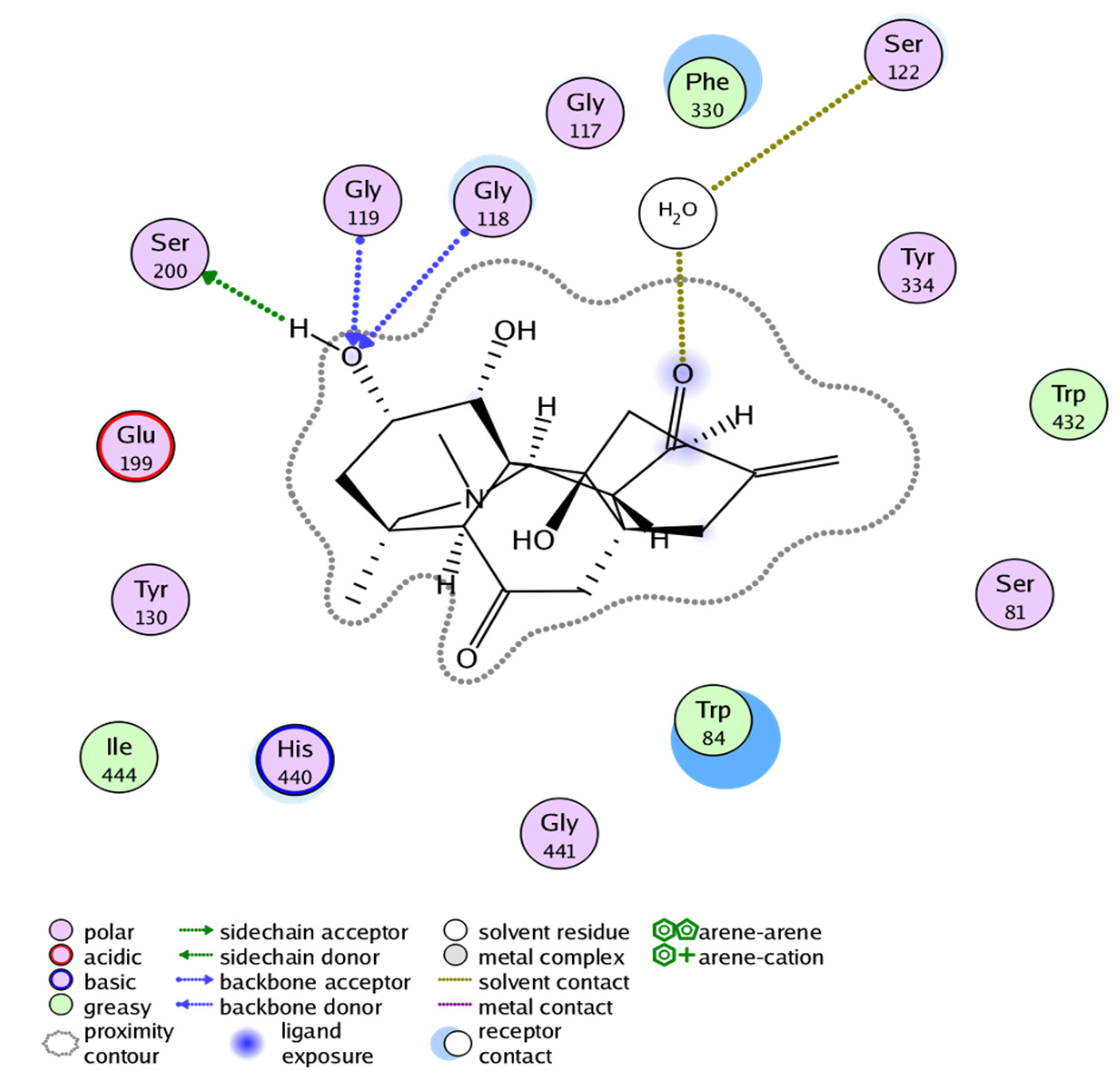
The ligand base docking result of Compound 1 against the acetylcholinesterase target showed binding free energy of −14.457 Kcal/mol (Table 5). The visual inspection of docked compound revealed that it interacts with four residues, i.e., SER200, GLY119, GLY118, and SER122 of acetylcholinesterase (Figure 7).
Details regarding chemical activity of Compound 1 were determined based on the correlation between calculated energies and quantum parameters. Quantum parameters, i.e., electronegativity (χ), electrophilicity (ω), hardness (η), and softness (S), are universal descriptors which are used to explain the chemical behavior of the molecules [22]. The hardness (η) value decides the resistance of an atom for the charge transfer to another atom. The electron-receiving ability of an atom is determined by the softness value. Electronegativity χ is the ability of molecules to attract electrons. The electrophilicity index ω is related to the electrophilic property of a molecule.
The significant orbitals found in molecules that affect the biological activity, molecular reactivity, and other electronic properties are HOMO and LUMO [23,24,25]. Deep insight into the biological mechanism of the active molecules can also be deduced on the basis of frontier orbital energy studies. Figure 5 shows that both HOMO and LUMO are localized at the tertiary nitrogen atom and hydroxyl group of Compound 1. This makes it clear that the activity related to this molecule could be attributed to the hydroxyl and tertiary nitrogen. Thus, HOMO and LUMO orbitals that penetrate the hydroxyl group will form interactions with active sites of the enzyme, giving rise to its reported biological activity.
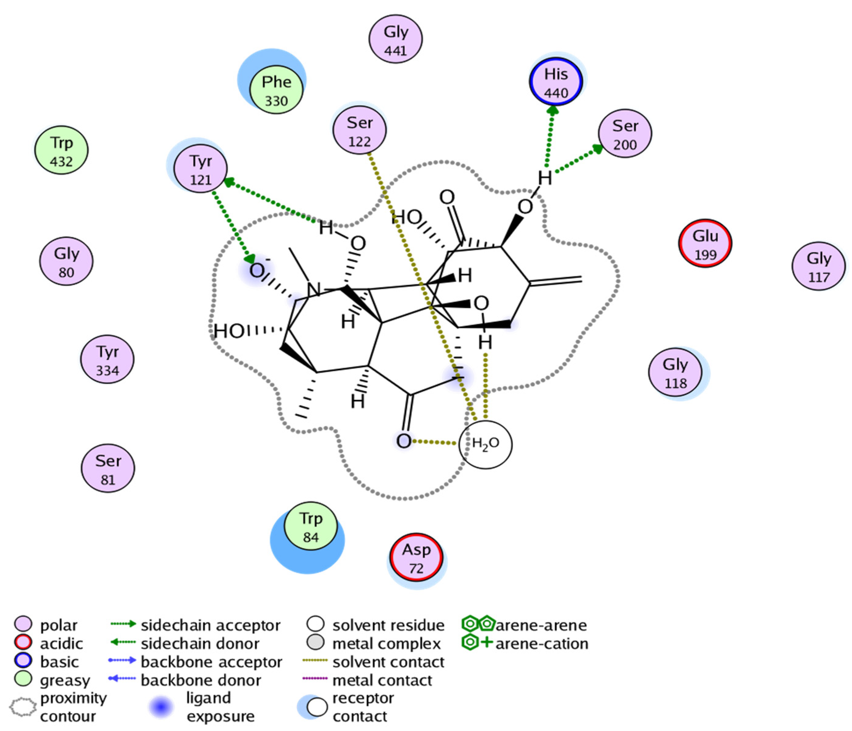
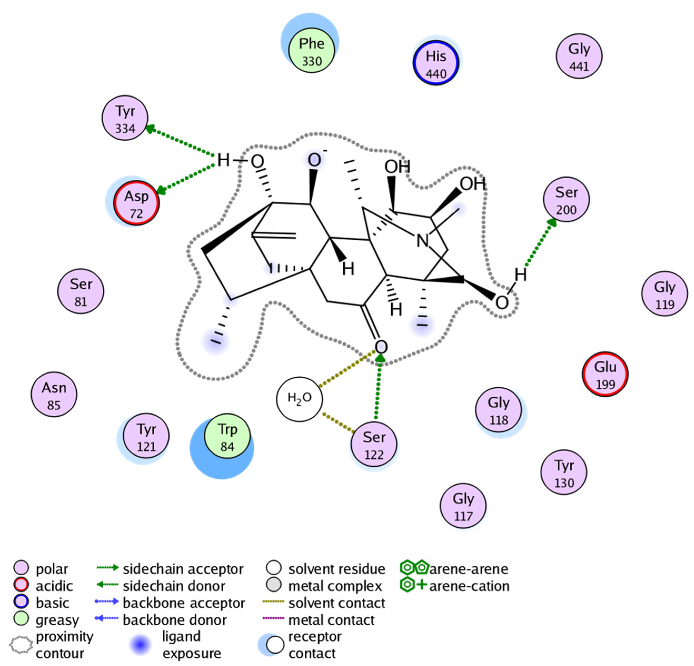
The docking result of Compound 2 showed binding free energy of −15.591 Kcal/mol and selective interactions with TYR121, SER122, HIS440, and SER200 residues (Figure 8). Likewise, the docking result of Compound 3 showed binding free energy of −16.400 Kcal/mol and interactions with TYR334, ASP72, SER122, and SER200 (Figure 9). All the Compounds 13 were found to develop interactions with the key residues of the gorge site of the acetylcholinesterase target and the in silico results are congruent with experimental findings, suggesting the competitive nature of Compounds 13. The differences in protein-ligand interaction and binding energies among these compounds are due to differences in basic chemical structures and receptor binding affinities.

3. Materials and Methods

3.1. General Procedures

The optical rotations ([α]25D) were obtained through a “JASCO DIP 360 polarimeter (Tokyo, Japan)” while the melting points (mps) were measured using “BioCote Stauart SMP10 (Tokyo, Japan)” melting point instrument. The mass spectral assignments were made from EI-MS/HR-EIMS spectra obtained through “JEOL JMS HX 110 (Tokyo, Japan)” while 1H-NMR/13C-NMR spectral measurements were carried out by using “Bruker NMR, Germany (500, 600 MHz for 1H-NMR; 125, 150 MHz for 13C-NMR (δ, ppm)), respectively. FT-IR analyses were determined on “JASCO-320-A spectrophotometer in KBr” as well as “Perkin–Elmer spectrophotometer”. All the solvents used in extraction and isolation of compounds were distilled before use, while the deuterated solvents were used for NMR analysis. Thin layer chromatography (TLC) was carried out using “silica gel F254 pre-coated aluminum sheets”. Visualization of TLC was conducted through a UV lamp at both 254 & 366 nm (λmax) as well as “Dragendorff’s reagent”. The solvent system; 20% acetone-hexane: 10 drops of diethylamine was used as developing solvent for TLC.

3.2. Plant Material

The aerial parts of D. chitralense were collected from Kumrat Valley (Latitude = 35.560654; Longitude = 72.200846; Altitude is 8100 feet) of Dir (U). A voucher specimen with number H.UOM. BG-161 was deposited in the herbarium of University of Malakand, Dir (L).

3.3. Extraction and Isolation

Standard procedure was adopted in the extraction and isolation processes with some modifications [15]. The shade dried powdered material of (10 kg) of D. chitralense was extracted for seven days with 80% methanol; thrice (3 × 20 L) in closed glass containers. This combined methanolic extract was filtered and concentrated in a vacuum on a rotary evaporator (Buchi, Flawil, Switzerland) at 40 °C to obtain 890 g methaloic crude. This methaloic crude was first pooled with 5% HCl solution (pH = 1–2) and then extracted with CHCl3 to separate the non-alkaloidal portion from acid-aqueous alkaloidal solution. This acidic portion was basified with 5% NaOH (pH= 8–10) to obtain free alkaloids in the solution. This solution was again re-extracted with CHCl3 to obtain alkaloidal portion (18 g). This alkaloidal portion was fractionated though a silica gel (360 g) column, which was eluted with increasing polarities of n-hexane (100%, DX-1) and n-hexane-chloroform and chloroform-methanol (up to 20% methanol) in gradient manner that afforded eight sub-fractions (DX-1 to DX-8). The sub-fraction DX-4 obtained from solvent system n-hexane-chloroform (50:50) showed interesting spots on TLC and was re-chromatographed on the flesh silica gel column eluted with n-hexane:acetone (80:20) with 10 drops of diethylamine (DEA/100 mL) to produce Compound 1 (top fraction) and Compound 2 (tail fraction). The fraction DX-5 which was obtained from major column with n-hexane-chloroform (60:40) yielded a semi pure compound, which was subjected to a flash column using gradient solvent system of n-hexane-acetone-10 drops of DEA. Elution with n-hexane-acetone (80:20) resulted in the isolation of pure Compound 3.

3.4. Physical and Spectroscopic Data of New Compounds

3.4.1. Chitralinine-A (1)

White crystal; m.p: 224–227 °C; [α]D30: −350 (c = 1, CHCl3); IR (υmax cm−1): 1720 & 1650 (C = O), 988 (C = CH2), 3460, 3422 (OH); 1HNMR (600 MHz, CDCl3): δ 5.07, 4.80 (2H, s, -CH2), δ 4.67, (1H, t, J = 4.7Hz, H-2), δ 4.22 (1H, brd s, H-1), δ 3.59 (1H, br s, H-20), δ 3.04 (2H, s, H-19), δ 2.66, (1H, t, J = 11 Hz, H-12), δ 2.59 (2H, d, J = 4 Hz, H-15), δ 2.44 (1H, s, H-5), δ 2.31 (2H, t, J = 2.1 Hz, H-11), δ 2.25 (3H, s, -NCH3), δ 2.19 (1H, d, J = 3 Hz, H-14), δ 2.08 (2H, s, H-7), δ 1.82 (2H, m, H-3), δ 1.17 (3H, s, H-18), HR-EIMS m/z: 373.4405 (C21H27NO5, calcd. 373.4417); 13C-NMR (150 MHz/CDCl3): See Table 1.

3.4.2. Chitralinine-B (2)

White powder; m.p: 252–255 °C; [α]D30: −250 (c = 1, CHCl3); IR υmax cm−1: 1660, 1450 (C = O), 3480, 3360, 3280 (OH) 1030 (C = CH2); 1HNMR (600 MHz, CDCl3): δ 5.02, 4.85 (2H, s-CH2), δ 5.0 (1H, s, H-19), δ 4.64 (1H, t, J = 4.9 Hz, H-2), δ 4.16 (1H, brd s, H-1), δ 3.59 (1H, br s, H-20), δ 3.36 (2H, s, H-11), δ 2.84 (1H, s, H-5), δ 2.46 (3H, s, -NCH3), δ 2.26 (1H, d, J = 2.5 Hz, H-14), δ 2.05 (2H, s, H-7), δ 1.92 (2H, m, H-3), δ 1.89 (2H, s, H-15), δ 1.16 (3H, s, H-18) HR-EIMS m/z: 421.4380 (C21H27NO8, calcd. 421.4396); 13C-NMR (150 MHz/CDCl3): See Table 1.

3.4.3. Chitralinine-C (3)

Amorphous powder; m.p: 233–238 °C; [α]D30: −230 (c = 1, CHCl3); IR υmax cm−1: 1658, 1446 (C = O), 3500, 3350 (br OH), 1032 (C = CH21HNMR (500 MHz, CDCl3): δ 5.02, 4.85 (2H, s-CH2), δ 4.97 (1H, s, H-19), δ 4.64 (1H, t, J = 4.8 Hz, H-2), δ 4.19 (1H, brd s, H-1), δ 3.57 (1H, br s, H-20), δ 3.27 (2H, s, H-11), δ 2.45 (3H, s, -NCH3), δ 2.30 (2H, s, H-7), δ 2.24 (1H, s, H-5), δ 1.95, 1.93 (2H, dd, 2H), δ 1.91 (2H, m, H-3). δ 1.89 (2H, s, H-15), δ 1.63 (1H, m, H-14), δ 1.15 (3H, s, H-18); HREI-MS m/z: 407.4550 (C21H29NO7, calcd. 407.4563); 13C (125 MHz/CDCl3): See Table 1.

3.5. X-ray Crystallography

The crystal structure data for Compound 1 was obtained from Single crystal X-ray crystallographic analysis using “STOE-IPDS II (Darmstadt, Germany); Graphite-monochromator at room temperature and Mo-Kα radiation (λ = 0.71073 Å)”. Data were captured by using charge-coupled device (CCD) area detector. The structure was solved and refined though SIR97 [26], SHELXL97 [27] and WinGX [28] programs.

3.6. Density Functional Theory (DFT) Calculations

The crystallographic data were used in the DFT calculations of 1 by means of two models of theory; “B3LYP-631G (p) and 6-311 + G(d,p)/wB97XD” [20,26,29]. Furthermore, the same methods were applied in obtaining other related information including HOMO-LUMO energy gap, optimized geometries, electron affinity, electrophilicity, ionization potential, and global hardness [30]. The data were manipulated through “Gauss-view molecule visualizer and GAUSSIAN-03 programs (Wallingford, CT, USA)”. Recently, this procedure has also been applied for DFT calculations and obtaining data of other parameters in C19 and C20 alkaloids [30].

3.7. Acetylcholinesterase (AChE) and Butyrylcholinesterase (BChE) Inhibition Assays

All the solvents used in this assays were of analytical grade while the chemicals/reagents such as AChE (Electric-eel EC 3.1.1.7), BChE (horse serum EC 3.1.1.8), DTNB, Acetyl choline iodide (AChI), butyryl choline chloride (BChI) and the reference, galantamine were purchased from Sigma–Aldrich (St. Louis, MO, USA). The inhibition was obtained through spectroscopic measurements [31]. Standard procedures and conditions of the assays were applied throughout the experiments [32]. Various dilutions of the tested compounds (62.5, 125, 250, 500 and 1000 μg/mL) were used in this assay. AChI and BChl were used as substates in this assay.
In brief, 880 µL of sodium phosphate buffer solution (62 mM, pH 8) containing 0.2 mM DNB was mixed with 40 µL solution of compound and 40 µL AChE or BChE solutions. This reaction mixture was incubated at 25 °C for 15 min followed by initiation of the reaction through addition of acetyl choline (ACh) or butyryl choline (BCh) (40 μL) in each experiment. The formation of yellow colored product (5-thio-2-nitorbenzoate anion) from reaction of DTNB with acetylcholine/butyrylcholine were observed through naked eyes, and the absorbance was measured at 412 nm using UV/Visible spectrophotometer (BMS-USA, New York, NY, USA). All the experiments were carried out in triplicate.

IC50 Values Determination

Various dilutions of the tested compounds (62.5, 125, 250, 500, and 1000 μg/mL) were used in this assay for IC50 determination. The data (activity in percent vs concentration) were fitted into a non-linear sigmoid plot using MS Excel (Microsoft, Redmond, WA, USA) program. The non-linear concentration-dependent inhibitory concentration of compounds were taken into account at low and high concentration leading to determination of IC50 values. The effective concentrations of Compounds 13 were represented in μM [33].

3.8. Molecular Docking Study

To perform molecular docking analysis, the choline esterase crystal structures (PDB ID: 1ACJ) was retrieved from the Protein Databank (PDB). The molecular docking studies were carried out in the presence of water molecules as they play crucial role in the enzymatic activities [34,35,36,37] while other ions were removed from the retrieved crystal structures using the Molecular Operating Environment (MOE) software (www.chemcomp.com, accessed on 10 May 2022). Hydrogen atoms were added to the protein structures by 3D protonation and then energy minimization was carried out using the default parameters of the MOE. The structures of the Compounds 13 were built and their energy minimization was performed using the default parameters of the MOE. The choline esterase target was allowed to dock to the Compounds 13 by the default parameters i.e., Placement: Triangle Matcher, Rescoring: London dG of MOE software. The binding pockets are identified by site-finder module of the MOE. For each ligand ten conformations were generated. The top-ranked conformation of each compound was used for subsequent analysis [38].

4. Conclusions

In the present research work, bioactivity-guided isolation, crystal structure determination, DFT calculation, anticholinesterase inhibitory potential, and molecular docking studies of diterpenoid alkaloids isolated from D. chitralense have been carried out. The structures of three new diterpenoids were established by spectral interpretation, including single X-ray crystallography. Some structural parameters of Compound 1 were calculated by means of DFT. All the isolated compounds were screened for their possible anticholinesterase inhibitory potential and were found to exhibit strong, competitive types of inhibition against cholinesterase as compared to the standard. Further in silico evaluation of isolated compounds exhibited possible binding modes and justified the experimental results. In addition, the negative binding energies of the isolated Compounds 13 showed proper relationships to the AChE and BChE enzymes. Thus, the present study validates a potential role of diterpenoid alkaloids from D. chitralense in the treatment of neurodegenerative disorders and suggests that they could be good natural candidates against AD.

Supplementary Materials

The following supporting information can be downloaded at: https://www.mdpi.com/article/10.3390/molecules27144348/s1. The CIF of the Compounds (1) were deposited at the Cambridge Crystallographic Data Centre as supplementary publication No. CCDC 1470285. These data can be retrieved free of charge from the Cambridge Crystallographic Data Centre, www.ccdc.cam.ac.uk/getstructures, accessed on 10 May 2022.

Author Contributions

Conceptualization, S.A., H.A. and M.A. (Manzoor Ahmad); methodology, S.A.A.S., N.M.K. and A.K.; software, Z. and M.H.; manuscript—writing and editing, F.A.K., H.A. Resources, funding acquisition, M.A. (Mazen Almehmadi). All authors have read and agreed to the published version of the manuscript.

Funding

Taif University: Taif, Saudi Arabia: TURSP-2020/80.

Institutional Review Board Statement

Not applicable.

Informed Consent Statement

Not applicable.

Data Availability Statement

All data contained within this article.

Acknowledgments

We are thankful to Taif University, Taif, Saudi Arabia, for their support (Taif University Researchers Support Project number: TURSP-2020/80).

Conflicts of Interest

All the authors have no financial interest regarding publishing scientific data in this article.

Sample Availability

Samples of the compounds are available from the authors. Will be provided on reasonable request.

References

  1. Shaheen, F.; Ahmad, M.; Rizvi, T.S.; Ali, L. Norditerpenoid alkaloids from Delphinium kohatense Munz. Rec. Nat. Prod. 2015, 9, 76–80. [Google Scholar]
  2. Qasem, A.M.A.; Zeng, Z.; Rowan, M.G.; Blagbrough, I.S. Norditerpenoid alkaloids from Aconitum and Delphinium: Structural relevance in medicine, toxicology, and metabolism. Nat. Prod. Rep. 2022, 39, 460–473. [Google Scholar] [CrossRef] [PubMed]
  3. Houghton, P.J.; Ren, Y.; Howes, M.J. Acetylcholinesterase inhibitors from plants and fungi. Nat. Prod. Rep. 2006, 23, 181–199. [Google Scholar] [CrossRef] [PubMed]
  4. Mukherjee, P.K.; Kumar, V.; Houghton, P.J. Screening of Indian medicinal plants for acetylcholinesterase inhibitory activity. Phyther. Res. 2007, 21, 1142–1145. [Google Scholar] [CrossRef]
  5. Hostettmann, K.; Borloz, A.; Urbain, A.; Marston, A. Natural Product Inhibitors of Acetylcholinesterase. Curr. Org. Chem. 2006, 10, 825–847. [Google Scholar] [CrossRef]
  6. Ranjan, N.; Ranjan, N.; Kumari, M. Acetylcholinesterase inhibition by medicinal plants: A Review. Ann. Plant Sci. 2017, 6, 1640–1644. [Google Scholar] [CrossRef] [Green Version]
  7. Yin, T.; Cai, L.; Ding, Z. An overview of the chemical constituents from the genus Delphinium reported in the last four decades. RSC Adv. 2020, 10, 13669–13686. [Google Scholar] [CrossRef] [Green Version]
  8. Wang, Y.; Sun, D.; Chen, Y.; Xu, J.; Xu, Y.; Yue, X.; Jia, J.; Li, H.; Chen, L. Alkaloids of Delphinium grandiflorum and their implication to H2O2-induced cardiomyocytes injury. Bioorganic Med. Chem. 2021, 37, 116113. [Google Scholar] [CrossRef]
  9. Xu, J.B.; Li, Y.Z.; Huang, S.; Chen, L.; Luo, Y.Y.; Gao, F.; Zhou, X.L. Diterpenoid alkaloids from the whole herb of Delphinium grandiflorum L. Phytochemistry 2021, 190, 112866. [Google Scholar] [CrossRef]
  10. Zhang, Z.T.; Jian, X.X.; Ding, J.Y.; Deng, H.Y.; Chao, R.B.; Chen, Q.H.; Chen, D.L.; Wang, F.P. Further studies on structure-cardiac activity relationships of diterpenoid alkaloids. Nat. Prod. Commun. 2015, 10, 2075–2084. [Google Scholar] [CrossRef] [Green Version]
  11. Noori, T.; Dehpour, A.R.; Sureda, A.; Sobarzo-Sanchez, E.; Shirooie, S. Role of natural products for the treatment of Alzheimer’s disease. Eur. J. Pharmacol. 2021, 898, 173974. [Google Scholar] [CrossRef] [PubMed]
  12. Sultzer, D.L.; Lim, A.C.; Gordon, H.L.; Yarns, B.C.; Melrose, R.J. Cholinergic receptor binding in unimpaired older adults, mild cognitive impairment, and Alzheimer’s disease dementia. Alzheimer’s Res. Ther. 2022, 14, 1–15. [Google Scholar] [CrossRef] [PubMed]
  13. MH, O.; PJ, H.; WK, W.; JH, C. Screening of Korean herbal medicines used to improve cognitive function for anti-cholinesterase activity. Phytomedicine 2004, 11, 544–548. [Google Scholar] [CrossRef]
  14. Schulz, V. Ginkgo extract or cholinesterase inhibitors in patients with dementia: What clinical trials and guidelines fail to consider. Phytomedicine 2003, 10 (Suppl. 4), 74–79. [Google Scholar] [CrossRef] [PubMed]
  15. Ahmad, H.; Ahmad, S.; Ali, M.; Latif, A.; Shah, S.A.A.; Naz, H.; ur Rahman, N.; Shaheen, F.; Wadood, A.; Khan, H.U.; et al. Norditerpenoid alkaloids of Delphinium denudatum as cholinesterase inhibitors. Bioorganic Chem. 2018, 78, 427–435. [Google Scholar] [CrossRef]
  16. Ahmad, H.; Ahmad, S.; Shah, S.A.A.; Khan, H.U.; Khan, F.A.; Ali, M.; Latif, A.; Shaheen, F.; Ahmad, M. Selective dual cholinesterase inhibitors from Aconitum laeve. J. Asian Nat. Prod. Res. 2017, 20, 172–181. [Google Scholar] [CrossRef]
  17. Mitić, M.; Lazarević-Pašti, T. Does the application of acetylcholinesterase inhibitors in the treatment of Alzheimer’s disease lead to depression? Expert Opin. Drug Metab. Toxicol. 2021, 17, 841–856. [Google Scholar] [CrossRef]
  18. Ahmad, S.; Ahmad, H.; Khan, H.U.; Shahzad, A.; Khan, E.; Ali Shah, S.A.; Ali, M.; Wadud, A.; Ghufran, M.; Naz, H.; et al. Crystal structure, phytochemical study and enzyme inhibition activity of Ajaconine and Delectinine. J. Mol. Struct. 2016, 1123, 441–448. [Google Scholar] [CrossRef]
  19. Ahmad, H.; Ahmad, S.; Khan, E.; Shahzad, A.; Ali, M.; Tahir, M.N.; Shaheen, F.; Ahmad, M. Isolation, crystal structure determination and cholinesterase inhibitory potential of isotalatizidine hydrate from Delphinium denudatum. Pharm. Biol. 2017, 55, 680–686. [Google Scholar] [CrossRef] [Green Version]
  20. Teixeira, A.M.R.; Santos, H.S.; Albuquerque, M.R.J.R.; Bandeira, P.N.; Rodrigues, A.S.; Silva, C.B.; Gusmão, G.O.M.; Freire, P.T.C.; Bento, R.R.F. Vibrational Spectroscopy of Xanthoxyline Crystals and DFT Calculations. Braz. J. Phys. 2012, 42, 180–185. [Google Scholar] [CrossRef]
  21. Ali Altaf, A.; Shahzad, A.; Gul, Z.; Rasool, N.; Gul, Z.; Badshah, A.; Lal, B.; Khan, E.; Khan, E. A Review on the Medicinal Importance of Pyridine Derivatives. J. Drug Des. Med. Chem. 2015, 1, 1–11. [Google Scholar] [CrossRef]
  22. Tsuneda, T.; Song, J.W.; Suzuki, S.; Hirao, K. On Koopmans’ theorem in density functional theory. J. Chem. Phys. 2010, 133, 174101. [Google Scholar] [CrossRef]
  23. Clare, B.W. Frontier orbital energies in quantitative structure-activity relationships: A comparison of quantum chemical methods. Theor. Chim. Acta 1994, 87, 415–430. [Google Scholar] [CrossRef]
  24. Clare, B.W. Charge transfer complexes and frontier orbital energies in QSAR: A congeneric series of electron acceptors. J. Mol. Struct. Theochem 1995, 337, 139–150. [Google Scholar] [CrossRef]
  25. Zhang, G.; Musgrave, C.B. Comparison of DFT methods for molecular orbital eigenvalue calculations. J. Phys. Chem. A 2007, 111, 1554–1561. [Google Scholar] [CrossRef] [PubMed]
  26. Altomare, A.; Burla, M.C.; Camalli, M.; Cascarano, G.L.; Giacovazzo, C.; Guagliardi, A.; Moliterni, A.G.G.; Polidori, G.; Spagna, R. SIR97: A new tool for crystal structure determination and refinement. J. Appl. Crystallogr. 1999, 32, 115–119. [Google Scholar] [CrossRef]
  27. Sheldrick, G.M. IUCr A short history of SHELX. Acta Crystallogr. Sect. A 2007, 64, 112–122. [Google Scholar] [CrossRef] [Green Version]
  28. Farrugia, L.J. WinGX suite for small-molecule single-crystal crystallography. J. Appl. Crystallogr. 1999, 32, 837–838. [Google Scholar] [CrossRef]
  29. Wang, H.; Wang, X.; Wang, H.; Wang, L.; Liu, A. DFT study of new bipyrazole derivatives and their potential activity as corrosion inhibitors. J. Mol. Model. 2006, 13, 147–153. [Google Scholar] [CrossRef]
  30. Ahmad, H.; Ahmad, S.; Shah, S.A.A.; Latif, A.; Ali, M.; Khan, F.A.; Tahir, M.N.; Shaheen, F.; Wadood, A.; Ahmad, M. Antioxidant and anticholinesterase potential of diterpenoid alkaloids from Aconitum heterophyllum. Bioorganic Med. Chem. 2017, 25, 3368–3376. [Google Scholar] [CrossRef]
  31. Ellman, G.L.; Courtney, K.D.; Andres, V.; Featherstone, R.M. A new and rapid colorimetric determination of acetylcholinesterase activity. Biochem. Pharmacol. 1961, 7, 88–95. [Google Scholar] [CrossRef]
  32. Batista, J.; Rocha, T.; Rocha, J.B.T.; Emanuelli, T.; Pereira, M.E. Effects of early undernutrition on kinetic parameters of brain acetylcholinesterase from adult rats. Acta Neurobiol. Exp. 1993, 53, 431. [Google Scholar]
  33. Burlingham, B.T.; Widlanski, T.S. An Intuitive Look at the Relationship of Ki and IC50: A More General Use for the Dixon Plot. J. Chem. Educ. 2003, 80, 214–218. [Google Scholar] [CrossRef]
  34. Henchman, R.H.; Tai, K.; Shen, T.; Andrew McCammon, J. Properties of water molecules in the active site gorge of acetylcholinesterase from computer simulation. Biophys. J. 2002, 82, 2671. [Google Scholar] [CrossRef]
  35. Daoud, I.; Melkemi, N.; Salah, T.; Ghalem, S. Combined QSAR, molecular docking and molecular dynamics study on new Acetylcholinesterase and Butyrylcholinesterase inhibitors. Comput. Biol. Chem. 2018, 74, 304–326. [Google Scholar] [CrossRef]
  36. Geromichalos, G.D.; Lamari, F.N.; Papandreou, M.A.; Trafalis, D.T.; Margarity, M.; Papageorgiou, A.; Sinakos, Z. Saffron as a source of novel acetylcholinesterase inhibitors: Molecular docking and in vitro enzymatic studies. J. Agric. Food Chem. 2012, 60, 6131–6138. [Google Scholar] [CrossRef]
  37. Berg, L.; Da Andersson, C.D.; Artursson, E.; Hörnberg, A.; Tunemalm, A.K.; Linusson, A.; Ekström, F. Targeting acetylcholinesterase: Identification of chemical leads by high throughput screening, structure determination and molecular modeling. PLoS ONE 2011, 6, e26039. [Google Scholar] [CrossRef] [Green Version]
  38. Babitha, P.; Sahila, M.; Bandaru, S.; Nayarisseri, A.; Sureshkumar, S. Molecular Docking and Pharmacological Investigations of Rivastigmine-Fluoxetine and Coumarin–Tacrine hybrids against Acetyl Choline Esterase. Bioinformation 2015, 11, 378–386. [Google Scholar] [CrossRef] [Green Version]
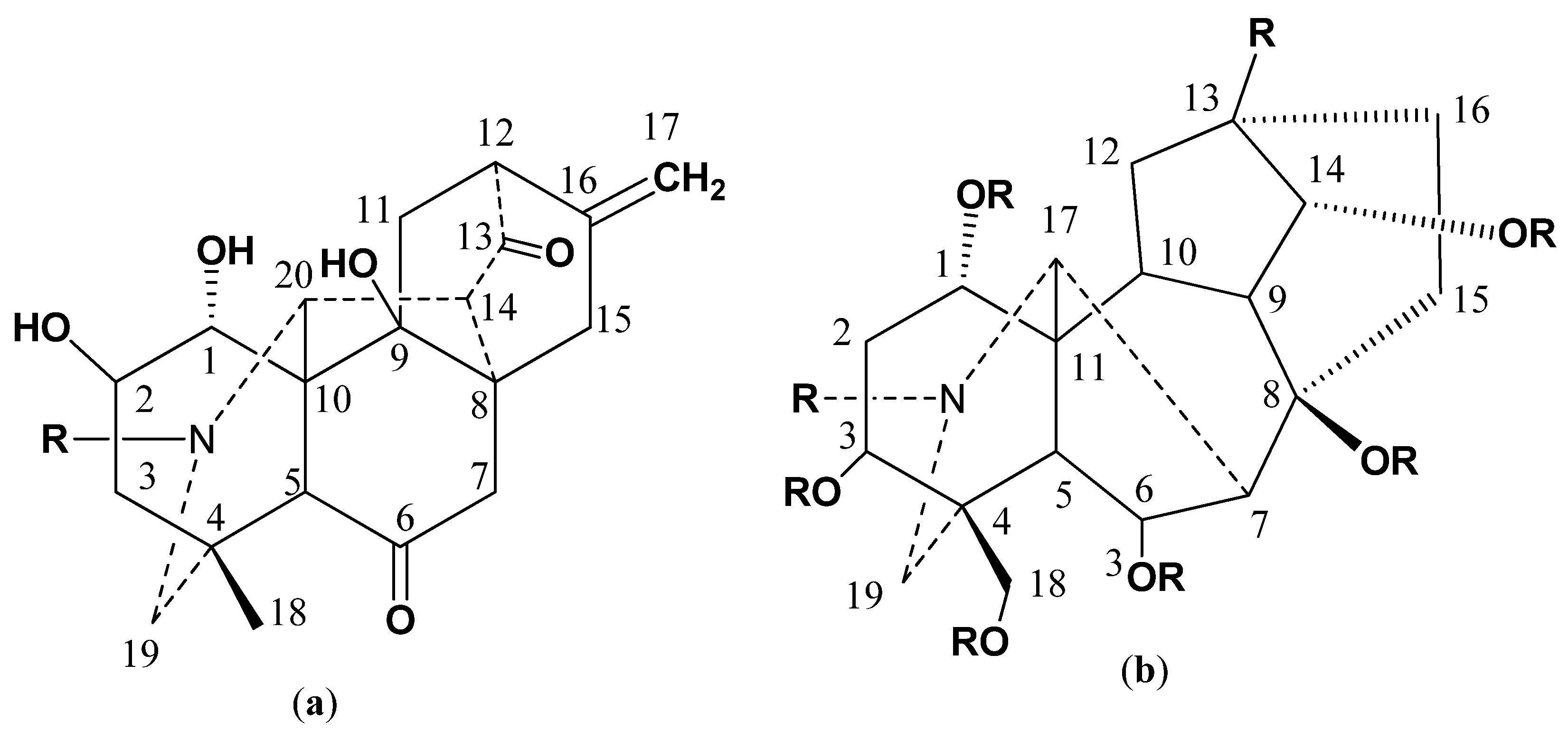
Figure 1. General structures of (a) C-20 diterpenoid alkaloids (b) C-19 diterpenoid alkaloids.
Molecules 27 04348 g001
Figure 2. Structures of Compounds 13.
Molecules 27 04348 g002
Figure 3. HMBC interaction in 13.
Molecules 27 04348 g003
Figure 4. Structural representation of Compound 1, with 50% probability of thermal ellipsoids, hydrogens were emitted for clarity.
Molecules 27 04348 g004
Figure 5. Structure of Compound 1, orientation of different rings and groups are shown accordingly.
Molecules 27 04348 g005
Figure 6. HOMO-LUMO of Compound 1 calculated at B3LYP/6-31þG (p).
Molecules 27 04348 g006
Figure 7. Docking pose of Compound 1 within cholinesterase target. (Docking score of = −14.457).
Molecules 27 04348 g007
Figure 8. Docking pose of Compound 2 within cholinesterase target (Docking score of = −15.591).
Molecules 27 04348 g008
Figure 9. Docking pose of Compound 3 within cholinesterase target (Docking score of = −16.400).
Molecules 27 04348 g009
Table 1. 13C NMR data of Compounds 13 in CDCl3.
PositionCompound 1 (150 MHz)Compound 2 (150 MHz)Compound 3 (125 MHz)
178.475.475.7
275.973.373.3
340.734.034.0
436.732.532.6
559.949.748.9
6209.4209.4209.6
749.744.044.2
84436.736.7
978.678.383.8
1039.743.644.0
1132.380.780.7
1253.489.378.3
13209.4209.534.8
1457.948.740.7
1534.930.826.9
16142141.9142
17110.3110.4110.4
1827.329.429.7
1963.194.294.2
2070.370.170.1
214838.738.7
Table 2. Crystal data and structure refinement of Compound 1.
Crystal Parameter Compound 1
Empirical formulaC21H27NO5Density (mg m−3)1.153
Formula weight373.43(h, k, l) min(−31, −5, −15)
Temperature (K)29.6(h, k, l) max(31, 9, 15)
Wavelength (Å)0.71073Theta (max)26.0
Crystal systemMonoclinicR (reflection)0.053(2408)
Space groupC2wR20.185
A25.726 (5) ÅNo of measured, independent and observed [I > 2σ(I)] reflections8474, 3521, 2408
B7.5766 (12) ÅRint0.053
C12.654 (2) Å(sin θ/λ)max−1)0.617
Volume Å32150.4 (6) Å3No. of reflections3521
μ (mm−1)0.08No. of parameters270
Z4No. of restraints1
Crystal size (mm)0.43 × 0.22 × 0.18Absolute structure parameter−0.7 (10)
Table 3. Calculated chemical parameters of Compound 1 computed at various level of DFT and basis sets, with values shown in atomic unit (au).
Compound-16-31G(d)/B3LYP6-311 + G(d,p)/wB97XD
EHOMO (au)−0.225−0.319
ELUMO (au)−0.034−0.027
ΔE = (ELUMO-EHOMO) (au)0.1910.292
IE = = −EHUMO (au)0.2250.319
EA = −ELUMO (au)0.0340.027
Global Hardness(η) = 1/2 (ELOMO-EHOMO)0.0950.146
Chemical Potential μ = 1/2 (EHOMO + ELUMO)−0.095−0.146
Global Electrophilicity ω = μ2/2η0.0480.073
Table 4. AChE and BChE inhibitory activities of alkaloids from D. Chitralense.
S. NoCompoundsAChE ± SEM a (μM)BChE ± SEM a (μM)Type of Inhibition
1Chitralinine A13.86 ± 0.3528.17 ± 0.92Competitive
2Chitralinine B11.64 ± 0.0824.31± 0.33Competitive
3Chitralinine-C12.11 ± 0.8226.35± 0.06Competitive
6Allanzanthane A8.23 ± 0.0118 ± 0.06
7Galanthamine b10.12 ±0.0620.62 ± 0.08
a Standard error of mean of five assays; b Positive control used in the assays.
Table 5. Interaction features of Compounds 13 against Cholinesterase.
InhibitorsMOE ScoreMOE Interactions ResiduesGorge Site Residues of Target
1−14.457Ser200, Gly119, Gly118, Ser122121(288)297(290)120(118)121(119)204(201)447(440)334(327)203(200)86(84)72(70)124(121)286(279)
2−15.591Tyr121, Ser122, His440, Ser200
3−16.400Tyr334, Asp72, Ser122, Ser200
Publisher’s Note: MDPI stays neutral with regard to jurisdictional claims in published maps and institutional affiliations.