Article

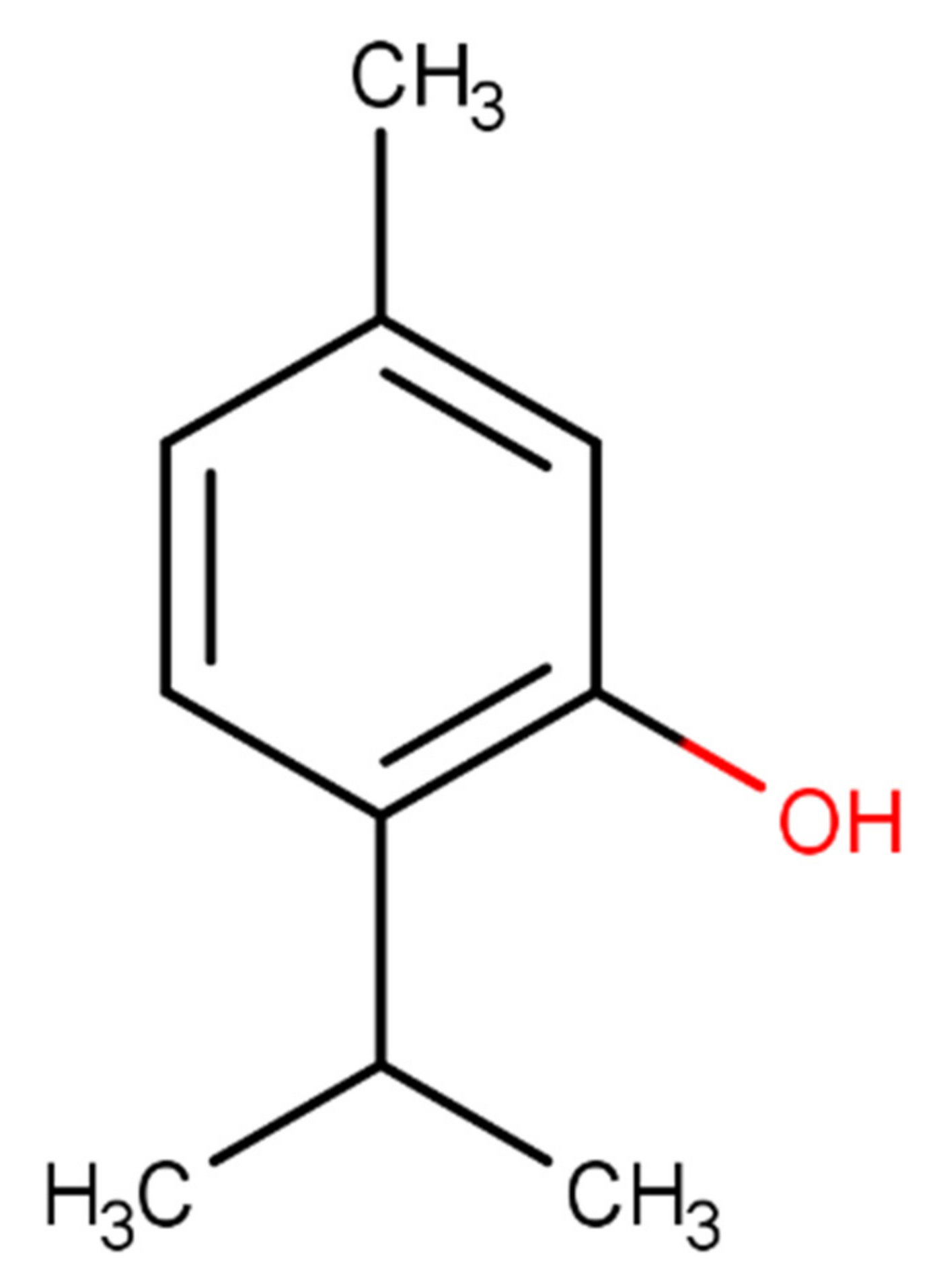
In Silico Evaluation of the Antimicrobial Activity of Thymol—Major Compounds in the Essential Oil of Lippia thymoides Mart. & Schauer (Verbenaceae)

by
Jorddy Neves Cruz
1,2,3,*,
Sebastião Gomes Silva
2,
Daniel Santiago Pereira
3,
Antônio Pedro da Silva Souza Filho
3,
Mozaniel Santana de Oliveira
2,
Rafael Rodrigues Lima
1 and
Eloisa Helena de Aguiar Andrade
2
1
Laboratory of Functional and Structural Biology, Institute of Biological Sciences, Federal University of Pará, Belém 66075-110, PA, Brazil
2
Adolpho Ducke Laboratory, Museu Paraense Emílio Goeldi, Belém 66077-830, PA, Brazil
3
Brazilian Agricultural Research Corporation (EMBRAPA), Belém 66095-100, PA, Brazil
*
Author to whom correspondence should be addressed.
Molecules 2022, 27(15), 4768; https://doi.org/10.3390/molecules27154768
Submission received: 22 June 2022 / Revised: 19 July 2022 / Accepted: 21 July 2022 / Published: 26 July 2022
(This article belongs to the Section Molecular Structure)

Abstract

:
In this paper, we evaluated the drug-receptor interactions responsible for the antimicrobial activity of thymol, the major compound present in the essential oil (EO) of Lippia thymoides (L. thymoides) Mart. & Schauer (Verbenaceae). It was previously reported that this EO exhibits antimicrobial activity against Candida albicans (C. albicans), Staphylococcus aureus (S. aureus), and Escherichia coli (E. coli). Therefore, we used molecular docking, molecular dynamics simulations, and free energy calculations to investigate the interaction of thymol with pharmacological receptors of interest to combat these pathogens. We found that thymol interacted favorably with the active sites of the microorganisms’ molecular targets. MolDock Score results for systems formed with CYP51 (C. albicans), Dihydrofolate reductase (S. aureus), and Dihydropteroate synthase (E. coli) were −77.85, −67.53, and −60.88, respectively. Throughout the duration of the MD simulations, thymol continued interacting with the binding pocket of the molecular target of each microorganism. The van der Waals (ΔEvdW = −24.88, −26.44, −21.71 kcal/mol, respectively) and electrostatic interaction energies (ΔEele = −3.94, −11.07, −12.43 kcal/mol, respectively) and the nonpolar solvation energies (ΔGNP = −3.37, −3.25, −2.93 kcal/mol, respectively) were mainly responsible for the formation of complexes with CYP51 (C. albicans), Dihydrofolate reductase (S. aureus), and Dihydropteroate synthase (E. coli).

1. Introduction

Essential oils (EOs), formed as secondary metabolites of aromatic plants, are biosynthesized in different plant organs such as flowers, leaves, and stems, among others [1,2]. These EOs chemical compositions and yields can change due to natural factors such as physiological, environmental, geographic, genetic, and plant evolution [3,4]. These oils play essential roles in plant protection and communication [5,6].
In the industry, EOs have been widely studied, mainly because of their potential applications as antimicrobials [7]. Over the years, the volatile compounds present in them have been employed for several pharmacological activities, such as antioxidant, anticancer, antiprotozoal, antimicrobial, anti-inflammatory, phytotoxic, and neuroprotective activities [8,9,10,11,12]. In a recent study, Tanrıkulu et al. [13] demonstrated that species such as Ocimum basilicum and Thymbra spicata show good antioxidant and antimicrobial activities against Staphylococcus aureus, Streptomyces murinus, Micrococcus luteus, Bacillus subtilis, Klebsiella pneumoniae, Pseudomonas aeruginosa, Yersina enterocolitica, Proteus algilus vulgaris, and Candida algilis, demonstrating the importance of EOs in the industry.
Although the antimicrobial action of EOs is not yet fully understood, it can be attributed to their ability to permeate the cell wall of microorganisms, which arises from their diverse and synergistic chemical compositions. The hydrophobic nature of EOs enables the partitioning of lipids from the cell membrane and mitochondria, making them more permeable; consequently, ions and critical cellular components (lipids, proteins, and nucleic acids) are extravasated from the cells, leading to eventual cell death. Generally, EOs have more action on gram-negative than on gram-positive bacterium, owing to the hydrophobic components of these oils interacting with the cell membranes of the former [14,15,16].
Different methods have been used to assess the antibacterial and antifungal properties of EOs. The most commonly used methods are the agar disk diffusion, minimum inhibition concentration (MIC), minimum bacterial concentration (MBC), and minimum fungicidal concentration (MFC) methods. Because the agar disk diffusion method is limited by the hydrophobic nature of EOs and plant extracts, preventing their uniform diffusion through the agar medium, most authors report their obtained results via the MIC, MBC, and MFC methods [17].
Silva et al. [18] evaluated the antimicrobial activity of EOs from Lippia thymoides Mart. & Schauer (Verbenaceae) against C. albicans, S. aureus, and E. coli. In this study, we used molecular modeling approaches to further analyze the investigations performed by Silva et al. We decided to conduct these investigations in silico to deepen our understanding of the interactions of volatile compounds with molecular targets that are vital for the viability of these microorganisms. Previous publications have shown that these approaches successfully reveal how drug-receptor interactions occur [19,20,21,22]. Therefore, we used molecular docking, molecular dynamics (MD) simulations, and affinity energy calculations to investigate how thymol (Figure 1)—the major compound of the EO from L. thymoides—interacts with the molecular targets from C. albicans, S. aureus, and E. coli.

2. Results and Discussions

2.1. Molecular Binding Mode

According to the molecular docking results, the thymol interacted favorably with the binding sites of the proteins (Table 1).
We identified the residues with which the ligand interacted and the chemical nature of these interactions (Figure 2).
After molecular docking, we performed MD simulations of 100 ns to evaluate how thymol can interact with the active site of the molecular targets. We then performed per-residue free energy decomposition using the MM/GBSA approach to evaluate the energetic contribution of the residues to the formation of the receptor-ligand complex, as shown in Figure 3.
EOs target the membrane of microorganisms such as C. albicans, S. aureus, and E. coli [23]. The total or partial rupture of the membrane allows the extravasation of intracellular liquid, causing a hydroelectrolytic imbalance capable of causing the death of the microorganism [24]. In addition, after passage through the membrane, EO compounds are free to interact with molecular targets essential for bacterial and fungal viability [25]. Thymol, for example, in our results, was shown to be able to interact with CYP51, Dihydrofolate reductase, and dihydropteroate synthase proteins that are critical for parasite viability. But in addition to these molecular targets, there are reports that this same compound can interact with other proteins. Dutta et al. concluded that the thymol in the EO of Trachyspermum ammi interacted with the pocket binding of glucosamine-6-phosphate synthase. The observed molecular interactions were essentially hydrophobic [26]. Some authors have identified that thymol can interact with dihydro-folate reductase from S. aureus [27,28]. Barbosa et al. (2021) determined that thymol can also inhibit NorA efflux pump inhibition in multidrug-resistant (MDR) S. aureus strains. In this paper, the authors evaluated that thymol establishes hydrophobic, hydrophilic, and electrostatic molecular interactions in the binding site [29]. Nagle et al. used thymol as a scaffold to perform a computer-aided drug design. Thymol and analogous compounds were investigated for their antimicrobial properties against E. coli and S. aureus. The molecular target chosen for the in silico studies was glucosamine-6-phosphate synthase GlcN-6-P synthase. The docking results showed that thymol and analogous showed a favorable interaction with the active site of the protein and their interactions were hydrophobic and electrostatic [30].
At the active site of CYP51 (C. albicans), the thymol established interactions with the heme group and Leu352. The interactions with the heme group were hydrophobic; one was of the π-π type and the other two were of the π-alkyl type. In addition to these three interactions, the heme group participated in the binding of the compound with the binding pocket, contributing with an energy value of −2.5 Kcal/mol. With Leu352, the interaction was of the alkyl type, and throughout the simulations, this residue provided an energy value of −1.24 Kcal/mol.
In the binding pocket of dihydrofolate reductase (S. aureus), thymol established a hydrogen bond with Phe92, and the energy value of its interaction was −3.1 Kcal/mol. Alkyl hydrophobic interactions were formed with Ile20, Ile31, and Ile50 with energy contributions of −0.98, −0.83, −0.67 Kcal/mol, respectively. The docking simulations demonstrated that NAPH established π-π interaction with thymol reaching energy value of −2.33 Kcal/mol throughout the MD simulations.
At the active site of dihydropteroate synthase (E. coli), the ligand formed alkyl type hydrophobic interactions with Arg63, Ile117, and Met139 with energy contribution values for a compost binding of −0.65, −0.58 e 0.61 Kcal/mol, respectively. With Arg225, the interaction occurred with energy value of −1.12 and a π-π interaction was established between the benzene ring and the guanidino group of the amino acid. Another π–π interaction was formed between the thymol and Phe190 side chain, exhibiting an energy value of −1.2 Kcal/mol.

2.2. DM Trajectory Stability

We used root-mean-square deviation (RMSD) calculations to assess each protein’s structural changes in the ligand and backbone. The RMSD of the ligand was calculated using its heavy atoms, while the Cα atoms were used for the RMSD plot of the protein backbone (Figure 4).
The RMSD values of the ligands in all systems showed minor variations along the trajectories. Thus, the ligand was accommodated in its binding site, preserving its interactions with the catalytic residues. The three-dimensional structures of the proteins did not undergo any change that would compromise the maintenance of the systems formed with thymol. Throughout the entire trajectory, the inhibitors remained in interaction with the proteins.

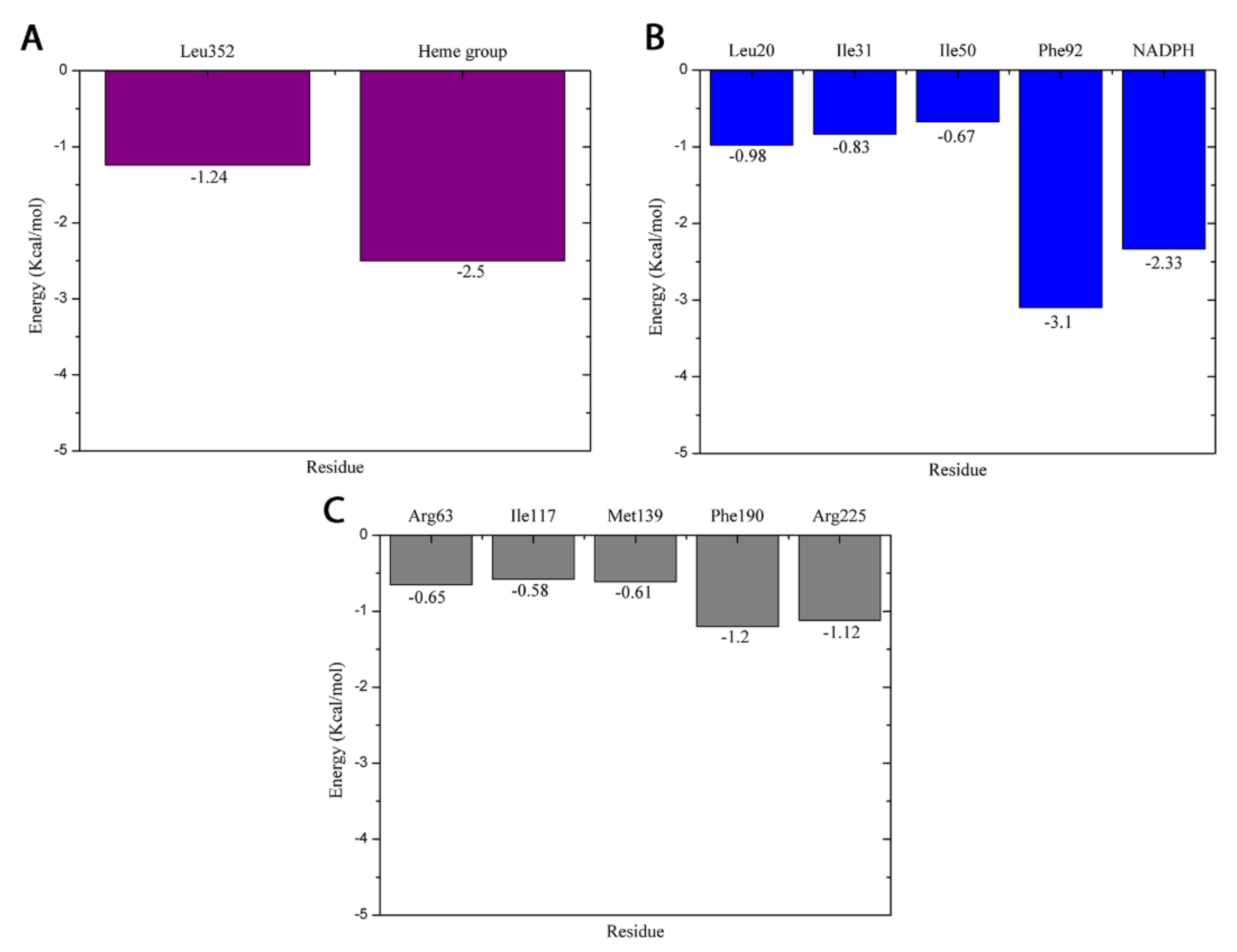
2.3. Free Energy Calculation of the Thymol-Receptor Complexes

In this study, the free energies of the thymol-receptor complexes were calculated to assess whether thymol interacts favorably with the target protein of each microorganism. For this, we used the MM/GBSA method, and the free energy and its energetic components were calculated using the last 500 frames of the MD trajectories. The results are presented in Table 2.
The main contributions to the formation of the complexes were the van der Waals (ΔEvdW), electrostatic (ΔEelectrostatic), and nonpolar solvation (ΔGnonpol) energies. The negative values of ΔGbind demonstrated that the interaction of the ligand with these molecular targets is favorable. Based on all our results, we can state that thymol is capable of interacting with and thereby inhibiting these molecular targets.

3. Materials and Methods

3.1. Molecular Docking

For the molecular docking study, thymol was selected since it was the major compound from the essential oil isolated from the leaves of L. thymoides Mart. & Schauer (Verbenaceae). Molecular docking was used to investigate the interaction between thymol and essential proteins of C. albicans, S. aureus, and E. coli. The proteins used as a molecular target are essential for the metabolic pathways of such microorganisms, in addition to being reported in the literature as targets for natural and synthetic products that combat these pathogens [31,32,33,34].
Thymol (2-Isopropyl-5-methylphenol) is a volatile substance present in the essential oil of several species. This substance is a monoterpenoid belongs being derived from a hydride of a p-cymene [35]. Thymol was drawn in GaussView 6 [36], and its structure was optimized via B3LYP/6-31G * [37] using the Gaussian quantum chemistry software 16 [38]. The three-dimensional structures of the proteins used as molecular targets were obtained from the Protein Data Bank (www.rcsb.org (1 January 2022). The corresponding PDB IDs are 5V5Z (C. albicans—SC5314) [39], 2W9H (S. aureus—ATCC) [33], and 1AJ2 (E. coli—ATCC) [34]. To study the interaction mode of this molecule with target proteins for drug action, the software Molegro Virtual Docker 5.5 [40]. The MolDock Score (GRID) function was used with a Grid resolution of 0.30 Å and radius of 7 Å, encompassing the entire crystallographic ligand-binding cavity found in the PDB of each protein. The MolDock SE algorithm was used with the number of runs equal to 10; 1500 max interactions, and max population size equal to 50. The full evaluation of 300 steps with neighbor distance factor equal to 1 and energy threshold equal to 100 was used during the molecular docking simulation. The RMSD limit for multiple cluster poses was set to <1.00 Å.

3.2. MD Simulations

The charges of the thymol atoms were calculated using the restrained electrostatic potential protocol using HF/6-31G * [41]. Parameter files were built using the Antechamber and General Amber Force Field [42]. The protonation states of the protein residues were studied from the results obtained using the PROPKA server [43].
The proteins were described by the ff14SB force field [44] in all simulations, with explicit water molecules described by the TIP3P model [45]. Each system was solvated in an octahedron periodic box with a 12 Å cutting radius in all directions from the solute. An adequate number of counterions were added to neutralize the partial charge of the systems.
The MD simulations were performed using the Amber 16 software [46,47]. Energy minimizations were performed with the sander module, while the heating, balance and production steps were performed with pmemd. CUDA.
The system’s energy minimization took place in three steps. In the first stage, 2000 cycles were executed using the steepest descent method and conjugate gradient algorithms, applying a harmonic force constant of 50 kcal·mol−1·Å−2 about the solute. In the second stage, the harmonic force constant applied to the solute was 25 kcal·mol−1·Å−2, and 1000 more cycles were run using the steepest descent method and conjugate gradient algorithms. In the last step, the constraints were removed, and 1000 cycles were run using the steepest descent method and conjugate gradient algorithm.
900 ps simulations were run to increase the system temperature from 0 to 300 k. Warming up was carried out in three steps. In the first, the solute was constrained with a harmonic force constant of 25 kcal·mol−1·Å−2, in this way, only the solvent and counterions were free to move. In the next two steps, the harmonic force constant was removed. To balance the complexes, we run 2 ns simulations at constant temperature and without restrictions. Then, for each complex, 100 ns of MD simulation with NVT ensemble were generated.
The particle mesh Ewald method [48] was used for calculating the electrostatic interaction energies, and the bonds involving hydrogen atoms were restricted with the SHAKE algorithm [49]. The temperature was controlled using a Langevin thermostat [50] with a collision frequency of 2 ps−1.

3.3. Free Energy Calculations

The free energy calculations were performed using the molecular mechanic’s generalized boron surface area (MM/GBSA) method [51,52,53]. For these calculations, we used the last 5 ns of the MD simulation trajectories. The free energy was calculated as follows:
ΔGbind = ΔH − TΔS ≈ ΔEMM + ΔGsolv − TΔS
where ΔGbind is the free energy of the complex, resulting from the sum of the molecular mechanics energy (ΔEMM), desolvation free energy (ΔGsolv), and entropy (−TΔS).
ΔEMM = ΔEinternal + ΔEelectrostatic + ΔEvdW
The energy of molecular gas phase mechanics (ΔEMM) can be described by the sum of the internal energy contributions (ΔEinternal); sum of the connection, angle, and dihedral energies; electrostatic contributions (ΔEelectrostatic); and van der Waals terms (ΔEvdW).
ΔGsolv = ΔGGB + ΔGnonpol
The desolvation free energy (ΔGsolv) is the sum of the polar (ΔGGB) and nonpolar (ΔGnonpol) contributions. The polar desolvation term was calculated using the implicit generalized born (GB) approach.

4. Conclusions

According to our results for the binding energies obtained using the MM/GBSA method, thymol interacts favorably with the molecular targets of microorganisms. The binding free energies (ΔGbind) for thymol interacting with CYP51, dihydrofolate reductase, and dihydropteroate synthase proteins demonstrate that the formation of the complexes is favorable; the ΔGbind values obtained were: −20.04, −24.73, −17.84 kcal/mol, respectively. During the 100 ns of MD simulations, thymol remained in interaction with the binding pockets of the enzymes. The RMSD values obtained over 100 ns of MD simulation showed that the thymol are stable in the binding pocket. The main interactions established by the ligand with the active site residues were found to be hydrophobic.

Author Contributions

Conceptualization, J.N.C.; methodology, J.N.C.; software, J.N.C.; validation, S.G.S.; formal analysis, M.S.d.O.; investigation, J.N.C.; resources, D.S.P. and A.P.d.S.S.F.; data curation, D.S.P. and A.P.d.S.S.F.; writing—original draft preparation, J.N.C.; writing—review and editing, D.S.P. and A.P.d.S.S.F.; visualization, M.S.d.O.; supervision, R.R.L. and E.H.d.A.A.; project administration, R.R.L. and E.H.d.A.A.; funding acquisition, R.R.L. and E.H.d.A.A. All authors have read and agreed to the published version of the manuscript.

Funding

This study was financed in part by the Coordenação de Aperfeiçoamento de Pessoal de Nível Superior-Brasil (CAPES)-Finance Code 001. Universidade Federal do Pará/Propesp/PROGRAMA DE APOIO À PUBLICAÇÃO QUALIFICADA-PAPQ-EDITAL 2022.

Institutional Review Board Statement

Not applicable.

Informed Consent Statement

Informed consent was obtained from all subjects involved in the study.

Data Availability Statement

Not applicable.

Acknowledgments

The author M.S.d.O., thanks PCI-MCTIC/MPEG, as well as CNPq for the scholarship process number: 301194/2021-1.

Conflicts of Interest

The authors declare no conflict of interest.

Sample Availability

Samples of the L. thymoides Mart. & Schauer (Verbenaceae) are available from the Museu Emilio Goeldi.

References

  1. Ferreira, O.O.; Neves da Cruz, J.; de Jesus Pereira Franco, C.; Silva, S.G.; da Costa, W.A.; de Oliveira, M.S.; de Aguiar Andrade, E.H. First Report on Yield and Chemical Composition of Essential Oil Extracted from Myrcia Eximia DC (Myrtaceae) from the Brazilian Amazon. Molecules 2020, 25, 783. [Google Scholar] [CrossRef] [PubMed] [Green Version]
  2. Hartmann, T. From Waste Products to Ecochemicals: Fifty Years Research of Plant Secondary Metabolism. Phytochemistry 2007, 68, 2831–2846. [Google Scholar] [CrossRef] [PubMed]
  3. Figueiredo, A.C.; Barroso, J.G.; Pedro, L.G.; Scheffer, J.J.C. Factors Affecting Secondary Metabolite Production in Plants: Volatile Components and Essential Oils. Flavour Fragr. J. 2008, 23, 213–226. [Google Scholar] [CrossRef]
  4. Ferreira, O.O.; Cruz, J.N.; de Moraes, Â.A.; de Jesus Pereira Franco, C.; Lima, R.R.; Anjos, T.O.; Siqueira, G.M.; Nascimento, L.D.; Cascaes, M.M.; de Oliveira, M.S.; et al. Essential Oil of the Plants Growing in the Brazilian Amazon: Chemical Composition, Antioxidants, and Biological Applications. Molecules 2022, 27, 4373. [Google Scholar] [CrossRef]
  5. Do Nascimento, L.D.; de Moraes, A.A.B.; da Costa, K.S.; Galúcio, J.M.P.; Taube, P.S.; Costa, C.M.L.; Cruz, J.N.; Andrade, E.H.d.A.; de Faria, L.J.G. Bioactive Natural Compounds and Antioxidant Activity of Essential Oils from Spice Plants: New Findings and Potential Applications. Biomolecules 2020, 10, 988. [Google Scholar] [CrossRef]
  6. Sodeifian, G.; Sajadian, S.A.; Saadati Ardestani, N. Experimental Optimization and Mathematical Modeling of the Supercritical Fluid Extraction of Essential Oil from Eryngium Billardieri: Application of Simulated Annealing (SA) Algorithm. J. Supercrit. Fluids 2017, 127, 146–157. [Google Scholar] [CrossRef]
  7. Cascaes, M.M.; Dos, O.; Carneiro, S.; Diniz Do Nascimento, L.; Antônio Barbosa De Moraes, Â.; Santana De Oliveira, M.; Neves Cruz, J.; Skelding, G.M.; Guilhon, P.; Helena De Aguiar Andrade, E.; et al. Essential Oils from Annonaceae Species from Brazil: A Systematic Review of Their Phytochemistry, and Biological Activities. Int. J. Mol. Sci. 2021, 22, 12140. [Google Scholar] [CrossRef]
  8. Santana de Oliveira, M.; Pereira da Silva, V.M.; Cantão Freitas, L.; Gomes Silva, S.; Nevez Cruz, J.; de Aguiar Andrade, E.H. Extraction Yield, Chemical Composition, Preliminary Toxicity of Bignonia Nocturna (Bignoniaceae) Essential Oil and in Silico Evaluation of the Interaction. Chem. Biodivers. 2021, 18, cbdv.202000982. [Google Scholar] [CrossRef]
  9. Benavides, S.; Villalobos-Carvajal, R.; Reyes, J.E. Physical, Mechanical and Antibacterial Properties of Alginate Film: Effect of the Crosslinking Degree and Oregano Essential Oil Concentration. J. Food Eng. 2012, 110, 232–239. [Google Scholar] [CrossRef]
  10. Leal, S.M.; Pino, N.; Stashenko, E.E.; Martínez, J.R.; Escobar, P. Antiprotozoal Activity of Essential Oils Derived from Piper Spp. Grown in Colombia. J. Essent. Oil Res. 2013, 25, 512–519. [Google Scholar] [CrossRef]
  11. Silva, F.V.; Guimarães, A.G.; Silva, E.R.S.; Sousa-Neto, B.P.; MacHado, F.D.F.; Quintans-Júnior, L.J.; Arcanjo, D.D.R.; Oliveira, F.A.; Oliveira, R.C.M. Anti-Inflammatory and Anti-Ulcer Activities of Carvacrol, a Monoterpene Present in the Essential Oil of Oregano. J. Med. Food 2012, 15, 984–991. [Google Scholar] [CrossRef]
  12. Da Silva Júnior, O.S.; Franco, C.d.J.P.; de Moraes, A.A.B.; Cruz, J.N.; da Costa, K.S.; do Nascimento, L.D.; Andrade, E.H.d.A. In Silico Analyses of Toxicity of the Major Constituents of Essential Oils from Two Ipomoea L. Species. Toxicon 2021, 195, 111–118. [Google Scholar] [CrossRef]
  13. Tanrıkulu, G.İ.; Ertürk, Ö.; Yavuz, C.; Can, Z.; Çakır, H.E. Chemical Compositions, Antioxidant and Antimicrobial Activities of the Essential Oil and Extracts of Lamiaceae Family (Ocimum Basilicum and Thymbra Spicata) from Turkey. Int. J. Second. Metab. 2017, 4, 340–348. [Google Scholar] [CrossRef]
  14. Calo, J.R.; Crandall, P.G.; O’Bryan, C.A.; Ricke, S.C. Essential Oils as Antimicrobials in Food Systems—A Review. Food Control 2015, 54, 111–119. [Google Scholar] [CrossRef]
  15. Akthar, M.S.; Degaga, B.; Azam, T. Antimicrobial Activity of Essential Oils Extracted from Medicinal Plants against the Pathogenic Microorganisms: A Review. Issues Biol. Sci. Pharm. Res. 2014, 2, 1–7. [Google Scholar]
  16. Solórzano-Santos, F.; Miranda-Novales, M.G. Essential Oils from Aromatic Herbs as Antimicrobial Agents. Curr. Opin. Biotechnol. 2012, 23, 136–141. [Google Scholar] [CrossRef]
  17. Bilia, A.R.; Santomauro, F.; Sacco, C.; Bergonzi, M.C.; Donato, R. Essential Oil of Artemisia Annua L.: An Extraordinary Component with Numerous Antimicrobial Properties. Evid. Based Complement. Altern. Med. 2014, 2014, 159819. [Google Scholar] [CrossRef] [Green Version]
  18. Silva, S.G.; de Oliveira, M.S.; Cruz, J.N.; da Costa, W.A.; da Silva, S.H.M.; Barreto Maia, A.A.; de Sousa, R.L.; Carvalho Junior, R.N.; de Aguiar Andrade, E.H. Supercritical CO2 Extraction to Obtain Lippia Thymoides Mart. & Schauer (Verbenaceae) Essential Oil Rich in Thymol and Evaluation of Its Antimicrobial Activity. J. Supercrit. Fluids 2021, 168, 105064. [Google Scholar] [CrossRef]
  19. Costa, E.B.B.; Silva, R.C.C.; Espejo-Román, J.M.M.; Neto, M.F.d.A.F.d.A.; Cruz, J.N.N.; Leite, F.H.A.H.A.; Silva, C.H.T.P.H.T.P.; Pinheiro, J.C.C.; Macêdo, W.J.C.J.C.; Santos, C.B.R.B.R. Chemometric Methods in Antimalarial Drug Design from 1,2,4,5-Tetraoxanes Analogues. SAR QSAR Environ. Res. 2020, 31, 677–695. [Google Scholar] [CrossRef]
  20. Castro, A.L.G.; Cruz, J.N.; Sodré, D.F.; Correa-Barbosa, J.; Azonsivo, R.; de Oliveira, M.S.; de Sousa Siqueira, J.E.; da Rocha Galucio, N.C.; de Oliveira Bahia, M.; Burbano, R.M.R.; et al. Evaluation of the Genotoxicity and Mutagenicity of Isoeleutherin and Eleutherin Isolated from Eleutherine Plicata Herb. Using Bioassays and in Silico Approaches. Arab. J. Chem. 2021, 14, 103084. [Google Scholar] [CrossRef]
  21. Galucio, N.C.d.R.; Moysés, D.d.A.; Pina, J.R.S.; Marinho, P.S.B.; Gomes Júnior, P.C.; Cruz, J.N.; Vale, V.V.; Khayat, A.S.; Marinho, A.M.d.R. Antiproliferative, Genotoxic Activities and Quantification of Extracts and Cucurbitacin B Obtained from Luffa Operculata (L.) Cogn. Arab. J. Chem. 2022, 15, 103589. [Google Scholar] [CrossRef]
  22. Rego, C.M.A.; Francisco, A.F.; Boeno, C.N.; Paloschi, M.V.; Lopes, J.A.; Silva, M.D.S.; Santana, H.M.; Serrath, S.N.; Rodrigues, J.E.; Lemos, C.T.L.; et al. Inflammasome NLRP3 Activation Induced by Convulxin, a C-Type Lectin-like Isolated from Crotalus Durissus Terrificus Snake Venom. Sci. Rep. 2022, 12, 4706. [Google Scholar] [CrossRef]
  23. Xu, J.; Zhou, F.; Ji, B.P.; Pei, R.S.; Xu, N. The Antibacterial Mechanism of Carvacrol and Thymol against Escherichia Coli. Lett. Appl. Microbiol. 2008, 47, 174–179. [Google Scholar] [CrossRef]
  24. Nazzaro, F.; Fratianni, F.; De Martino, L.; Coppola, R.; De Feo, V. Effect of Essential Oils on Pathogenic Bacteria. Pharmaceuticals 2013, 6, 1451–1474. [Google Scholar] [CrossRef]
  25. Chouhan, S.; Sharma, K.; Guleria, S. Antimicrobial Activity of Some Essential Oils—Present Status and Future Perspectives. Medicines 2017, 4, 58. [Google Scholar] [CrossRef] [Green Version]
  26. Dutta, S.; Kundu, A.; Saha, S.; Prabhakaran, P.; Mandal, A. Characterization, Antifungal Properties and in Silico Modelling Perspectives of Trachyspermum Ammi Essential Oil. LWT 2020, 131, 109786. [Google Scholar] [CrossRef]
  27. Arokiyaraj, S.; Choi, S.H.; Lee, Y.; Bharanidharan, R.; Hairul-Islam, V.I.; Vijayakumar, B.; Oh, Y.K.; Dinesh-Kumar, V.; Vincent, S.; Kim, K.H. Characterization of Ambrette Seed Oil and Its Mode of Action in Bacteria. Molecules 2015, 20, 384–395. [Google Scholar] [CrossRef] [PubMed] [Green Version]
  28. Bishoyi, A.K.; Mahapatra, M.; Paidesetty, S.K.; Padhy, R.N. Design, Molecular Docking, and Antimicrobial Assessment of Newly Synthesized Phytochemical Thymol Mannich Base Derivatives. J. Mol. Struct. 2021, 1244, 130908. [Google Scholar] [CrossRef]
  29. Dos Santos Barbosa, C.R.; Scherf, J.R.; de Freitas, T.S.; de Menezes, I.R.A.; Pereira, R.L.S.; dos Santos, J.F.S.; de Jesus, S.S.P.; Lopes, T.P.; de Sousa Silveira, Z.; de Morais Oliveira-Tintino, C.D.; et al. Effect of Carvacrol and Thymol on NorA Efflux Pump Inhibition in Multidrug-Resistant (MDR) Staphylococcus Aureus Strains. J. Bioenerg. Biomembr. 2021, 53, 489–498. [Google Scholar] [CrossRef] [PubMed]
  30. Nagle, P.; Pawar, Y.; Sonawane, A.; Bhosale, S.; More, D. Docking Simulation, Synthesis and Biological Evaluation of Novel Pyridazinone Containing Thymol as Potential Antimicrobial Agents. Med. Chem. Res. 2014, 23, 918–926. [Google Scholar] [CrossRef]
  31. Dos Santos, K.L.B.; Cruz, J.N.; Silva, L.B.; Ramos, R.S.; Neto, M.F.A.; Lobato, C.C.; Ota, S.S.B.; Leite, F.H.A.; Borges, R.S.; da Silva, C.H.T.P.; et al. Identification of Novel Chemical Entities for Adenosine Receptor Type 2a Using Molecular Modeling Approaches. Molecules 2020, 25, 1245. [Google Scholar] [CrossRef] [Green Version]
  32. Keniya, M.V.; Sabherwal, M.; Wilson, R.K.; Woods, M.A.; Sagatova, A.A.; Tyndall, J.D.A.; Monk, B.C. Crystal Structures of Full-Length Lanosterol 14α-Demethylases of Prominent Fungal Pathogens Candida Albicans and Candida Glabrata Provide Tools for Antifungal Discovery. Antimicrob. Agents Chemother. 2018, 62, e01134-18. [Google Scholar] [CrossRef] [Green Version]
  33. Heaslet, H.; Harris, M.; Fahnoe, K.; Sarver, R.; Putz, H.; Chang, J.; Subramanyam, C.; Barreiro, G.; Miller, J.R. Structural Comparison of Chromosomal and Exogenous Dihydrofolate Reductase from Staphylococcus Aureus in Complex with the Potent Inhibitor Trimethoprim. Proteins Struct. Funct. Bioinforma 2009, 76, 706–717. [Google Scholar] [CrossRef]
  34. Achari, A.; Somers, D.O.; Champness, J.N.; Bryant, P.K.; Rosemond, J.; Stammers, D.K. Crystal Structure of the Anti-Bacterial Sulfonamide Drug Target Dihydropteroate Synthase. Nat. Struct. Biol. 1997, 4, 490–497. [Google Scholar] [CrossRef]
  35. Escobar, A.; Pérez, M.; Romanelli, G.; Blustein, G. Thymol Bioactivity: A Review Focusing on Practical Applications. Arab. J. Chem. 2020, 13, 9243–9269. [Google Scholar] [CrossRef]
  36. Dennington, R.; Keith, T.; Millam, J. GaussView, Version 5; Semichem Inc.: Shawnee Mission, KS, USA, 2009. [Google Scholar]
  37. Becke, A.D. Density-Functional Thermochemistry. III. The Role of Exact Exchange. J. Chem. Phys. 1993, 98, 5648–5652. [Google Scholar] [CrossRef] [Green Version]
  38. Frisch, M.J.; Trucks, G.W.; Schlegel, H.B.; Scuseria, G.E.; Robb, M.A.; Cheeseman, J.R.; Scalmani, G.; Barone, V.; Petersson, G.A.; Nakatsuji, H.; et al. Gaussian 16 Revision 16.A.03; Gaussian Inc.: Wallingford, CT, USA, 2016. [Google Scholar]
  39. Monk, B.C.; Keniya, M.V.; Sabherwal, M.; Wilson, R.K.; Graham, D.O.; Hassan, H.F.; Chen, D.; Tyndall, J.D.A. Azole Resistance Reduces Susceptibility to the Tetrazole Antifungal VT-1161. Antimicrob. Agents Chemother. 2019, 63, e02114-18. [Google Scholar] [CrossRef] [Green Version]
  40. Thomsen, R.; Christensen, M.H. MolDock: A New Technique for High-Accuracy Molecular Docking. J. Med. Chem. 2006, 49, 3315–3321. [Google Scholar] [CrossRef]
  41. Wang, J.; Cieplak, P.; Kollman, P.A. How Well Does a Restrained Electrostatic Potential (RESP) Model Perform in Calculating Conformational Energies of Organic and Biological Molecules? J. Comput. Chem. 2000, 21, 1049–1074. [Google Scholar] [CrossRef]
  42. Wang, J.; Wolf, R.M.; Caldwell, J.W.; Kollman, P.A.; Case, D.A. Development and Testing of a General Amber Force Field. J. Comput. Chem. 2004, 25, 1157–1174. [Google Scholar] [CrossRef]
  43. Dolinsky, T.J.; Nielsen, J.E.; McCammon, J.A.; Baker, N.A. PDB2PQR: An Automated Pipeline for the Setup of Poisson-Boltzmann Electrostatics Calculations. Nucleic Acids Res. 2004, 32, W665–W667. [Google Scholar] [CrossRef]
  44. Maier, J.A.; Martinez, C.; Kasavajhala, K.; Wickstrom, L.; Hauser, K.E.; Simmerling, C. Ff14SB: Improving the Accuracy of Protein Side Chain and Backbone Parameters from Ff99SB. J. Chem. Theory Comput. 2015, 11, 3696–3713. [Google Scholar] [CrossRef] [Green Version]
  45. Jorgensen, W.L.; Chandrasekhar, J.; Madura, J.D.; Impey, R.W.; Klein, M.L. Comparison of Simple Potential Functions for Simulating Liquid Water. J. Chem. Phys. 1983, 79, 926–935. [Google Scholar] [CrossRef]
  46. Santos, C.B.R.; Santos, K.L.B.; Cruz, J.N.; Leite, F.H.A.; Borges, R.S.; Taft, C.A.; Campos, J.M.; Silva, C.H.T.P. Molecular Modeling Approaches of Selective Adenosine Receptor Type 2A Agonists as Potential Anti-Inflammatory Drugs. J. Biomol. Struct. Dyn. 2020, 39, 3115–3127. [Google Scholar] [CrossRef]
  47. Araújo, P.H.F.; Ramos, R.S.; da Cruz, J.N.; Silva, S.G.; Ferreira, E.F.B.; de Lima, L.R.; Macêdo, W.J.C.; Espejo-Román, J.M.; Campos, J.M.; Santos, C.B.R. Identification of Potential COX-2 Inhibitors for the Treatment of Inflammatory Diseases Using Molecular Modeling Approaches. Molecules 2020, 25, 4183. [Google Scholar] [CrossRef]
  48. Darden, T.; York, D.; Pedersen, L. Particle Mesh Ewald: An N·log(N) Method for Ewald Sums in Large Systems. J. Chem. Phys. 1993, 98, 10089–10092. [Google Scholar] [CrossRef] [Green Version]
  49. Ryckaert, J.P.; Ciccotti, G.; Berendsen, H.J.C. Numerical Integration of the Cartesian Equations of Motion of a System with Constraints: Molecular Dynamics of n-Alkanes. J. Comput. Phys. 1977, 23, 327–341. [Google Scholar] [CrossRef] [Green Version]
  50. Lzaguirre, J.A.; Catarello, D.P.; Wozniak, J.M.; Skeel, R.D. Langevin Stabilization of Molecular Dynamics. J. Chem. Phys. 2001, 114, 2090–2098. [Google Scholar] [CrossRef]
  51. Hou, T.; Wang, J.; Li, Y.; Wang, W. Assessing the Performance of the MM/PBSA and MM/GBSA Methods. 1. The Accuracy of Binding Free Energy Calculations Based on Molecular Dynamics Simulations. J. Chem. Inf. Model. 2011, 51, 69–82. [Google Scholar] [CrossRef] [PubMed]
  52. Leão, R.P.; Cruz, J.V.J.N.; da Costa, G.V.; Cruz, J.V.J.N.; Ferreira, E.F.B.; Silva, R.C.; de Lima, L.R.; Borges, R.S.; Dos Santos, G.B.; Santos, C.B.R. Identification of New Rofecoxib-Based Cyclooxygenase-2 Inhibitors: A Bioinformatics Approach. Pharmaceuticals 2020, 13, 209. [Google Scholar] [CrossRef] [PubMed]
  53. Lima, A.R.J.A.d.M.; Siqueira, A.S.; Möller, M.L.S.; Souza, R.C.D.; Cruz, J.N.; Lima, A.R.J.A.d.M.; da Silva, R.C.; Aguiar, D.C.F.; Junior, J.L.d.S.G.V.; Gonçalves, E.C. In Silico Improvement of the Cyanobacterial Lectin Microvirin and Mannose Interaction. J. Biomol. Struct. Dyn. 2020, 40, 1064–1073. [Google Scholar] [CrossRef]
Figure 1. The molecular structure of thymol.
Molecules 27 04768 g001
Figure 2. Intermolecular interactions of the drug-receptor systems. Molecular binding mode of thymol interacting with the active site residues of (A) CYP51 (C. albicans), (B) dihydrofolate reductase (S. aureus), and (C) dihydropteroate synthase (E. coli).
Molecules 27 04768 g002
Figure 3. Per-residue free energy decomposition: (A) CYP51 (C. albicans), (B) dihydrofolate reductase (S. aureus), and (C) dihydropteroate synthase (E. coli).
Molecules 27 04768 g003
Figure 4. Intermolecular interactions of the drug-receptor systems. Molecular binding mode of thymol interacting with the active site residues of (A) CYP51 (C. albicans), (B) dihydrofolate reductase (S. aureus), and (C) dihydropteroate synthase (E. coli).
Molecules 27 04768 g004
Table 1. Scoring functions obtained with the MolDock score.
Drug TargetMolDock Score (Kcal/mol)
CYP51 (C. albicans)−77.85
Dihydrofolate reductase (S. aureus)−67.53
Dihydropteroate synthase (E. coli)−60.88
Table 2. Binding energy values (ΔGbind) of the drug-receptor systems. ΔEvdW, van der Waals contributions; ΔEelectrostatic, electrostatic energy; ΔGGB, polar solvation energy; ΔGnonpol, nonpolar solvation energy. All values are in kcal/mol.
SystemΔEvdWΔEeleΔGGBΔGNPΔGbind
CYP51(C. albicans)−24.88−3.9412.15−3.37−20.04
dihydrofolate reductase (S. aureus)−26.44−11.0716.03−3.25−24.73
dihydropteroate synthase (E. coli)−21.71−12.4319.23−2.93−17.84
Publisher’s Note: MDPI stays neutral with regard to jurisdictional claims in published maps and institutional affiliations.

