Article

Photodynamic Inactivation of Microorganisms Using Semisynthetic Chlorophyll a Derivatives as Photosensitizers

by
Marciana Pierina Uliana
1,2,3,*,
Andréia da Cruz Rodrigues
3,
Bruno Andrade Ono
1,
Sebastião Pratavieira
1,
Kleber Thiago de Oliveira
2 and
Cristina Kurachi
1
1
Instituto de Física de São Carlos, Universidade de São Paulo, São Carlos, São Paulo CEP 13560-970, Brazil
2
Departamento de Química, Universidade Federal de São Carlos, Rodovia Washington Luís, km 235-SP-310, São Carlos, São Paulo CEP 13565-905, Brazil
3
Universidade Federal da Integração Latino-Americana, Foz do Iguaçu CEP 85866-000, Brazil
*
Author to whom correspondence should be addressed.
Molecules 2022, 27(18), 5769; https://doi.org/10.3390/molecules27185769
Submission received: 11 July 2022 / Revised: 29 August 2022 / Accepted: 1 September 2022 / Published: 6 September 2022
(This article belongs to the Special Issue Porphyrin-Based Compounds: Synthesis and Application)

Abstract

:
In this study, we describe the semisynthesis of cost-effective photosensitizers (PSs) derived from chlorophyll a containing different substituents and using previously described methods from the literature. We compared their structures when used in photodynamic inactivation (PDI) against Staphylococcus aureus, Escherichia coli, and Candida albicans under different conditions. The PSs containing carboxylic acids and butyl groups were highly effective against S. aureus and C. albicans following our PDI protocol. Overall, our results indicate that these nature-inspired PSs are a promising alternative to selectively inactivate microorganisms using PDI.

1. Introduction

Photodynamic reactions have been demonstrated to be an efficient alternative for the treatment of cancer [1,2,3,4,5], psoriasis [6], herpes [7], dermatological treatment [8], periodontics [9], canine otitis [10], control of cariogenic bacteria [11,12], and pneumonia [13]. In particular, antimicrobial photodynamic therapy (aPDT) or photodynamic inactivation (PDI) represents an interesting alternative for microbiological control because this technique is minimally toxic, noninvasive, minimizes the use of antibiotics, and due to its action over a broad spectrum of biomolecules, the risk of resistance is unlikely [14,15,16,17,18].
PDI has been used in the treatment of diseases caused by various microorganisms, including Gram-positive and Gram-negative bacteria and fungi [19], such as the inactivation of Candida albicans, which causes diseases in patients with low immunity [20,21]. It has also been used in the inactivation of Aedes aegypti mosquito larvae, a vector of the dengue, Zika, and chikungunya arboviruses [22,23,24,25,26]. Furthermore, PDI has gained attention in the inactivation of viruses, which have also shown resistance to drugs, such as an alternative to antiviral treatments against human papillomavirus and hepatitis B virus [27]. Recent studies have also shown the use of PDI for the treatment of severe acute respiratory syndrome caused by the new coronavirus (COVID-19) [28]. As PDI can inactivate DNA- or RNA-based viruses, these studies suggest considerable potential for use in virus photoinactivation in the future [29].
In general, photodynamic reactions require the presence of a photosensitizer, which is activated by light at a specific wavelength, allowing the production of reactive oxygen species (ROS). The main ROS are singlet oxygen, superoxide anions, hydroxyl radicals, and hydrogen peroxide. The quantum yields of each of these ROS depend on the PS and the conditions of the medium. After the formation of ROS, they interact with the target cells, causing death [30,31,32,33,34].
The most commonly used photosensitizers are methylene blue [35], porphyrins [36], chlorins [16,37], curcuminoids [38,39], and bacteriochlorins [40]. However, natural product derivatives, such as chlorin-e6 (chl-e6) obtained from chlorophyll a, are relatively low-cost photosensitizers and present many advantages in terms of pharmacokinetics, as they are easily eliminated from the body [11,41].
Chlorophyll a is formally a chlorin derivative, with four nitrogen atoms surrounding a central magnesium atom, along with numerous attached side chains and a hydrocarbon chain. Chlorins are excellent photosensitizers, and several synthetic chlorin analogues, such as m-tetrahydroxyphenylchlorin and mono-L aspartyl chlorin e6, have been used. Some substances, such as porphyrins, chlorins, and bacteriochlorins, stand out for their application in PDI: they present selected photophysical characteristics, allowing structural modifications to promote better solubility and amphiphilicity and improve their properties in several treatments [42]. Natural products are sources of inspiration for the development of several drugs [43]. The natural chlorophyll pigments from the cyanobacterium Spirulina maxima are abundant and easy to obtain. According to our protocol, [44] once dried, S. maxima is treated with methanol with 5% sulfuric acid, it gives rise to methyl pheophorbide a, and, after treatment with different amines or molecular oxygen, generates methyl pheophorbide a derivatives, including purpurin-18.
In this study, we propose the diverse semisynthesis of chlorophyll a derivatives, using simple reactions to evaluate these photosensitizers against microorganisms. All the chemical modifications performed with chlorophyll a were also aimed at conferring amphiphilicity to the PS, as this strategy has succeeded well in many approaches for PDT or PDI studies [45,46,47,48].

2. Results and Discussion

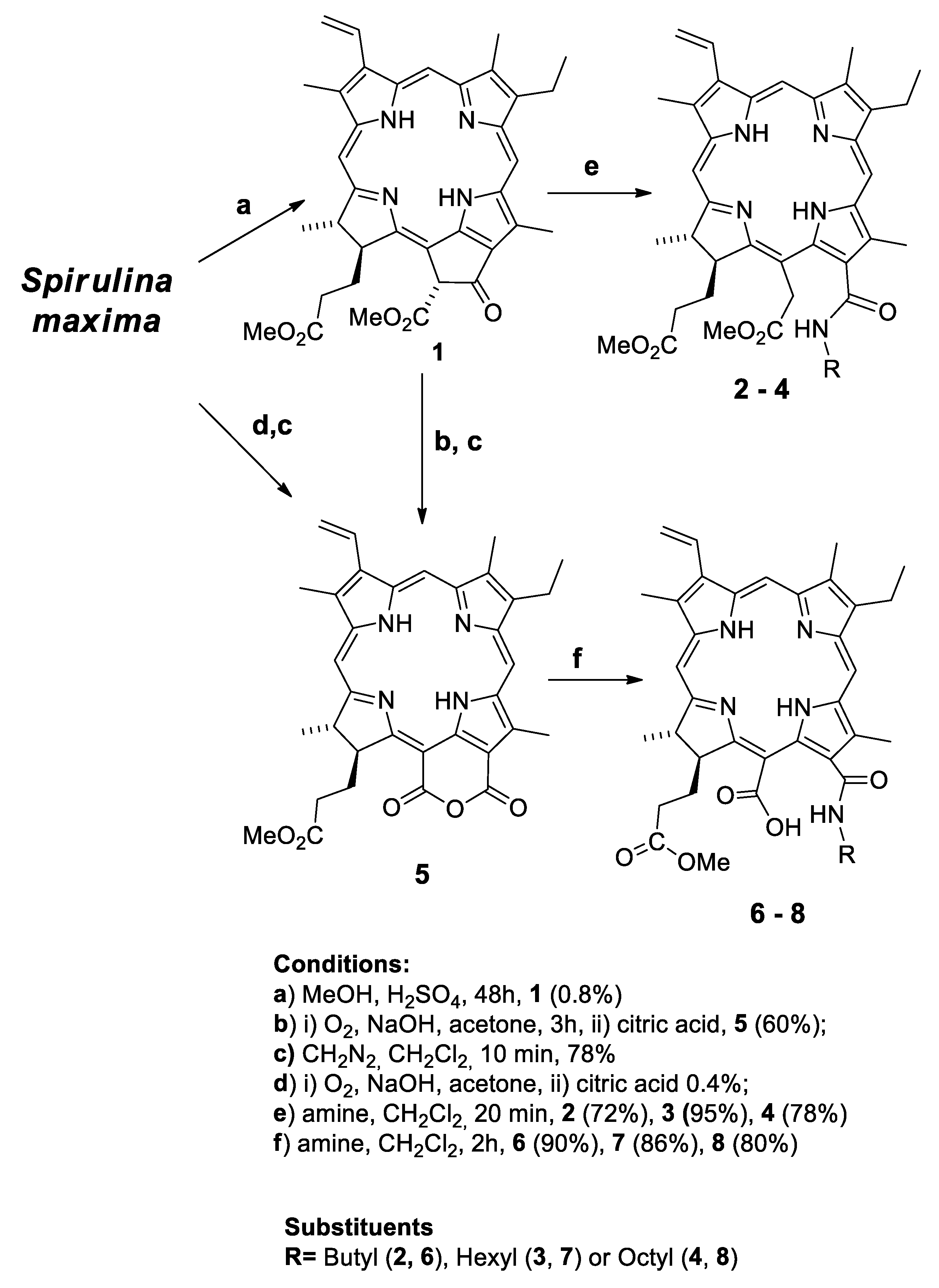
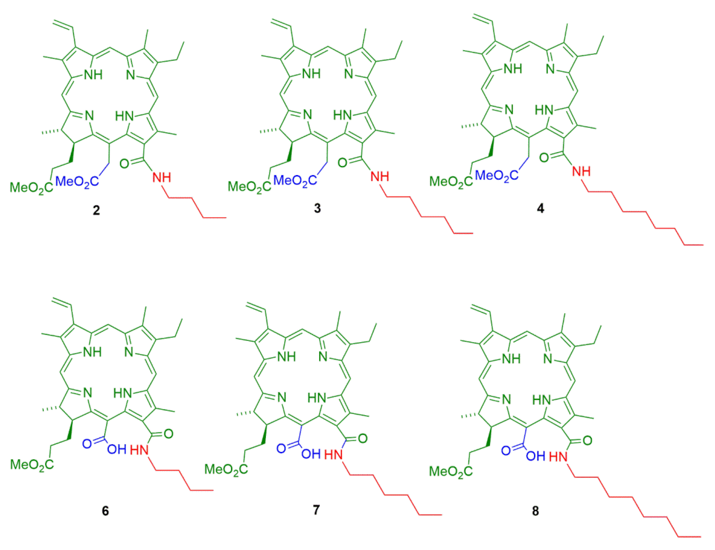
2.1. Semisynthesis of Photosensitizers

All the compounds were prepared using previously described methods from the literature. Initially, methyl-pheophorbide a (1) was obtained as previously described by our research group [44] (Scheme 1). Subsequently, primary aliphatic amines (butylamine, hexylamine, and octylamine) were reacted with 1 to give compounds 24, all with absorption bands around 660 nm [49,50,51].
The purpurin-18 methyl ester (5) was also obtained from the oxidation of methyl pheophorbide a (1) [52,53], which allowed the addition of different aliphatic amines (butylamine, hexylamine, and octylamine) into the anhydride ring, resulting in products 68. Compound 5 exhibited a characteristic absorption at 700 nm, which became approximately 660 nm when the anhydride ring was opened by the insertion of the amines (products 68) [54,55,56].
The obtained PS 24 and 68, were characterized using NMR, UV-vis, and high-resolution mass spectrometry (HRMS Q-TOFF) and presented absorption bands in the red region. See more details on the characterizations in Supplementary Materials, in which we present our compound band wavelengths and additional literature data (Table S1). Overall, these photosensitizers were semisynthesized at a low cost because we performed small structural modifications in the natural chlorophyll a, having the methyl-pheophorbide a (1) as a direct and versatile molecular template. With these modifications, we obtained six chlorin derivatives, 24 and 68, all with absorption bands near 660 nm, different substituents, and amphiphilicity, which are desirable for use in PDT (Figure 1 and Figure 2) [57]. The photostability of all compounds was also checked, showing that the photobleaching was not measurable even after 10 min of irradiation (see Supplementary Materials, Figure S18).

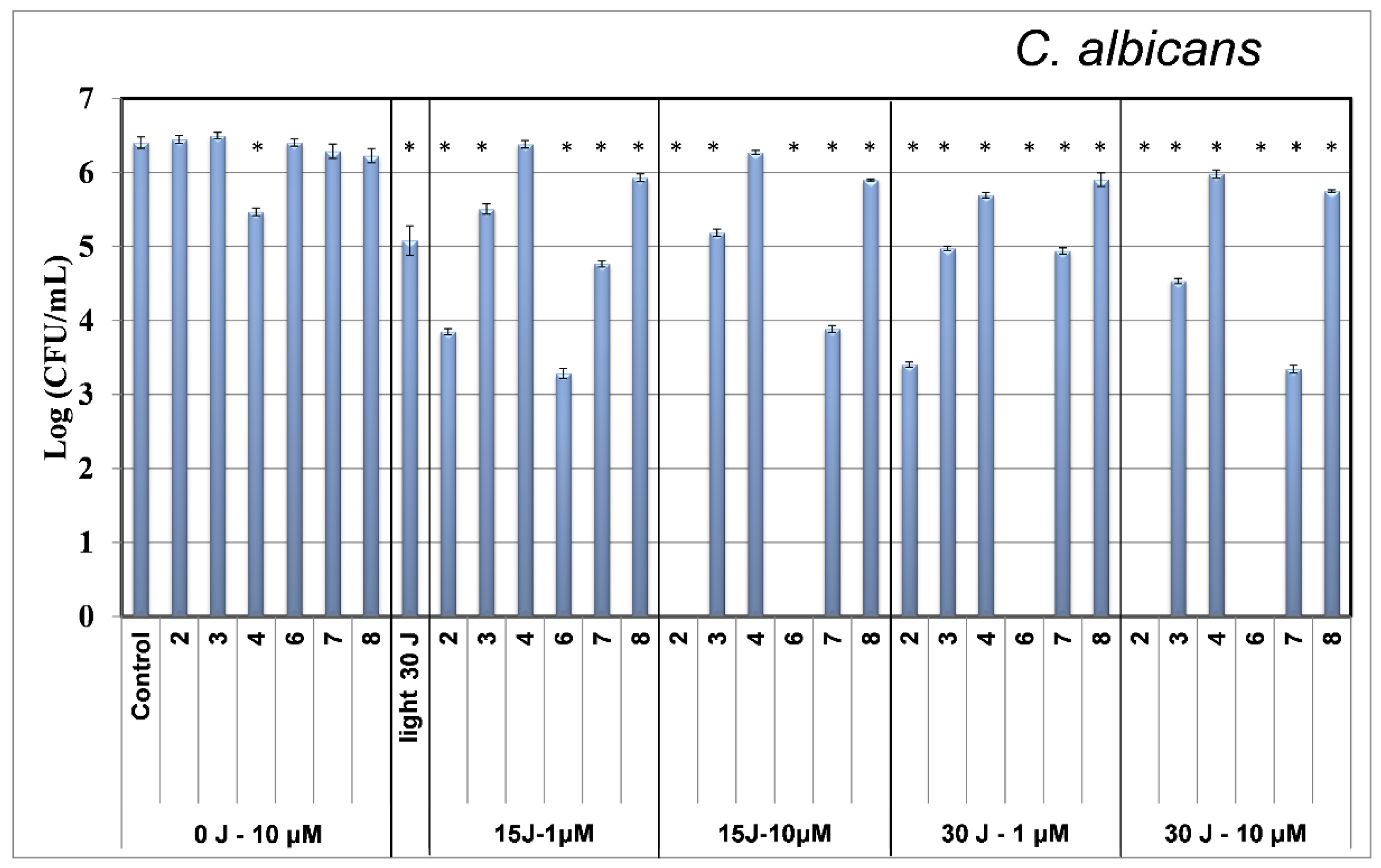
2.2. Photodynamic Inactivation

The compounds 24 and 68 were evaluated against three microorganisms—S. aureus, C. albicans, and E. coli—for the inactivation of these microorganisms. First, we investigated the dark toxicity of the chlorophyll derivatives. The microorganisms were incubated for 20 min in the dark with the respective photosensitizer (10 μM) and their mortality was evaluated after 24 h. No mortality was observed after 24 h with only the irradiation of the microorganisms without photosensitizers (30 Jcm−2). The inactivation study was then performed with each microorganism. All photosensitizers 24 and 68 were used in the evaluation of S. aureus (Gram-positive bacteria), and different concentrations of photosensitizer (1 μM and 10 μM) and light fluences (15 Jcm−2 and 30 Jcm−2) were utilized. The results obtained from these initial photoinactivation studies (Figure 3) show that with the increase in the carbon chain (from four to eight C atoms), the photosensitizers presented a decrease in the inactivation levels for the derivatives 24 and 68.
In addition, we observed that the photoinactivation of both S. aureus (Figure 3) and C. albicans (Figure 4) was influenced by light fluence and photosensitizer concentration, with increased photoinactivation at high concentrations and fluences.
Evaluating the photoinactivation of S. aureus (Figure 3), we observed that at 1 µM and 15 Jcm−2, the methyl pheophorbide derivatives 24 did not present relevant inactivation, whereas the purpurin-18 derivatives 68 allowed significant inactivation, with a reduction of 3 log using PS 8, 3.5 log with PS 7, and 4 log with PS 6. Maintaining the same concentration of photosensitizer and increasing the dose of light from 15 Jcm−2 to 30 Jcm−2 resulted in the inactivation of microorganisms by derivatives 24; however, the photoinactivation promoted by PS 8 was approximately 4 log, and that by derivatives 6 and 7 was approximately 5 log.
It is possible to observe in Figure 3 that at 10 µM, both light doses (15 Jcm−2 and 30 Jcm−2) were not very effective in the photoinactivation of S. aureus with photosensitizers 24. In contrast, purpurin-18 derivatives 68 completely inhibited the growth of these microorganisms, proving that these derivatives are much more effective than those derived from methyl pheophorbide 24.
These results suggest that the purpurin-18 derivatives 68, due to the presence of a carboxylic acid group in the molecules may facilitate their microorganism uptake, whereas the derivatives of methyl pheophorbide 24 have an ester group with lower uptake.
The results presented in Figure 4 for C. albicans were similar to those obtained for S. aureus, with purpurin-18 derivatives 68 presenting better results in terms of photoactivation than methyl pheophorbide a derivatives 24. However, PS 2 with the ester and butyl group also showed promising inactivation of C. albicans.
Overall, the results were similar to those for S. aureus and the higher the carbon chain present in the photosensitizers, the more hydrophobic the PS is used, and lower microorganism uptake was observed. As a consequence, we observed lower photoinactivation.
Photosensitizers 24 and 68 were also tested against E. coli, (Gram-negative bacteria), at 500 μM and light dose of 45 Jcm−2. No photoinactivation by the photosensitizers 24 and 68 was observed, even using high doses of light, which was completely expected as Gram-negative microorganisms are preferentially inactivated by cationic photosensitizers [58,59]. Compared with our previous results using chlorin e6 (chl-e6) [44], we observed that S. aureus was completely inactivated at chl-e6 concentrations of 1.6 μM and 16 μM when using a light dose of 30 Jcm−2, which is very similar to the results obtained with derivatives 6 and 7. However, considering C. albicans when compared with chl-e6, we found total inactivation at concentrations equivalent to 33 μM (20 μg mL−1) and 50 μM (30 μg mL−1) using a light dose of 30 Jcm−2. We obtained the best results with derivative 6, which completely inactivated C. albicans at a concentration of 10 μM (15 Jcm−2) and at concentrations of 1 μM and 10 μM with 30 Jcm−2. Chlorin e6 and 2, 3, 4, 6, 7, and 8 did not show efficacy against E. coli [44].
Comparing among photosensitizers, when methylene blue was evaluated against the microorganism S. aureus at a concentration of 50 μM and a light dose of 9 J at 660 nm [60], a reduction of approximately 1.5 log CFU was obtained, whereas the photosensitizers 6, 7, and 8 used in this study completely inactivated the S. aureus microorganism at a concentration of 10 μM and a light dose of 15 Jcm−2, demonstrating it was more effective using these semisynthetic PS. When using 50 μM and a light dose of 9 J to perform photodynamic inactivation with methylene blue for C. albicans, a reduction of approximately 1 log CFU was obtained, whereas in this study, PS 7 at a concentration of 10 μM and a light dose of 15 Jcm−2 allowed a reduction of approximately 2 log CFU. Using the PS 3, we obtained a reduction of approximately 1 log CFU, and PS 2 and 6 completely inactivated the microorganism.
Indocyanine green (ICG) [61], was also used as a photosensitizer for the photoinactivation of S. aureus. At a concentration of 25 μg mL−1 (32 μM) ICG and exposure to 411 Jcm−2 with near-infrared (NIR) light (808 nm laser), a significant reduction in the viable count was achieved (5.56 log10); complete inactivation of the microorganism occurred at a concentration of 200 μg mL−1 (258 μM) and 411 Jcm−2. In another study using ICG [62], PDI against S. aureus with an ICG concentration of 8 μg mL−1 (10 μM) at an energy dose of 84 Jcm−2 resulted in 100% inactivation of this microorganism. In the current study, PS 6, 7, and 8 also showed complete inactivation of S. aureus at a concentration of 10 μM using 15 Jcm−2 and 30 Jcm−2 (660 nm).
In an in vitro study, the inactivation of C. albicans was evaluated with ICG at 1 μg mL−1 using a light dose of 228 J/cm−2 (810 nm); a satisfactory result showing a reduction of 1.2 log was obtained, similar to the results with nystatin. Compared to the control, the elimination of C. albicans increased by 92% when treated with ICG (1 mg/mL) with infrared (IR) laser irradiation (810 nm, 55 J/cm−2).

3. Materials and Methods

Nuclear magnetic resonance (NMR) analyses were performed on a Bruker Avance 400 spectrometer at 400.15 MHz (1H) and 100.13 MHz (13C). Tetramethylsilane was used as an internal reference.
High-resolution mass spectrometry (HRMS) was performed using ESI-TOF (Waters Xevo G2-S Qtof). Ultraviolet-visible spectrophotometry analyses were performed using a Lambda 25 spectrometer (PerkinElmer, Waltham, MA, USA).
Spirulina maxima powder was purchased from Pharma Nostra (Rio de Janeiro, Brazil), and other reagents were purchased from Sigma-Aldrich (St. Louis, MO, USA).

3.1. Semisynthesis of Photosensitizers

Isolation of methyl pheophorbide a (1) from Spirulina maxima (Mepheo a): 300 g of the Spirulina maxima was treated with a 1.5 L of the 5% methanolic solution of H2SO4 for 24 h at room temperature. This mixture was filtered and washed with methanol (900 mL) and ethyl acetate (900 mL), and the organic phases were evaporated under reduced pressure. After that, 150 g of crushed ice was added to the crude residue. The residue was neutralized with solid NaHCO3 and placed on a silica gel plug. The chlorophyll derivatives were retained on the plug, and the residual proteins and peptides (pale yellow in color) were eluted with water. The chlorophyll derivatives were then eluted with ethyl acetate (900 mL) and washed with water (3 × 400 mL). The organic phase was separated, dried over Na2SO4, and the solvent evaporated under reduced pressure. The methyl pheophorbide a was purified by silica gel flash chromatography using as eluent toluene:ethyl acetate (9:1), yielding the methyl pheophorbide a (1) (2.4 g, 3.9 mmol, 0.8% yield from natural dried Spirulina maxima) [44,63,64].
UV-vis (CH2Cl2): λmax (nm): 666, 609, 534, 505, 409 [65].
1H-NMR (CDCl3, 400 MHz) δ: 9.51 (s, 1H, H-10); 9.37 (s, 1H, H-5); 8.56 (s, 1H, H-20); 7.97 (dd, 1H, J = 11.6, J = 17.8 Hz, H-31); 6.25 (s, 1H, H-132); 6.24 (dd, 1H, J = 1.5 and J = 17.8 Hz, H-32B); 6.16 (dd, 1H, J = 1.5 and J = 17.8 Hz, H-32a); 4.48–4.43 (m, 1H, H-181); 4.21–4.19 (m, 1H, H-17); 3.88 (s, 3H, H-134); 3.68 (s, 3H, H-121); 3.60 (q, 2H, J = 7.7 Hz, H-81); 3.57 (s, 3H, H-174); 3.40 (s, 3H, H-21); 3.22 (s, 3H, H-71); 2.68–2.48 (m, 2H, H-171); 2.36–2.20 (m, 2H, H-172); 1.81 (d, 3H, J = 7.3 Hz, H-182); 1.69 (t, 3H, J = 7.6 Hz, H-82); 0.54 (br. s, 1H, H-21); −1.63 (br. s, 1H, H-23).
13C-NMR (CDCl3, 100 MHz) δ: 192.1, 174.7, 172.5, 169.4, 162.9, 155.9, 151.1, 150.1, 145.3, 142.3, 137.9, 136.8, 136.4, 136.3, 132.0, 129.1, 129.0, 127.9, 122.9, 104.6, 104.5, 97.5, 93.4, 65.9, 65.1, 63.3, 52.2, 51.1, 50.3, 30.8, 30.0, 23.2, 19.4, 17.4, 12.1, 11.3
13-(Butylcarbamoyl)-chlorin e6 15,17-dimethyl ester (2). To a solution of 30 mg (0.049 mmol) of 1 in 3 mL of dry tetrahydrofuran, 0.5 mL of butylamine was added and the reaction was stirred for 20 min at room temperature. After that, the solvent was evaporated, the mixture was diluted with dichloromethane and washed with HCl solution (1%). The resulting solution was dried over anhydrous sodium sulfate, and the solvent evaporated under reduced pressure. Compound 2 was isolated by chromatography over silica gel using toluene:ethyl acetate (9:1) as eluent (22 mg, 72% yield) [50,51].
UV-vis (CH2Cl2) λmax (nm): 662, 607, 528, 498, 399 [66].
1H-NMR (CDCl3, 400 MHz) δ: 9.69 (s, 1H); 9.64 (s, 1H); 8.80 (s, 1H); 8.08 (dd, 1H, J = 17.8 and 11.6 Hz); 6.36–6.40 (m, 1H, CONH); 6.33 (dd, 1H, J = 16.7, 1.4 Hz); 6.15 (dd, 1H, J = 11.6, 1.4 Hz); 5.54 and 5.26 (d, 1H each, J = 18.9 Hz); 4.46 (q, 1H, J = 7.2 Hz); 4.35 (d, 1H, J = 9.5 Hz); 3.91–3.76 (m, 4H); 3.81 (s, 3H); 3.60 (s, 3H); 3.56 (s, 3H); 3.49 (s, 3H); 3.32 (s, 3H); 2.56–2.09 (m, 4H); 1.82–1.69 (m, 10H); 1.06 (t, 3H, J = 7.3 Hz); −1.61 (br.s, NH); −1.83 (br.s, NH).
HRMS (ESI): m/z calculated [M + H]+ for C40H50O5N5+ = 680.38065; found 680.38379.
13-(Hexylcarbamoyl)chlorin e6 15,17-dimethyl ester (3). To a solution of 40 mg (0.066 mmol) of 1 in 3 mL of dry tetrahydrofuran, 0.5 mL of hexylamine was added and the reaction was stirred for 20 min at room temperature. After that, the solvent was evaporated, the mixture was diluted with dichloromethane and washed with HCl solution (1%). The resulting solution was dried over anhydrous sodium sulfate, and the solvent evaporated under reduced pressure. Compound 3 was isolated by chromatography over silica gel using toluene:ethyl acetate (9:1) as eluent (45 mg, 95% yield) [50,51].
UV-vis (CH2Cl2): λmax (nm): 663, 605, 527, 497, 398 [66].
1H-NMR (CDCl3, 400 MHz) δ: 9.70 (s, 1H), 9.64 (s, 1H), 8.80 (s, 1H), 8.10 (dd,1H, J = 11.5 and 17.8 Hz), 6.36–6.39 (m, CONH), 6.35 (dd, 1H, J = 1.4 and 17.8 Hz), 6.13 (dd, 1H, J = 1.4 and 11.5 Hz), 5.52 (d, 1H, J = 18.9 Hz), 5.25 (d, 1H, J = 19.0 Hz), 4.46 (q, 1H, J = 7.3 Hz), 4.35 (d, 1H, J = 11.2 Hz), 3.90–3.68 (m, 4H), 3.80 (s, 3H), 3.60 (s, 3H), 3.57 (s, 3H), 3.49 (s, 3H), 3.32 (s, 3H), 2.56–2.07 (m, 4H), 1.83–1.37 (m, 16H), 0.94 (t, 1H, J = 7.2 Hz), −1.63 (NH), −1.83 (NH).
HRMS (ESI): m/z calculated [M + H]+ for C42H54O5N5+ = 708.41195; found 708.41516.
13-(Octylcarbamoyl)chlorin e6 15,17-dimethyl ester (4). To a solution of 30 mg (0.049 mmol) of 1 in 3 mL of dry tetrahydrofuran, 0.5 mL of octylamine was added and the reaction was stirred for 20 min at room temperature. After that, the solvent was evaporated, the mixture was diluted with dichloromethane and washed with HCl solution (1%). The resulting solution was dried over anhydrous sodium sulfate, and the solvent evaporated under reduced pressure. Compound 4 was isolated by chromatography over silica gel using toluene:ethyl acetate (9:1) as eluent (28 mg, 78% yield) [51].
UV-vis (CH2Cl2): λmax (nm): 663, 609, 527, 497, 399 [66].
1H-NMR (CDCl3, 400 MHz) δ: 9.70 (s, 1H); 9.64 (s, 1H); 8.80 (s, 1H); 8.09 (dd, 1H, J = 17.8, 11.5 Hz); 6.36–6.39 (m, 1H, CONH); 6.36 (dd, 1H, J = 18.2, 1.4 Hz); 6.14 (dd, 1H, J = 11.5, 1.4 Hz), 5.54 and 2.25 (d, 1H each, J = 18.9 Hz); 4.47 (q, 1H, J = 7.1 Hz); 4.36 (d. 1H, J = 7.6 Hz); 3.90–3.77 (m, 4H); 3.80 (s, 3H); 3.60 (s, 3H); 3.57 (s, 3H); 3.49 (s, 3H); 3.32 (s, 3H); 2.52–2.09 (m, 4H); 1.83–1.69 (m, 10 H); 1.46–1.33 (m, 8H); 0.90 (t, 3H, J = 7.1 Hz); −1.62 (br, s, NH); −1.83 (br, s, NH).
HRMS (ESI): m/z calculated [M + H]+ for C44H58O5N5+ = 736.44325; found 736.44147.
Purpurin-18 Methyl Ester (5) Method 1: Methyl pheophorbide a 1 (151 mg, 0.25 mmol) was dissolved in 500 mL of diethyl ether and 5 mL of pyridine. After, a potassium hydroxide solution in 1-propanol (2 g of KOH was dissolved in 10 mL of 1-propanol) was added into the first solution and oxygen was bubbled into the resulting reaction mixture for 1 h. The reaction mixture was extracted with water (500 mL). The aqueous layer was collected and the pH adjusted to 2–4 using cold H2SO4 solution (25%). The aqueous layer was extracted with CH2Cl2 and the solvent evaporated to give a purple residue. The product was purified by chromatography over silica gel using hexane:ethyl acetate 3:1 as eluent. After that, the carboxylic acid precursor was obtained in 55% yield (0.078 g, 0.119 mmol). This product was further reacted with a diazomethane solution in dichloromethane to produce purpurin-18 methyl ester (5) for 10 min at 0 ºC. The residue was crystallized with dichloromethane/hexane, thus obtaining 5 in 60% yield (63.0 mg, 0.109 mmol) as purple red crystals [51]. Method 2: Pigments were extracted twice from S. maxima dried powder (10 g) with acetone (4 × 100 mL) under magnetic stirring at 60 °C (4 × 30 min). The dark green extract was filtered off and the filtrate (ca. 400 mL) was reduced to 200 mL by partial evaporation under reduced pressure. NaOH (40 mL, 6 M) was added to the previous extract (200 mL); the mixture was vigorously stirred and oxygen was bubbled during 3 h. The solution was then acidified with concentrated HCl. The oxidized extract was evaporated to dryness. Carotenoids and part of xanthophylls were removed by extraction with petroleum ether (2 × 100 mL). The resulting residue was purified by flash chromatography (eluent CH2Cl2:MeOH 8:2) obtaining 40.0 mg (0.070 mmol, 0.4% yield) of the purpurin-18 carboxylic acid. The product was esterified with diazomethane obtaining 32.0 mg (0.055 mmol, 78% yield) of the 5 [53,67].
UV-vis (CH2Cl2): λmax (nm): 699, 642, 546, 508, 478, 410.
1-H NMR (400 MHz, CDCl3, ppm): 9.57 (s, 1H, H-10); 9.36 (s, 1H, H-5); 8.56 (s, 1H, H-20); 7.89 (dd, 1H, J = 17.8, 11.6 Hz, H-31); 6.27 (dd, 1H, J = 17.8 and J = 9.4, 2.5 Hz, H-32B); 6.20 (dd, 1H, J = 11.6 and J = 9.4, 2.5 Hz, H-32a); 4.39 (q, 1H, J = 7.3 Hz, H-181); 3.77 (s, 3H, H-121); 3.64 (q, 2H, J = 7.9 Hz, H-81); 3.60 (s, 3H, H-174); 3.35 (s, 3H, H-21); 3.15 (s, 3H, H-71); 2.78–2.70 (m, 2H H-171); 2.49–2.43 (m, 2H, H-172); 1.74 (d, 3H, J = 7.3 Hz, H-182); 1.65 (t, 3H, J = 7.6 Hz, H-82); 0.21 (br s, 1H, H-21); −0.08 (br s, 1H, H-23).
13C-NMR (CDCl3, 100 MHz) δ: 178.6, 173.5, 169.8, 165.4, 163.4, 158.4, 154.2, 146.9, 145.1, 141.9, 140.0, 137.9, 135.5, 133.5, 132.8, 132.8, 131.4, 128.0, 125.6, 107.4, 102.5, 97.9, 94.5, 55.5, 51.7, 49.6, 32.4, 31.4, 20.4, 19.6, 16.9, 12.9, 12.1, 11.2
Chlorin-p, 6-N-Butylamide-7-methyl Ester (6). To a solution of 5 (40.0 mg, 0.069 mmol) in dichloromethane (5 mL), butylamine (0.5 mL) was added and the reaction mixture was stirred at room temperature in the dark under nitrogen for 2 h. After that, the UV-vis analysis and TLC showed the absence of starting material. The product was purified by crystallization with dichloromethane-hexane to give the product 6 (48.0 mg, 0.070 mmol, 90% yield) [54,55,56].
UV-vis (CH2Cl2): λmax (nm): 662, 606, 526, 498, 398.
1H-NMR (CDCl3, 400 MHz) δ: 9.67(s, 1H), 9.62 (s, 1H), 8.74 (s, 1H), 8.12 (dd, J = 18, 11 Hz 1H), 6.96–7.05 (m, NH), 6.37 (d, J = 18 Hz, 1H), 6.15 (d, J = 11 Hz, 1H), 5.06 (d, J = 7.7 Hz,1H), 4.33–4.37 (m, 1H) 3.74–3.78 (m, 4H), 3.57 (s, 3H), 3.46 (s, 3H), 3.31 (s, 3H), 3.23 (s, 3H), 2.49–2.27 (m, 2H), 1.70 (d, 3H, J = 7.2Hz), 1.68 (t, 3H, J = 7.5Hz), 0.79–1.10 (m, 4H), 0.95 (t, 3H, J = 7.2Hz), 0.63(t, 3H, J = 7.2Hz),−1.66 (s, NH) and −1.94 (s, NH).
HRMS (ESI): m/z calculated [M + H]+ for C38H46O5N5+ = 652.34935; found 652.35046.
Chlorin-p, 6-N-hexylamide-7-methyl Ester (7). To a solution of 5 (40 mg, 0.069 mmol) in dichloromethane (0.3 mL), hexylamine (0.2 mL) was added and the reaction mixture was stirred at room temperature in the dark under nitrogen for 2 h. After that, UV-vis analysis and TLC showed the absence of starting material. The product was purified by crystallization with dichloromethane-hexane to give the product 7 (42.0 mg, 0.062 mmol, 86% yield) [54,55,56].
UV-vis (CH2Cl2): λmax (nm): 666, 608, 528, 497, 398.
1H-NMR (CDCl3, 400 MHz) δ: 9.66 (s, 1H), 9.55 (s, 1H), 8.70 (s, 1H), 7.98 (dd, 1H, J = 11.5 and 17.6 Hz), 6.33–6.37 (m, NH), 6.28 (d, 1H, J = 16.8Hz), 6.10 (d, 1H, J = 12.8Hz), 4.93 (d, 1H, J = 7.7Hz), 4.40 (q, 1H, J = 7.0Hz), 4.13 (s, 3H), 3.82–3.60 (m, 4H), 3.56 (s, 3H), 3.40 (s, 3H), 3.22 (s, 3H), 2.70–2.12 (m, 4H), 1.77 (d, 3H, J = 7.2Hz), 1.64 (t, 3H, J = 7.5Hz), 0.92–0.77 (m, 9H), 0.67 (t, 3H, J = 7.2Hz), −1.32 (s, NH), −1.54 (s, NH).
HRMS (ESI): m/z calculated [M + H]+ for C40H50O5N5+ = 680.38065; found 680.38251.
Chlorin-p, 6-N-octylamide-7-methyl Ester (8). To a solution of 5 (22.0 mg, 0.038 mmol) in dichloromethane (1.0 mL), octylamine (0.2 mL) was added. The reaction mixture was stirred at room temperature in the dark under nitrogen for 2 h. After that, UV-vis analysis and TLC showed the absence of starting material. The product was purified by crystallization with dichloromethane-hexane to give the product 8 (21.0 mg, 0.030 mmol, 80% yield) [54,55,56].
UV-vis (CH2Cl2): λmax (nm): 663, 605, 528, 499, 399.
1H-NMR (CD3)2CO, 400 MHz) δ: 9.71 (s, 1H), 9.41 (s, 1H), 9.04 (s, 1H), 8.17 (dd, 1H), 7.78–7.82 (m, NH), 6.37 (d, 1H), 6.15 (d, 1H), 5.16 (d, 1H), 4.54 (q, 1H), 3.67 (s, 3H), 3.60–3.64 (m, 4H), 3.48 (s, 3H), 3.40 (s, 3H), 3.25 (s, 3H), 2.70–2.12 (m, 6H), 1.78 (d, 3H), 1.63 (t, 3H, J = 7.5Hz), 0.92–0.77 (m, 9H), 0.85 (t, 3H, J = 7.2Hz), −1.46 (s, NH), −1.82 (s, NH).
HRMS (ESI): m/z calculated [M + H]+ for C42H54O5N5+ = 708.41195; found 708.41364.

3.2. Photodynamic Inactivation

Staphylococcus aureus (American Type Culture Collection, ATCC 25923) and Escherichia coli (ATCC 25922) were grown in brain and heart infusion media. Candida albicans (ATCC 10231) was grown in Sabouraud dextrose broth. For experimental purposes, the microorganism concentration was adjusted to 107–108 cells/mL in sterile distilled water, and 500 µL of each microorganism culture was added to 24-well plates with photosensitizers 24 and 68. Solutions were prepared by diluting the photosensitizer powder (1 mg for S. aureus and C. albicans and 2 mg for E. coli) in 100 μL of dimethyl sulfoxide (DMSO) (to dissolve the PS) and 900 μL of sterile water; the initial concentration of DMSO in the stock solutions was 10%. After dilution of these stock solutions to final concentrations of 1 µM and 10 µM for S. aureus and C. albicans, respectively, and of 500 μM for E. coli, the final concentration of DMSO was less than 2% in all the solutions. After preparing the solutions, they were protected from light. The 24-well plates were kept in the dark at 37 °C for 20 min.
A homemade LED-based device with emission centered at 660 nm was used to irradiate the culture plates. The 24-well plates were irradiated at 30 mWcm−2 using this device for 8, 16, and 25 min, resulting in fluences of 15, 30, and 45 Jcm−2, respectively, which were used in the PDI against microorganisms. The fluence levels used were 15 Jcm−2 and 30 Jcm−2 for S. aureus and C. albicans, and 45 Jcm−2 for E. coli. After irradiation, 10-fold serial dilutions were performed and cells were cultured in agar plates. The colony-forming units (CFUs) were determined 24 h after initiation of the experimental procedure. All experiments were performed in triplicate.
In the control group (no treatment), 24-well plates were maintained at room temperature for 32 min. Using the same incubation time, the dark toxicity of the photosensitizers was evaluated in 24-well plates covered with aluminum foil to avoid light exposure. Phototoxicity was determined by irradiation at 30 Jcm−2 for S. aureus and C. albicans, and 45 Jcm−2 for E. coli.
Survival fractions (SFs) were expressed as ratios of CFUs of treated groups to the control group. The SF at 0 J/cm2 provides a measure of the dark toxicity of chlorins.

3.3. Statistical Analysis

All the results reported in Figure 3 and Figure 4 were statistically analyzed using the RStudio software (R version 4.1.1 (10 August 2021), R Core Team (2021), R: A language and environment for statistical computing; R Foundation for Statistical Computing, Vienna, Austria (https://www.R-project.org, accessed on 10 August 2021)) using a significance level of at least p < 0.05 and a confidence level of approximately 95%. The data were analyzed and approved by the normality test. Comparisons between the experimental groups were verified by Tukey’s test.

4. Conclusions

The photosensitizers 24 and 67 derived from chlorophyll a were successfully semisynthesized. The characterization of the compounds is in accordance with data described in the literature and the main photophysical data are compiled and organized in Table S1 (SI). Overall, methyl pheophorbide a or purpurin-18 derivatives with different side chains (from the butyl to octyl groups) were prepared and studied. Subsequently, we investigated the photoinactivation of S. aureus and observed that the methyl pheophorbide derivatives 24 did not show great inactivation, whereas the purpurin-18 derivatives 68 allowed significant PDT inactivation. For C. albicans, the purpurin-18 derivative with the butyl group showed relevant inactivation, and the methyl pheophorbide with the butyl group also exhibited PDI. Photosensitizers 24 and 68 were also tested against the Gram-negative bacterium E. coli; however, no significant photoinactivation was observed. In general, the higher the carbon chain present in the photosensitizers, the more hydrophobic the compounds, with consequently lower photoinactivation efficacy. These results suggest that the use of chlorin derivatives with lower hydrophobic properties can be more effective for the photoinactivation of such microorganisms as S. aureus and C. albicans.

Supplementary Materials

The following supporting information can be downloaded at: https://www.mdpi.com/article/10.3390/molecules27185769/s1, Figure S1: 1H-NMR (CDCl3) spectrum of methyl pheophorbide a (1); Figure S2: 13C-NMR (CDCl3) spectrum of methyl pheophorbide a (1); Figure S3: 1H-NMR (CDCl3) spectrum of 13-(Butylcarbamoyl)chlorin e6 15,17-dimethyl ester (2); Figure S4: HRMS (ESI) of 13-(Butylcarbamoyl)chlorin e6 15,17-dimethyl ester (2); Figure S5: 1H-NMR (CDCl3) spectrum of 13-(Hexylcarbamoyl)chlorin e6 15,17-dimethyl ester (3); Figure S6: HRMS (ESI) of 13-(Hexylcarbamoyl)chlorin e6 15,17-dimethyl ester (3); Figure S7: 1H-NMR (CDCl3) spectrum of 13-(Octylcarbamoyl)chlorin e6 15,17-dimethyl ester (4); Figure S8: HRMS (ESI) of 13-(Octylcarbamoyl)chlorin e6 15,17-dimethyl ester (4); Figure S9: 1H-NMR (CDCl3) spectrum of Purpurin-18 Methyl Ester (5); Figure S10: 13C-NMR (CDCl3) spectrum of Purpurin-18 Methyl Ester (5); Figure S11: 1H-NMR (CDCl3) spectrum of Chlorin-p, 6-N-Butylamide-7-methyl Ester (6); Figure S12: HRMS (ESI) of Chlorin-p, 6-N-Butylamide-7-methyl Ester (6); Figure S13: 1H-NMR (CDCl3) spectrum of Chlorin-p, 6-N-hexylamide-7-methyl Ester (7); Figure S14: HRMS (ESI) of Chlorin-p, 6-N-hexylamide-7-methyl Ester (7); Figure S15: 1H-NMR (CDCl3) spectrum of Chlorin-p, 6-N-octylamide-7-methyl Ester (8); Figure S16: HRMS (ESI) of Chlorin-p, 6-N-octylamide-7-methyl Ester (8); Figure S17: Homemade engineered Biotable model for PDI studies (660 nm). Figure S18: Photodegradation experiment at 660 nm at 63.7 mWcm-2 using ethyl acetate as solvent. Table S1: Wavelengths data and Quantum yield of Singlet Oxygen 1O2 data of the literature for some of the compounds.

Author Contributions

Formal analysis, M.P.U., A.d.C.R. and B.A.O.; Methodology, M.P.U.; Writing—original draft, M.P.U., A.d.C.R., B.A.O. and S.P.; Writing—review and editing, M.P.U., K.T.d.O. and C.K. All authors have read and agreed to the published version of the manuscript.

Funding

The authors thank FAPESP (São Paulo Research Foundation) grant numbers: 2013/06532-4, 2011/19720-8, 2011/13993-2, 2008/57858-9, 2009/54035-4 (EMU) and 2013/07276-1 (CEPOF); 2019/27176-8 (KTO), 2020/06874-6 (KTO); PRPPG-UNILA (80/2019/PRPPG, 104/2020/PRPPG and 105/2020/PRPPG); CNPq (465360/2014-9 and 306919/2019-2) and CAPES for financial support and fellowships.

Institutional Review Board Statement

Not applicable.

Informed Consent Statement

Not applicable.

Data Availability Statement

Not applicable.

Acknowledgments

Thanks are also due to Waters laboratory in Brazil for ESI-TOF measurements, and P. T. Weis and V. A. Kuana for the technical appointment.

Conflicts of Interest

The authors declare no conflict of interest.

References

  1. Agostinis, P.; Berg, K.; Cengel, K.A.; Foster, T.H.; Girotti, A.W.; Gollnick, S.O.; Hahn, S.M.; Hamblin, M.R.; Juzeniene, A.; Kessel, D.; et al. Photodynamic Therapy of Cancer: An Update. CA-A Cancer J. Clin. 2011, 61, 250–281. [Google Scholar] [CrossRef] [PubMed]
  2. Betrouni, N.; Boukris, S.; Benzaghou, F. Vascular targeted photodynamic therapy with TOOKAD (R) Soluble (WST11) in localized prostate cancer: Efficiency of automatic pre-treatment planning. Lasers Med. Sci. 2017, 32, 1301–1307. [Google Scholar] [CrossRef] [PubMed]
  3. Mazor, O.; Brandis, A.; Plaks, V.; Neumark, E.; Rosenbach-Belkin, V.; Salomon, Y.; Scherz, A. WST11, a novel water-soluble bacteriochlorophyll derivative; Cellular uptake, pharmacokinetics, biodistribution and vascular-targeted photodynamic activity using melanoma tumors as a model. Photochem. Photobiol. 2005, 81, 342–351. [Google Scholar] [CrossRef] [PubMed]
  4. Mfouo-Tynga, I.S.; Dias, L.D.; Inada, N.M.; Kurachi, C. Features of third generation photosensitizers used in anticancer photodynamic therapy: Review. Photodiagn. Photodyn. Ther. 2021, 34, 102091. [Google Scholar] [CrossRef] [PubMed]
  5. Alzeibak, R.; Mishchenko, T.A.; Shilyagina, N.Y.; Balalaeva, I.V.; Vedunova, M.V.; Krysko, D.V. Targeting immunogenic cancer cell death by photodynamic therapy: Past, present and future. J. Immunother. Cancer 2021, 9, e001926. [Google Scholar] [CrossRef]
  6. Zhang, P.; Wu, M.X. A clinical review of phototherapy for psoriasis. Lasers Med. Sci. 2018, 33, 173–180. [Google Scholar] [CrossRef]
  7. Marotti, J.; Aranha, A.C.C.; Eduardo, C.D.; Ribeiro, M.S. Photodynamic Therapy Can Be Effective as a Treatment for Herpes Simplex Labialis. Photomed. Laser Surg. 2009, 27, 357–363. [Google Scholar] [CrossRef]
  8. Issa, M.C.A.; Manela-Azulay, M. Photodynamic therapy: A review of the literature and image documentation. An. Bras. Dermatol. 2010, 85, 501–511. [Google Scholar] [CrossRef]
  9. De Carvalho, G.G.; Sanchez-Puetate, J.C.; Donatoni, M.C.; Huacho, P.M.M.; Rastelli, A.N.D.; de Oliveira, K.T.; Spolidorio, D.M.P.; Zandim-Barcelos, D.L. Photodynamic inactivation using a chlorin-based photosensitizer with blue or red-light irradiation against single-species biofilms related to periodontitis. Photodiagn. Photodyn. Ther. 2020, 31, 101916. [Google Scholar] [CrossRef]
  10. Seeger, M.G.; Ries, A.S.; Gressler, L.T.; Botton, S.A.; Iglesias, B.A.; Cargnelutti, J.F. In vitro antimicrobial photodynamic therapy using tetra-cationic porphyrins against multidrug-resistant bacteria isolated from canine otitis. Photodiagn. Photodyn. Ther. 2020, 32, 101982. [Google Scholar] [CrossRef]
  11. Hirose, M.; Yoshida, Y.; Horii, K.; Hasegawa, Y.; Shibuya, Y. Efficacy of antimicrobial photodynamic therapy with Rose Bengal and blue light against cariogenic bacteria. Arch. Oral Biol. 2021, 122, 105024. [Google Scholar] [CrossRef] [PubMed]
  12. Reis, A.C.M.; Regis, W.F.M.; Rodrigues, L.K.A. Scientific evidence in antimicrobial photodynamic therapy: An alternative approach for reducing cariogenic bacteria. Photodiagn. Photodyn. Ther. 2019, 26, 179–189. [Google Scholar] [CrossRef] [PubMed]
  13. Geralde, M.C.; Leite, I.S.; Inada, N.M.; Salina, A.C.G.; Medeiros, A.I.; Kuebler, W.M.; Kurachi, C.; Bagnato, V.S. Pneumonia treatment by photodynamic therapy with extracorporeal illumination—An experimental model. Physiol. Rep. 2017, 5, e13190. [Google Scholar] [CrossRef] [PubMed]
  14. Babu, B.; Sindelo, A.; Mack, J.; Nyokong, T. Thien-2-yl substituted chlorins as photosensitizers for photodynamic therapy and photodynamic antimicrobial chemotherapy. Dyes Pigm. 2021, 185, 108886. [Google Scholar] [CrossRef]
  15. Kate, C.; Blanco, N.M.I.; Carbinatto, F.M.; Bagnato, V.S. Antimicrobial Efficacy of Curcumin Formulations by Photodynamic Therapy. J. Pharm. Pharmacol. 2017, 5, 506–511. [Google Scholar]
  16. Amos-Tautua, B.M.; Songca, S.P.; Oluwafemi, O.S. Application of Porphyrins in Antibacterial Photodynamic Therapy. Molecules 2019, 24, 2456. [Google Scholar] [CrossRef] [PubMed]
  17. Sampaio, L.S.; de Annunzio, S.R.; de Freitas, L.M.; Dantas, L.O.; de Boni, L.; Donatoni, M.C.; de Oliveira, K.T.; Fontana, C.R. Influence of light intensity and irradiation mode on methylene blue, chlorin-e6 and curcumin-mediated photodynamic therapy against Enterococcus faecalis. Photodiagn. Photodyn. Ther. 2020, 31, 101925. [Google Scholar] [CrossRef]
  18. Mesquita, M.Q.; Dias, C.J.; Neves, M.; Almeida, A.; Faustino, M.A.F. Revisiting Current Photoactive Materials for Antimicrobial Photodynamic Therapy. Molecules 2018, 23, 2424. [Google Scholar] [CrossRef] [PubMed]
  19. da Silva, A.P.; Uliana, M.P.; Guimaraes, F.E.G.; de Oliveira, K.T.; Blanco, K.C.; Bagnato, V.S.; Inada, N.M. Investigation on the in vitro anti-Trichophyton activity of photosensitizers. Photochem. Photobiol. Sci. 2022, 21, 1185–1192. [Google Scholar] [CrossRef]
  20. Habermeyer, B.; Guilard, R. Some activities of PorphyChem illustrated by the applications of porphyrinoids in PDT, PIT and PDI. Photochem. Photobiol. Sci. 2018, 17, 1675–1690. [Google Scholar] [CrossRef]
  21. Ziganshyna, S.; Guttenberger, A.; Lippmann, N.; Schulz, S.; Bercker, S.; Kahnt, A.; Rffer, T.; Voigt, A.; Gerlach, K.; Werdehausen, R. Tetrahydroporphyrin-tetratosylate (THPTS)-based photodynamic inactivation of critical multidrug-resistant bacteria in vitro. Int. J. Antimicrob. Agents 2020, 55, 105976. [Google Scholar] [CrossRef]
  22. De Souza, L.M.; Inada, N.M.; Venturini, F.P.; Carmona-Vargas, C.C.; Pratavieira, S.; de Oliveira, K.T.; Kurachi, C.; Bagnato, V.S. Photolarvicidal effect of curcuminoids from Curcuma longa Linn. against Aedes aegypti larvae. J. Asia-Pac. Entomol. 2019, 22, 151–158. [Google Scholar] [CrossRef]
  23. Oriel, S.; Nitzan, Y. Photoinactivation of Candida albicans by Its Own Endogenous Porphyrins. Curr. Microbiol. 2010, 60, 117–123. [Google Scholar] [CrossRef] [PubMed]
  24. Huang, L.; Dai, T.; Hamblin, M.R. Antimicrobial photodynamic inactivation and photodynamic therapy for infections. Methods Mol. Biol. 2010, 635, 155–173. [Google Scholar] [CrossRef] [PubMed]
  25. Gois, M.M.; Kurachi, C.; Santana, E.J.B.; Mima, E.G.O.; Spolidorio, D.M.P.; Pelino, J.E.P.; Bagnato, V.S. Susceptibility of Staphylococcus aureus to porphyrin-mediated photodynamic antimicrobial chemotherapy: An in vitro study. Lasers Med. Sci. 2010, 25, 391–395. [Google Scholar] [CrossRef] [PubMed]
  26. De Souza, L.M.; Inada, N.M.; Pratavieira, S.; Corbi, J.J.; Kurachi, C.; Bagnato, V.S. Efficacy of Photogem® (Hematoporphyrin Derivative) as a Photoactivatable Larvicide against Aedes aegypti (Diptera: Culicidae) Larvae. J. Life Sci. 2017, 11, 74–81. [Google Scholar] [CrossRef]
  27. Wiehe, A.; O’Brien, J.M.; Senge, M.O. Trends and targets in antiviral phototherapy. Photochem. Photobiol. Sci. 2019, 18, 2565–2612. [Google Scholar] [CrossRef]
  28. Conrado, P.C.V.; Sakita, K.M.; Arita, G.S.; Galinari, C.B.; Goncalves, R.S.; Lopes, L.D.G.; Lonardoni, M.V.C.; Teixeira, J.J.V.; Bonfim-Mendonca, P.S.; Kioshima, E.S. A systematic review of photodynamic therapy as an antiviral treatment: Potential guidance for dealing with SARS-CoV-2. Photodiagn. Photodyn. Ther. 2021, 34, 102221. [Google Scholar] [CrossRef]
  29. Svyatchenko, V.A.; Nikonov, S.D.; Mayorov, A.P.; Gelfond, M.L.; Loktev, V.B. Antiviral photodynamic therapy: Inactivation and inhibition of SARS-CoV-2 in vitro using methylene blue and Radachlorin. Photodiagn. Photodyn Ther. 2021, 33, 102112. [Google Scholar] [CrossRef]
  30. Sternberg, E.D.; Dolphin, D.; Bruckner, C. Porphyrin-based photosensitizers for use in photodynamic therapy. Tetrahedron 1998, 54, 4151–4202. [Google Scholar] [CrossRef]
  31. Ben Amor, T.; Jori, G. Sunlight-activated insecticides: Historical background and mechanisms of phototoxic activity. Insect Biochem. Mol. Biol. 2000, 30, 915–925. [Google Scholar] [CrossRef]
  32. Nesi-Reis, V.; Lera-Nonose, D.; Oyama, J.; Silva-Lalucci, M.P.P.; Demarchi, I.G.; Aristides, S.M.A.; Teixeira, J.J.V.; Silveira, T.G.V.; Lonardoni, M.V.C. Contribution of photodynamic therapy in wound healing: A systematic review. Photodiagn. Photodyn. Ther. 2018, 21, 294–305. [Google Scholar] [CrossRef] [PubMed]
  33. Scoditti, S.; Chiodo, F.; Mazzone, G.; Richeter, S.; Sicilia, E. Porphyrins and Metalloporphyrins Combined with N-heterocyclic Carbene (NHC) Gold (I) Complexes for Photodynamic Therapy Application. What Is the Weight of the Heavy Atom Effect? Molecules 2022, 27, 4046. [Google Scholar] [CrossRef] [PubMed]
  34. Ma, Q.L.; Sun, X.; Wang, W.L.; Yang, D.L.; Yang, C.J.; Shen, Q.; Shao, J.J. Diketopyrrolopyrrole-derived organic small molecular dyes for tumor phototheranostics. Chin. Chem. Lett. 2022, 33, 1681–1692. [Google Scholar] [CrossRef]
  35. Sampaio, F.J.P.; de Oliveira, S.; Crugeira, P.J.L.; Monteiro, J.S.C.; Fagnani, S.; Pepe, I.M.; de Almeida, P.F.; Pinheiro, A.L.B. aPDT using nanoconcentration of 1,9-dimethylmethylene blue associated to red light is efficacious in killing Enterococcus faecalis ATCC 29212 in vitro. J. Photochem. Photobiol. B 2019, 200, 11654. [Google Scholar] [CrossRef]
  36. Sobotta, L.; Skupin-Mrugalska, P.; Piskorz, J.; Mielcarek, J. Porphyrinoid photosensitizers mediated photodynamic inactivation against bacteria. Eur. J. Med. Chem. 2019, 175, 72–106. [Google Scholar] [CrossRef]
  37. Yook, K.; Kim, J.; Song, W. In vitro study on the effects of photodynamic inactivation using methyl pheophorbide a, PhotoMed, PhotoCure, and 660 nm diode laser on Candida albicans. Photodiagn. Photodyn. Ther. 2022, 38, 102871. [Google Scholar] [CrossRef]
  38. Cieplik, F.; Deng, D.M.; Crielaard, W.; Buchalla, W.; Hellwig, E.; Al-Ahmad, A.; Maisch, T. Antimicrobial photodynamic therapy—What we know and what we don’t. Crit. Rev. Microbiol. 2018, 44, 571–589. [Google Scholar] [CrossRef]
  39. Santezi, C.; Reina, B.D.; Dovigo, L.N. Curcumin-mediated Photodynamic Therapy for the treatment of oral infections—A review. Photodiagn. Photodyn. Ther. 2018, 21, 409–415. [Google Scholar] [CrossRef]
  40. Pratavieira, S.; Uliana, M.P.; Lopes, N.S.D.; Donatoni, M.C.; Linares, D.R.; Anibal, F.D.; de Oliveira, K.T.; Kurachi, C.; de Souza, C.W.O. Photodynamic therapy with a new bacteriochlorin derivative: Characterization and in vitro studies. Photodiagn. Photodyn. Ther. 2021, 34, 102251. [Google Scholar] [CrossRef]
  41. Kharkwal, G.B.; Sharma, S.K.; Huang, Y.Y.; Dai, T.H.; Hamblin, M.R. Photodynamic Therapy for Infections: Clinical Applications. Lasers Surg. Med. 2011, 43, 755–767. [Google Scholar] [CrossRef] [PubMed]
  42. Simplicio, F.I.; Maionchi, F.; Hioka, N. Photodynamic therapy: Pharmacological aspects, applications and news from medications development. Quim. Nova 2002, 25, 801–807. [Google Scholar] [CrossRef]
  43. Kubrak, T.P.; Kołodziej, P.; Sawicki, J.; Mazur, A.; Koziorowska, K.; Aebisher, D. Some Natural Photosensitizers and Their Medicinal Properties for Use in Photodynamic Therapy. Molecules 2022, 27, 1192. [Google Scholar] [CrossRef]
  44. Uliana, M.P.; Pires, L.; Pratavieira, S.; Brocksom, T.J.; de Oliveira, K.T.; Bagnato, V.S.; Kurachi, C. Photobiological characteristics of chlorophyll a derivatives as microbial PDT agents. Photochem. Photobiol. Sci. 2014, 13, 1137–1145. [Google Scholar] [CrossRef] [PubMed]
  45. De Oliveira, K.T.; Silva, A.M.S.; Tomé, A.C.; Neves, M.G.P.M.S.; Neri, C.R.; Garcia, V.S.; Serra, O.A.; Iamamoto, Y.; Cavaleiro, J.A.S. Synthesis of new amphiphilic chlorin derivatives from protoporphyrin-IX dimethyl ester. Tetrahedron 2008, 64, 8709–8715. [Google Scholar] [CrossRef]
  46. Petrilli, R.; Praça, F.S.G.; Carollo, A.R.H.; Medina, W.S.G.; de Oliveira, K.T.; Fantini, M.C.A.; Neves, M.G.P.M.S.; Cavaleiro, J.A.S.; Serra, O.A.; Yassuko Iamamoto, Y.; et al. Nanoparticles of Lyotropic Liquid Crystals: A Novel Strategy for the Topical Delivery of a Chlorin Derivative for Photodynamic Therapy of Skin Cancer. Curr. Nanosci. 2013, 9, 434–444. [Google Scholar] [CrossRef]
  47. Dos Santos, F.A.B.; Uchoa, A.F.; Baptista, M.S.; Iamamoto, Y.; Serra, O.A.; Brocksom, T.J.; de Oliveira, K.T. Synthesis of functionalized chlorins sterically-prevented from self-aggregation. Dyes Pigm. 2013, 99, 402–411. [Google Scholar] [CrossRef]
  48. Uchoa, A.F.; de Oliveira, K.T.; Baptista, M.S.; Bortoluzzi, A.J.; Iamamoto, Y.; Serra, O.A. Chlorin Photosensitizers Sterically Designed To Prevent Self-Aggregation. J. Org. Chem. 2011, 76, 8824–8832. [Google Scholar] [CrossRef]
  49. Gushchina, O.I.; Larkina, E.A.; Nikolskaya, T.A.; Mironov, A.F. Synthesis of amide derivatives of chlorin e(6) and investigation of their biological activity. J. Photochem. Photobiol. B 2015, 153, 76–81. [Google Scholar] [CrossRef]
  50. Belykh, D.V.; Pushkareva, E.I. Amidation of the Ester Group of Methylpheoforbide a with Sterically Nonhindered Primary and Secondary Aliphatic Amines. Russ. J. Gen. Chem. 2011, 81, 1216–1221. [Google Scholar] [CrossRef]
  51. Belykh, D.V.; Kopylov, E.A.; Gruzdev, I.V.; Kuchin, A.V. Opening of the extra ring in pheophorbide a methyl ester by the action of amines as a one-step method for introduction of additional fragments at the periphery of chlorin macroring. Russ. J. Org. Chem. 2010, 46, 577–585. [Google Scholar] [CrossRef]
  52. Louda, J.W.; Li, J.; Liu, L.; Winfree, M.N.; Baker, E.W. Chlorophyll-a degradation during cellular senescence and death. Org. Geochem. 1998, 29, 1233–1251. [Google Scholar] [CrossRef]
  53. Drogat, N.; Barriere, M.; Granet, R.; Sol, V.; Krausz, P. High yield preparation of purpurin-18 from Spirulina maxima. Dyes Pigm. 2011, 88, 125–127. [Google Scholar] [CrossRef]
  54. Lee, S.J.H.; Jagerovic, N.; Smith, K.M. Use of the chlorophyll derivative, purpurin-18, for syntheses of sensitizers for use in photodynamic therapy. J. Chem. Soc.-Perkin Trans. 1 1993, 19, 2369–2377. [Google Scholar] [CrossRef]
  55. Zheng, G.; Potter, W.R.; Camacho, S.H.; Missert, J.R.; Wang, G.S.; Bellnier, D.A.; Henderson, B.W.; Rodgers, M.A.J.; Dougherty, T.J.; Pandey, R.K. Synthesis, photophysical properties, tumor uptake, and preliminary in vivo photosensitizing efficacy of a homologous series of 3-(1 ’-alkyloxy) ethyl-3-devinaylpurpurin-18-N-alkylimides with variable lipophilicity. J. Med. Chem. 2001, 44, 1540–1559. [Google Scholar] [CrossRef] [PubMed]
  56. Kozyrev, A.N.; Zheng, G.; Zhu, C.F.; Dougherty, T.J.; Smith, K.M.; Pandey, R.K. Syntheses of stable bacteriochlorophyll-a derivatives as potential photosensitizers for photodynamic therapy. Tetrahedron Lett. 1996, 37, 6431–6434. [Google Scholar] [CrossRef]
  57. Ash, C.; Dubec, M.; Donne, K.; Bashford, T. Effect of wavelength and beam width on penetration in light-tissue interaction using computational methods. Lasers Med. Sci. 2017, 32, 1909–1918. [Google Scholar] [CrossRef]
  58. Moreira, X.; Santos, P.; Faustino, M.A.F.; Raposo, M.M.M.; Costa, S.P.G.; Moura, N.M.M.; Gomes, A.; Almeida, A.; Neves, M. An insight into the synthesis of cationic porphyrin-imidazole derivatives and their photodynamic inactivation efficiency against Escherichia coli. Dyes Pigm. 2020, 178, 108330. [Google Scholar] [CrossRef]
  59. Ladeira, B.M.F.; Dias, C.J.; Gomes, A.T.P.C.; Tomé, A.C.; Neves, M.G.P.M.S.; Moura, N.M.; Almeida, A.; Faustino, M.A.F. Cationic Pyrrolidine/Pyrroline-Substituted Porphyrins as Efficient Photosensitizers against E. coli. Molecules 2021, 26, 464. [Google Scholar] [CrossRef]
  60. De Oliveira, B.P.; Lins, C.C.d.S.A.; Diniz, F.A.; Melo, L.L.; de Castro, C.M.M.B. In Vitro antimicrobial photoinactivation with methylene blue in different microorganisms. Braz. J. Oral Sci. 2014, 13, 53–57. [Google Scholar] [CrossRef]
  61. Omar, G.S.; Wilson, M.; Nair, S.P. Lethal photosensitization of wound-associated microbes using indocyanine green and near-infrared light. BMC Microbiol. 2008, 8, 10. [Google Scholar] [CrossRef] [PubMed]
  62. Topaloglu, N.; Gulsoy, M.; Yuksel, S. Antimicrobial Photodynamic Therapy of Resistant Bacterial Strains by Indocyanine Green and 809-nm Diode Laser. Photomed. Laser Surg. 2013, 31, 155–162. [Google Scholar] [CrossRef]
  63. Hargus, J.A.; Fronczek, F.R.; Graca, M.; Vicente, H.; Smith, K.M. Mono-(L)-aspartylchlorin-e(6). Photochem. Photobiol. 2007, 83, 1006–1015. [Google Scholar] [CrossRef]
  64. Ma, L.; Dolphin, D. Nucleophilic reaction of 1,8- diazabicyclo[5.4.0]undec-7-ene and 1,5- diazabicyclo[4.3.0]non-5-ene with methyl pheophorbide a. Unexpected products. Tetrahedron 1996, 52, 849–860. [Google Scholar] [CrossRef]
  65. Kustov, A.V.; Belykh, D.V.; Startseva, O.M.; Kruchin, S.O.; Venediktov, E.A.; Berezin, D.B. New Sensitizers Developed on a Methylpheophorbide a Platform for Photodynamic Therapy: Synthesis, Singlet Oxygen Generation and Modeling of Passive Membrane Transport. Pharm. Anal. Acta 2016, 7, 480. [Google Scholar] [CrossRef]
  66. Belykh, D.V.; Tarabukinaa, I.S.; Gruzdevb, I.V.; Kodessc, M.I.; Kutchin, A.V. Aminomethylation of chlorophyll a derivatives using bis(N,N-dimethylamino)methane. J. Porphyr. Phthalocyanines 2009, 13, 949–956. [Google Scholar] [CrossRef]
  67. Redmond, R.W.; Gamlin, J.N. A Compilation of Singlet Oxigen Yields from Biologically Relevant Molecules. Photochem. Photobiol. 1999, 70, 391–475. [Google Scholar] [CrossRef]
Scheme 1. Semisynthesis of chlorophyll a derivatives.
Molecules 27 05769 sch001
Figure 1. Chlorophyll a derivatives 24 and 68.
Molecules 27 05769 g001
Figure 2. UV-vis absorption spectra of chlorophyll a derivatives 24 and 68 in ethyl acetate.
Molecules 27 05769 g002
Figure 3. Staphyloccocus aureus counts (log CFU/mL) for 1 µM and 10 µM of photosensitizers 24 and 68 with the light doses of 0 Jcm−2 (control), 15 Jcm−2 and 30 Jcm−2. Results are presented as mean ± SE. All the experiments were statistically evaluated (Tukey’s test) and are in agreement with significance level of at least p < 0.05. Experiments pointed out with (*) represent p < 0.05 when compared to the control.
Molecules 27 05769 g003
Figure 4. Candida albicans counts (log CFU/mL) for 1 µM and 10 µM of photosensitizers 24 and 68 with the light doses of 0 Jcm−2 (control), 15 Jcm−2 and 30 Jcm−2. Results are presented as mean ± SE. All the experiments were statistically evaluated (Tukey’s test) and are in agreement with significance level of at least p < 0.05. Experiments pointed out with (*) represent p < 0.05 when compared to the control.
Molecules 27 05769 g004
Publisher’s Note: MDPI stays neutral with regard to jurisdictional claims in published maps and institutional affiliations.

