Article

Chemical Constituents and Anti-Angiogenic Principles from a Marine Algicolous Penicillium sumatraense SC29

1
Institute of Fisheries Science, National Taiwan University, Taipei 10617, Taiwan
2
Institute of Biomedical Sciences, MacKay Medical College, New Taipei City 252, Taiwan
3
Department of Medicine, MacKay Medical College, New Taipei City 252, Taiwan
4
Graduate Institute of Natural Products, College of Pharmacy, Kaohsiung Medical University, Kaohsiung 807, Taiwan
5
Department of Biochemistry and Molecular Cell Biology, School of Medicine, College of Medicine, Taipei Medical University, Taipei 11031, Taiwan
6
Graduate Institute of Medical Science, College of Medicine, Taipei Medical University, Taipei 11031, Taiwan
7
Graduate Institute of Cancer Biology and Drug Discovery, College of Medical Science and Technology, Taipei Medical University, Taipei 11031, Taiwan
8
Institute of Marine Biology and Centre of Excellence for the Oceans, National Taiwan Ocean University, Keelung 202301, Taiwan
9
The Department of Life Science, Fu Jen Catholic University, New Taipei City 242304, Taiwan
10
Graduate institute of Applied Science and Engineering, College of Science and Engineering, Fu Jen Catholic University, New Taipei City 242304, Taiwan
11
Department of Chinese Medicine, MacKay Memorial Hospital, Taipei 10449, Taiwan
12
Department of Cosmetic Science, College of Human Ecology, Chang Gung University of Science and Technology, Taoyuan 33303, Taiwan
13
Department of Chinese Pharmaceutical Sciences and Chinese Medicine Resources, China Medical University, Taichung 40447, Taiwan
14
Department of Biotechnology, Asia University, Taichung 41354, Taiwan
15
Chinese Medical Research Center, China Medical University, Taichung 40447, Taiwan
*
Author to whom correspondence should be addressed.
Molecules 2022, 27(24), 8940; https://doi.org/10.3390/molecules27248940
Submission received: 3 November 2022 / Revised: 7 December 2022 / Accepted: 13 December 2022 / Published: 15 December 2022
(This article belongs to the Section Natural Products Chemistry)

Abstract

:
In this study, a marine brown alga Sargassum cristaefolium-derived fungal strain, Penicillium sumatraense SC29, was isolated and identified. Column chromatography of the extracts from liquid fermented products of the fungal strain was carried out and led to the isolation of six compounds. Their structures were elucidated by spectroscopic analysis and supported by single-crystal X-ray diffraction as four previously undescribed (R)-3-hydroxybutyric acid and glycolic acid derivatives, namely penisterines A (1) and C–E (35) and penisterine A methyl ether (2), isolated for the first time from natural resources, along with (R)-3-hydroxybutyric acid (6). Of these compounds identified, penisterine E (5) was a unique 6/6/6-tricyclic ether with an acetal and two hemiketal functionalities. All the isolates were subjected to in vitro anti-angiogenic assays using a human endothelial progenitor cell (EPCs) platform. Among these, penisterine D (4) inhibited EPC growth, migration, and tube formation without any cytotoxic effect. Further, in in vivo bioassays, the percentages of angiogenesis of compound 3 on Tg (fli1:EGFP) transgenic zebrafish were 54% and 37% as the treated concentration increased from 10.2 to 20.4 µg/mL, respectively, and the percentages of angiogenesis of compound 4 were 52% and 41% as the treated concentration increased from 8.6 to 17.2 µg/mL, respectively. The anti-angiogenic activity of penisterine D (4) makes it an attractive candidate for further preclinical investigation.

Graphical Abstract

1. Introduction

Sargassum pallidum and S. fusiforme, two species of marine brown algae, have long been used as traditional Chinese medicines for the treatments of phlegm elimination and detumescence [1]. With abundant and highly diversified bioactive secondary metabolites as allelochemicals or defensive strategy [2,3], the Sargassum spp. usually dominate in the subtidal zone of the Central Indo-Pacific region during the spring season [4]. It was also reported that the alga-associated microorganisms could exert remarkable effects to improve the algal ability to survive in harsh environments [5]. That implied the microorganisms derived from Sargassum spp. could be a promising source for bioactive natural products. So far, a number of secondary metabolites with anti-inflammatory activity have been disclosed from subtropical Sargassum spp., whereas studies on the tropical species focused mainly on the polysaccharides [3]. However, chemical investigations of both tropical and subtropical algicolous microorganisms have not been conducted intensively [5].
Cell growth and tube formation by endothelial progenitor cells (EPCs) are drivers of angiogenesis, which enable new blood vessels to develop in existing vasculature [6], and EPCs are players in cancer progression, and they contribute to both tumor germination and maintaining an inflammatory state [7]. Therefore, anti-angiogenic therapy has been employed as an anti-cancer strategy in recent years, aiming to block the growth of tumor blood vessels, thereby inhibiting tumor growth [8]. Moreover, the zebrafish model has been gradually applied to the studies of many diseases because of its easy observation characteristics [9]. Zebrafish have also been widely used in the tests of drug efficacy and toxicity and active substance screening. Friend leukemia integration 1 (fli1) is a gene closely related to angiogenesis, which is expressed in vascular endothelial cells. The fli1:EGFP recombinant gene sequence is inserted into the genome of Tg (fli1:EGFP) transgenic zebrafish, and under the regulation of the fli1 promoter, the green fluorescent protein EGFP in this gene is expressed, and the endothelial cells of all blood vessels were fluorescent under the fluorescence microscope, which could be directly applied to observe the angiogenesis of zebrafish [10]. The Tg (fli1:EGFP) transgenic zebrafish has become an anti-angiogenic, high-throughput drug screening model and has been used to evaluate the anti-angiogenic activity of natural products [11].
Due to the location at tropical and subtropical regions, the resources of marine algae are abundant in Taiwan, and at least eight Sargassum species have been identified locally [12]. As they have been developed potentially for functional foods or dietary supplements during the past decade, fucose-containing sulfated polysaccharides (fucoidans) were considered to be the active principles of local Sargassum spp. and to exert a wide array of bioactivities, such as antioxidant, anti-inflammatory, antilipogenic, immune promoting, and anti-infection activities [13,14,15]. As in other tropical and subtropical regions, the chemical investigations of the local Sargassum-derived microorganisms still remain rare. Thus, efficient agar-based isolation, small-scale liquid fermentation, and screening by anti-angiogenic platform were performed sequentially for pursuing bioactive fungal strains from S. cristaefolium collected in Taiwan. In an attempt to unravel the bioactive principles of P. sumatraense SC29 isolated from S. cristaefolium, a series of fungal cultivation, compound separation, and structural determination was thus undertaken and resulted in the identification of five (R)-3-hydroxybutyric acid and glycolic acid derivatives 15 (Figure 1), together with (R)-3-hydroxybutyric acid. The in vitro and in vivo anti-angiogenic evaluation of 15 in human endothelial progenitor cells (EPCs) and embryonic zebrafish model were also performed.

2. Results and Discussion

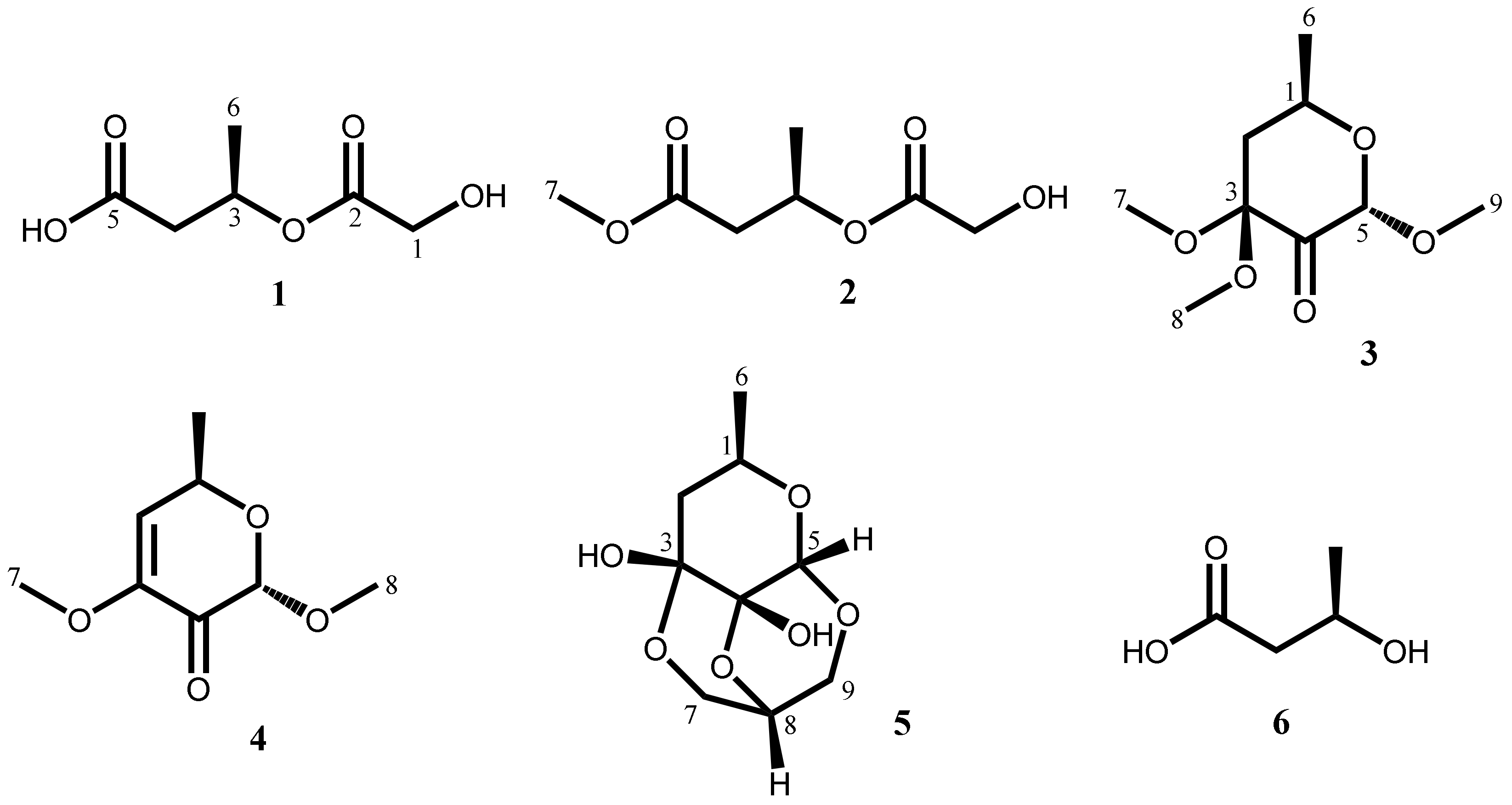
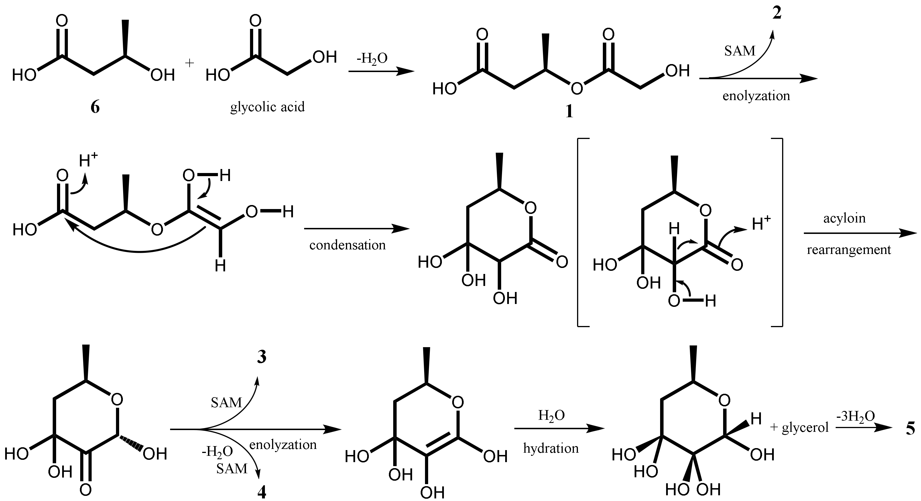
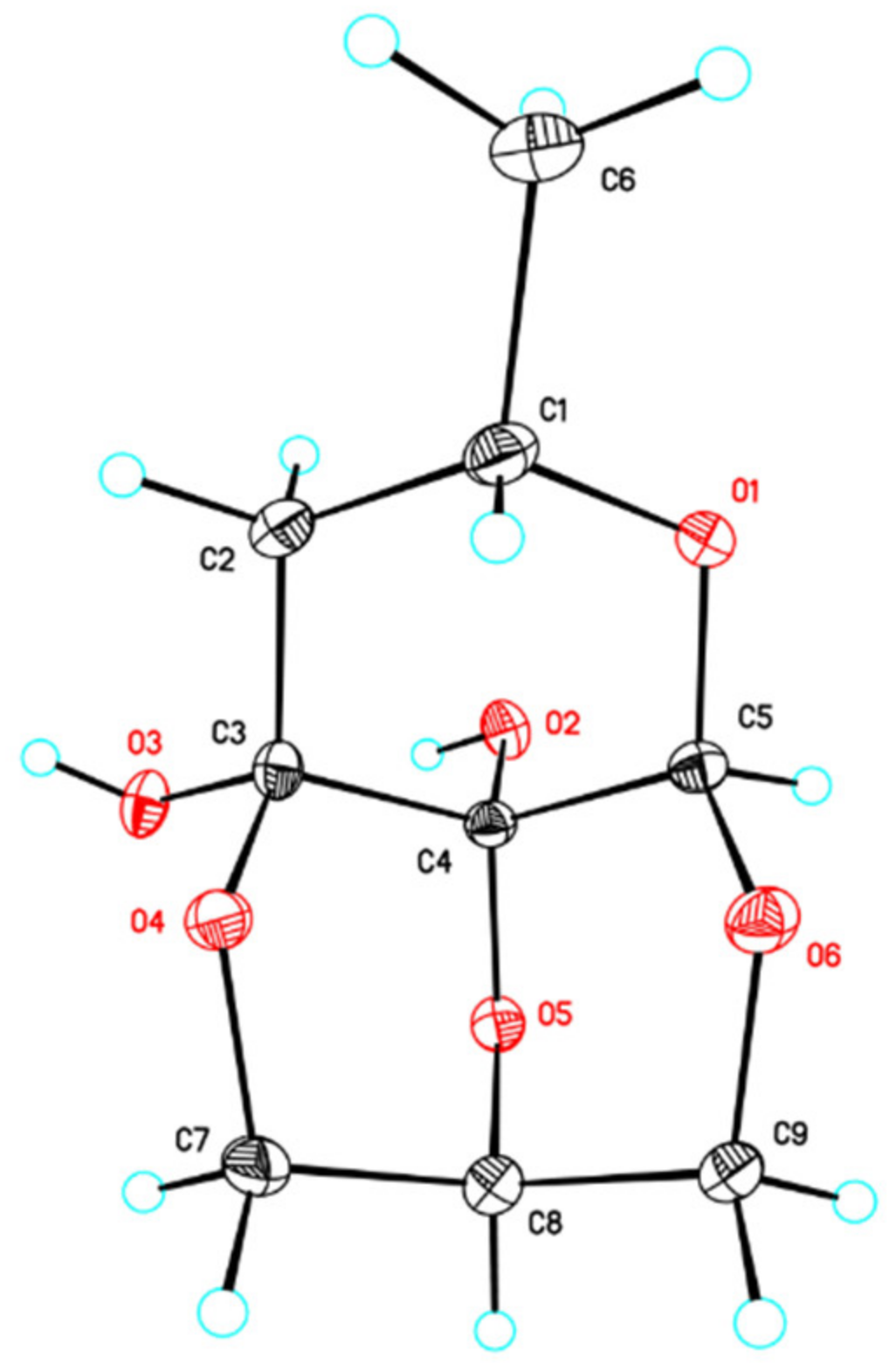
2.1. Isolation and Characterization of Secondary Metabolites

In this study, the brown alga S. cristaefolium-derived fungal strain P. sumatraense SC29 was cultured in potato dextrose broth and malt extract, and six compounds—including four unreported (1 and 35) and the previously described penisterine A methyl ether (2), which was isolated for the first time from natural resource, along with (R)-3-hydroxybutyric acid (6)—were purified from the fermented products. Of these, (R)-3-hydroxybutyric acid has been reported to exhibit antibiotic activity and could possibly serve as a chiral building block for the synthesis of fine chemicals such as antibiotics, vitamins, aromatics, and pheromones [16]. It was also used as monomer to produce poly[(R)-3-hydroxybutyrate], a kind of biodegradable plastic with properties comparable to those of polypropylene [17].
Compound 1 was obtained as a colorless oil. The quasi-molecular ion peak [M − H] at m/z 161.0450 (calcd. 161.0450 for C6H9O5) in the HRESIMS and supported by 13C NMR of 1 (Table 1), indicating a molecular formula of C6H10O5. The IR spectrum indicated the presence of a carboxylic acid (3400–2400 cm−1) and a ketone carbonyl functionality (1728 cm−1). The 1H NMR spectrum of 1 (Table 2) showed signals of a methyl proton at δH 1.31 (3H, d, J = 6.6 Hz, H-6), two methylene protons at δH 2.56 (1H, dd, J = 16.2, 5.4 Hz, Ha-4) and 2.63 (1H, dd, J = 16.2, 8.4 Hz, Hb-4) and δH 4.07 (2H, br s, H-1), and a methine proton at δH 5.31 (1H, dqd, J = 8.4, 6.6, 5.4 Hz, H-3). The 13C NMR spectrum, coupled with DEPT and phase-sensitive HSQC, of 1 (Table 1) showed six carbon signals, including a methyl at δC 20.1 (C-6), two methylenes at δC 41.4 (C-4) and 61.2 (C-1), an oxygenated methine at δC 69.6 (C-3), and two carbonyl carbon signals at δC 173.8 (C-2) and 174.1 (C-5). Key cross-peaks from H3-6/H-3 and H-3/H2-4 in the COSY spectrum in combination with key cross-peaks from H2-4/C-5, H-3/C-2, and H2-1/C-2 in the HMBC spectrum (Figure 2) established the gross structure of 1 as shown. Alkaline hydrolysis of 1, followed by HPLC purification, gave 3-hydroxybutyric acid (6) as evidenced by comparing its 1H NMR and 13C NMR (Figures S38 and S39) with those of (R)-3-hydroxybutyric acid (6). The sign of optical rotation ([α]26D = −20.2) of 6 was consistent with that of (R)-3-hydroxybutyric acid ([α]25D = −16.0) in the literature [18]. The configuration of C-3 in 1 was thus determined to be R.
The spectroscopic data of 2 resembled those of 1 except for the presence of an additional methyl group (δH 3.67s/ δC 52.4) (Table 1 and Table 2). The additional methyl group was assigned to be attached at OH-5 of 1 to form a methoxyl moiety on 2 based on a key cross-peak of H3-7/C-5 in the HMBC spectrum of 2 (Figure 2). Thus, compound 2 was determined to be the methyl analogue of 1. In the Scifindern database (Chemical Abstracts Service, American Chemical Society, Columbus, Ohio, USA), it was shown that 2 seemed to be a purchasable chemical (No. 1841321-02-5); however, no reference and spectroscopic data were provided. Therefore, we present the 1H and 13C NMR data of 2.
Compound 3, obtained as a brown oil, was determined to have a molecular formula of C9H16O5, as evidenced by its HRESIMS analysis and 13C NMR spectrum (Table 1). The IR absorption band at 1739 cm−1 indicated the presence of a ketone carbonyl group. The 1H NMR data along with the HSQC spectrum of 3 showed a methyl signal at δH 1.31 (d, J = 6.6 Hz, H3-6), three methoxyl signals at δH 3.18 (s, H3-7), 3.31 (s, H3-8), and 3.39 (s, H3-9), a set of nonequivalent methylene signals at δH 2.36 (dd, J = 13.5, 3.0 Hz, Ha-2) and 2.65 (dd, J = 13.5, 11.4 Hz, Hb-2), and two oxygenated methine signals at δH 4.11 (dqd, J = 11.4, 6.6, 3.0 Hz, H-1) and 4.94 (s, H-5) (Table 2). The 13C NMR data of 3 accompanied with its phase-sensitive HSQC spectrum exhibited a methyl carbon at δC 21.6 (C-6); three methoxyl carbons at δC 48.4 (C-8), 50.7 (C-7), and 55.4 (C-9); a methylene carbon at δC 48.4 (C-2); a nonprotonated ketal carbon at δC 99.4 (C-3); a dioxygenated methine carbon at δC 102.1 (C-5); and a ketone carbonyl at δC 202.9 (C-4) (Table 1). Correlations from δH 1.31 (H-6)/δH 4.11 (H-1) and δH 4.11 (H-1)/δH 2.36 and 2.65 (H2-2) in the COSY spectrum of 3 together with key correlations from δH 2.36 and 2.65 (H2-2)/δC 202.9 (C-4), δH 2.36 (Ha-2)/δC 99.4 (C-3), δH 4.94 (H-5)/δC 67.4 (C-1) and 202.9 (C-4), δH 3.18 (H3-7)/δC 99.4 (C-3), δH 3.31 (H3-8)/δC 99.4 (C-3), and δH 3.39 (H3-9)/δC 102.1 (C-5) in the HMBC spectrum of 3 (Figure 2) established the planar structure of 3. The relative configurations of C-1 and C-5 in 3 were deduced to be R* and S*, respectively, based on a key correlation of δH 4.11 (H-1)/δH 3.39 (H3-9) in the NOESY spectrum of 3 (Figure 2). H3-8 and H-5 were determined to be located at the same side due to a cross-peak of δH 3.31 (H3-8)/δH 4.94 (H-5) in the NOESY spectrum (Figure 2). Since compound 3 was speculated reasonably to be derived originally from (R)-3-hydroxybutyric acid (6) and glycolic acid and was further synthesized via sequential enolyzation, condensation, acyloin rearrangement [19], and methylation (Scheme 1), the absolute configurations of its C-1 were thus assigned as the R form and the C-5 was then established as the S form.
The 1H, 13C, and HSQC data of compound 4 were almost identical to those of compound 3 except that a methylene at C-2 and a methoxyl group at C-3 in 3 was replaced by an olefinic functionality at [δH 6.02 (d, J = 1.8 Hz, H-2); δC 121.2 (C-2)] and δC 148.2 (C-3) in 4. Complete assignments of COSY, HMBC, and NOESY spectra of 4 (Figure 2) allowed the elucidation of its planar structure as shown in Figure 1. That was further corroborated by a quasi-molecular ion [M + H]+ at m/z 173.0808 (calcd. 173.0814 for C8H13O4) in the HRESIMS and a carbonyl signal at 1739 cm−1 in the IR spectrum of 3 shifted to 1707 cm−1 in that of 4 due to olefinic conjugation effect. Compound 4 was also inferred to originate from compound 6 and glycolic-acid-like compound 3 (Scheme 1), and the absolute configurations of C-1 and C-5 in 4 were deduced to be the same as those of 3.
The molecular formula of compound 5 was deduced to be C9H14O6 by a quasi-molecular ion [M − H] at m/z 217.0715 (calcd. 217.0711 for C9H13O6) and supported by its 13C NMR data (Table 1), indicating a double bond equivalence (DBE) value of three. The 1H and 13C NMR data of A ring (C-1–C-6) of 5 were consistent with those of 3 except that the carbonyl (δC 202.9, C-4) in 3 disappeared (Figure 2), and instead a hemiketal carbon signal δC 93.1 (C-4) in 5 was observed in addition to three methoxyl groups (δH 3.18, 3.31, and 3.39; δC 48.4, 50.7, and 55.4) in 3 replaced by two oxygenated methylenes (δH 3.72, 4.03, 4.07, and 4.34; δC 63.6 and 69.2) and an oxygenated methine (δH 3.77; δC 72.5) in 5 (Table 1 and Table 2). Key cross-peaks from δH 1.14 (H3-6)/δH 4.38 (H-1) and δH 4.38 (H-1)/δH 1.67 and 1.79 (H2-2) in the COSY spectrum along with key correlations of δH 1.67 and 1.79 (H2-2)/δC 89.4 (C-3) and 93.1 (C-4) and δH 4.62 (H-5)/δC 66.6 (C-1) and 93.1 (C-4) established the ring A of 5 as shown in Figure 2. The other signals at [δH 3.72 (d, J = 12.0 Hz, Ha-7), 3.77 (t, J = 3.0 Hz, H-8), 4.03 (d, J = 12.0 Hz, Ha-9), 4.07 (dt, J = 12.0, 3.0 Hz, Hb-9), and 4.34 (dt, J = 12.0, 3.0 Hz, Hb-7)] as well as [δC 63.6 (C-7), 72.5 (C-8) and 69.2 (C-9)] observed in the 1H and 13C NMR spectra of 5, respectively, were attributed to be a set of glycerol moieties. Long-range correlations from δH 4.03 (Ha-9) and 4.07 (Hb-9)/δC 98.8 (C-5) and δH 3.77 (H-8)/δC 93.1 (C-4) in the HMBC spectrum confirmed the existence of ring B of 5 (Figure 2), and C-7 was thus proposed to be connected with C-3 via an ether linkage to form ring C to fit the DBE value of 5. For determining the absolute configuration of compound 5 in this study, a single-crystal X-ray diffraction experiment with Cu radiation (λ = 0.154 nm) was employed (Figure 3). The chiralities of C-1, -3, -4, -5, and -8 in 5 were determined to be 1R, 3R, 4S, 5S, and 8R, respectively, which were consistent with those proposed in the biosynthetic pathway of compound 5 as shown in Scheme 1.

2.2. Anti-Angiogenesis Activities in Human Endothelial Progenitor Cells

Compounds 15 were evaluated for anti-angiogenic activity in human endothelial progenitor cells (EPCs) with sorafenib as the positive control [6]. As shown in Table 3, penisterine D (4) exhibited inhibition of EPC growth with IC50 values of 28.5 ± 2.2 µg/mL. Data from the tube formation and migration assay validated the anti-angiogenic effects of 4 on EPCs. It was found that 4 suppressed the capillary-like tube formation and migration of EPCs (Figure 4A,B, and S40). To determine whether these finding were caused by the potential cytotoxicity of 4, we measured LDH release by EPCs after 4 treatments. No statistical difference was observed between the control group and EPCs-treated with 4, which therefore excluded the possibility of cytotoxicity in the anti-angiogenic effect of 4 (Figure 4C). Collectively, these findings reveal that 4 displays the most active anti-angiogenic properties by blocking cell growth, migration, and tube formation of EPCs.

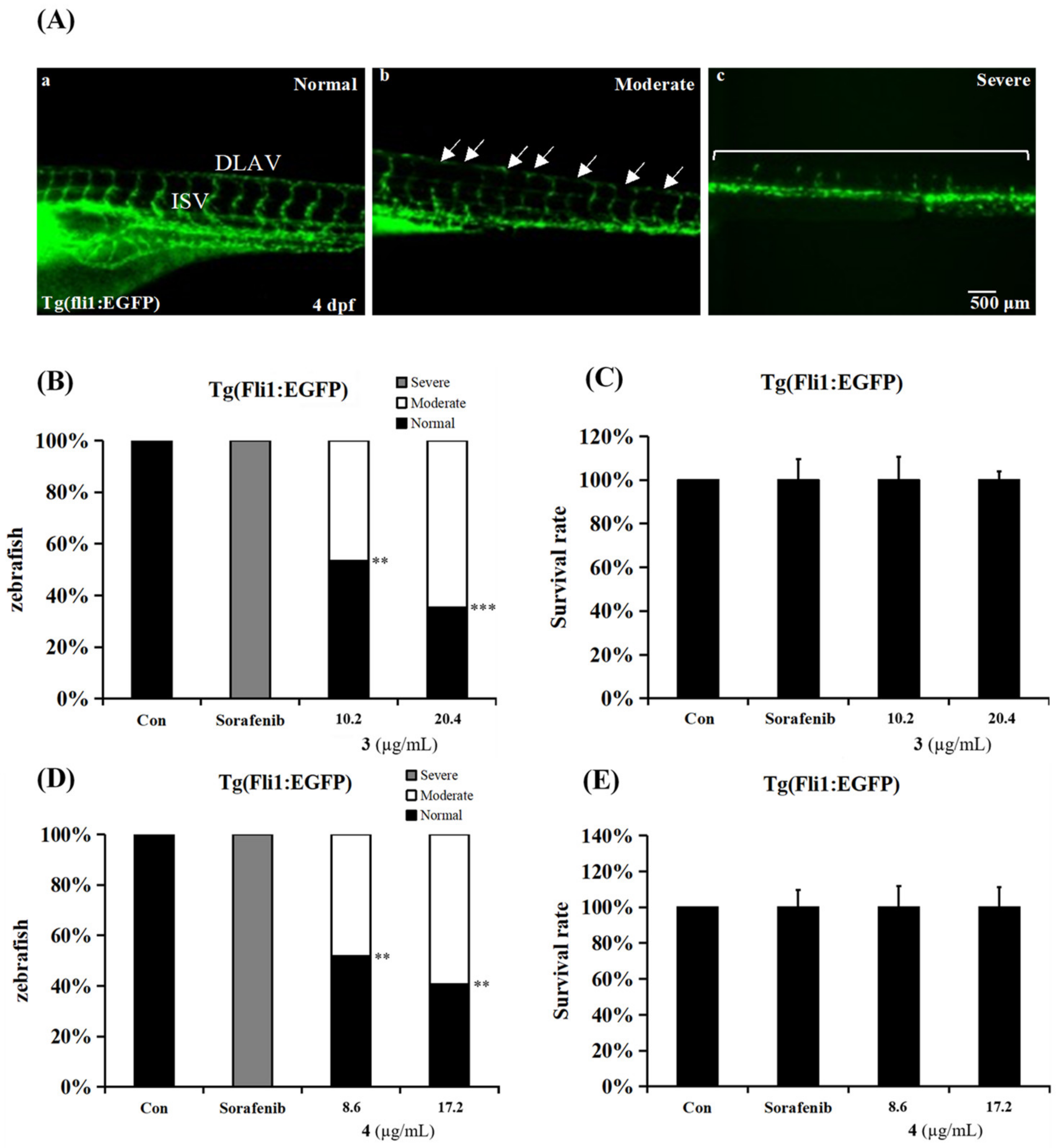
2.3. Anti-Angiogenesis Activities in an In Vivo Zebrafish Model

Vascular development in zebrafish is very similar to that of higher vertebrates such as humans, starting during gastrulation and continuing throughout life [20]. Since the amino acid sequences of some genes in humans and zebrafish are highly conserved in vertebrate evolution, the mechanism of human angiogenesis can be explored by studying zebrafish [21]. To monitor the in vivo anti-angiogenesis activity, we applied a transgenic zebrafish Tg (fli1:EGFP), which was expressed EGFP in the vasculature during development. Zebrafish embryos were incubated with 3 and 4 at 1 day post-fertilization (dpf) and evaluated the effect on angiogenesis at 4 dpf. Anti-angiogenesis was grouped into normal, mild, and severe (Figure 5A) according to the effects on intersegmental vessel (ISV) and dorsal longitudinal anastomtic vessel (DLAV) formation. Results showed that the angiogenesis percentages of zebrafish embryos were 54% and 37% by the treatment with 10.2 and 20.4 μg/mL of 3 (Figure 5B), respectively, and 52% and 41% by the treatment with 8.6 and 17.2 μg/mL of 4 (Figure 5D), respectively. We observed that no lethality occurred after 72 h of incubation with 3 and 4 (Figure 5C,E). These data suggest that 3 and 4 showed anti-angiogenic activity.

3. Materials and Methods

3.1. General Experimental Procedures

Optical rotation, ultraviolet, and IR spectra were measured on a JASCO P-2000 polarimeter (Tokyo, Japan), a Thermo UV–visible Heλios α spectrophotometer (Bellefonte, CA, USA), and a JASCO FT/IR 4100 spectrometer (Tokyo, Japan), respectively. 1H and 13C NMR spectra were obtained using an Agilent 600 MHz DD2 NMR spectrometer (Agilent Technologies, Santa Clara, CA, USA). High-resolution electrospray ionization mass spectra were obtained using an Orbitrap QE Plus mass spectrometer MS000100 (Thermo Fisher Scientific Inc., Waltham, MA, USA). Sephadex LH-20 (Sigma-Aldrich, St. Louis, MO, USA) was used for open column chromatography. Thin-layer chromatography was performed using silica gel 60 F254 plates (0.2 mm) (Merck, Darmstadt, Germany). An L-7100 HPLC pump (Hitachi, Tokyo, Japan) equipped with a refractive index detector (Bischoff, Leonberg, Germany) was employed for compound purification. All spectroscopic data are presented in the Supplementary Materials.

3.2. Algal Material

The algal material was collected in July 2021 off the coast of Badouzi (25°08′50.9″ N 121°47′42.3″ E), Keelung, Taiwan. Alga specimen was identified as Sargassum cristaefolium by T.-H.L. A voucher specimen (No. SC-IFS-2021) was deposited at Institute of Fisheries Science, National Taiwan University, Taipei, Taiwan.

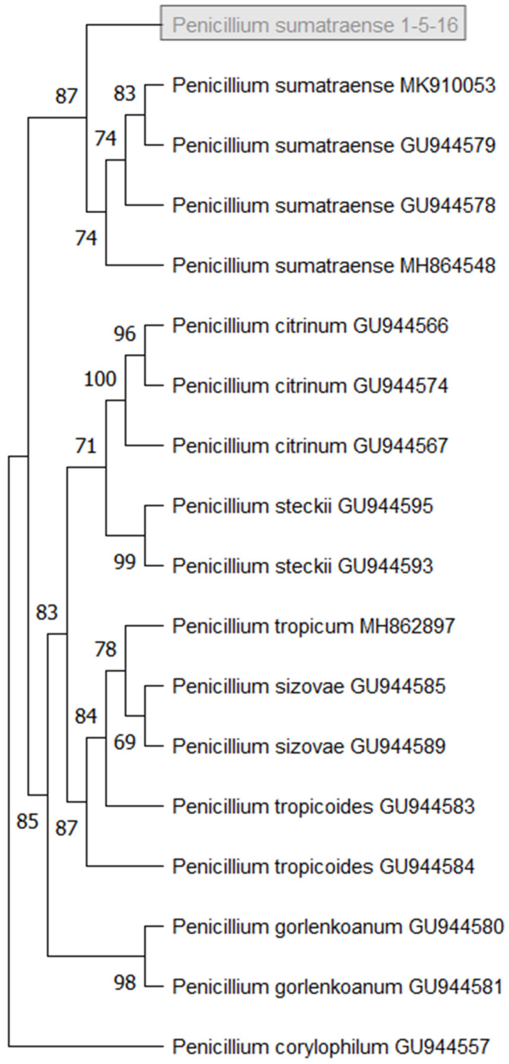
3.3. Isolation and Identification of Fungal Stain

The alga material was soaked in 75% EtOH followed by 0.01% NaOClaq and treated with ddH2O for surface cleaning. The disinfected alga was cut into circles of approximately 5 mm2. The sample was placed into the seawater PDA (potato dextrose agar) medium and incubated at 28 °C. A single fungal strain was obtained after continuous separation and purification. The mycelium of fungus was lyophilized and ground. The DNA of powdered material was extracted using DNeasy Plant Mini Kit (Qiagen, Venlo, The Netherlands) following the manufacturer’s protocol. Two sets of primers ITS4 (forward: 5«-TCCTCCGCTTATTGATATGC-3«) and ITS5 (reverse: 5«-GGAAGTAAAAGTCAAGG-3«) were used to amplify the ITS rRNA. The PCR products were analyzed by Genomic Co., Ltd. (New Taipei City, Taiwan). According to BLAST and phylogenetic analysis based on ITS rRNA gene sequences, the strain SC29 was identified as Penicillium sumatraense. The sequence was deposited in GenBank under the accession number ON685565. This stain is currently preserved in Institute of Fisheries Science, National Taiwan University, Taipei, Taiwan. We performed a most parsimonious tree (MPT) using 8 species (Figure 6). The evolutionary history was inferred using the maximum parsimony method. Tree #1 out of the 5 most parsimonious trees (length = 77) is shown. The consistency index is 0.961039 (0.938776), the retention index is 0.976923 (0.976923), and the composite index is 0.938861 (0.917111) for all sites and parsimony-informative sites (in parentheses). The percentage of replicate trees in which the associated taxa clustered together in the bootstrap test (1000 replicates) is shown next to the branches [22]. The MP tree was obtained using the tree-bisection-regrafting (TBR) algorithm (p. 126, [23]) with search level 1, in which the initial trees were obtained by the random addition of sequences (5 replicates). This analysis involved 18 nucleotide sequences. There were a total of 629 positions in the final dataset. Evolutionary analyses were conducted in MEGA11 [24].

3.4. Extraction and Isolation of Secondary Metabolites

The liquid-state mass cultures of P. sumatraense SC29 were carried out in seawater PDB (potato dextrose broth) medium. A single colony of the strain from the agar plate was inoculated into the 250 mL flask containing 100 mL PDB medium and incubated at 28 °C for 14 days on a rotary shaker at 180 rpm. In total, 7.2 L fermentation broth was harvested and partitioned using EtOAc and water three times. The EtOAc extract (3.6 g) was further subjected to size exclusion chromatography on a Sephadex LH-20 column (2.8 cm i.d. × 68 cm) and eluted with 100% EtOH at a flow rate of 2.0 mL/min to give 25 fractions. Each fraction (25 mL) collected was checked for its composition by TLC using DCM/MeOH (10:1) for development, and dipping in vanillin-H2SO4 was used in the detection of compounds with similar skeletons. All fractions were combined into four portions I–IV. Portion II (frs. 14–16) was rechromatographed on a semipreparative reversed-phase HPLC (Phenomenex Luna 5 μ PFP, 10 × 250 mm) with MeCN/H2O (3:7, v/v) as eluent to yield eight subfractions (F2A–F2H). F2A (1.05 g) was separated on semipreparative HPLC (Phenomenex Luna 5 μ C18, 10 × 250 mm) with 5% MeCNaq containing 0.1% formic acid as eluent to afford 1 (7.12 mg, tR = 14.0 min), 2 (33.62 mg, tR = 27.8 min), 5 (22.7 mg, tR = 19.6 min), and 6 (42.24 mg, tR = 8.9 min). Compound 3 (6.61 mg, tR = 10.0 min) was isolated from F2B by semipreparative reversed-phase HPLC (Phenomenex Luna 5 μ C18, 10 × 250 mm) using 40% MeCNaq as eluent. Compound 4 (12.1 mg, tR = 22.0 min) was isolated from F2D by semipreparative reversed-phase HPLC (Phenomenex Luna 5 μ C18, 10 × 250 mm) using 30% MeCNaq containing 0.1% formic acid as eluent.
Penisterine A (1): colorless oil; [α]26D -9.8 (c = 0.02, MeOH); IR (ZnSe) νmax: 3426, 1728, 1387, 1288, 1207, 1138, 1090, 1055 cm−1; 1H and 13C NMR spectroscopic data: see Table 1 and Table 2; HRESIMS m/z 161.0450 (calcd. 161.0450 for C6H9O5)
Penisterine B (2): colorless oil; [α]26D -14.4 (c = 0.02, MeOH); IR (ZnSe) νmax: 3445, 1737, 1439, 1384, 1308, 1266, 1201, 1138, 1096, 1055, 1001 cm−1; 1H and 13C NMR spectroscopic data: see Table 1 and Table 2; HRESIMS m/z 175.0506 (calcd. 175.0507 for C7H11O5)
Penisterine C (3): brown oil; [α]26D -33.2 (c = 0.02, MeOH); IR (ZnSe) νmax: 1739, 1115, 1098, 1062 cm−1; 1H and 13C NMR spectroscopic data: see Table 1 and Table 2; HRESIMS m/z 205.1069 (calcd. 205.1076 for C9H17O5)
Penisterine D (4): Yellowish oil; [α]26D 59.0 (c = 0.02, MeOH); UV (MeOH) λmax (log ε) 264 (2.7) nm; IR (ZnSe) νmax: 2933, 1707, 1632, 1455, 1375, 1318, 1244, 1199, 1172, 1153, 1091, 1057, 992, 970, 848, 832 cm−1; 1H and 13C NMR spectroscopic data: see Table 1 and Table 2; HRESIMS m/z 173.0808 (calcd. 173.0814 for C8H13O4)
Penisterine E (5): amorphous white powder; [α]26D -40.8 (c = 0.02, MeOH); IR (ZnSe) νmax: 3411, 2928, 1641, 1122, 1078, 1031, 975 cm−1; 1H and 13C NMR spectroscopic data: see Table 1 and Table 2; HRESIMS m/z 217.0715 (calcd. 217.0711 for C9H13O6)

3.5. X-ray Diffraction Analysis

The crystal data were acquired on an Oxford Gemini Dual System diffractometer. The data of compound 5 were acquired with Cu Kα radiation, and the crystal data and experimental details are listed in Tables S1–S4.
Crystallographic Data for Compound 5. (CCDC 2180439) The crystal was obtained from methanol-n-hexane–acetone (4:2:1). Crystal data: a = 5.9346(2) Å, b = 10.4559(4) Å, c = 15.4630(6) Å, α = 90°, β = 90°, γ = 90°, V = 959.50(6) Å3, μ(Cu Kα) = 1.102 mm−1. Flack parameter = 0.12(5).

3.6. Alkaline Hydrolysis of 1

Compound 1 (10.0 mg) and 2 mL of a CH3OH/0.5 M NaOH (1:1) were mixed and stirred at room temperature for 15 h, then the reaction mixtures were dried using rotary evaporator to remove CH3OH and neutralized by the addition of HCl. The reaction mixtures were then extracted with EtOAc, and the organic layer was evaporated in vacuo, and the residue was purified by HPLC (Phenomenex Luna 5 μ PFP, 10 × 250 mm) using MeCN/H2O containing 0.1% formic acid (5:95) with flow rate of 2 mL/min as eluent to afford (R)-3-hydroxybutanoic acid (6) (3.7 mg, tR = 14.8 min).
(R)-3-Hydroxybutanoic acid (6): clear oil; [α]26D -20.2 (c = 0.02, MeOH); 1H NMR (600 MHz, CD3OD): δH 1.20 (3H, d, J = 6.0 Hz), 2.40 (2H, m), 4.15 (1H, m); 13C NMR (150 MHz, CD3OD): δC 23.4, 44.7, 65.7, 175.6.

3.7. Isolation and Cultivation of Human EPCs

Human EPCs were isolated and cultured by the protocols as previously described [25]. Ethical approval for the collection of human EPCs was granted by the Institutional Review Board of Mackay Medical College, New Taipei City, Taiwan (P1000002).

3.8. Cell Growth Assay

EPCs were cultured in 96-well plates at a density of 5 × 10³ cells in each well. After 24 h of incubation, the culture medium was replaced with fresh MV2 complete medium containing 2% FBS in the presence of either vehicle (DMSO) or compounds. After 48 h of treatment, the survival rate of EPCs was assayed by SRB staining according to previously described procedure [6].

3.9. Capillary Tube Formation Assay

The capillary tube formation assay was carried out on Matrigel-coated 96-well plates. EPCs were seeded at the density of 1.25 × 10⁴ cells per well and incubated in MV2 complete medium with 2% FBS and the indicated concentration of tested compound for 24 h at 37 °C. EPCs differentiation and capillary-like tube formation was performed in three wells for each condition. The long axis of each tube was measured with MacBiophotonics Image J software in 3 randomly chosen fields per well.

3.10. Cell Migration Assay

Transwell inserts (8 μm pore size, Costar, NY, USA) were used for migration determination. EPCs migratory ability was assayed by the method based on our previous work [26].

3.11. Cytotoxicity Assay

EPCs (5 × 10³ cells/well) were seeded onto 96-well plates and incubated with MV2 complete medium containing 2% FBS in the presence of vehicle (DMSO) or penisterine D. Release of lactate dehydrogenase (LDH) into the medium was measured using a cytotoxicity assay kit (Promega, Madison, WI, USA).

3.12. Zebrafish

Zebrafish (Danio rerio) and embryos were maintained at 28 °C. All animal procedures were approved by the Institutional Animal Care and Use Committee or Panel (IACUC/IACUP) (protocol No.: LAC-2021-0181). The methods were carried out in accordance with the approved guidelines.

3.13. Transgenic Zebrafish Lines

The transgenic zebrafish line Tg (fli1:EGFP) was used in this study. The Tg (fli1:EGFP) containing fli1 (friend leukemia integration 1 transcription factor, 15 kb) promoter, driving the expression of enhanced green fluorescent protein (EGFP) in all blood vessels throughout embryogenesis [27], enables anti-angiogenesis readout for drug treatment.

3.14. Embryo Collection

One day prior to fertilization, male and female adult zebrafish were placed individually into mating tanks with inner mesh. Male and female fish were separated by a separator and left in mating cages overnight. The next morning after the removal of the separator, the couple zebrafish stimulated by the light started to chase each other and lay eggs and sperm. After 1 h, the embryos were collected and transferred to a 100 mm dish with E3 solution (5 mM NaCl, 0.17 mM KCl, 0.33 mM CaCl2, and 0.33 mM MgSO4, pH 7.0) [28] and incubated at 28 °C for 6 h. The unfertilized and dead embryos were removed, and the remaining live embryos were replenished with fresh E3 solution and kept for incubation.

3.15. Angiogenesis Inhibition Drug Screening Platform

At 1 day post-fertilization (dpf), the Tg (fli1:EGFP) embryos were distributed into 6-well culture plates with 25 embryos per well containing 3 mL E3/PTU (1-phenyl-2-thiourea, 0.003%) buffer. The embryos were treated with drugs at various concentrations. The embryos were anesthetized with tricaine (ethyl 3-aminobenzoate methanesulfonate, MS-222 (Sigma-Aldrich In., St. Louis, MO, USA) final concentration 0.016%) to prevent movement. Antiangiogenic effects of embryos were analyzed and images were captured with a fluorescence phase-contrast Zeiss Axio Vert.A1 inverted microscope (Zeiss, Jena, Germany) and a Leadview 2800AM-FL camera (Leadview, Taipei, Taiwan) at 4 dpf. Sorafenib was used as a positive control and DMSO (0.1%) was used as a negative control.

3.16. Survival Test

Tg (fli1:EGFP) embryos were used in the survival assay. At 4 dpf, 25 embryos were placed into 1 well of the 6-well plates wih 3 mL E3 medium supplement with drugs. The DMSO control and different compounds were serially diluted to determine the survival rate. Two days after exposure, the embryos were counted and the survival curves were measured. The heartbeat was used to evaluate mortality of zebrafish.

3.17. Statistical Analysis

Statistical analysis was performed from three independent experiments and analyzed the mean ± standard deviation (SD). One-way analysis of variation (AVOVA) followed by Tukey’s test was used to analyze the statistical significance and indicated by * p < 0.05, ** p < 0.01, and *** p < 0.001.

4. Conclusions

As a result, previously unreported compounds penisterine A (1), penisterines C–E (35), and penisterine A methyl ether (2) were isolated for the first time from natural resources with a known compound and were identified from a marine alga-derived fungus Penicillium sumatraense SC29. Among these, penisterine E (5) was a unique 6/6/6-tricyclic ether containing an acetal and two hemiketal functionalities. In addition, a possible biosynthetic pathway of 15 from the known compounds, (R)-3-hydroxybutanoic acid (6) and glycolic acid, was proposed. Penisterine D (4) shows anti-angiogenesis activity in both human EPCs and a Tg zebrafish model. The angiogenesis activity of penisterine D (4) makes it an attractive candidate for further preclinical investigation. Although penisterine C (3) did not have a significant effect on EPC at the dose seen with penisterine D (4), it did possess an anti-angiogenic effect in zebrafish. Thus, further investigation is required to understand the mechanism for the ability of penisterine C (3) to inhibit vessel development in embryonic zebrafish.

Supplementary Materials

The following supporting information can be downloaded at: https://www.mdpi.com/article/10.3390/molecules27248940/s1, Figure S1: title; Table S1: title. Figure S1. 1H NMR (600 MHz, MeOH-d4) of 1. Figure S2. 13C NMR (150 MHz, MeOH-d4) of 1. Figure S3. HSQC of 1. Figure S4. COSY of 1. Figure S5. HMBC of 1. Figure S6. IR spectrum of 1. Figure S7. HRESIMS spectrum of 1. Figure S8. 1H NMR (600 MHz, MeOH-d4) of 2. Figure S9. 13C NMR (150 MHz, MeOH-d4) of 2. Figure S10. HSQC of 2. Figure S11. COSY of 2. Figure S12. HMBC of 2. Figure S13. IR spectrum of 2. Figure S14. HRESIMS spectrum of 2. Figure S15. 1H NMR (600 MHz, MeOH-d4) of 3. Figure S16. 13C NMR (150 MHz, MeOH-d4) of 3. Figure S17. HSQC of 3. Figure S18. COSY of 3. Figure S19. HMBC of 3. Figure S20. NOESY of 3. Figure S21. IR spectrum of 3. Figure S22. HRESIMS spectrum of 3. Figure S23. 1H NMR (600 MHz, MeOH-d4) of 4. Figure S24. 13C NMR (150 MHz, MeOH-d4) of 4. Figure S25. HSQC of 4. Figure S26. COSY of 4. Figure S27. HMBC of 4. Figure S28. IR spectrum of 4. Figure S29. HRESIMS spectrum of 4. Figure S30. 1H NMR (600 MHz, MeOH-d4) of 5. Figure S31. 13C NMR (150 MHz, MeOH-d4) of 5. Figure S32. HSQC of 5. Figure S33. COSY of 5. Figure S34. HMBC of 5. Figure S35. NOESY of 5. Figure S36. IR spectrum of 5. Figure S37. HRESIMS spectrum of 5. Figure S38. 1H NMR (600 MHz, MeOH-d4) of 6. Figure S39. 13C NMR (150 MHz, MeOH-d4) of 6. Figure S40. Effects of compounds 1, 2, 3, and 5 on tube formation of human endothelial progenitor cells. Figure S41. Effects of compounds 1-5 on cell migration of human endothelial progenitor cells. Figure S42. Effect of compound 4 on the apoptotic cell death of human endothelial progenitor cells. Figure S43. Antiangiogenesis of compound 1, 2, and 5 using transgenic zebrafish. Figure S44. ITS rDNA sequences. Figure S45. Agarose gel electrophoresis. Figure S46. BLASTn results of the isolated fungus. Table S1. Crystal data and experimental details for 5. Table S2. Atomic coordinates (x 104) and equivalent isotropic displacement parameters (Å2 x 10³). Table S3. Bond lengths [Å] and angles [°] for 5. Table S4. Anisotropic displacement parameters (Å2 x 10³) for 5.

Author Contributions

H.-Y.H. performed the experiments and wrote the manuscript. S.-W.W., C.-H.C. and C.-Y.H. performed the biological assays. K.-L.P. and Y.-H.K. performed the experiments. J.-Y.L., S.-H.C. and Y.-T.L. prepared the published work. T.-H.L. performed the data curation, supervision, and methodology. All authors have read and agreed to the published version of the manuscript.

Funding

This work was supported by a grant from the National Science and Technology Council (MOST110-2320-B-002-023-MY3) of Taiwan to T.-H.L.

Institutional Review Board Statement

The animal study protocol was approved by Institutional Animal Care and Use Committee or Panel (IACUC/IACUP) (protocol No.: LAC-2021-0181).

Informed Consent Statement

Informed consent was obtained from all subjects involved in the study. Written informed consent has been obtained from the patient(s) to publish this paper.

Data Availability Statement

Not applicable.

Acknowledgments

We thank S.-Y.S. and A.G. in the Instrumentation Center of the College of Science, National Taiwan University and the Instrumentation Center of Taipei Medical University for the MS and NMR data acquisition, respectively.

Conflicts of Interest

The authors declare no conflict of interest.

Sample Availability

Not applicable.

References

  1. Liu, L.; Heinrich, M.; Myers, S.; Dworjanyn, S.A. Towards a better understanding of medicinal uses of the brown seaweed Sargassum in Traditional Chinese Medicine: A phytochemical and pharmacological review. J. Ethnopharmacol. 2012, 142, 591–619. [Google Scholar] [CrossRef]
  2. Sun, S.; Hu, S.; Zhang, B.; Sun, X.; Xu, N. Allelopathic effects and potential allelochemical of Sargassum fusiforme on red tide microalgae Heterosigma akashiwo. Mar. Pollut. Bull. 2021, 170, 112673. [Google Scholar] [CrossRef] [PubMed]
  3. Puspo Edi Giriwono, S.; Iskandriati, D.; Tan, C.P.; Andarwulan, N. Sargassum seaweed as a source of anti-inflammatory substances and the potential insight of the tropical species: A review. Mar. Drugs 2019, 17, 590. [Google Scholar]
  4. Yip, Z.T.; Quck, R.Z.B. Historical biogeography of the widespread macroalga Sargassum (Fucales, Phaeophyceae). J. Phycol. 2020, 56, 300–309. [Google Scholar] [CrossRef] [PubMed] [Green Version]
  5. Hsiao, G.; Wang, S.-W.; Chiang, Y.-R.; Chi, W.-C.; Kuo, Y.-H.; Phong, D.A.; Chen, C.-Y.; Lee, T.-H. Anti-inflammatory effects of peptides from a marine algicolous fungus Acremonium sp. NTU492 in BV-2 microglial cells. J. Food Drug Anal. 2020, 28, 89–97. [Google Scholar] [CrossRef]
  6. Yang, C.-Y.; Chen, C.; Lin, C.-Y.; Chen, Y.-H.; Lin, C.-Y.; Chi, C.-W.; Chen, Y.-J.; Liu, S.-C.; Chang, T.-K.; Tang, C.-H. Garcimultiflorone K inhibits angiogenesis through Akt/eNOS-and mTOR-dependent pathways in human endothelial progenitor cells. Phytomedicine 2019, 64, 152911. [Google Scholar] [CrossRef]
  7. Flamini, V.; Jiang, W.G.; Lane, J.; Cui, Y.-X. Significance and therapeutic implications of endothelial progenitor cells in angiogenic-mediated tumour metastasis. Crit. Rev. Oncol. Hematol. 2016, 100, 177–189. [Google Scholar] [CrossRef] [Green Version]
  8. Chen, S.-R.; Wang, S.-W.; Lin, Y.-C.; Yu, C.-L.; Yen, J.-Y.; Chen, Y.-F.; Cheng, Y.-B. Additional alkaloids from Zoanthus vietnamensis with neuroprotective and anti-angiogenic effects. Bioorg. Chem. 2021, 109, 104700. [Google Scholar] [CrossRef]
  9. Mimeault, M.; Batra, S.K. Emergence of zebrafish models in oncology for validating novel anticancer drug targets and nanomaterials. Drug Discov. Today 2013, 18, 128–140. [Google Scholar] [CrossRef] [PubMed] [Green Version]
  10. Howe, K.; Clark, M.D.; Torroja, C.F.; Torrance, J.; Berthelot, C.; Muffato, M.; Collins, J.E.; Humphray, S.; McLaren, K.; Matthews, L.; et al. The zebrafish reference genome sequence and its relationship to the human genome. Nature 2013, 496, 498–503. [Google Scholar] [CrossRef] [Green Version]
  11. Lin, C.-Y.; Chiang, C.-Y.; Tsai, H.-J. Zebrafish and Medaka: New model organisms for modern biomedical research. J. Biomed. Sci. 2016, 23, 1–11. [Google Scholar] [CrossRef] [PubMed]
  12. Wang, W.L.; Liu, S.L.; Lin, Y.B. 225 Seaweeds of Kenting National Park; Kenting National Park Headquarters: Pingtung, Taiwan, 2020; pp. 207–214. [Google Scholar]
  13. Hsiao, W.-C.; Hong, Y.-H.; Tsai, Y.-H.; Lee, Y.-C.; Patel, A.K.; Guo, H.-R.; Kuo, C.-H.; Huang, C.-Y. Extraction, biochemical characterization, and health effects of native and degraded fucoidans from Sargassum crispifolium. Polymers 2022, 14, 1812. [Google Scholar] [CrossRef] [PubMed]
  14. Wang, S.-H.; Huang, C.-Y.; Chen, C.-Y.; Chang, C.-C.; Huang, C.-Y.; Dong, C.-D.; Chang, J.-S. Structure and biological activity analysis of fucoidan isolated from Sargassum siliquosum. ACS Omega 2020, 5, 32447–32455. [Google Scholar] [CrossRef] [PubMed]
  15. Chen, B.-R.; Li, W.-M.; Li, T.-L.; Chan, Y.-L.; Wu, C.-J. Fucoidan from Sargassum hemiphyllum inhibits infection and inflammation of Helicobacter pylori. Sci. Rep. 2022, 12, 429. [Google Scholar] [CrossRef]
  16. Seebach, D.; Albert, M.; Arvidsson, P.I.; Rueping, M.; Schreiber, J.V. From the biopolymer PHB to biological investigations of unnatural beta- and gamma-peptides. Chimia 2001, 55, 345–353. [Google Scholar] [CrossRef]
  17. Tokiwa, Y.; Ugwu, C.U. Biotechnological production of (R)-hydroxybutyric acid monomer. J. Biotechnol. 2007, 132, 264–272. [Google Scholar] [CrossRef]
  18. Wu, Z.; Liu, D.; Huang, J.; Proksch, P.; Zhu, K.; Lin, W. Hansforesters A–M, polyesters from sponge-associated fungus Hansfordia sinuosea with antibacterial activities. RSC Adv. 2018, 8, 39756–39768. [Google Scholar] [CrossRef] [Green Version]
  19. Matsumoto, S.; Matsuo, M.; Iitaka, Y. Oxygenations of vitamin E (α-tocopherol) and its model compound, 2, 2, 5, 7, 8-pentamethylchroman-6-ol, in the presence of potassium superoxide suspended in tetrahydrofuran, and unusual acyloin rearrangements. J. Org. Chem. 1986, 51, 1435–1440. [Google Scholar] [CrossRef]
  20. Childs, S.; Chen, J.-N.; Garrity, D.M.; Fishman, M.C. Patterning of angiogenesis in the zebrafish embryo. Development 2002, 129, 973–982. [Google Scholar] [CrossRef]
  21. Bowley, G.; Kugler, E.; Wilkinson, R.; Lawrie, A.; van Eeden, F.; Chico, T.J.; Evans, P.C.; Noël, E.S.; Serbanovic-Canic, J. Zebrafish as a tractable model of human cardiovascular disease. Br. J. Pharmacol. 2022, 179, 900–917. [Google Scholar] [CrossRef]
  22. Felsenstein, J. Confidence limits on phylogenies: An approach using the bootstrap. Evolution 1985, 39, 783–791. [Google Scholar] [PubMed]
  23. Nei, M.; Kumar, S. Molecular Evolution and Phylogenetics; Oxford University Press: New York, NY, USA, 2000. [Google Scholar]
  24. Tamura, K.; Stecher, G.; Kumar, S. MEGA 11: Molecular Evolutionary Genetics Analysis Version 11. Mol. Biol. Evol. 2021, 38, 3022–3027. [Google Scholar] [CrossRef] [PubMed]
  25. Chen, W.-C.; Chung, C.-H.; Lu, Y.-C.; Wu, M.-H.; Chou, P.-H.; Yen, J.-Y.; Lai, Y.-W.; Wang, G.-S.; Liu, S.-C.; Cheng, J.-K. BMP-2 induces angiogenesis by provoking integrin α6 expression in human endothelial progenitor cells. Biochem. Pharmacol. 2018, 150, 256–266. [Google Scholar] [CrossRef]
  26. Wang, G.-S.; Shen, Y.-S.; Chou, W.-Y.; Tang, C.-H.; Yeh, H.-I.; Wang, L.-Y.; Yen, J.-Y.; Huang, T.-Y.; Liu, S.-C.; Yang, C.-Y. Senescence Induces Dysfunctions in Endothelial Progenitor Cells and Osteoblasts by Interfering Translational Machinery and Bioenergetic Homeostasis. Int. J. Mol. Sci. 2018, 19, 1997. [Google Scholar] [CrossRef] [Green Version]
  27. Lawson, N.D.; Weinstein, B.M. In vivo imaging of embryonic vascular development using transgenic zebrafish. Dev. Biol. 2002, 248, 307–318. [Google Scholar] [CrossRef] [PubMed] [Green Version]
  28. Vogt, A.; Codore, H.; Day, B.W.; Hukriede, N.A.; Tsang, M. Development of automated imaging and analysis for zebrafish chemical screens. J. Vis. Exp. 2010, 40, e1900. [Google Scholar] [CrossRef]
Figure 1. Chemical structures of 16.
Molecules 27 08940 g001
Figure 2. Key COSY, HMBC, and NOESY of compounds 15.
Molecules 27 08940 g002
Scheme 1. Proposed biosynthetic pathway of compounds 15.
Molecules 27 08940 sch001
Figure 3. Ortep diagram of 5.
Molecules 27 08940 g003
Figure 4. Effects of penisterine D (4) on tube formation and cytotoxicity of human EPCs. Cells were treated with the indicated concentrations of penisterine D and sorafenib for 24 h. (A) The inverted phase-contrast microscope was utilized for recording tubular morphogenesis. (B) The tube formation of EPCs was quantified by measuring the length of tubes by Image-J. (C) Cells were treated with 4 for 24 h and cytotoxicity was determined using the LDH assay. Data represent the mean ± SEM. * p < 0.05, compared with the control group.
Molecules 27 08940 g004
Figure 5. Penisterine C (3) and penisterine D (4) exert anti-angiogenic capacities in vivo. (A) Representative images of the trunk vasculature in Tg (fli1:EGFP) zebrafish embryos incubated with compounds or sorafenib at 4 days post-fertilization (dpf). Arrows indicate impaired at intersegmental vessel (ISV) and dorsal longitudinal anastomic vessel (DLAV). (B) Quantitative analysis presents percentage of fish embryos incubated with 3 with defective vasculature. ** p < 0.01, and *** p < 0.001 compared with the control group. (C) Quantitative analysis presents percentage of survival rate of fish embryos incubated with 3. (D) Quantitative analysis presents percentage of fish embryos incubated with 4 with defective vasculature. ** p < 0.01 compared with the control group. (E) Quantitative analysis presents percentage of survival rate of fish embryos incubated with 4. The zebrafish treated with 1% DMSO was used as a negative control, and 10 µg/mL sorafenib was used as a positive control.
Molecules 27 08940 g005
Figure 6. Maximum parsimonious tree based on sequences of rDNA ITS region. Isolated strain SC29 is highlighted in the grey box.
Molecules 27 08940 g006
Table 1. 13C NMR spectroscopic data for 15 (δ in ppm, mult.).
No.1 a,b2 a,b3 a,b4 a,b5 a,b
161.2, CH261.2, CH267.4, CH66.3, CH66.6, CH
2173.8, C172.6, C48.4, CH2121.2, CH44.1, CH2
369.6, CH69.4, CH99.4, C148.2, C89.4, C
441.4, CH241.3, CH2202.9, C186.7, C93.1, C
5174.1, C173.8, C102.1, CH100.7, CH98.8, CH
620.1, CH320.2, CH321.6, CH322.0, CH320.8, CH3
7 52.4, CH350.7, CH355.5, CH363.6, CH2
8 48.4, CH357.1, CH372.5, CH
9 55.4, CH3 69.2, CH2
a Measured in CD3OD (150 MHz). b Carbon types were determined from DEPT and phase-sensitive HSQC experiments.
Table 2. 1H NMR spectroscopic data for 15 (δ in ppm, mult., J in Hz).
No.1 a2 a3 a4 a5 a
14.07, brs4.07, brs4.11, dqd (11.4, 6.6, 3.0)4.82, qd (6.6, 1.8)4.38, dqd (11.4, 6.6, 2.4)
2a 2.36, dd (13.5, 3.0)6.02, d (1.8)1.67, dd (13.5, 2.4)
2b 2.65, dd (13.5, 11.4) 1.79, dd (13.5, 11.4)
35.31, dqd (8.4, 6.6, 5.4)5.31, dqd (7.8, 6.6, 5.4)
4a2.56, dd (16.2, 5.4)2.60, dd (15.9, 5.4)
4b2.63, dd (16.2, 8.4)2.66, dd (15.9, 7.8)
5 4.94, s4.74, s4.62, s
61.31, d (6.6)1.31, d (6.6)1.31, d (6.6)1.38, d (6.6)1.14, d (6.6)
7a 3.67, s3.18, s3.61, s3.72, d (12.0)
7b 4.34, dt (12.0, 3.0)
8 3.31, s3.50, s3.77, t (3.0)
9a 3.39, s 4.03, d (12.0)
9b 4.07, dt (12.0, 3.0)
a Measured in CD3OD (600 MHz).
Table 3. Anti-angiogenic effects of isolated compounds in human EPCs.
Compound a
(µg/mL)
EPCs Growth
2040
1>100%>100%
2>100%>100%
399.3 ± 1%>100%
465 ± 5%34 ± 1%
5>100%>100%
a EPCs were treated with the indicated compounds at concentrations of 20 and 40 µg/mL for 48 h, and anti-angiogenic effects were elucidated in a cell growth assay (n = 3). Data are expressed as the mean ± SEM.
Publisher’s Note: MDPI stays neutral with regard to jurisdictional claims in published maps and institutional affiliations.

