Article

Phytochemical and In Silico ADME/Tox Analysis of Eruca sativa Extract with Antioxidant, Antibacterial and Anticancer Potential against Caco-2 and HCT-116 Colorectal Carcinoma Cell Lines

by
Amir Mahgoub Awadelkareem
1,
Eyad Al-Shammari
1,
Abd Elmoneim O. Elkhalifa
1,
Mohd Adnan
2,
Arif Jamal Siddiqui
2,
Mejdi Snoussi
2,3,
Mohammad Idreesh Khan
4,
Z R Azaz Ahmad Azad
5,
Mitesh Patel
6 and
Syed Amir Ashraf
1,*
1
Department of Clinical Nutrition, College of Applied Medical Sciences, University of Hail, Hail P.O. Box 2440, Saudi Arabia
2
Department of Biology, College of Science, University of Hail, Hail P.O. Box 2440, Saudi Arabia
3
Laboratory of Genetic, Biodiversity and Valorization of Bioresources, Higher Institute of Bio-Technology of Monastir, University of Monastir, Avenue Taher Hadded BP 74, Monastir 5000, Tunisia
4
Department of Clinical Nutrition, College of Applied Health Sciences in Arras, Qassim University, Buraydah 52571, Saudi Arabia
5
Department of Post-Harvest Engineering and Technology, Aligarh Muslim University, Aligarh 202002, India
6
Bapalal Vaidya Botanical Research Centre, Department of Biosciences, Veer Narmad South Gujarat University, Surat 395007, India
*
Author to whom correspondence should be addressed.
Molecules 2022, 27(4), 1409; https://doi.org/10.3390/molecules27041409
Submission received: 18 January 2022 / Revised: 12 February 2022 / Accepted: 17 February 2022 / Published: 19 February 2022
(This article belongs to the Special Issue Bioactivities and In Silico Study of Phytochemicals)

Abstract

:
Eruca sativa Mill. (E. sativa) leaves recently grabbed the attention of scientific communities around the world due to its potent bioactivity. Therefore, the present study investigates the metabolite profiling of the ethanolic crude extract of E. sativa leaves using high resolution-liquid chromatography-mass spectrometry (HR-LC/MS), including antibacterial, antioxidant and anticancer potential against human colorectal carcinoma cell lines. In addition, computer-aided analysis was performed for determining the pharmacokinetic properties and toxicity prediction of the identified compounds. Our results show that E. sativa contains several bioactive compounds, such as vitamins, fatty acids, alkaloids, flavonoids, terpenoids and phenols. Furthermore, the antibacterial assay of E. sativa extract showed inhibitory effects of the tested pathogenic bacterial strains. Moreover, the antioxidant activity of 2,2-diphenyl-1-picrylhydrazyl (DPPH) and hydrogen peroxide (H2O2) were found to be IC50 = 66.16 μg/mL and 76.05 μg/mL, respectively. E. sativa also showed promising anticancer activity against both the colorectal cancer cells HCT-116 (IC50 = 64.91 μg/mL) and Caco-2 (IC50 = 83.98 μg/mL) in a dose/time dependent manner. The phytoconstituents identified showed promising pharmacokinetics properties, representing a valuable source for drug or nutraceutical development. These investigations will lead to the further exploration as well as development of E. sativa-based nutraceutical products.

1. Introduction

Eruca sativa Mill. (Eruca vesicaria) is a very popular species of the Brassicaceae family, which is commonly known as arugula, jarjeer or rocket leaves [1,2]. It is also popularly used in salad preparations. E. sativa leaves are considered as a valuable source of nutrition due to the presence of several important nutrients, such as dietary fiber, oligosaccharides, amino acids, peptides, proteins, polyunsaturated fatty acids, vitamins, carbohydrates, L-ascorbic acid and mineral content. Furthermore, it has been noticed that E. sativa is valued by dieticians for its low calorific content along with high nutritional values [3,4].
In recent years, E. sativa has come into the limelight due to its rich content of phytochemicals and its significance in various biological activities. Different parts of the E. sativa plant possess diverse phytochemicals, such as flavonoids, glucosinolates, phenolics, saponins, tannins and essential oils [5]. Additionally, several phytoconstituents were reported in various studies, namely, isothiocyanates, derivatives of butane, octane, nonane, 4-methylthiobutyl isothiocyanate, cis-3-hexen-1-ol,5-methylthiopentylisothiocyanate, cis-3-hexenyl 2-methylbutanoate, 5-methyl thiopentane nitrile [6], quercetin, kampferol, rutin, myricetin rhamnetin and kaempferol-3-O-galactoside [7,8], which indicates that E sativa is a rich source of flavonoids that possesses several other important bioactive compounds.
The E. sativa seed was investigated for its essential oil content and reported to contain a significant amount of sulfur and nitrogen compounds. Active compounds produced by essential oils, as well as other phytoconstituents during secondary vegetal metabolism are usually considered responsible for biological activities, such as antimicrobial and antioxidant [9]. Additionally, a wide range of phytochemicals present in E. sativa were reported to have various biological properties, including antimicrobial, antigenotoxic, antidiuretic, stimulant, stomach disorders analgesic, antioxidant, antiulcer, hepatoprotective activities, antidiabetic, antiacne, antihyperlipidemic, antihyperglycemic and anti-inflammatory [2,10,11,12,13,14]. E. sativa seed oil, which is commonly known as taramira oil or jamba oil in Central Asia, is used for massages, hair treatments and as an anti-influenza medication [10].
Therefore, based upon its several uses and therapeutic benefits, E. sativa grabbed the attention of scientific communities that aimed to further explore, identify and characterize the phytochemicals present in E. sativa leaves by using the high resolution-liquid chromatography-mass spectrometry (HR-LC/MS) technique. HR-LC/MS analysis is one of the novel chromatographic techniques usually applied for the identification and quantification of phytochemicals present in plant extracts. HR-LC/MS instrumentation is advantageous for conducting non-targeted analyses, when the compounds of interest are unknown, and it acquires data over a large mass range (e.g., m/z 100–1500). [15].
An abundance of bioactive components present in E. sativa makes it an important leafy vegetable with the potential for nutraceutical purposes and other therapeutic uses. In recent years, a new term, “nutraceuticals”, was introduced by merging two scientific disciplines, “nutrition” and “pharmaceuticals”, which represents any food or part of a food that not only imparts health benefits, but also contributes to the prevention, management or treatment of various diseases [16]. Moreover, in broad terms, nutraceuticals can be summarized as bioactive components, which play a vital role in human beings by maintaining their normal physiological functions and well-being. Hence, E. sativa could be further explored for its nutraceutical potential.
Therefore, based upon the nutraceutical potential and therapeutic benefits, the present study is designed to identify the phytochemicals or bioactive compounds present in E. sativa extract using the HR-LC/MS technique. Furthermore, E. sativa extract was investigated for several other biological activities, such as antibacterial, antioxidant and anticancer, against the colorectal carcinoma cell lines. Based upon the identification of phytochemicals, computer-aided technology was applied to understand the pharmacokinetic properties as well as toxicity prediction of the identified compounds. According to our report, no study to date has ever reported the potentiality of E. sativa crude extract in the management of colorectal cancer with a toxicity prediction of all the phytochemicals identified by HR-LCMS. The obtained results further support in vivo studies for the product’s possible use as a therapeutic, as well as the nutraceutical use of E. sativa against various diseases for the management and treatment possibilities of colorectal cancer.

2. Results and Discussion

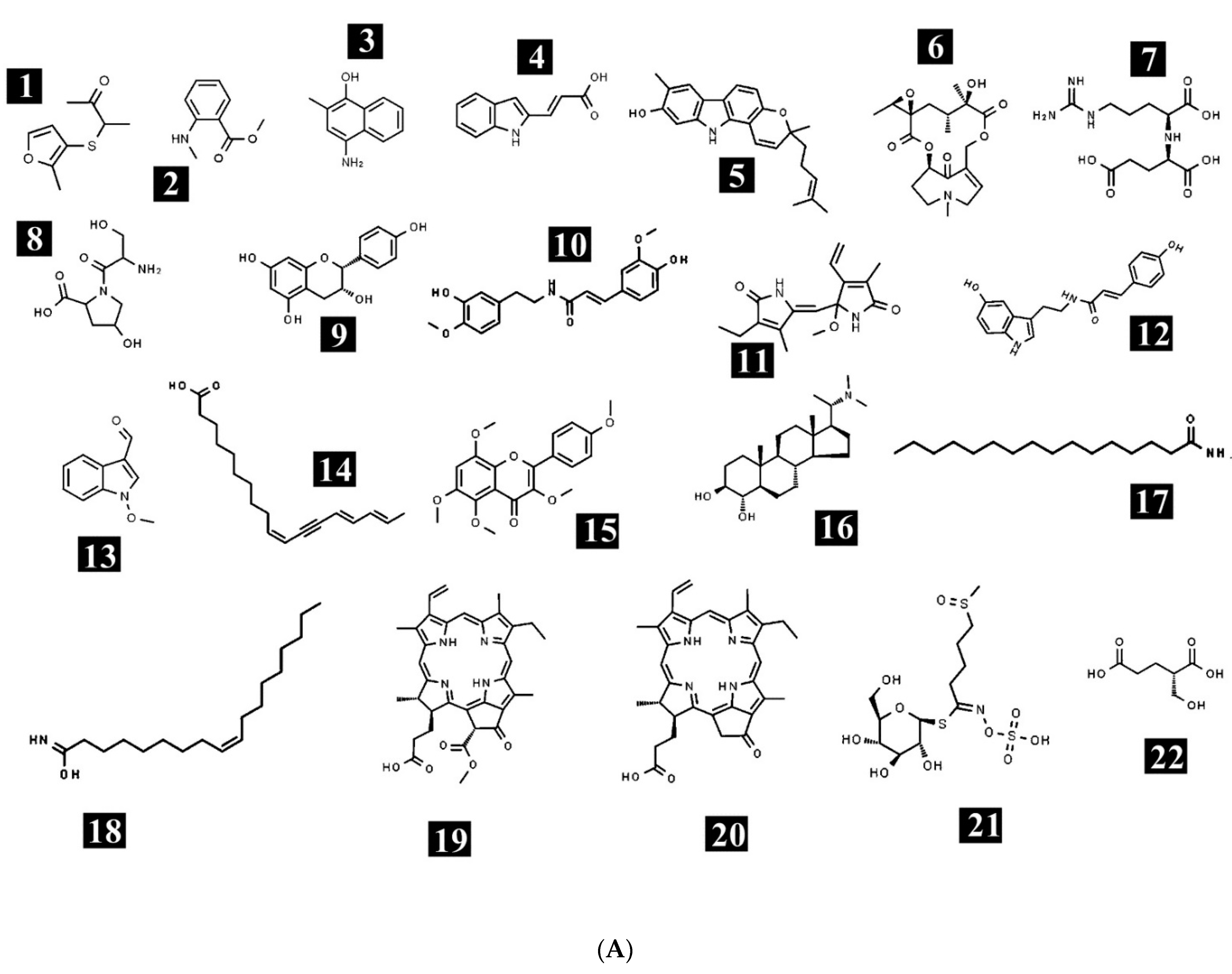
2.1. Phytochemical Profiling of E. sativa

The ethanolic crude extract of E. sativa was used for tentative phytochemical analysis via HR-LCMS. The% yield of the extract was 4.26%, i.e., 42.60 mg/g of dry weight of the whole plant (w/w). With the obtained retention times, absorbance spectra and the data of MS, we determined that the chemical composition of the crude extract of E. sativa possesses different bioactive compounds (Figure 1). The identified bioactive compounds belong to several classes, such as amino acids, vitamins, fatty acids, alkaloids, flavonoids, terpenoids and phenols. The chemical formula, mass and retention time of the identified compounds are listed in Table 1. The chemical structures of the identified compounds are presented in Figure 2A,B. Some of the bioactive compounds were already identified from different parts of E. sativa, such as, 1-methoxy-1H-indole-3-carboxaldehyde from the flower, leaf and seed [17]; 3,4′,5,6,8-pentamethoxyflavone from the fresh leaves [6]; glucoraphanin from the leaves [18]; rutin from the leaves [3,8,18]; kaempferol from the leaves [19] and from the whole plant except the root [7,20,21]; 9Z-Octadecenedioic acid, 16-Hydroxy hexadecanoic acid and (10Z,14E,16E)-10,14,16-Octadecatrien-12-ynoic acid (decanoic acid) from the flower, leaves and seed oil [6,17]; and linolenic acid from the seed oil [1,22,23] and from leaves [19]. In this study, phytoconstituents, namely (+/−)-3-[(2-methyl-3-furyl)thio]-2-butanone; methyl N-methylanthranilate; 4-amino-2-methyl-1-naphthol; indoleacrylic acid; pyrafoline D; petasitenine; nopaline; serinyl-Hydroxyproline; afzelechin; N-trans-feruloyl-4-O-methyldopamine; (±)-rollipyrrole; N6-cis-p-coumaroylserotonin; terminaline; palmitic amide; oleamide; pheophorbide a; pyropheophorbide a; (S)-2-(hydroxymethyl)glutarate; 2-deoxy-scyllo-inosose; artomunoxanthentrione epoxide; fraxidin; N-(6-oxo-6H-dibenzo[b,d]pyran-3-yl)maleamic acid; sciadopitysin; 5′-butyrylphosphoinosine; evoxine; lactucin; 1,4-dimethoxyglucobrassicin; pubesenolide; corchorifatty acid F; linifolin A; N2-(2-carboxymethyl-2-hydroxysuccinoyl)arginine; trilobolide; thalidasine; α-linolenic acid; 16-hydroxy hexadecanoic acid; 4-(3-hydroxy-7-phenyl-6-heptenyl)-1,2-benzenediol; and (6beta,8betaOH)-6,8-dihydroxy-7(11)-eremophilen-12,8-olide, were reported, to our knowledge, for the first time to be present in E. sativa. The differences in the phytochemistry are varied, possibly due to the season, habitat or the ecological conditions of the plant.
The presence of different types of phytochemicals, such as flavonoids, phenolics, tannins, saponins and essential oils, are also reported in the literature [1,19,24,25]. The chemodiversity of E. sativa essential oils possess 4-methylthiobutyl isothiocyanate, 5-methylthiopentanonitrile, as well as a large amount of sulfur- and nitrogen-containing compounds. Erucic acid is a major component of the plant, along with glucosinolatemethyl sulphinyl butyl isothiocyanate [22]. Furthermore, Jirovetz et al. (2002) analyzed the aromatic compounds of the fresh leaves E. sativa using gas chromatography. They reported the presence of various components, such as iso-thiocyanates, derivatives of butane, octane, nonane, 4-methylthiobutyl isothiocyanate, cis-3-hexen-1-ol,5-methylthiopentylisothiocyanate, cis-3-hexenyl 2-methylbutanoate, and 5-methyl thiopentone nitrile [6]. Zhang and Tang (2007) found that E. sativa leaves contain volatile components and the major constituents identified were 4-methylthiobutyl isothiocyanate, 5-methylthio-pentanonitrile and abundant amounts of sulfur- and nitrogen-possessing compounds. They also reported that isothiocyanates (isothiocyanate and erucin) had cytoprotective effects [26]. Nazif et al. (2010) investigated a 70% alcoholic extract of E. sativa seeds, which led to the isolation of three flavonoids, quercetin, rhamnetin and kaempferol-3-O-galactoside, in addition to two glucosinolates; 4-(methylthio)butyl-glucosinolate (glucoerucin) and 3-methylsulfinylpropyl-glucosinolate (glucoiberin) [7]. Michael et al. (2011) determined that the phytochemical analysis of the aqueous extract of E. sativa fresh leaves showed nine natural flavonoid compounds. The isolated and identified flavonoids were kaempferol-3-O-beta-d-glucoside, rhamnocitrin 3-O-(2′′-O-methylmalonyl-β-d-glucopyranoside)-4′-O-β-d-glucopyranoside, 3-O-glucopyranoside, 4′-O-glucopyranoside, rhamnocitrin-3-O-glucopyranoside, 4′-O-glucopyranoside, kaempferol and rhamnocitrin [19].
Arora et al. (2014) revealed that glucosinolates and their hydrolytic products form a significant class of plant secondary metabolites involved in numerous plant defense-linked mechanisms. They exploit the volatile nature of the glucosinolates and develop a method that not only enhance the yield of glucosinolate hydrolytic products, but also reduce the amount of undesired compounds. Among all the tested protocols, the hydro-distillation method using the Clevenger apparatus was considered as the best procedure, which was evident from an enhanced yield as well as an increased number of hydrolytic products when compared to the other methods as observed by gas chromatography-mass spectrometry (GC-MS) [27]. Hussein et al. (2014) revealed that naturally occurring polyphenolic compounds, such as flavonoids, were abundantly present in E. sativa, which has a wide range of therapeutic properties (for instance, antimicrobial, anti-allergic, anti-inflammatory, antioxidant and anticancer). Among different classes of flavonoids, the flavonol class was found to be the most important and widely spread flavonoid, in which quercetin, kampferol, rutin and myricetin were found to be the most potent [8]. Cavaiuolo et al. (2014) described E. sativa as an important leafy vegetable crop and a good source of antioxidants and anticancer molecules, such as glucosinolates and other sulfur compounds. E. sativa is also a hyper-accumulator of nitrates, which have been considered for a long time as the main factors that cause gastro-intestinal cancer. Moreover, recent studies carried out on E. sativa determined that the consumption of leafy vegetable would reduce the risk of contracting cancer and other cardiovascular diseases [28].
Bell et al. (2014) concluded that all parts of the E. sativa plant, including the seeds, leaves and flowers, contain the chief flavonoids aglycones kaempferol, quercetin and isorhamnetin. The aglycones quercetin and kaempferol are typically found attached to a sugar molecule and acylated. This introduces variations in the biological activities of flavonols. In addition, they reported other major flavonoids, such as kaempferol-di-O-glycoside and its isomer, quercetin-di-O-glycoside, quercetin-tri-O-glycoside, isorhamnetindin-O-glycoside, quercetin-3-O-glucoside and quercetin-monosinapoyl di-O-glycoside, in the E. sativa extract [21]. Jalil (2016) estimated four important flavonols (quercetin, kaempferol, rutin and myricetin) and evaluated the antioxidant activity of the methanolic–ethanolic extracts of E. sativa leaves. The qualitative and quantitative estimations of quercetin, kaempferol, rutin and myricetin were reported by thin layer chromatography (TLC) and high-performance liquid chromatography (HPLC), revealing that kaempferol had the highest concentration followed by myricetin and rutin, while quercetin had the lowest. It was reported that the antioxidant activity of the E. sativa extract was more reactive at a concentration of 20 μg/mL, and less active at a concentration of 2.5 μg/mL (IC50 33 and 47 μg/mL, respectively). Moreover, E. sativa has come into the limelight due to its high and significant amount of phytochemicals, which is still under investigation [3].

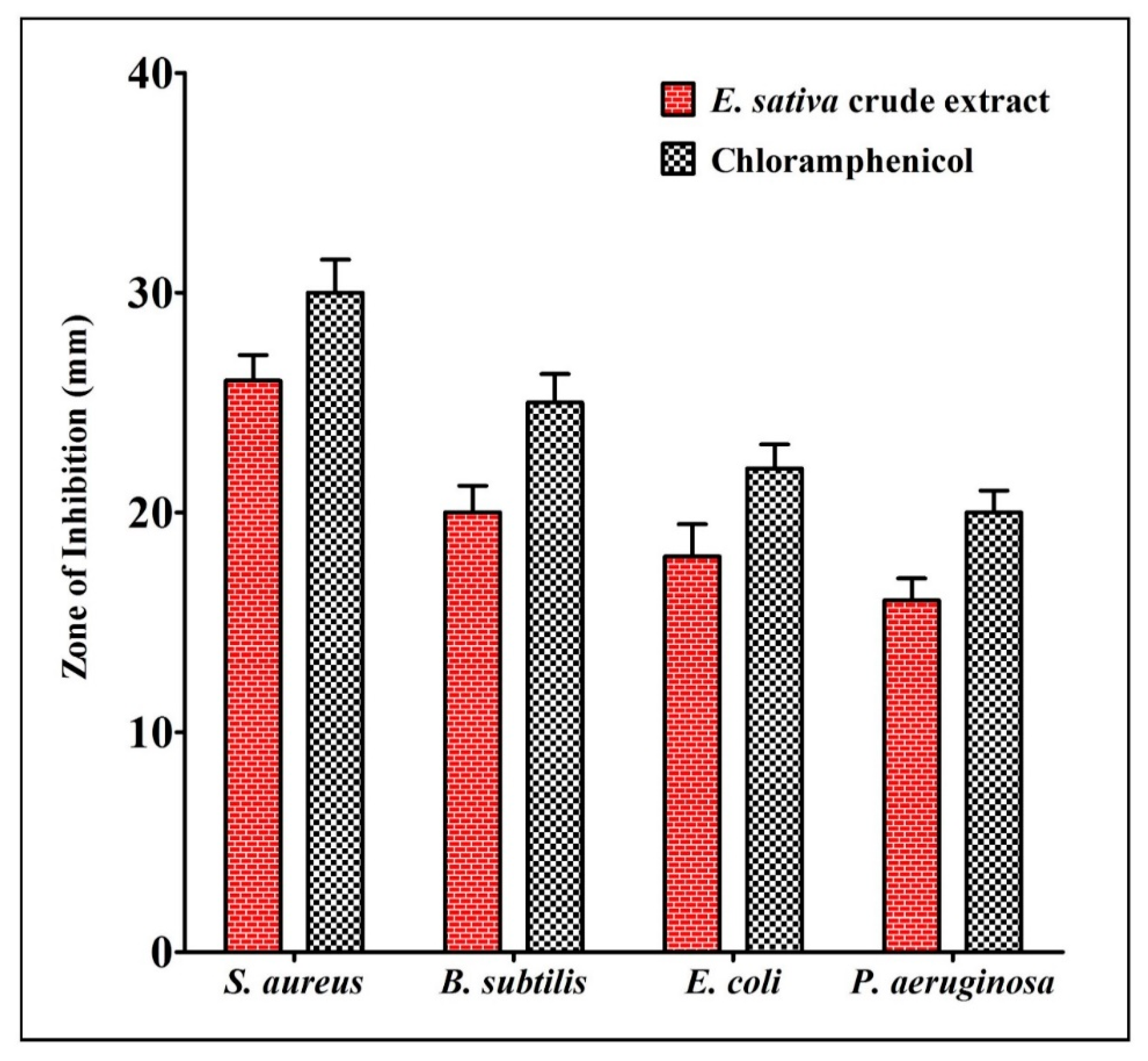
2.2. Antibacterial Activity of E. sativa Crude Extract

The antibacterial activity of E. sativa crude extract was evaluated via the agar well-diffusion assay method against two strains of Gram-positive pathogenic bacteria (B. subtilis and S. aureus) and two strains of Gram-negative pathogenic bacteria (E. coli and P. aeruginosa). Our results show that the crude extract of E. sativa exhibits a substantial inhibition of all the tested pathogens. The results of the antibacterial activity as well as the depiction of the crude extract preparation are presented in Figure 3. The obtained results reveal that E. sativa crude extract is potentially effective in controlling bacterial growth, and E. sativa crude extract shows a maximum zone of inhibition against S. aureus, followed by B. subtilis, E. coli and P. aeruginosa.
Currently, the antibiotic resistance of pathogenic bacteria is a major concern around the globe, where multidrug-resistant bacteria are continuously emerging and spreading causing a great challenge to the healthcare system [9]. Therefore, there is always a great demand for the search for an effective novel antimicrobial agent, which can fight against multidrug-resistant bacteria. Plants, especially medicinal/aromatic, are considered as a potential source of antimicrobial agents, as they consist of a high number of diverse phytochemicals [1]. In the present study, the antibacterial activity of E. sativa crude extract was evaluated using the agar well-diffusion method against different pathogenic strains of bacteria. Among them, the E. sativa crude extract exhibited the considerable inhibition of all the tested pathogens.
Using the agar cup diffusion method, Kauba et al. (2015) reported an inhibition zone (IZ) similar to the one obtained in our results with E. sativa ethanolic extract against S. typhimurium (IZ = 16.7 mm), B. subtilis (IZ = 16.6 mm), E. coli (IZ = 16.0 mm) and B. thuringiensis (IZ = 15.6 mm) [14]. Additionally, Khoobchandani et al. (2010) reported the antimicrobial activity of crude extracts of different parts of E. sativa against two Gram-positive and three Gram-negative bacteria [29]. Among them, a higher activity was reported for the seed oil against Gram-positive bacteria compared to Gram-negative bacteria. Additionally, Qaddoumi and El-banna (2019) reported the antagonistic activity of the aqueous extract of E. sativa towards E. coli (IZ = 19.0 mm) and S. aureus (IZ = 12.0 mm) [30]. In the same study, the antimicrobial activity of the crude extract of ethyl acetate presented no antimicrobial activity towards the tested pathogens. In another study, Rizwan et al. (2016) reported the antimicrobial activities of ethanol, methanol, ethyl acetate, acetone and chloroform extracts from E. sativa against different Gram-positive and Gram-negative bacteria [31]. Among them, a higher inhibition activity was found in the ethyl acetate and chloroform extracts against S. aureus (IZ = 25.66 mm, 23.16 mm), respectively, followed by methanol and ethanol (IZ = 16 mm, 14.33 mm).

2.3. The Antioxidant Activity of E. sativa Crude Extract

Free radicals, such as reactive oxygen species (ROS) and reactive nitrogen species (RNS), are linked with oxygen, which possess a strong reaction activity with other molecules apart from oxygen. Frequently, such free radicals are raised as a by-product of different metabolic activity inside the cell and ionize radiation in the form of the hydroxyl radical (HO•); superoxide ion, O2−; singlet oxygen (O2); hydrogen peroxide (H2O2); lipid peroxyl radical (ROO·); lipid alkoxyl radical (RO); lipid hydroperoxide (ROOH); nitrogen di-oxide (·NO2); nitric oxide (•NO); peroxynitrite (ONOO-) and thiol radical (RS) [32,33].
These free radicals generate oxidative stress and damage proteins, lipids and nucleic acids. Therefore, the role of free radicals is postulated in different disease conditions, such as the process of aging, various cancers, inflammatory conditions (adult respiratory diseases, arthritis and vasculitis), neurological disorders (Alzheimer’s disease, Parkinson’s disease) and ischemic diseases (stroke, heart diseases and intestinal ischemia) [34].
The antioxidant activity of E. sativa crude extract was evaluated using very well- established techniques, viz., DPPH and H2O2 assay. The results of the antioxidant assay reveal that E. sativa crude extract can scavenge the radicals to a great extent. The scavenging efficacy of crude extract on DPPH (IC50 = 66.16 µg/mL) radicals was better when compared to H2O2 (IC50 = 76.05 µg/mL). Furthermore, the antioxidant potential of E. sativa crude extract is dose dependent (Figure 4).
Antioxidants are substances that protect the cells from free radicals. Inside the cell, antioxidant molecules are present at low concentrations, which considerably reduces the oxidative stress and prevents the cells from the damage of free radicals [35]. Apart from endogenous antioxidants, they can also be available exogenously from the diet or as dietary supplements. An efficient antioxidant can readily absorb and remove free radicals and chelate metals at a physiologically appropriate level. Usually, endogenous antioxidants maintain optimal cellular functions, but under the condition of oxidative stress, endogenous antioxidants are not enough and exogenous antioxidants are needed to maintain optimal cellular functions [36]. Plants are considered as the potent source of antioxidant molecules, as they synthesize and accumulate various non-enzymatic antioxidants, such as ascorbic acid, glutathione and phenolics and flavonoids.
The antioxidant potential of E. sativa crude extract was analyzed against DPPH and H2O2 molecules in comparison to ascorbic acid. The crude extract from E. sativa exhibited notable free radical scavenging activity against both DPPH and H2O2 molecules. Previously, the antioxidant activity of E. sativa seeds was tested by Kishore et al. (2016) by using various in vitro antioxidant methods, such as phenolic content, total antioxidant capacity, reducing power, hydrogen peroxide, nitric oxide and superoxide dismutase scavenging activity. According to the previous study, the total phenol content of alcohol extract and hydro-alcohol extract was found to be 216.0 and 229.00 mg/g of gallic acid equivalents, respectively, and the total antioxidant capacity was determined to be 111.00 and 230.60 µM/g of ascorbic acid equivalents. Alcohol extracts and hydro-alcohol extracts were found to be able to scavenge DPPH radicals, hydrogen peroxide radicals, nitric oxide radicals and superoxide radicals, respectively. The IC50 values were found to be 3.28 and 3.53 µg/mL, 188.11 and 181.56 µg/mL, 73.05 and 64.33 µg/mL, and 87.91 and 41.12 mg/mL, respectively [37].
Koubaa et al. (2015) reported the antioxidant potential of E. sativa leaf extract and concluded that antioxidant activity is present due to the high concentration of phenolics(kaempferol 3,4-di-O-glucoside, kaempferol 3-glucosyl, quercetin 3-glucosyl and isorhamnetin 3-glucosyl) [14]. In one more study, Maia et al. (2015) reported the antioxidant potential of the seed extract of E. sativa by scavenging H2O2 and alkyl hydroperoxides accumulated in the cells and peripheral blood via acting as a precursor of sulforaphane, due to the presence of phenolics and glucosinolates in high concentrations in the seed [38].

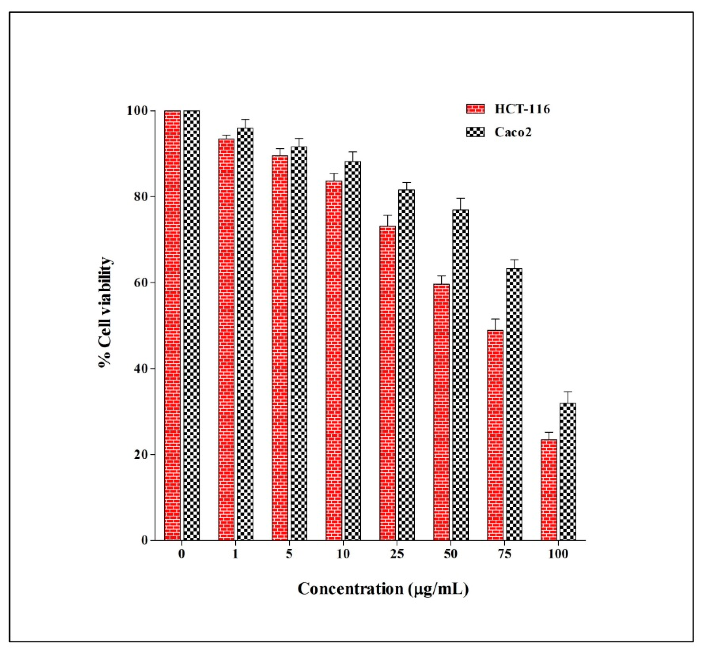
2.4. The Anticancer Activity of E. sativa Crude Extract

The anticancer potential of E. sativa crude extract was evaluated against two different human colorectal cancer cells (HCT-116 and Caco-2) via MTT assay. The results of the anticancer activity reveal that E. sativa crude extract could significantly inhibit the cell viability of both cells in a dose-dependent manner. The viability of the HCT-116 cell line after the treatment of crude extract was found to be higher, and the IC50 value was calculated, i.e., 64.91 μg/mL compared to Caco-2 with the IC50 value of 83.98 μg/mL (Figure 5).
After cardiovascular diseases, cancer has been reported to be the most common cause of mortality in several countries. In males, lung, liver, colorectal, prostate and stomach cancer are found to be the most common, whereas in females, breast, cervical, lung, thyroid and colorectal cancer are the most common [39]. Therefore, there is an urgent need to find alternate and cost-effective types of cancer treatments with lesser or no side effects. Natural products came to the attention of scientists due to their ability to serve as therapeutic as well as preventive agents over the past decade. Almost 60% of all conventional drugs used in cancer treatment are directly or indirectly extracted from plants, which encourages the discovery of medicinal plant-based novel drugs [39]. There are literatures which states, that the regular consumption of the cruciferous vegetable E. sativa can reduce the risk of different types of cancer development in the body.
Several phytochemicals, such as glucosinolates, sulfur containing plant secondary metabolites and isothiocyanate, including sulforaphane and erucin, are believed to be present in E. sativa. Erucin [1-isothiocyanato-4-(methylthio)butane], which is metabolically and structurally similar to sulforaphane, which is also reported to be present in large quantities in E. sativa [40]. The promising anticancer effect of erucin was already reported through many in vitro and in vivo studies. In order to observe the protective effect of erucin against cancer, in vivo research was conducted for the first time by a group of researches on the different mouse tissues mediated through the induction of several detoxification enzymes [41]. To date, many different studies confirmed the effectiveness of erucin against several human cancer cells. The anticancer effect of erucin was reported for different human cancer cell lines, such as the lung, liver, colon, and prostate, with the help of different mechanisms, viz., cell cycle regulation, apoptosis and the inhibition of proliferation as well as mitochondrial depolarization.
One of the studies stated that the erucin can induce apoptosis and also cell cycle arrest on human leukemia cells, together with the multidrug-resistant alternatives [19]. Jakubikova et al. (2005) investigated the anticancer property of erucin, which, similar to sulforaphane, induces the phase II detoxification enzymes and inhibits phase I enzymes. In addition, erucin arrests cell cycle progression and induces apoptosis in human lung carcinomas, hepatomas and leukemia cell lines. Erucin increases the expression of multidrug resistance transporters in human carcinoma cell lines [42]. Nazif et al. (2010) evaluated the cytotoxic activity of the total alcoholic extract of the defatted seeds and the isolated compounds against several types of tumor cell lines using the SRB assay. The total alcoholic extract and the aglucones of the isolated compounds glucoerucin and glucoiberin exhibited significant cytotoxic activity for HCT116 (the colon carcinoma cell line) (IC50 = 0.74, 2.42 and 0.94 μg/mL), respectively, while the IC50 was >10 μg/mL for Hela (the cervix carcinoma cell line), HEPG2 (the liver carcinoma cell line), MCF7 (the breast carcinoma cell line) and U251 (the brain carcinoma cell line) [7].
Melchini and Maria (2010) revealed that the intake of vegetables is associated with a reduced risk in the development of various types of cancer. This is attributed to the bioactive hydrolysis products that are derived from these vegetables, namely isothiocyanates. Isothiocyanates are characterized as small organic compounds synthesized as glucosinolates with R–N=C=S functional groups. Isothiocyanates present in cruciferous vegetables have a higher amount of anti-cancerous properties and can inhibit cell proliferation. Isothiocyanates suppress cancer cell proliferation by inhibiting the proteins involved in tumor initiation and proliferation pathways. Meanwhile, isothiocyanate treatment stimulates the reactive oxygen species (ROS), cell cycle arrest, programmed cell death and autophagy. More than 20 isothiocyanates are reported as having anticarcinogenic properties against tumorigenesis [43]. Erucin, the isothiocyanate product derived from E. sativa, showed chemoprevention activity against cancer cells in animal models. The mechanism of action showed the modulation of phase I, II and III detoxification, the regulation of cell growth by the induction of apoptosis, cell cycle arrest, the induction of ROS mechanisms and regulation androgen receptor pathways [13]. Michael et al. (2011) reported the potent anticancer activity of a 70% ethanolic extract of E. sativa against different human tumor cell lines, such as HepG2 (liver carcinoma), MCF7 (breast carcinoma), HCT116 (colon carcinoma) and Hep2 (larynx carcinoma) [19]. Khoobchandani et al. (2011) investigated the anticancer potential of solvent extracts prepared from the aerial roots and seed oil of E. sativa against melanoma cells. The seed oil (isothiocyanates rich) was found to significantly reduce the tumor growth and angiogenesis in mice without any major toxicity [19]. Azarenko et al. (2014) found that erucin prevents the proliferation of MCF7 (breast cancer cells) (IC50 = 28 mM) in parallel with cell cycle arrest at mitosis (IC50 = 13 mM) and apoptosis by a mechanism consistent with the impairment of microtubule dynamics [40]. However, a further exploration using different solvent system extractions and the in vitro anticancer activity of selected cancer cell lines is still required for the therapeutic implications to improve human health.

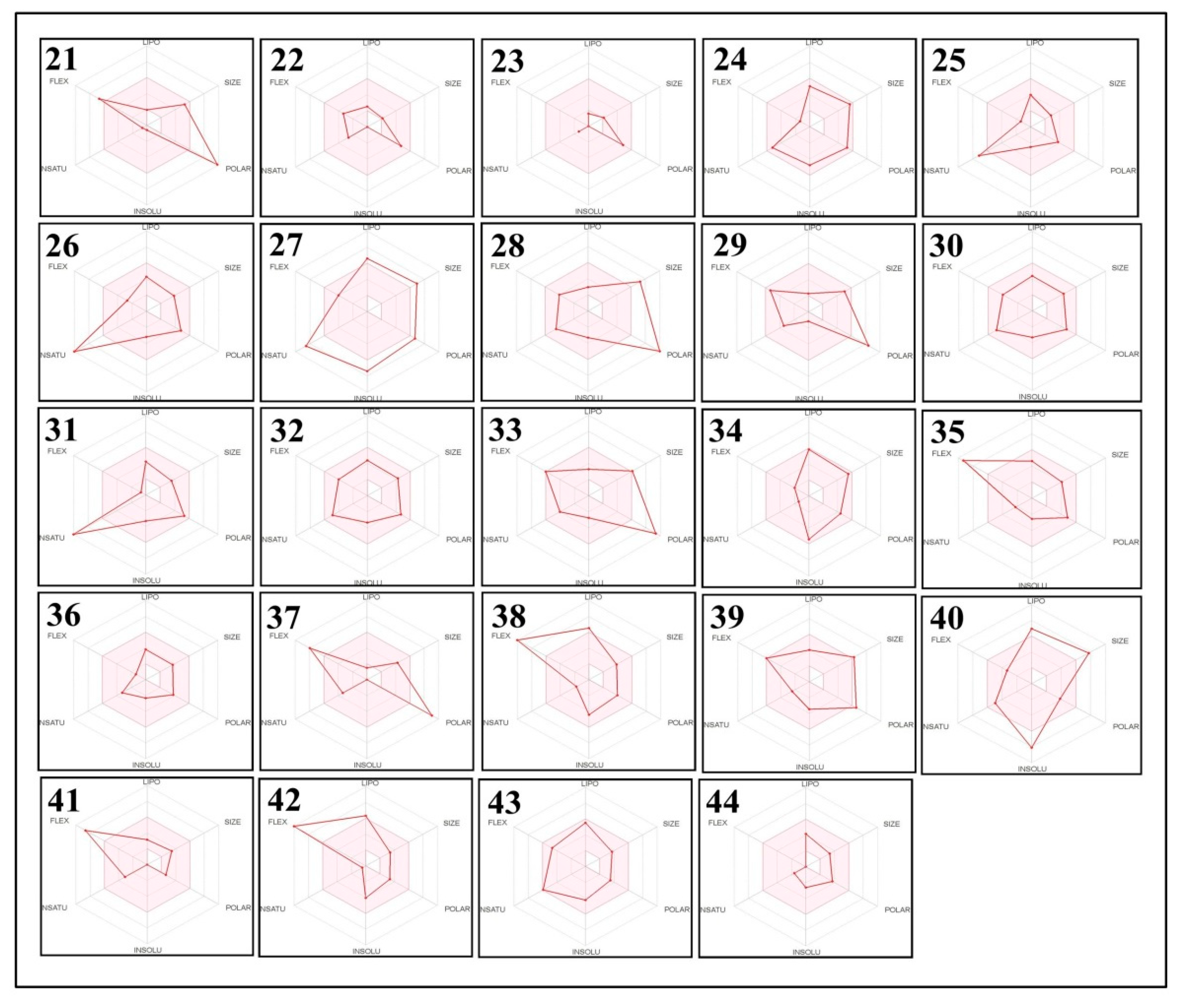
2.5. The Pharmacokinetic and Toxicity (ADMET) Profiles of the Identified Phytoconstituents from E. sativa Ethanolic Crude Extract

The potential of a good drug could be ruined because of the limited absorption, distribution, metabolism, excretion and toxicity (ADMET) characteristics. Furthermore, the major drawback of drug discovery in clinical trials is believed to be its pharmacokinetic properties, by virtue of which it becomes very expensive. Therefore, ADMET parameters were estimated using in silico tools to determine the probability of the E. sativa ethanolic crude extract becoming a potential candidate for the development of drugs (Table 2 and Table 3). Interestingly, the majority of the phytoconstituents were found to meet the Lipinski’s rule of five, and some of them were also found to follow Ghose, Veber and Egan filters, with most of them attaining a good score of bioavailability. One more important attribute is the solubility for the absorption of the compound and its distribution in the body, which was specified via the value of aqueous solubility. In the results, it can be observed that most of the compounds are highly soluble in water.
It is important to assess the skin’s permeability, i.e., the rate of a molecule penetrating the stratum corneum, to determine the potential for creating a form of transdermal drug delivery. It is considered that a molecule will penetrate the skin at the log Kp value of more than −2.5 cm/h. All of the identified phytochemical constituents from the crude extract were found to possess moderate-to-good skin penetrability. Caco-2 is the human epithelial colorectal adenocarcinoma cell and its permeability can calculate the intake of oral drugs. Most of the compounds were found to have moderate-to-potent Caco-2 permeability values (log Papp values > 0.90 cm/s).
ADMET analysis is a computer-based drug designing approach, which can lead to the initial stage of drug discovery [44,45,46]. The main motive behind this in silico approach is to lower the cost and time factors involved in comparison to standard ADMET profiling, as the in silico approach can screen more than 20,000 compounds within a minute; the wet lab will take more than 20 weeks to perform the same task [47,48,49]. Therefore, recently, after the establishment of ADMET data in the 1990s, the majority of pharmaceutical companies used this computational approach for the screening of drugs. In the present study, we also performed a computer-aided prediction of pharmacokinetic properties and the safety profile of the identified phytoconstituents via SwissADME and pkCSM tools. The results of the computational analysis can be useful for researchers to advance the development of prospective semi-synthetic and synthetic drugs for miscellaneous use.
Most of the identified phytochemical compounds were not found as a P-gp inhibitor/substrate. P-gp is the main element of ATP-binding cassette transporters or ABC-transporters, which is utilized to protect the central nervous system (CNS) from xenobiotics and is a prime method used to determine active efflux through biological membranes. The majority of the phytochemical compounds were found to be absorbed by the intestine during intestinal absorption analysis. The prediction of skin permeation was carried out by LogKP, by which phytochemical constituents were identified and found to penetrate the skin at a moderate or high level, which confirms their drug-like feature. Compounds 1, 3, 5 and 16 have a log BB (logarithm value of the brain-to-plasma concentration ratio, 0.1 to 0.3) and were determined to have more potential to cross the brain blood barrier (BBB). Only fewer compounds have the ability to penetrate the CNS. The compounds 9, 28, 31 and 43 are amongst the phytoconstituents that are more successfully distributed, with a distribution volume (logVDss) of 0.562 L/kg and 1.444 L/kg, respectively, in the tissues.
The isoforms of human cytochrome P450 (CYP), which are integrated in the metabolism of drugs inside the liver were also assessed. The foremost clinically appropriate drug-metabolizing enzyme in the human body is CYP3A4. The inhibition of CYP3A4 could direct lead to drug toxicity, drug–drug interactions and other adverse effects. Some of them were non-inhibitors/substrates of isoenzymes. Most of the compounds were found to be non-inhibitors of CYP3A4, the isoenzyme responsible for the metabolism of about 60% of xenobiotics, including drugs, carcinogens, steroids and eicosanoids.
To predict the route of excretion, the total clearance (CLTOT) for both hepatic and renal and renal organic cation transporter 2 (OCT2) substrates was expressed as the log mL/min/kg that was predicted. The results revealed that the majority of the phytochemical constituents displayed a positive total clearance value and can be simply be excreted. The AMES toxicity, hepatotoxicity, hERG potassium channel inhibition and skin sensitization parameters were predicted to find out the toxicity profile of the identified phytochemical constituents from the E. sativa crude extract. The obtained results reveal that only a few of the compounds have a deviated mutagenic and hepatic toxicity potential, which means that the majority of the compounds are devoid of any risk of toxicity (99% have no hERG I inhibition and 72% exert no skin-sensitive effects).
To obtain further information about the improved bioavailability and drug-likeness of the identified phytochemicals, we estimated the results of the bioavailability radar. The results are represented by the lipophilicity: XLOGP3 between −0.7 to +5.0; polarity: TPSA between 20–130 Å2; size: molecular weight 150–500 g/mol; saturation: fraction of carbons in the sp3 hybridization not less than 0.25; solubility: log S not higher than 6; and flexibility: no more than 9 rotatable bonds with the colored zone representing the desired physico-chemical space for good oral bioavailability, and they all display significant drug-likeness properties (Figure 6 and Figure 7).
The examples of the brain or intestinal estimated permeation (BOILED Egg model) estimation of gastrointestinal (GI) absorption and BBB permeation for all the identified compounds were conducted. The results show that the compound indicated with a red point in the yellow ellipse has the potential for brain penetration and is a non-substrate of P-gp (PGP-) (Figure 8).
Some of the plant families, such as Brassicaceae, Capparaceae and Resedaceae, were reported to contain glucosinolates, which are considered as one of the most significant secondary metabolites. Subsequently, in addition to the enzyme myrosinase, secondary metabolites are broken down into various hydrolytic products [50,51]. E. sativa, a member of the Brassicaceae family, is reported to be a rich source of glucosinolates, and its different hydrolytic products are reported to exert different bioactive properties, such as anticancer, antimutagenic, bioherbicidal, antimicrobial, antigenotoxic and antitumor activities [52,53,54,55,56]. In the present study, two glucosinolates, glucoraphanin and 4-dimethoxyglucobrassicin, were identified, which could be the major constituents of the E. sativa crude extract and are possibly responsible for various potent biological activities.

3. Materials and Methods

3.1. Media and Chemicals

All the microbiological media used in the present study were purchased from the local suppliers of Hi-Media®, Mumbai, India. Chemicals, drugs and solvents were purchased from Sigma-Aldrich®, Bangalore, India. Analytical grade solvents were used.

3.2. Plant Material and Extraction Preparation

E. sativa plants were cultivated in a field for the purpose of the present study. Firstly, the plants of E. sativa were grown in silty clay with sand soil type at an average temperature of 30–35 °C, and were watered once every day. Later, the whole plant leaves were collected after maturity, washed under running tap water and oven-dried. Dry plant leaves were then grounded into a fine powder with an electrical grinder and stored in airtight containers. Next, E. sativa powder (25 g) was soaked in 85% ethanol at 37 °C for 24 h with vigorous shaking at 120 rpm. The ethanolic extract was filtered using Whatman no. 1 filter paper and the obtained extract was concentrated using a rotary evaporator. The obtained dried residues were dissolved in 10% dimethyl sulfoxide (DMSO) to make up 1 mg/mL of plant leaf extract concentration to carry out various biological assays [57].

3.3. High Resolution Liquid Chromatograph Mass Spectrometery Analysis

The HR-LCMS analysis of the ethanolic extract was carried out by a UHPLC-PDA-Detector Mass Spectrophotometer (HR-LC/MS 1290 Infinity UHPLC System), Agilent Technologies®, Santa Clara, CA, USA, and consisted of an HiP sampler, binary gradient solvent pump, column compartment and Quadrupole Time of Flight Mass Spectrometer (MS Q-TOF) with a dual Agilent Jet Stream Electrospray (AJS ES) ion source. A total of 1% formic acid in deionized water (solvent A) and acetonitrile (solvent B) was used as a solvent. The flow rate of 0.350 mL/min was used, while MS detection was performed in MS Q-TOF. Compounds were identified via their mass spectra and their unique mass fragmentation patterns. Compound Discoverer 2.1, ChemSpider and PubChem were used as the main tools for the identification of the phytochemical constituents [58].

3.4. Antibacterial Assay

All pathogenic bacterial test strains, Pseudomonas aeruginosa (P. aeruginosa) (MTCC 741), Bacillus subtilis (B. subtilis) (MTCC 121), Escherichia coli, (E. coli) (MTCC 9537) and Staphylococcus aureus (S. aureus) (MTCC 96), were obtained from the Microbial Type Culture Collection (MTCC), India, and Muller–Hinton Agar (MHA) was used to maintain the bacterial culture. The antibacterial activity of E. sativa crude extract was carried out via the agar cup/well diffusion method. Firstly, bacterial cultures were grown overnight at 37 °C in fresh medium and a total of 0.5 McFarland standard 108 colony-forming units/mL (CFU/mL) was matched by culture turbidity adjustment using 0.9% of sterile saline solution. Bacterial suspension was evenly spread all over the plates, and the wells were made with a sterile cork borer. A total of 60 µL of crude extract (1 mg/mL) was then inoculated into each respective well and the plates were incubated at 37 °C for 24 h. Antibacterial activity was noted in the form of the zone of inhibition. A total of 1000 µg/mL of chloramphenicol and 10% of DMSO was used as a positive and negative control, respectively [58].

3.5. Antioxidant Assays

3.5.1. DPPH Scavenging Activity

The antioxidant potential of E. sativa crude extract was determined in terms of its radical scavenging capability against DPPH free radicals [59]. Crude extracts of different concentrations (1–100 µg/mL) were mixed in the tubes containing 2 mL of DPPH solution (6 × 10⁻⁵ M) in DMSO. The tubes were mixed well and incubated in the dark for 1 h. At the end of the incubation, the absorbance was read out at 517 nm. DMSO was used as a blank, DPPH solution without any crude extract was used as a control, and ascorbic acid was used as a standard [60]. The calculation of the percentage of scavenging of DPPH free radicals was calculated as follows:
DPPH scavenging activity (%) = (A0 − A1)/A0 × 100
where A0 = absorbance of the control; A1 = absorbance of the sample.

3.5.2. Hydrogen Peroxide Scavenging Activity

The antioxidant potential of E. sativa crude extract against H2O2 was carried out according to the described method [59]. Crude extract of different concentrations (1–100 µg/mL) were mixed in the tubes having 1 mL of H2O2 (1 mL, 2 mM) solution, prepared in a phosphate buffer (0.1M, pH 7.4). The tubes were then incubated for 10 min at room temperature. At the end of the incubation, the absorbance was read out at 230 nm against a blank solution (phosphate buffer without H2O2), while ascorbic acid was used as a positive control [61]. The following formula was then used to calculate the percentage of H2O2 scavenged:
Inhibition (%) = (A0 − A1)/A0) × 100
where A0 = absorbance of the control; A1 = absorbance of the extract/standard

3.6. Anticancer Assay (MTT Assay)

The anticancer activity of E. sativa crude extract was carried out against human colon cancer cell lines (HCT-116 and Caco-2). Both cells were acquired from the National Centre for Cell Science (NCCS), India, and propagated in 25 cm2 flask containing Dulbecco’s Modified Eagle’s Medium (DMEM) supplemented with 10% Fetal Bovine Serum (FBS) and antibiotic solution in a humidified (5% CO2) atmosphere at 37 °C. To perform the MTT assay, cells were grown up to 80% confluence and then seeded at a density of more than 1 × 10⁵ cells per well in 96-well plates and incubated in conditions, as mentioned above. Trypan Blue (0.4%) was used to stain the cells, and the viability was calculated by using a hemocytometer. Cells were then treated with different concentrations of E. sativa crude extract (1–100 μg/mL) for 24 h. Cells were washed with PBS solution and subjected to 100 μL of MTT solution (3-(4,5-dimethylthiazolyl-2)-2,5 diphenyltetrazolium bromide) (5 mg/mL) and incubated for 4 h. Then, the medium was removed and 100 μL of DMSO was added to solubilize the formazan crystals. The amount of formazan crystal was determined by measuring the absorbance at 570 nm using an ELISA reader. Cisplatin was used as a positive control. All assays were performed in triplicate and 50% cytotoxic concentration (IC50) was calculated [62,63].

3.7. ADMET Analysis

Assumption of the toxicity and pharmacokinetics of the compounds identified from E. sativa crude extract via HR-LC/MS was carried out via SwissADME (http://www.swissadme.ch/) and pkCSM (http://biosig.unimelb.edu.au/pkcsm/prediction) online tools accessed on 7 September 2021 [64,65,66].

4. Conclusions

From the present study, it can be concluded that E. sativa displays potent antibacterial activity against the different human pathogenic bacterial strains. Additionally, E. sativa also shows promising antioxidant and anticancer activity. Based on our results, E. sativa can also be utilized for the production and development of nutraceutical or functional food. Therefore, further investigation warrants in vivo pharmacological and toxicological research to demonstrate the unexplored and valuable aspect of E. sativa. The computational analysis shows that E. sativa possesses a significant pharmacokinetic and safety profile of the phytochemical constituents identified using HR-LC/MS. Additionally, it also reveals the ethanolic crude extract of E. sativa as a prospective drug candidate for the treatment and management of various diseases, as well as for therapeutic purposes. However, the current computational models are severely constrained due to the lack of understanding of the underlying molecular processes of the diseases. Therefore, to overcome these limitations, one way to formulate more effective strategies is to incorporate ligand, target, phenotype and biological network-based approaches, as well as a deeper reinforcement of learning methods, which are more likely to increase predictability. This will overcome the shortcomings of the existing computational approaches and improve the drug development process.

Author Contributions

Conceptualization, A.M.A. and S.A.A.; methodology, E.A.-S. and A.E.O.E.; validation, A.J.S., M.S. and M.A.; formal analysis, M.I.K. and S.A.A.; investigation, A.J.S., S.A.A. and A.M.A.; data curation, E.A.-S., S.A.A. and M.I.K.; writing—original draft preparation, M.P., S.A.A. and M.A.; writing—review and editing, S.A.A., A.J.S. and M.A.; visualization, M.P., E.A.-S., M.S., A.E.O.E. and Z.R.A.A.A.; supervision, M.A., S.A.A., E.A.-S., Z.R.A.A.A. and A.M.A.; project administration, A.M.A., S.A.A. and M.A. All authors have read and agreed to the published version of the manuscript.

Funding

This research was funded by the Scientific Research Deanship at University of Ha’il- Saudi Arabia through project number RG-20151.

Institutional Review Board Statement

Not applicable.

Informed Consent Statement

Not applicable.

Data Availability Statement

All data generated or analyzed during this study are included in this article.

Conflicts of Interest

The authors declare no conflict of interest.

Sample Availability

Samples of the extract is available from the authors.

References

  1. Gulfraz, M.; Sadiq, A.; Tariq, H.; Imran, M.; Qureshi, R. Phytochemical analysis and antibacterial activity of Eruca sativa seed. Pak. J. Bot. 2011, 43, 1351–1359. [Google Scholar]
  2. Alqasoumi, S.; Al-Sohaibani, M.; Al-Howiriny, T.; Al-Yahya, M.; Rafatullah, S. Rocket “Eruca sativa”: A salad herb with potential gastric anti-ulcer activity. World J. Gastroenterol. 2009, 15, 1958–1965. [Google Scholar] [CrossRef] [PubMed]
  3. Abdul-Jalil, T.Z. Phytochemicals Screening by GC/MS and Determination of Some Flavonol in Cultivated Iraqi Eruca sativa Dried Leaves Extract and its Biological Activity as Antioxidant. Int. J. Pharmacogn. Phytochem. Res. 2016, 8, 1722–1730. [Google Scholar]
  4. Nurzyńska-Wierdak, R. Nutritional and energetic value of Eruca sativa Mill. leaves. Acta Sci. Pol. Hortorum Cultus 2015, 14, 191–199. [Google Scholar]
  5. Di Gioia, F.; Avato, P.; Serio, F.; Argentieri, M.P. Glucosinolate profile of Eruca sativa, Diplotaxis tenuifolia and Diplotaxis erucoides grown in soil and soilless systems. J. Food Compos. Anal. 2018, 69, 197–204. [Google Scholar] [CrossRef]
  6. Jirovetz, L.; Smith, D.; Buchbauer, G. Aroma compound analysis of Eruca sativa (Brassicaceae) SPME headspace leaf samples using GC, GC-MS, and olfactometry. J. Agric. Food Chem. 2002, 50, 4643–4646. [Google Scholar] [CrossRef]
  7. Nazif, N.M.; Habib, A.A.E.; Tawfik, W.A.M.; Hassan, R.A. Chemical composition and cytotoxic activity of Eruca sativa L. Seeds cultivated in Egypt. Asian J. Chem. 2010, 22, 2407–2416. [Google Scholar]
  8. Hussein, S.A. Phytochemical Study of Vsome Medicinal Compounds Present in Hedera helix L. Plant Cultivated in Iraq. Master’s Thesis, Baghdad University, Baghdad, Iraq, 2014. [Google Scholar]
  9. Ashraf, S.A.; Al-Shammari, E.; Hussain, T.; Tajuddin, S.; Panda, B.P. In-vitro antimicrobial activity and identification of bioactive components using GC-MS of commercially available essential oils in Saudi Arabia. J. Food Sci. Technol. 2017, 54, 3948–3958. [Google Scholar] [CrossRef]
  10. Sastry, E.V.D. Taramira (Eruca sativa) and its improvement A review. Agric. Rev. 2003, 24, 235–249. [Google Scholar]
  11. Bukhsh, E.; Malik, S.; Ahmad, S. Estimation of Nutritional Value and Trace elements Content of Carthamus oxyacantha, Eruca stiva and Plantago ovata. Pak. J. Bot. 2007, 39, 1181–1187. [Google Scholar]
  12. Yehuda, H.; Khatib, S.; Sussan, I.; Musa, R.; Vaya, J.; Tamir, S. Potential skin antiinflammatory effects of 4-methylthiobutylisothiocyanate (MTBI) isolated from rocket (Eruca sativa) seeds. BioFactors 2009, 35, 295–305. [Google Scholar] [CrossRef]
  13. Melchini, A.; Traka, M.H. Biological profile of erucin: A new promising anticancer agent from cruciferous vegetables. Toxins 2010, 2, 593–612. [Google Scholar] [CrossRef] [Green Version]
  14. Koubaa, M.; Driss, D.; Bouaziz, F.; Ghorbel, R.; Chaabouni Ellouz, S. Antioxidant and antimicrobial activities of solvent extract obtained from rocket (Eruca sativa L.) flowers. Free Radic. Antioxid. 2015, 5, 29–34. [Google Scholar] [CrossRef] [Green Version]
  15. Allen, D.R.; McWhinney, B.C. Quadrupole Time-of-Flight Mass Spectrometry: A Paradigm Shift in Toxicology Screening Applications. Clin. Biochem. Rev. 2019, 40, 135–146. [Google Scholar] [CrossRef]
  16. Elkhalifa, A.E.O.; Alshammari, E.; Adnan, M. Okra (Abelmoschus Esculentus) as a Potential Dietary Medicine with Nutraceutical Importance for Sustainable Health Applications. Molecules 2021, 26, 696. [Google Scholar] [CrossRef]
  17. Blažević, I.; Mastelić, J. Free and bound volatiles of rocket (Eruca sativa Mill.). Flavour Fragr. J. 2008, 23, 278–285. [Google Scholar] [CrossRef]
  18. Villatoro-Pulido, M.; Priego-Capote, F.; Álvarez-Sánchez, B.; Saha, S.; Philo, M.; Obregón-Cano, S.; De Haro-Bailón, A.; Font, R.; Del Río-Celestino, M. An approach to the phytochemical profiling of rocket [Eruca sativa (Mill.) Thell]. J. Sci. Food Agric. 2013, 93, 3809–3819. [Google Scholar] [CrossRef] [PubMed]
  19. Michael, H.; Shafik, R.; Rasmy, G. Studies on the chemical constituents of fresh leaf of Eruca sativa extract and its biological activity as anticancer agent in vitro. J. Med. Plants Res. 2011, 5, 1184–1191. [Google Scholar]
  20. Bennett, R.N.; Rosa, E.A.; Mellon, F.A.; Kroon, P.A. Ontogenic profiling of glucosinolates, flavonoids, and other secondary metabolites in Eruca sativa (salad rocket), Diplotaxis erucoides (wall rocket), Diplotaxis tenuifolia (wild rocket), and Bunias orientalis (Turkish rocket). J. Agric. Food Chem. 2006, 54, 4005–4015. [Google Scholar] [CrossRef] [PubMed]
  21. Bell, L.; Wagstaff, C. Glucosinolates, myrosinase hydrolysis products, and flavonols found in rocket (Eruca sativa and Diplotaxis tenuifolia). J. Agric. Food Chem. 2014, 62, 4481–4492. [Google Scholar] [CrossRef] [PubMed]
  22. Uğur, A.; Süntar, I.; Aslan, S.; Orhan, I.E.; Kartal, M.; Sekeroğlu, N.; Eşiyok, D.; Sener, B. Variations in fatty acid compositions of the seed oil of Eruca sativa Mill. caused by different sowing periods and nitrogen forms. Pharmacogn. Mag. 2010, 6, 305–308. [Google Scholar]
  23. Elfakir, C.; Dreux, M. Simultaneous analysis of intact and desulfated glucosinolates with a porous graphitized carbon column. J. Chromatogr. A 1996, 727, 71–82. [Google Scholar] [CrossRef]
  24. Miyazawa, M.; Maehara, T.; Kurose, K. Composition of the essential oil from the leaves of Eruca sativa. Flavour Fragr. J. 2002, 17, 187–190. [Google Scholar] [CrossRef]
  25. Hussein, Z.F. Study the Effect of Eruca sativa Leaves Extract on Male Fertility in Albino Mice. J. Al-Nahrain Univ. -Sci. 2013, 16, 143–146. [Google Scholar] [CrossRef]
  26. Zhang, Y.; Tang, L. Discovery and development of sulforaphane as a cancer chemopreventive phytochemical. Acta Pharmacol. Sin. 2007, 28, 1343–1354. [Google Scholar] [CrossRef]
  27. Arora, R.; Sharma, D.; Kumar, R.; Singh, B.; Vig, A.P.; Arora, S. Evaluating extraction conditions of glucosinolate hydrolytic products from seeds of Eruca sativa (Mill.) Thell. using GC-MS. J. Food Sci. 2014, 79, C1964–C1969. [Google Scholar] [CrossRef]
  28. Cavaiuolo, M.; Ferrante, A. Nitrates and glucosinolates as strong determinants of the nutritional quality in rocket leafy salads. Nutrients 2014, 6, 1519–1538. [Google Scholar] [CrossRef] [PubMed] [Green Version]
  29. Khoobchandani, M.; Ojeswi, B.K.; Ganesh, N.; Srivastava, M.M.; Gabbanini, S.; Matera, R.; Iori, R.; Valgimigli, L. Antimicrobial properties and analytical profile of traditional Eruca sativa seed oil: Comparison with various aerial and root plant extracts. Food Chem. 2010, 120, 217–224. [Google Scholar] [CrossRef]
  30. Qaddoumi, S.Q.; El-Banna, N. Antimicrobial Activity of Arugula (Eruca sativa) Leaves on Some Pathogenic Bacteria. Int. J. Biol. 2019, 11, 10. [Google Scholar] [CrossRef]
  31. Rizwana, H.; Alwhibi, M.; Khan, F.A.; Soliman, D.A. Chemical Composition and Antimicrobial Activity of Eruca sativa Seeds Against Pathogenic Bacteria and Fungi. J. Anim. Plant Sci. 2016, 26, 1859–1871. [Google Scholar]
  32. Kurutas, E.B.; Ciragil, P.; Gul, M.; Kilinc, M. The Effects of Oxidative Stress in Urinary Tract Infection. Mediat. Inflamm. 2005, 2005, 528064. [Google Scholar] [CrossRef] [PubMed]
  33. Valko, M.; Leibfritz, D.; Moncol, J.; Cronin, M.T.; Mazur, M.; Telser, J. Free radicals and antioxidants in normal physiological functions and human disease. Int. J. Biochem. Cell Biol. 2007, 39, 44–84. [Google Scholar] [CrossRef]
  34. Stefanis, L.; Burke, R.E.; Greene, L.A. Apoptosis in neurodegenerative disorders. Curr. Opin. Neurol. 1997, 10, 299–305. [Google Scholar] [CrossRef] [PubMed]
  35. Sharifi-Rad, M.; Anil Kumar, N.V.; Zucca, P.; Varoni, E.M.; Dini, L.; Panzarini, E.; Rajkovic, J.; Tsouh Fokou, P.V.; Azzini, E.; Peluso, I.; et al. Lifestyle, Oxidative Stress, and Antioxidants: Back and Forth in the Pathophysiology of Chronic Diseases. Front. Physiol. 2020, 11, 694. [Google Scholar] [CrossRef] [PubMed]
  36. Halliwell, B. Biochemistry of oxidative stress. Biochem. Soc. Trans. 2007, 35 Pt 5, 1147–1150. [Google Scholar] [CrossRef]
  37. Kishore, S. Evaluation of Antioxidant Activity and Total Phenolic Content of Eruca sativa L., Seeds. Int. J. Toxicol. Pharm. Res. 2016, 8, 146–151. [Google Scholar]
  38. Maia, M.L.; Correia-Sá, L.; Coelho, A.; Barroso, M.F.; Domingues, V.F.; Delerue-Matos, C. Eruca sativa: Benefits as antioxidants source versus risks of already banned pesticides. J. Environ. Sci. Health Part B 2015, 50, 338–345. [Google Scholar] [CrossRef]
  39. Adnan, M.; Siddiqui, A.J.; Hamadou, W.S.; Patel, M.; Ashraf, S.A.; Jamal, A.; Awadelkareem, A.M.; Sachidanandan, M.; Snoussi, M.; De Feo, V. Phytochemistry, Bioactivities, Pharmacokinetics and Toxicity Prediction of Selaginella repanda with Its Anticancer Potential against Human Lung, Breast and Colorectal Carcinoma Cell Lines. Molecules 2021, 26, 768. [Google Scholar] [CrossRef]
  40. Azarenko, O.; Jordan, M.A.; Wilson, L. Erucin, the major isothiocyanate in arugula (Eruca sativa), inhibits proliferation of MCF7 tumor cells by suppressing microtubule dynamics. PLoS ONE 2014, 9, e100599. [Google Scholar] [CrossRef] [Green Version]
  41. Melchini, A.; Costa, C.; Traka, M.; Miceli, N.; Mithen, R.; De Pasquale, R.; Trovato, A. Erucin, a new promising cancer chemopreventive agent from rocket salads, shows anti-proliferative activity on human lung carcinoma A549 cells. Food Chem. Toxicol. Int. J. Publ. Br. Ind. Biol. Res. Assoc. 2009, 47, 1430–1436. [Google Scholar] [CrossRef]
  42. Jakubikova, J.; Bao, Y.; Sedlak, J. Isothiocyanates induce cell cycle arrest, apoptosis and mitochondrial potential depolarization in HL-60 and multidrug-resistant cell lines. Anticancer. Res. 2005, 25, 3375–3386. [Google Scholar] [PubMed]
  43. Soundararajan, P.; Kim, J.S. Anti-Carcinogenic Glucosinolates in Cruciferous Vegetables and Their Antagonistic Effects on Prevention of Cancers. Molecules 2018, 23, 2983. [Google Scholar] [CrossRef] [PubMed] [Green Version]
  44. Lipinski, C.A.; Lombardo, F.; Dominy, B.W.; Feeney, P.J. Experimental and computational approaches to estimate solubility and permeability in drug discovery and development settings. Adv. Drug Deliv. Rev. 2001, 46, 3–26. [Google Scholar] [CrossRef]
  45. Lombardo, F.; Gifford, E.; Shalaeva, M.Y. In silico ADME prediction: Data, models, facts and myths. Mini Rev. Med. Chem. 2003, 3, 861–875. [Google Scholar] [CrossRef]
  46. Gleeson, M.P.; Hersey, A.; Hannongbua, S. In-silico ADME models: A general assessment of their utility in drug discovery applications. Curr. Top. Med. Chem. 2011, 11, 358–381. [Google Scholar] [CrossRef]
  47. DiMasi, J.A.; Hansen, R.W.; Grabowski, H.G. The price of innovation: New estimates of drug development costs. J. Health Econ. 2003, 22, 151–185. [Google Scholar] [CrossRef] [Green Version]
  48. Hodgson, J. ADMET--turning chemicals into drugs. Nat. Biotechnol. 2001, 19, 722–726. [Google Scholar] [CrossRef] [PubMed]
  49. Darvas, F.; Keseru, G.; Papp, A.; Dormán, G.; Urge, L.; Krajcsi, P. In Silico and Ex silico ADME approaches for drug discovery. Curr. Top. Med. Chem. 2002, 2, 1287–1304. [Google Scholar] [CrossRef]
  50. Heaney, R.K.; Fenwick, G.R. Identifying toxins and their effects: Glucosinolates. In Natural Toxicants in Food: Progress and Prospects; Watson, D.H., Ed.; Ellis Horwood: Chichester, UK, 1987; pp. 76–109. [Google Scholar]
  51. Brown, P.D.; Tokuhisa, J.G.; Reichelt, M.; Gershenzon, J. Variation of glucosinolate accumulation among different organs and developmental stages of Arabidopsis thaliana. Phytochemistry 2003, 62, 471–481. [Google Scholar] [CrossRef]
  52. Zhang, Y.; Talalay, P. Anticarcinogenic activities of organic isothiocyanates: Chemistry and mechanisms. Cancer Res. 1994, 54 (Suppl. S7), 1976s–1981s. [Google Scholar]
  53. Brown, P.D.; Morra, M.J. Glucosinolate-containing plant tissues as bioherbicides. J. Agric. Food Chem. 1995, 43, 3070–3074. [Google Scholar] [CrossRef]
  54. Fahey, J.W.; Talalay, P. Antioxidant functions of sulforaphane: A potent inducer of Phase II detoxication enzymes. Food Chem. Toxicol. Int. J. Publ. Br. Ind. Biol. Res. Assoc. 1999, 37, 973–979. [Google Scholar] [CrossRef]
  55. Vig, A.P.; Rampal, G.; Thind, T.S.; Arora, S. Bio-protective effects of glucosinolates–A review. LWT-Food Sci. Technol. 2009, 42, 1561–1572. [Google Scholar] [CrossRef]
  56. Fimognari, C.; Hrelia, P. Sulforaphane as a promising molecule for fighting cancer. Mutat. Res. 2007, 635, 90–104. [Google Scholar] [CrossRef]
  57. Adnan, M.; Patel, M.; Deshpande, S.; Alreshidi, M.; Siddiqui, A.J.; Reddy, M.N.; Emira, N.; De Feo, V. Effect of Adiantum philippense Extract on Biofilm Formation, Adhesion With Its Antibacterial Activities Against Foodborne Pathogens, and Characterization of Bioactive Metabolites: An in vitro-in silico Approach. Front. Microbiol. 2020, 11, 823. [Google Scholar] [CrossRef]
  58. Muzaffer, U.; Paul, V.I. Phytochemical analysis, in vitro antioxidant and antimicrobial activities of male flower of Juglans regia L. Int. J. Food Prop. 2018, 21, 345–356. [Google Scholar] [CrossRef] [Green Version]
  59. Adnan, M.; Patel, M.; Reddy, M.N.; Alshammari, E. Formulation, evaluation and bioactive potential of Xylaria primorskensis terpenoid nanoparticles from its major compound xylaranic acid. Sci. Rep. 2018, 8, 1740. [Google Scholar] [CrossRef] [PubMed]
  60. Orhan, I.; Aslan, M. Appraisal of scopolamine-induced antiamnesic effect in mice and in vitro antiacetylcholinesterase and antioxidant activities of some traditionally used Lamiaceae plants. J. Ethnopharmacol. 2009, 122, 327–332. [Google Scholar] [CrossRef]
  61. Ruch, R.J.; Cheng, S.J.; Klaunig, J.E. Prevention of cytotoxicity and inhibition of intercellular communication by antioxidant catechins isolated from Chinese green tea. Carcinogenesis 1989, 10, 1003–1008. [Google Scholar] [CrossRef]
  62. Reddy, M.N.; Adnan, M.; Alreshidi, M.M.; Saeed, M.; Patel, M. Evaluation of Anticancer, Antibacterial and Antioxidant Properties of a Medicinally Treasured Fern Tectaria coadunata with its Phytoconstituents Analysis by HR-LCMS. Anti-Cancer Agents Med. Chem. 2020, 20, 1845–1856. [Google Scholar] [CrossRef]
  63. Lombardi, V.R.; Carrera, I. In Vitro Screening for Cytotoxic Activity of Herbal Extracts. Evid.-Based Complementary Altern. Med. 2017, 2017, 2675631. [Google Scholar] [CrossRef] [PubMed]
  64. Kadri, A.; Aouadi, K. In vitro antimicrobial and α-glucosidase inhibitory potential of enantiopure cycloalkylglycine derivatives: Insights into their in silico pharmacokinetic, druglikeness, and medicinal chemistry properties. J. Appl. Pharm. Sci. 2020, 10, 107–115. [Google Scholar]
  65. Othman, I.M.M.; Gad-Elkareem, M.A.M.; Hassane Anouar, E.; Aouadi, K.; Kadri, A.; Snoussi, M. Design, synthesis ADMET and molecular docking of new imidazo[4,5-b]pyridine-5-thione derivatives as potential tyrosyl-tRNA synthetase inhibitors. Bioorg. Chem. 2020, 102, 104105. [Google Scholar] [CrossRef]
  66. Ghannay, S.; Kadri, A.; Aouadi, K. Synthesis, in vitro antimicrobial assessment, and computational investigation of pharmacokinetic and bioactivity properties of novel trifluoromethylated compounds using in silico ADME and toxicity prediction tools. Mon. Chem.-Chem. Mon. 2020, 151, 267–280. [Google Scholar] [CrossRef]
Figure 1. HR-LC/MS spectrum peak of E. sativa crude extract showing the chromatogram intensity against the acquisition time, (A) positive analysis and (B) negative analysis.
Molecules 27 01409 g001
Figure 2. (A). Chemical structures of the identified compounds in E. sativa crude extract. (1) (+/−)-3-[(2-methyl-3-furyl)thio]-2-butanone; (2) methyl N-methylanthranilate; (3) 4-Amino-2-methyl-1-naphthol; (4) indoleacrylic acid; (5) pyrafoline D; (6) petasitenine; (7) nopaline; (8) Serinyl-Hydroxyproline; (9) afzelechin; (10) N-trans-Feruloyl-4-O-methyldopamine; (11) (±)-rollipyrrole; (12) N6-cis-p-Coumaroylserotonin; (13) 1-Methoxy-1H-indole-3-carboxaldehyde; (14) (10Z,14E,16E)-10,14,16-Octadecatrien-12-ynoic acid; (15) 3,4′,5,6,8-Pentamethoxyflavone; (16) terminaline; (17) palmitic amide; (18) oleamide; (19) pheophorbide a; (20) pyropheophorbide a; (21) glucoraphanin; and (22) (S)-2-(Hydroxymethyl)glutarate. (B). Chemical structures of the identified compounds in E. sativa crude extract. (23) 2-Deoxy-scyllo-inosose; (24) artomunoxanthentrione epoxide; (25) fraxidin; (26) N-(6-Oxo-6H-dibenzo[b,d]pyran-3-yl) maleamic acid; (27) sciadopitysin; (28) rutin; (29) 5′-Butyrylphosphoinosine; (30) evoxine; (31) kaempferol 3-O-β-d-galactoside; (32) lactucin; (33) 1,4-Dimethoxyglucobrassicin; (34) pubesenolide; (35) corchorifatty acid F; (36) linifolin A; (37) N2-(2-Carboxymethyl-2-hydroxysuccinoyl)arginine; (38) 9Z-Octadecenedioic acid; (39) trilobolide; (40) thalidasine; (41) α-linolenic acid; (42) 16-Hydroxy hexadecanoic acid; (43) 4-(3-Hydroxy-7-phenyl-6-heptenyl)-1,2-benzenediol; and (44) (6beta,8betaOH)-6,8-Dihydroxy-7(11)-eremophilen-12,8-olide.
Molecules 27 01409 g002aMolecules 27 01409 g002b
Figure 3. Antibacterial activity (zone of inhibition) against E. coli, P. aeruginosa, B. subtilis and S. aureus in comparison with positive control chloramphenicol. The test was carried out in triplicate and the data represent the mean ± SD, n = 3.
Molecules 27 01409 g003
Figure 4. Antioxidant activity of E. sativa crude extract against (A) DPPH molecules and (B) H2O2 molecules. The activity was carried out in triplicate and the data represent the mean ± SD.
Molecules 27 01409 g004
Figure 5. Anticancer activity of E. sativa crude extract on human colorectal cancer cell lines (HCT-116 and Caco-2) via MTT assay and the results are expressed in a dose-dependent manner. The activity was carried out in triplicate and the data represent the mean ± SD.
Molecules 27 01409 g005
Figure 6. Bioavailability radar of the phytochemical compounds of E. sativa based on the physicochemical indices ideal for oral bioavailability. The pink area represents the optimal range for each property (lipophilicity: XLOGP3 between −0.7 and +5.0; size: MW between 150 and 500 g/mol; polarity: TPSA between 20 and 130 Å2; solubility: log S not higher than 6; saturation: fraction of carbons in the sp3 hybridization not less than 0.25; and flexibility: no more than 9 rotatable bonds). (1) (+/−)-3-[(2-methyl-3-furyl)thio]-2-butanone; (2) methyl N-methylanthranilate; (3) 4-Amino-2-methyl-1-naphthol; (4) indoleacrylic acid; (5) pyrafoline D; (6) petasitenine; (7) nopaline; (8) Serinyl-Hydroxyproline; (9) afzelechin; (10) N-trans-Feruloyl-4-O-methyldopamine; (11) (±)-rollipyrrole; (12) N6-cis-p-Coumaroylserotonin; (13) 1-Methoxy-1H-indole-3-carboxaldehyde; (14) (10Z,14E,16E)-10,14,16-Octadecatrien-12-ynoic acid; (15) 3,4′,5,6,8-Pentamethoxyflavone; (16) terminaline; (17) palmitic amide; (18) oleamide; (19) pheophorbide; and (20) pyropheophorbide a.
Molecules 27 01409 g006
Figure 7. Bioavailability radar of the phytochemical compounds of E. sativa based on the physicochemical indices ideal for oral bioavailability. The pink area represents the optimal range for each property (lipophilicity: XLOGP3 between −0.7 and +5.0; size: MW between 150 and 500 g/mol; polarity: TPSA between 20 and 130 Å2; solubility: log S not higher than 6; saturation: fraction of carbons in the sp3 hybridization not less than 0.25; and flexibility: no more than 9 rotatable bonds). (21) Glucoraphanin; (22) (S)-2-(Hydroxymethyl)glutarate;, (23) 2-Deoxy-scyllo-inosose; (24) artomunoxanthentrione epoxide; (25) fraxidin; (26) N-(6-Oxo-6H-dibenzo[b,d]pyran-3-yl) maleamic acid; (27) sciadopitysin; (28) rutin; (29) 5′-Butyrylphosphoinosine; (30) evoxine; (31) kaempferol 3-O-β-d-galactoside; (32) lactucin; (33) 1,4-Dimethoxyglucobrassicin; (34) pubesenolide; (35) corchorifatty acid F; (36) linifolin A; (37) N2-(2-Carboxymethyl-2-hydroxysuccinoyl)arginine; (38) 9Z-Octadecenedioic acid; (39) trilobolide; (40) thalidasine; (41) α-linolenic acid; (42) 16-Hydroxy hexadecanoic acid; (43) 4-(3-Hydroxy-7-phenyl-6-heptenyl)-1,2-benzenediol; and (44) (6beta,8betaOH)-6,8-Dihydroxy-7(11)-eremophilen-12,8-olide.
Molecules 27 01409 g007
Figure 8. BOILED Egg model of the phytochemical compounds of E. sativa using the Swiss ADME predictor. BOILED Egg allows for the intuitive evaluation of passive gastrointestinal absorption (HIA) and brain penetration (BBB) in function of the position of the molecules in the WLOGP-versus-TPSA referential. The white region indicates the high probability of passive absorption by the gastrointestinal tract, and the yellow region (yolk) indicates the high probability of brain penetration. The yolk and white areas are not mutually exclusive. In addition, the points are colored in blue if predicted as actively effluxed by P-gp (PGP+) and in red if predicted as non-substrate of P-gp (PGP-).
Molecules 27 01409 g008
Table 1. Identified tentative phytoconstituents from the ethanolic crude extract of E. sativa using HR-LC/MS.
Compound NumberAnalysis
Mode
NameClassFormulaMassm/zRT
1Positive(+/−)-3-[(2-methyl-3-furyl)thio]-2-butanoneFurans-aryl thioethersC9H12O2S184.0576185.06490.816
2PositiveMethyl N-methylanthranilateMethyl esterC9H11NO2165.078166.08522.06
3Positive4-Amino-2-methyl-1-naphtholVitaminC11H11NO173.0859174.09372.55
4PositiveIndoleacrylic acidIndolesC11H9NO2187.0623188.06963.189
5PositivePyrafoline DCarbazolesC23H25NO2347.1928348.20013.705
6PositivePetasitenineSpiro-epoxideC19H27NO7381.1773382.18464.232
7PositiveNopalineAmino acidC11H20N4O6304.1444305.1525.05
8PositiveSerinyl-HydroxyprolineDipeptideC8H14N2O5218.0894219.09745.443
9PositiveAfzelechinFlavonoidC15H14O5274.079275.08736.004
10PositiveN-trans-Feruloyl-4-O-methyldopamineCinnamamidesC19H21NO5343.1368344.1456.22
11Positive(±)-RollipyrrolePyrrolinesC16H20N2O3288.1463289.15366.853
12PositiveN6-cis-p-CoumaroylserotoninN-acylserotoninsC19H18N2O3322.1306323.13797.024
13Positive1-Methoxy-1H-indole-3-carboxaldehydeIndolesC10H9NO2175.0625176.06977.09
14Positive(10Z,14E,16E)-10,14,16-Octadecatrien-12-ynoic acidFatty acidsC18H26O2274.192275.19918.672
15Positive3,4′,5,6,8-PentamethoxyflavoneFlavonoidsC20H20O7372.1191373.126411.085
16PositiveTerminalineCorticosteroidC23H41NO2363.3122364.319512.545
17PositivePalmitic amideFatty amideC16H33NO255.2554256.262716.876
18PositiveOleamideFatty amideC18H35NO281.2705282.277817.242
19PositivePheophorbide a-C35H36N4O5592.2665593.273817.474
20PositivePyropheophorbide a-C33H34N4O3534.2615535.268817.918
21NegativeGlucoraphaninThia-glucosinolic acidC12H23NO10S3437.0439436.03671.124
22Negative(S)-2-(Hydroxymethyl)glutarate-C6H10O5162.0501161.04291.202
23Negative2-Deoxy-scyllo-inososeCyclohexanoneC6H10O5162.0502161.04311.485
24NegativeArtomunoxanthentrione epoxidePyranoxanthonesC26H22O8462.1337461.12653.084
25NegativeFraxidinHydroxycoumarinsC11H10O5222.0532221.04633.883
26NegativeN-(6-Oxo-6H-dibenzo[b,d]pyran-3-yl)maleamic acidCoumarinsC17H11NO5309.067354.06594.262
27NegativeSciadopitysinFlavonoidC33H24O10580.1389625.13735.015
28NegativeRutinFlavonoidC27H30O16610.1504609.14325.015
29Negative5′-Butyrylphosphoinosine-C14H19N4O9P418.0875463.08565.735
30NegativeEvoxineAlkaloidC18H21NO6347.1374392.1366.06
31NegativeKaempferol 3-O-β-d-galactoside-C21H20O11448.0983447.09116.223
32NegativeLactucinGamma butyrolactonesC15H16O5276.1009321.09946.227
33Negative1,4-DimethoxyglucobrassicinIndole glucosinolateC18H24N2O11S2508.0794507.07226.294
34NegativePubesenolide-C28H42O5458.2963457.28917.746
35NegativeCorchorifatty acid FFatty acidC18H32O5328.2228327.21568.681
36NegativeLinifolin ATerpenoidC17H20O5304.1319349.13048.903
37NegativeN2-(2-Carboxymethyl-2-hydroxysuccinoyl)arginineAmino acidC12H20N4O8348.1297393.128610.612
38Negative9Z-Octadecenedioic acidFatty acidC18H32O4312.2278311.220411.488
39NegativeTrilobolideTerpenoidC27H38O10522.2548521.249512.359
40NegativeThalidasineAlkaloidC39H44N2O7652.3179711.333912.591
41Negativeα-linolenic acidFatty acidC18H30O2278.2223277.215116.479
42Negative16-Hydroxy hexadecanoic acidFatty acidC16H32O3272.2328271.225616.487
43Negative4-(3-Hydroxy-7-phenyl-6-heptenyl)-1,2-benzenediol-C19H22O3298.1575297.150517.381
44Negative(6beta,8betaOH)-6,8-Dihydroxy-7(11)-eremophilen-12,8-olideTerpenoidsC15H22O4266.1527265.145817.524
Table 2. Absorption, distribution, metabolism, excretion and toxicity (ADMET) properties of the identified phytochemical compounds of E. sativa: (1) (+/-)-3-[(2-methyl-3-furyl)thio]-2-butanone; (2) methyl N-methylanthranilate; (3) 4-Amino-2-methyl-1-naphthol; (4) indoleacrylic acid; (5) pyrafoline D; (6) petasitenine; (7) nopaline; (8) serinyl-hydroxyproline; (9) afzelechin; (10) N-trans-Feruloyl-4-O-methyldopamine; (11) (±)-rollipyrrole; (12) N6-cis-p-Coumaroylserotonin; (13) 1-Methoxy-1H-indole-3-carboxaldehyde; (14) (10Z,14E,16E)-10,14,16-Octadecatrien-12-ynoic acid; (15) 3,4′,5,6,8-Pentamethoxyflavone; (16) terminaline; (17) palmitic amide; (18) oleamide; (19) pheophorbide a; and (20) pyropheophorbide a.
Entry0102030405060708091011121314151617181920
Drug-Likeness
LipinskiYesYesYesYesYesYesYesYesYesYesYesYesYesYesYesYesYesYesYesYes
Bioavailability score0.550.550.550.560.550.550.110.550.550.550.550.550.550.850.550.550.550.550.560.56
Absorption
Water
solubility
−2.128−1.264−2.845−3.419−5.033−3.47−2.892−2.236−3.254−3.438−3.044−3.73−1.522−5.475−4.782−3.24−6.511−7.074−4.432−4.517
Caco2
permeability
1.6381.7471.1830.9031.1590.639−0.55−0.3821.0771.0310.6840.8121.861.5971.2451.1421.5251.550.5380.603
Intestinal
absorption
(human)
95.16693.33491.70391.23990.17283.2280.0029.52191.48290.30294.95690.37997.48794.64898.58188.1990.39990.21870.99483.4
Skin
Permeability
−2.164−2.165−2.743−2.717−2.752−2.777−2.735−2.735−2.735−2.786−3.917−2.738−2.107−2.717−2.672−3.106−2.565−2.725−2.735−2.734
P-glycoprotein substrateNoNoYesYesYesYesYesYesYesYesNoYesYesNoNoYesNoNoNoNo
P-glycoprotein I inhibitorNoNoNoNoNoNoNoNoNoNoNoNoNoNoYesYesNoNoNoNo
P-glycoprotein II inhibitorNoNoNoNoYesNoNoNoNoNoNoNoNoNoYesNoNoNoYesYes
Distribution
VDss (human)0.0006−0.150.322−0.9050.190.3530.007−0.8420.5620.053−0.0670.0530.088−0.702−0.228−0.2660.3190.281−0.678−0.434
BBB
permeability
0.22−0.0870.333−0.7460.163−0.701−1.329−0.793−0.818−0.834−0.567−0.8080.094−0.036−1.0260.182−0.332−0.389−0.888−0.722
CNS
permeability
−2.759−1.785−1.873−2.411−1.361−3.053−4.282−4.104−2.473−2.682−2.992−2.326−2.126−1.387−3.022−2.298−1.813−1.651−2.116−1.779
Metabolism
CYP2D6
substrate
NoNoNoNoNoNoNoNoNoNoNoYesNoNoNoNoNoNoNoNo
CYP3A4
substrate
NoNoYesNoYesNoNoNoNoYesNoYesNoYesYesYesYesYesYesYes
CYP1A2
inhibitor
NoNoYesNoYesNoNoNoNoNoNoYesYesYesYesNoYesYesNoNo
CYP2C19
inhibitor
NoNoNoNoYesNoNoNoNoNoNoYesNoNoYesNoNoNoNoNo
CYP2C9
inhibitor
NoNoNoNoYesNoNoNoNoNoNoYesNoNoYesNoNoNoNoNo
CYP2D6
inhibitor
NoNoNoNoNoNoNoNoNoNoNoNoNoNoNoYesNoNoNoNo
CYP3A4
inhibitor
NoNoNoNoYesNoNoNoNoNoNoYesNoYesYesNoNoNoNoNo
Excretion
Total clearance0.3710.750.3020.6440.3430.627−0.1710.3390.2550.2710.5830.4540.2971.9170.7690.2061.8371.9590.1350.213
Renal OCT2 substrateNoNoNoNoNoNoNoNoNoNoNoNoNoNoNoNoNoNoNoNo
Toxicity (Compound Number)
AMES toxicityNoNoYesNoYesYesNoNoNoNoNoNoNoNoNoNoNoNoNoNo
HepatotoxicityNoNoNoNoYesYesNoNoNoNoNoYesNoYesNoNoNoNoYesYes
hERG I inhibitorsNoNoNoNoNoNoNoNoNoYesNoNoNoNoNoNoNoNoNoNo
Skin SensitizationYesYesYesNoYesNoNoNoNoNoNoNoYesYesNoNoYesYesNoNo
Table 3. Absorption, distribution, metabolism, excretion and toxicity (ADMET) properties of the identified phytochemical compounds of E. sativa: (21) glucoraphanin; (22) (S)-2-(Hydroxymethyl)glutarate; (23) 2-Deoxy-scyllo-inosose; (24) artomunoxanthentrione epoxide; (25) fraxidin; (26) N-(6-Oxo-6H-dibenzo[b,d]pyran-3-yl) maleamic acid; (27) sciadopitysin; (28) rutin; (29) 5′-Butyrylphosphoinosine; (30) evoxine; (31) kaempferol 3-O-β-d-galactoside; (32) lactucin; (33) 1,4-Dimethoxyglucobrassicin; (34) pubesenolide; (35) corchorifatty acid F; (36) linifolin A; (37) N2-(2-Carboxymethyl-2-hydroxysuccinoyl)arginine; (38) 9Z-Octadecenedioic acid; (39) trilobolide; (40) thalidasine; (41) α-linolenic acid; (42) 16-Hydroxy hexadecanoic acid; (43) 4-(3-Hydroxy-7-phenyl-6-heptenyl)-1,2-benzenediol; and (44) (6beta,8betaOH)-6,8-Dihydroxy-7(11)-eremophilen-12,8-olide.
Entry2122232425262728293031323334353637383940
Drug-Likeness
LipinskiYesYesYesYesYesYesYesNoYesYesNoYesNoYesYesYesNoYesYesYes
Bioavailability
score
0.110.560.550.560.550.560.550.170.110.550.170.550.110.550.560.550.110.850.550.55
Absorption
Water
solubility
−2.338−0.839−1.509−3.683−2.659−3.633−3.02−2.892−2.848−3.23−2.863−2.337−2.811−5.093−3.539−3.27−2.654−3.298−4.682−4.011
Caco2
permeability
−0.675−0.386−0.1810.8790.4870.129−0.229−0.949−0.6161.1950.3060.484−0.6220.8760.7471.336−0.530.2520.3150.468
Intestinal
absorption
(human)
0.0025.10540.25199.4295.17866.63798.32223.44628.68194.25148.05258.6910.10294.47641.903100.000.0093.188100.0094.002
Skin
permeability
−2.735−2.735−3.121−2.889−3.023−2.734−2.735−2.735−2.735−2.806−2.735−4.388−2.735−3.641−2.728−3.337−2.735−2.735−2.833−2.735
P-glycoprotein
substrate
YesNoNoNoNoYesYesYesYesYesYesNoYesYesNoNoYesNoYesYes
P-glycoprotein I
inhibitor
NoNoNoYesNoNoYesNoNoNoNoNoNoYesNoNoNoNoYesYes
P-glycoprotein II
inhibitor
NoNoNoNoNoNoYesNoNoNoNoNoNoYesNoNoNoNoNoYes
Distribution
VDss
(human)
−0.572−1.009−0.026−0.234−0.056−0.929−1.284−1.6630.683−0.151.444−0.098−0.83−0.367−1.174−0.003−1.001−1.452−0.384−0.297
BBB
permeability
−1.774−0.879−0.611−0.845−0.254−0.424−1.851−1.889−2.225−0.823−1.514−0.169−1.874−0.568−1.032−0.293−1.353−0.441−0.739−0.171
CNS
permeability
−3.935−3.155−3.205−3.099−2.462−2.326−3.239−5.178−4.043−3.272−3.908−3.031−4.424−2.094−3.545−2.892−4.274−3.007−3.479−2.421
Metabolism
CYP2D6
substrate
NoNoNoNoNoNoNoNoNoNoNoNoNoNoNoNoNoNoNoNo
CYP3A4
substrate
NoNoNoYesNoYesYesNoNoNoNoNoNoYesNoYesNoYesNoYes
CYP1A2
inhibitor
NoNoNoNoNoNoNoNoNoYesNoNoNoNoNoNoNoNoNoNo
CYP2C19
inhibitor
NoNoNoNoNoNoYesNoNoNoNoNoNoNoNoNoNoNoNoNo
CYP2C9
inhibitor
NoNoNoNoNoNoYesNoNoNoNoNoNoNoNoNoNoNoNoNo
CYP2D6
inhibitor
NoNoNoNoNoNoNoNoNoNoNoNoNoNoNoNoNoNoNoNo
CYP3A4
inhibitor
NoNoNoNoNoNoNoNoNoNoNoNoNoNoNoNoNoNoNoYes
Excretion
Total
clearance
0.3940.7940.581-0.1190.7160.76-0.833-0.3690.4860.6380.4620.3680.5070.5672.0190.417-0.0431.8350.820.693
Renal OCT2
substrate
NoNoNoNoNoNoNoNoNoNoNoNoNoNoNoNoNoNoNoNo
Toxicity (Compound Number)
AMES
toxicity
NoNoNoNoNoNoNoNoNoNoNoYesNoNoNoYesNoNoYesNo
HepatotoxicityNoNoNoNoNoYesYesNoNoYesNoNoYesYesNoNoNoNoNoNo
hERG I
inhibitors
NoNoNoNoNoNoNoNoNoNoNoNoNoNoNoNoNoNoNoNo
Skin
sensitization
NoNoNoNoNoNoNoNoNoNoNoNoNoNoYesNoNoYesNoNo
Entry41424344
Drug-Likeness
LipinskiYesYesYesYes
Bioavailability
score
0.850.850.550.55
Absorption
Water
solubility
−5.787−4.518−3.487−3.338
Caco2
permeability
1.577−1.458−1.1080.955
Intestinal
absorption
(human)
92.83691.16990.30695.298
Skin
permeability
−2.722−2.719−2.736−3.799
P-glycoprotein
substrate
NoNoYesNo
P-glycoprotein I
inhibitor
NoNoNoNo
P-glycoprotein II
inhibitor
NoNoYesNo
Distribution
VDss
(human)
−0.617−0.7960.635−0.148
BBB
permeability
−0.115−0.360.014−0.315
CNS
permeability
−1.547−2.99−2.236−2.252
Metabolism
CYP2D6
substrate
NoNoNoNo
CYP3A4
substrate
YesYesYesNo
CYP1A2
inhibitor
YesNoYesNo
CYP2C19
inhibitor
NoNoYesNo
CYP2C9
inhibitor
NoNoYesNo
CYP2D6
inhibitor
NoNoNoNo
CYP3A4
inhibitor
YesNoNoNo
Excretion
Total
clearance
1.9911.7860.136−1.018
Renal OCT2
substrate
NoNoNoYes
Toxicity (Compound Number)
AMES
toxicity
NoNoNoYes
HepatotoxicityYesNoNoNo
hERG I
inhibitors
NoNoNoNo
Skin
sensitization
YesYesNoNo
Publisher’s Note: MDPI stays neutral with regard to jurisdictional claims in published maps and institutional affiliations.

