Article

A Significant Fluorescence Turn-On Probe for the Recognition of Al3+ and Its Application

1
School of Laboratory Medicine, Youjiang Medical University for Nationalities, Baise 533000, China
2
Department of Reproductive Medicine, Affiliated Hospital of Youjiang Medical University for Nationalities, Baise 533000, China
3
Environmental Health Risk Assessment and Prevention Engineering Center of Ecological Aluminum Industry Base, Youjiang Medical University for Nationalities, Baise 533000, China
4
Institute of Science and Technology Information, Baise 533000, China
*
Authors to whom correspondence should be addressed.
Molecules 2022, 27(8), 2569; https://doi.org/10.3390/molecules27082569
Submission received: 11 February 2022 / Revised: 11 April 2022 / Accepted: 12 April 2022 / Published: 15 April 2022

Abstract

:
An easy prepared probe, BHMMP, was designed and synthesized, which displayed a significant fluorescence enhancement (over 38-fold) and obvious color change in the recognition of Al3+. The binding ratio of probe BHMMP to Al3+ was determined as 1:1, according to Job plot. The binding mechanism was fully clarified by the experiments, such as FT-IR spectrum, ESI–MS analysis, and 1H NMR titration. A DFT study further confirmed the binding mode of BHMMP to Al3+. The limit of detection (LOD) for Al3+ was determined as low as 0.70 µM, based on the fluorescence titration of BHMMP. Moreover, the results from real sample experiments, including real water samples, test papers, and cell images, well-demonstrated that BHMMP was capable of sensing Al3+ in environmental and biological systems.

Graphical Abstract

1. Introduction

As is known to us, aluminum exists widely in the earth and keeps in close touch with our daily life, such as packaging materials, electrical devices, kitchenware, and pharmaceutical synthesis [1,2,3,4]. Nevertheless, ingested aluminum ions can accumulate in different organs and cause significant toxicity to damage creatures [5,6]. Some researchers have indicated that high levels of aluminum ions in soil and water resources can impede plant growth and severely influence marine life [7,8,9]. In addition, it also can damage the human’s nervous system and immune system, while its accumulation exceeds the tolerable level in human body, thus leading to serious diseases, such as Alzheimer’s disease and dialysis dementia syndrome [10,11,12,13]. Therefore, it is essential to detect Al3+ by qualitative and quantitative analyses for further environmental protection and biological health maintenance.
There are many analytical methods available for monitoring metal ions, including atomic absorption spectrometry [14,15], inductively coupled plasma mass spectrometry [16], and anodic stripping voltammetry [17]. Compared with the above detection methods, fluorescence analysis has gradually become an effective tool in the field of analysis and detection, not only due to its advantages of high sensitivity, as well as low detection limit, but also owing to its visual recognition and low intracellular toxicity [18,19,20,21,22,23,24,25,26,27,28,29]. Considering some Al3+ fluorescent probes suffer from unacceptable factors, such as the complex synthesis routes, interferences by other co-existence metal ions, and insolubility in water, the development of an efficient Al3+ fluorescent probe with high sensitivity, as well as good application prospects, still attracts worldwide attention [2,30,31,32,33,34,35].
Benzothiazole is an excellent fluorophore in design fluorescent probes, as it has good photostability and high fluorescence quantum yield [36,37,38,39]. Meanwhile, Schiff base ligands are coordinated to specific metal ions, which leads to the prevention of C=N isomerization, thereby enhancing fluorescence [40,41,42]. So, the introduction of a special sp2-hybridized nitrogen (CH=N) functional group was used to compensate for the lack of spectral characteristics, inadequate coordination, and strong hydration ability of aluminum ions [43,44]. Enlightened by our previous work, a novel Schiff base fluorescent probe (E)-4-(benzo[d]thiazol-2-yl)-2-(((2-hydroxyphenyl)imino)methyl)-6-methoxyphenol (BHMMP) was easily synthesized and systematically investigated (Scheme 1), presenting a turn-on fluorescent response towards Al3+, which was ascribed to the inhibition of C=N isomerization and photo-induced electron transfer (PET) processes. A comparison of the probe BHMMP with the ones with the similar group was provided in Table S1 [45,46,47,48,49,50,51]. The unique advantages of BHMMP showed high sensitivity, good water solubility, significant recognition signal, and excellent potential application capabilities.

2. Results and Discussion

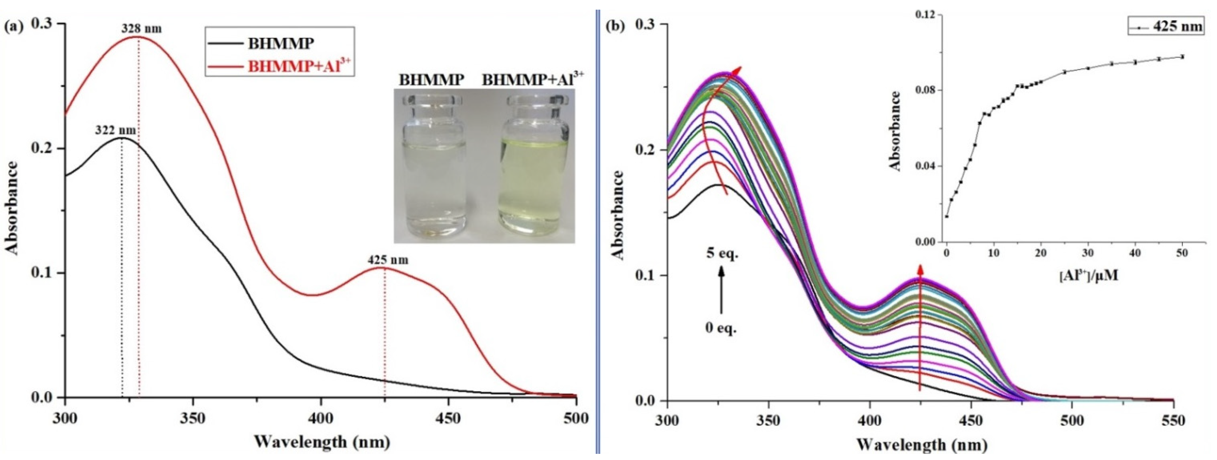
2.1. Fluorescence and UV–Vis Spectral Response of BHMMP to Al3+

Firstly, one of the most essential characteristics of fluorescent probe is its excellent selectivity; so, a fluorescence selectivity experiment was conducted on the fluorescence emission spectrum of the solution of probe BHMMP (10 μM) with different metal ions (50 μM) (including Al3+, K+, Mg2+, Mn2+, Na+, Ni2+, Ag+, Ca2+, Cd2+, Co2+, Cr3+, Cu2+, Fe2+, Pb2+, Zn2+, Hg2+, and Fe3+) in EtOH/H2O (2/3, v/v, 0.01 M HEPES, pH = 5) at room temperature. As depicted in Figure 1a, there was a weak fluorescence emission for BHMMP alone, with a fluorescence quantum yield (Φf, quinine sulfate as standard) as low as 0.005 [52]. In the case of the existence of Al3+ in BHMMP solution, the fluorescence spectrum displayed a significant enhancement with the maximum emission peak at 522 nm (Φf = 0.11), alongside fluorescence color, which was changed from colorless to yellow-green under ultraviolet light. It was conjectured that the increase of fluorescence might be associated with the coordination of BHMMP to Al3+, resulting in the enhancement of structure rigidity of the probe, which could prevent PET and C=N isomerization processes [53,54]. Under the same conditions, other metal ions did not cause significant changes in fluorescence intensity, indicating that the probe showed a highly sensitive “turn-on” fluorescence sensing behavior in the presence of Al3+. Competition experiments were measured to validate the fluorescence sensing property of BHMMP to Al3+. Adding the same amount of other metal cations and the mixture (all tested ions) into the BHMMP solution containing Al3+ (5 eq.), the fluorescence intensity at 522 nm had almost no obvious changes (Figure 1b), which confirmed that BHMMP was more intimate with Al3+ in the presence of other ions and could be viewed as a selective fluorescent probe for measuring Al3+ in complex environments.
Fluorescence titration experiments were performed to study the quantitative fluorescence sensing ability of BHMMP to Al3+. During the process of titration, a fluorescence emission peak at 522 nm raised gradually with the increase of Al3+ concentration, and the fluorescence intensity stabilized a maximum value in the presence of two equiv. of Al3+ (Figure 2). According to the titration data mentioned above, it was found that the emission intensity of BHMMP was linearly related to the Al3+ concentration, in the range of 1.0–10.0 μM (R2 = 0.9908), which indicated that BHMMP could be successfully used as a quantitative tool to evaluate Al3+ (Figure S1). The detection limit (LOD) was obtained from the result of fluorescence titration as 7.04 × 10⁻⁷ M (3σ/S, where σ is the standard deviation of the blank solution, and S is slope from plotting the fluorescence intensity versus the concentration of Al3+), which was below the guideline value of Al3+ in drinking water prescribed by WHO (the maximum concentration was 7.4 μM) [55,56].
In the UV spectrum experiment (Figure 3a) showed that, after adding five equiv. of Al3+ to the BHMMP solution, a slight red-shift of the absorbance peak from 322 to 328 nm, along with the appearance of a new peak at 425 nm, was observed, which suggested the stable complex formation between BHMMP and Al3+. At the same time, the presence of Al3+ resulted in the increase of the conjugated degree, thus causing the solution to alter obviously from colorless to pale yellow. Furthermore, it was worthwhile to mention that the absorption value of BHMMP–Al3+ at 425 nm was positively correlated with the concentration of Al3+, from which the LOD was calculated to be 4.18 × 10⁻⁷ M (Figure 3b). The above results confirmed that the potential application of BHMMP, in the quantitative determination of Al3+, was expected in analytical chemistry and biological systems.

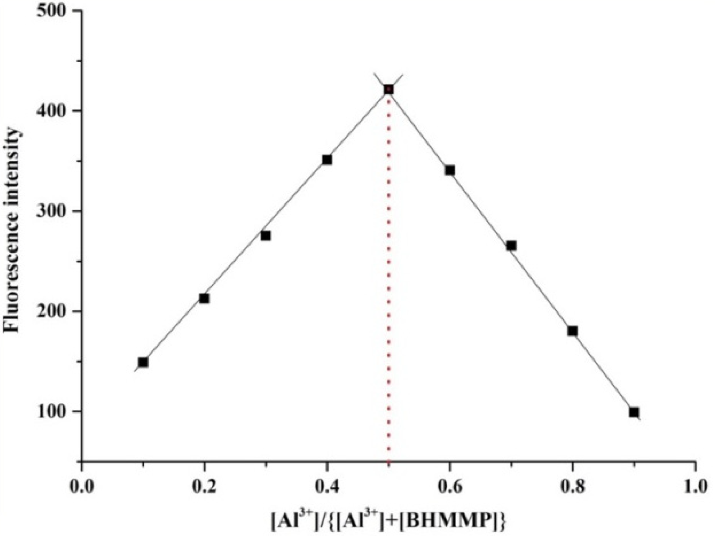
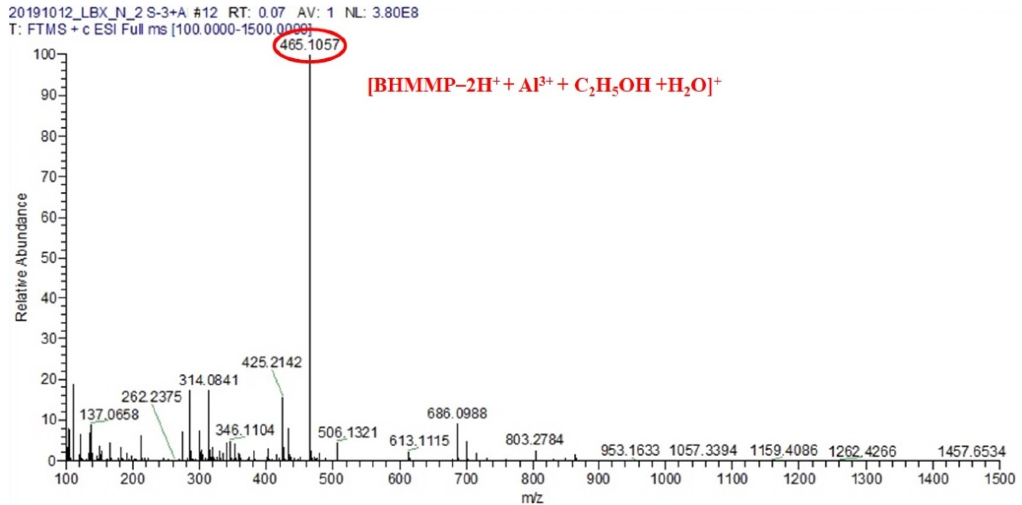
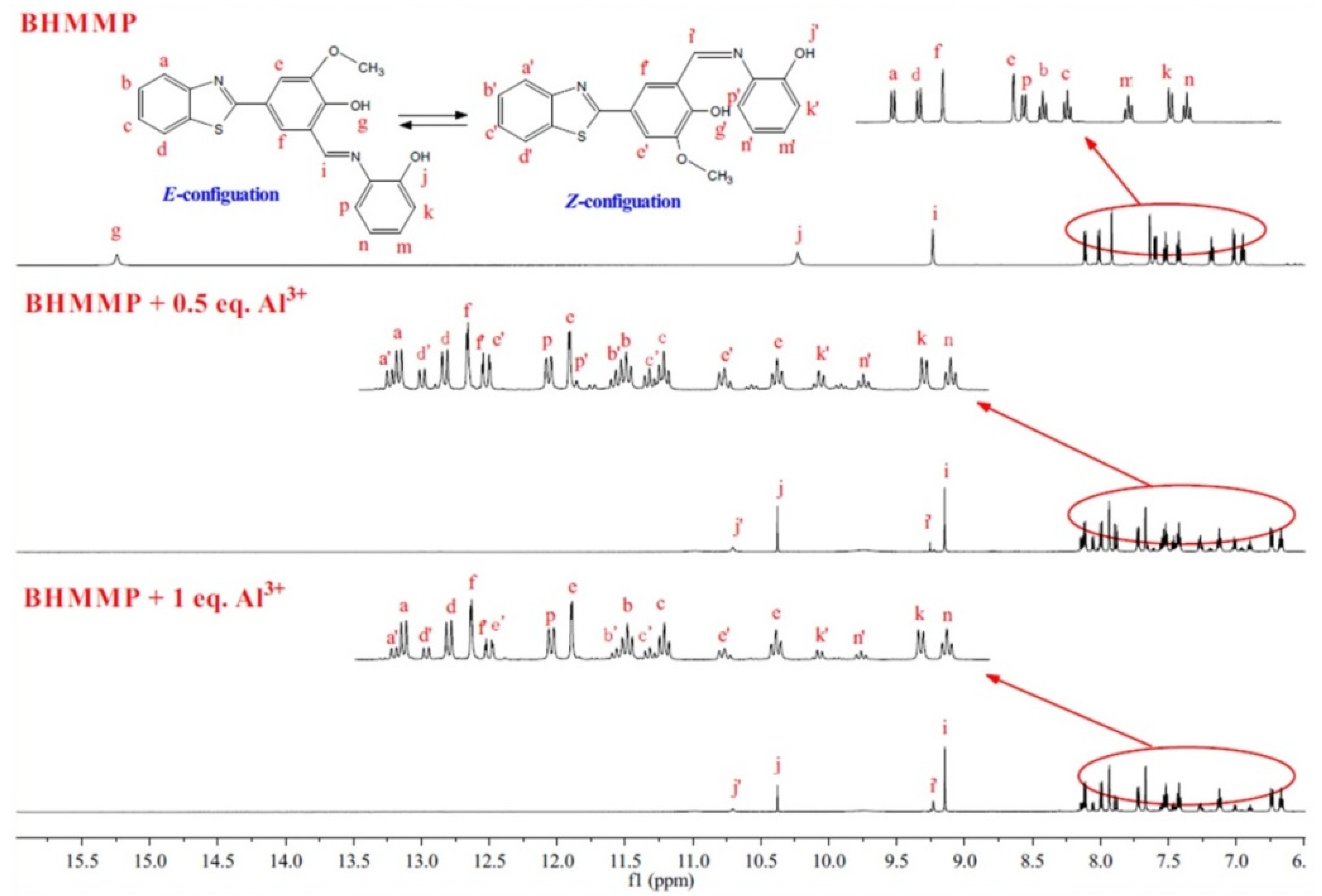
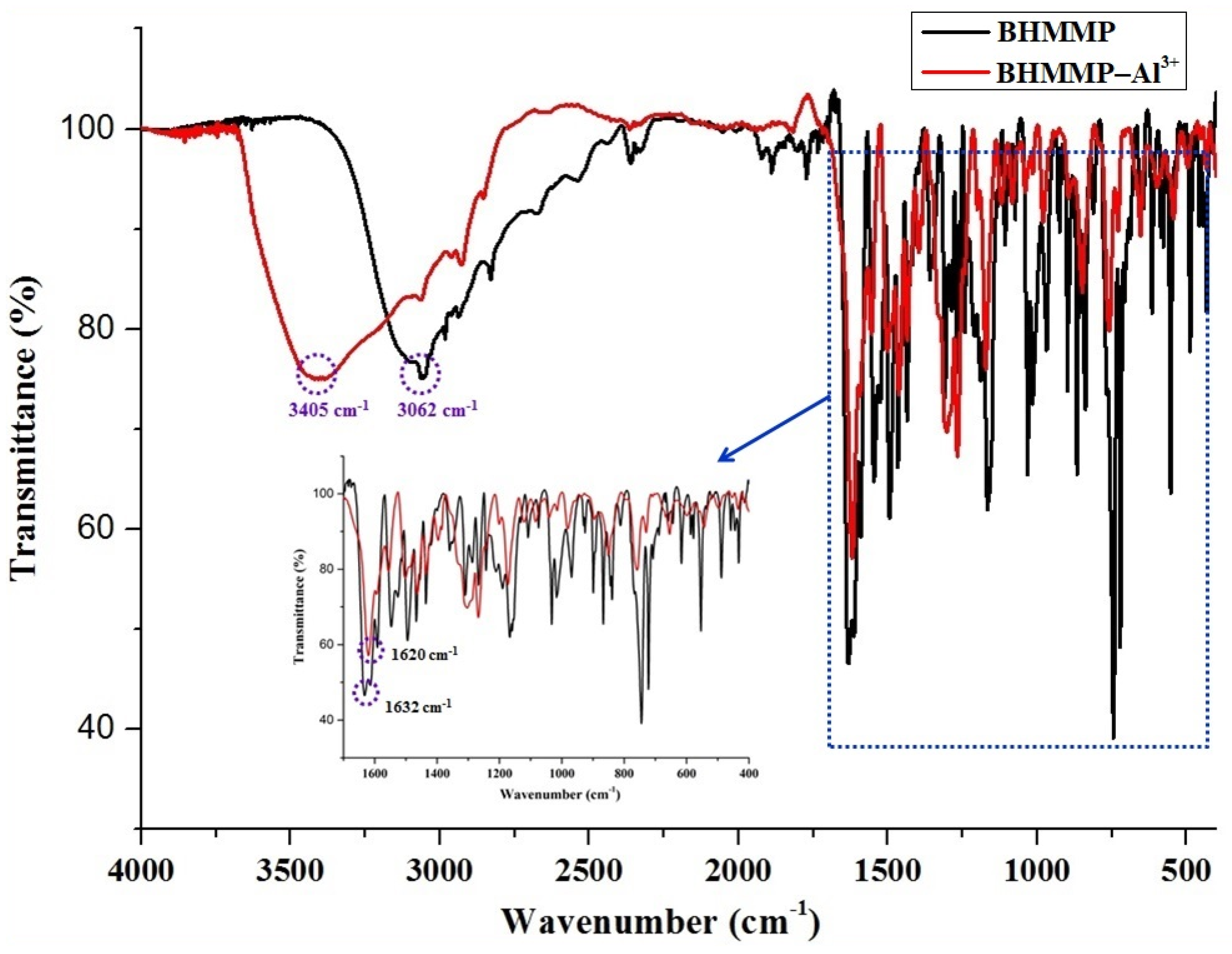
2.2. Binding Mode Studied
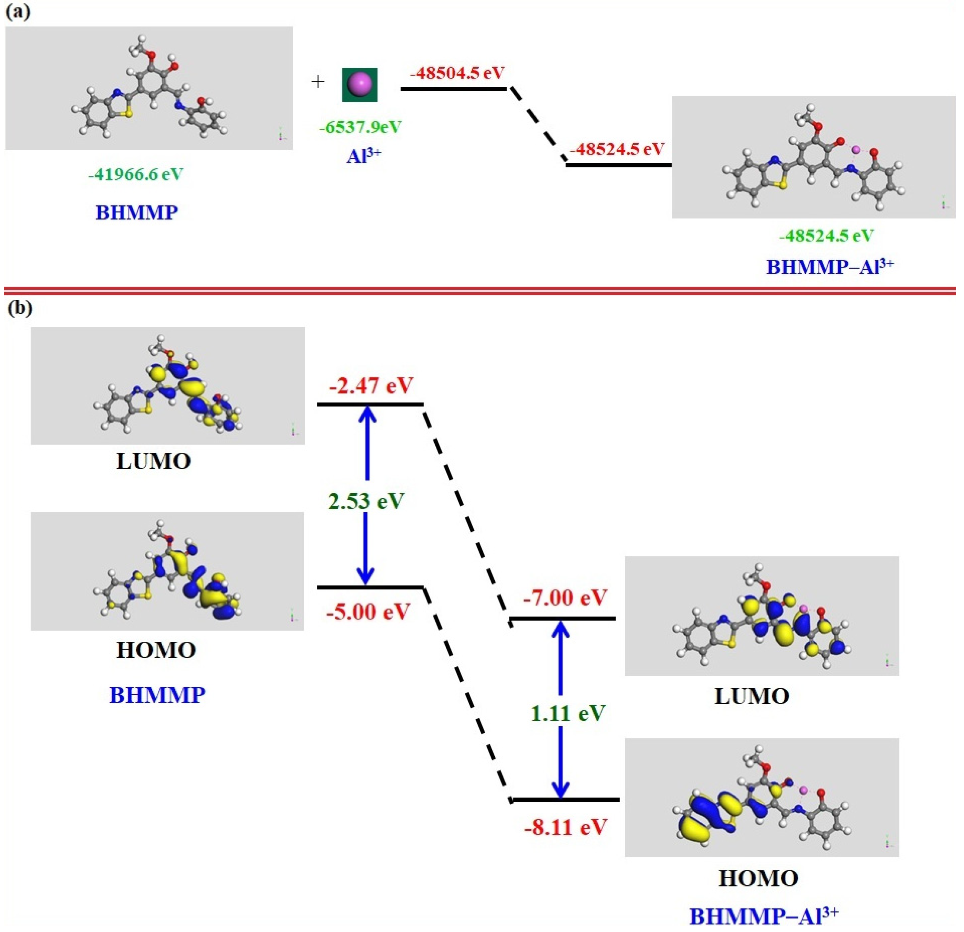
The Job plot method was adopted to infer the stoichiometric relationship between BHMMP and Al3+ (Figure 4). To this end, nine groups of solutions, with continuously varying mole fraction of guest [Al3+]/([BHMMP+Al3+]), were prepared by maintaining the total concentration of the mixed system at 5 × 10⁻⁵ M. The emission intensity reached the maximum when the abscissa of the Job curve was 0.5, indicating the coordination ratio of BHMMP–Al3+ complex was 1:1 in EtOH/H2O (2/3, v/v, 0.01 M HEPES, pH = 5), which was further supported by mass spectroscopy analysis, as follows. A mass spectra signal at 465.1056 m/z was attributed to [BHMMP+Al3++C2H5OH+H2O-2H]+ (calcd: 465.1065), which proved that the stoichiometry of the chelate is 1:1 and revealed that the coordination sphere of Al3+ was composed of solution components, such as water and ethanol (Figure 5). There was a strong binding affinity of BHMMP with Al3+, and the association constants calculated from the results of fluorescence and absorption titration experiments were 3.10 × 10⁴ (Figure S2) and 2.55 × 10³ M−1 (Figure S3), by the Benesi–Hildebrand plot, respectively [57,58,59].
To clarify detailed information about the interaction mode between BHMMP and Al3+, the 1H NMR and FT-IR spectrum of BHMMP were recorded. The result of 1H NMR titration experiment was obtained by adding different equivalent Al3+ to several BHMMP, as illustrated in Figure 6. By comparison, the resonance signal of phenolic hydroxyl group (Hg), at around 15.24 ppm, was completely curtailed, supporting the occurrence of deprotonation upon the combination of BHMMP with Al3+. The peaks at 10.23 and 9.23 ppm, belonging to the hydroxyl (Hj) and imine (Hi) groups, were gradually shortened; meanwhile, their corresponding signals were appeared at 10.71 (Hj’) and 9.25 ppm (Hi’), due to the transition of (Z)-configuration to (E)-configuration, caused by the formation of complex. In addition, the chemical shifts of protons in the aromatic ring (from 6.94 ppm to 8.12 ppm) showed obvious changes, which provided strong evidence for the existence of the two spatial configurations. These observations indicated that nitrogen atom on imine and oxygen atoms on two phenolic hydroxyl groups participated, in coordination with Al3+. As for the FT-IR spectra of BHMMP and BHMMP–Al3+ complex, the stretching vibration peak, attributed to C=N, was shifted from 1634 to 1620 cm−1, which was consistent with the conclusion that imine was taking part in the complexation process (Figure 7).

2.3. DFT Study

The comparison of total energy between BHMMP (BHMMP = −41,966.6 eV) and the BHMMP–Al3+ complex (BHMMP–Al3+ = −48,524.5 eV) indicated that the BHMMP–Al3+ complex was highly stable (Figure 8a). Moreover, the energy of either the highest occupied molecular orbital (HOMO) or the lowest unoccupied molecular orbital (LUMO) of the BHMMP–Al3+ complex was significantly lower than that of free BHMMP (Figure 8b), and the energy gap of HOMO-LUMO of BHMMP–Al3+ (calculated as 1.11 eV) was decreased in comparison with that of BHMMP (calculated as 2.53 eV), which indicated that the complex formation of BHMMP and Al3+ was more stable than BHMMP. Meanwhile, Tauc plots (Figure S4) were illustrated, according to the absorption spectra data of BHMMP and BHMMP–Al3+, and the optical energy gap of BHMMP–Al3+ (estimated as 2.5 eV) also decreased, in comparison with that of BHMMP (estimated as 3.1 eV). Based on the above analysis, the experimental and theoretical results were consistent with the conclusion that the energy gap of BHMMP will decrease after the addition of Al3+.
Combining the above fluorescence and IR spectroscopy, HRMS and 1H NMR titration, and DFT study, the possible structure of BHMMP–Al3+ was reasonably speculated (Scheme 2), and the mechanism of fluorescent enhancement was attributed to the hindrance of the PET and C=N isomerization processes by the establishment of the complex.

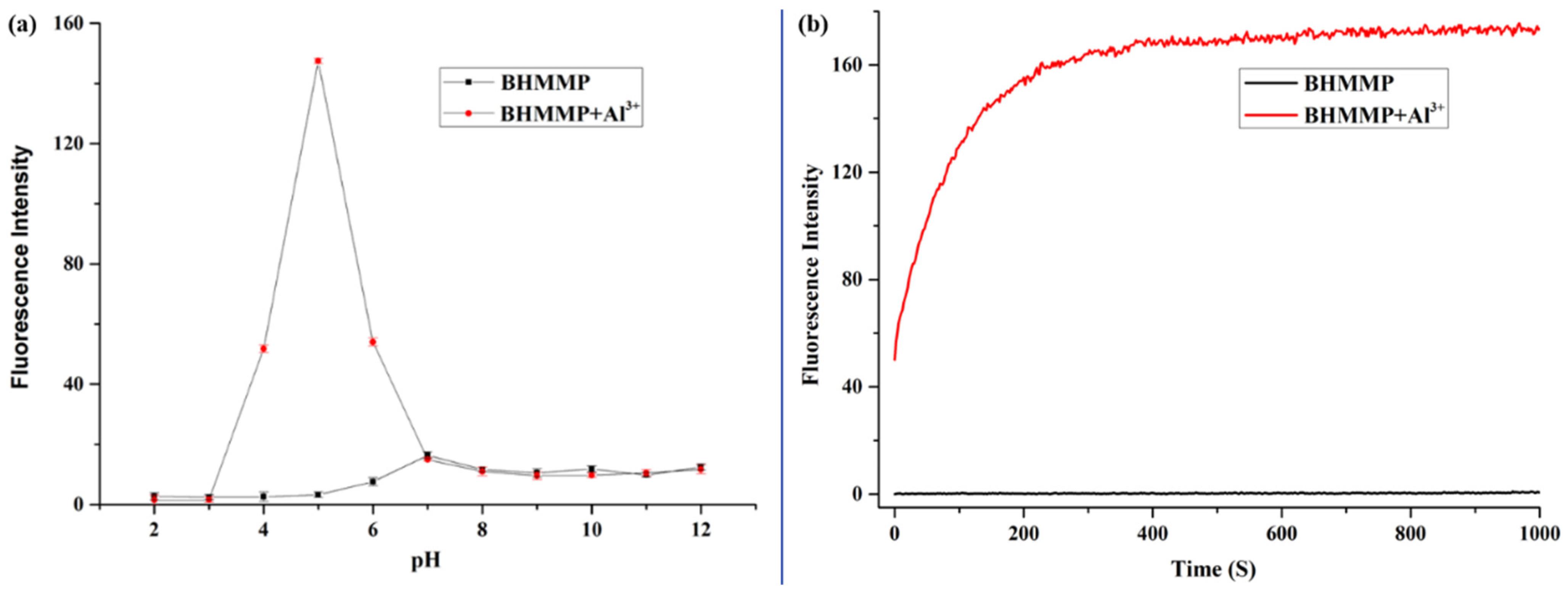
2.4. Effect of pH and Response Time Study

Considering that the probe is usually affected by the proton concentration in the medium during the recognition of ions, the effect of pH on the fluorescence spectrum of BHMMP in the absence and presence of Al3+ was explored at different pH values (Figure 9a). The fluorescence intensity of free probe BHMMP at 522 nm was weak, in a pH range from 2 to 12, indicating that the probe was insensitive to H+/OH. However, a significant increase in the emission intensity of BHMMP between pH 4 and 6 was observed, so the probe was suitable for detecting Al3+ in faintly acidic medium.
The response time enabled us to reflect the sensitivity and stability of the probe. The changing law of the fluorescence intensity with time was monitored in medium EtOH/H2O (v/v, 2/3, 0.01 M HEPES, pH = 5) (Figure 9b). After the addition of Al3+, the fluorescence signal of BHMMP responded instantaneously, reaching the maximum at 3 min, and maintained constant at more than a quarter of an hour. The result sufficiently confirmed that the complexing process between BHMMP and Al3+ was rapid and stable.

2.5. Reversibility Study

The reversibility of BHMMP (10 µM) was investigated in the EtOH/H2O (v/v, 2/3, 0.01 M HEPES, pH = 5) solution by adding EDTA, which was a good chelating agent with Al3+ (Figure S5). Upon the addition of Al3+ (50 µM) into the solution of BHMMP, the fluorescence spectrum had significant changes, compared with the correspondence spectrum of BHMMP itself. However, after the addition of EDTA (50 µM) to the solutions of BHMMP–Al3+, the fluorescence spectra of the solution of BHMMP–Al3+ showed much more similar correspondence to that of BHMMP in the absence of Al3+, indicating the recovering of the BHMMP (Figure S5a). This result was also supported by the bonding constant of BHMMP with Al3+, calculated as 3.10 × 10⁴, which was far lower than that of EDTA, with Al3+ calculated as 1.99 × 10¹⁶ M−1 [60]. However, the alternate addition of Al3+ (50 µM) and EDTA (50 µM) to the above-mentioned solution, the fluorescence intensity at 522 nm was almost constant with that of BHMMP itself (Figure S5b). This result indicated that BHMMP was a non-reusable probe in sensing Al3+.

3. Practical Applications

3.1. Quantitative Detection of Al3+ in Actual Water Samples

To measure the feasibility of probe BHMMP to quantitatively detect Al3+ in real aqueous sample, tap water and Songhua River water samples were spiked with different concentrations of Al3+ solution and analyzed by the proposed fluorimetric method (Figure S6). The fluorescence intensity in the above experiment was collected, and the recoveries were within the range of 97–102%, indicating that the probe had the potential capacity to conduct trace analysis on Al3+ in environmental water samples (Table S2).

3.2. Monitoring Al3+ on Test Paper

The color changes of the mixed solution containing different concentrations of Al3+ were observed clearly under sunlight and 365 nm ultraviolet light (Figure 10). Enlightened by this, we conducted colorimetric experiments on Al3+ by loading BHMMP on the test paper [61,62]. The test paper was soaped into BHMMP solutions, with various amounts of Al3+, and then dried naturally. As the concentration of Al3+ increased, the color of the test strip changed from colorless to yellow-green under 365 nm UV light, which indicated that BHMMP was expected to become a portable tool for detecting Al3+.

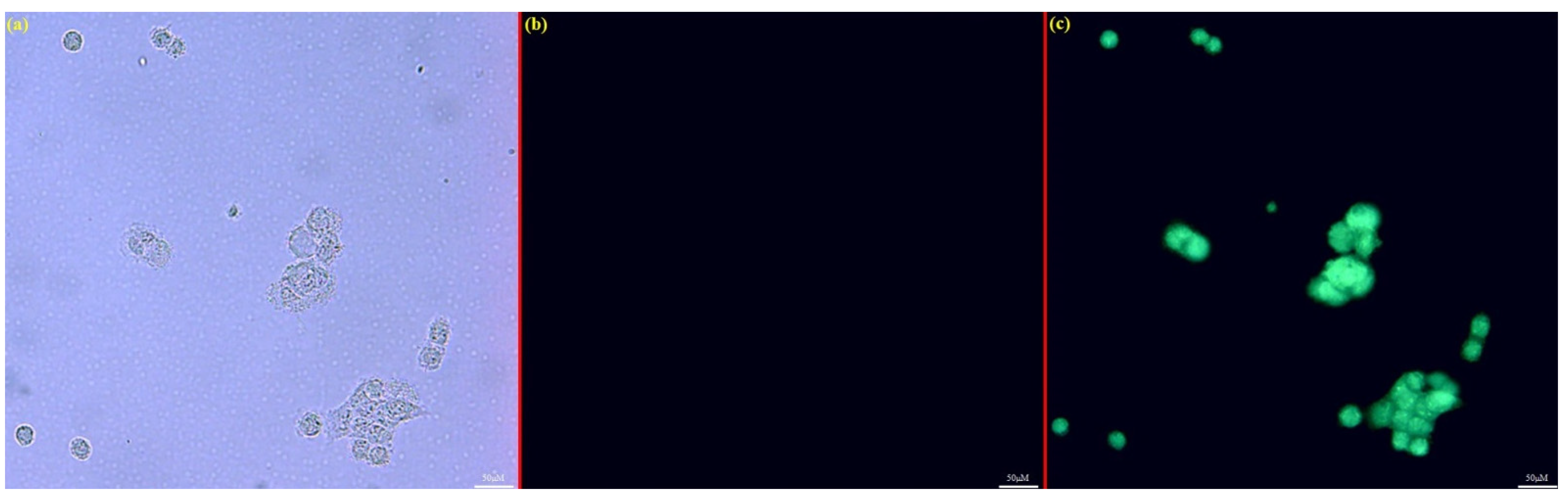
3.3. Cellular Imaging Experiments

Due to the serious toxicity of aluminum ions on living organisms, successful imaging in biological systems is an essential practical application capability for excellent Al3+ probes. Fluorescence images of human stromal cells (HSC) treated with Al3+ were acquired by a fluorescence microscopy [63,64]. As depicted in Figure 11, it could be observed that either the cells themselves (Figure 11A), or incubated with BHMMP (Figure 11B), did not all emit fluorescence; however, the unique light-green fluorescence in cells cultured with BHMMP (10 μM) and Al3+ (50 μM) was monitored, which might be the coordination of BHMMP and Al3+ inside the cells (Figure 11C). Hence, the probe BHMMP was still capable of sensing Al3+ in cells.

4. Materials and Methods

4.1. Materials and Instruments

Materials (such as vanillin, 2-aminophenol and 2-aminothiophenol) for synthesis and metal salts were obtained from Energy Chemical (Shanghai, China). The required solvents and reagents were of analytical or spectroscopic reagent grades during the overall experiments. The structure of the target compound and its coordination mode with metal ions were studied on the Bruck AV-600 MHz system, Waters Xevo UPLC/G2-SQ Tof MS, and Bruker ALPHA-T spectrometers. The spectral properties of the compound are explored, in detail, through a Shimadzu UV-2700 UV–vis spectrometer and Perkin Elmer LS55 fluorescence spectrometer. Test solutions of different pH values at room temperature were prepared by a pHS-3C acidometer.

4.2. Analytical Procedures for Spectroscopic Experiments

The nitrate, chloride, or perchlorate salt of various cationic ions were processed into solution by dissolving in ultrapure water with concentration of 10 mM. The stock solution of BHMMP was prepared, according to this procedure, through dissolving BHMMP in pure ethanol and then diluting to 0.01 mM, in which 40% of the component was ethanol solvent. For spectrum measurement, a certain amount of metal ions was precisely added to the BHMMP solution with a pipetting gun, and the spectrum of the test solution, after uniform mixing, was recorded by UV–Vis and fluorescence spectrometers. In the fluorescence experiment, the excitation wavelength of the test solution was 370 nm, and the excitation, as well as emission slit widths, were all fixed to 10 nm.

4.3. DFT Investigation

In this paper, all DFT calculations were performed by the Dmol3 module of Materials Studio [65]. The Perdew–Burke–Ernzerholf (PBE) version of generalized gradient approximation (GGA) was utilized to treat the exchange correlation interaction [66]. To describe the long-range weak interactions, such as van der Waals force, the Grimme dispersion correction was used [67], and a double numerical basis set with polarization functions (DNP+) was applied for this system. The tolerance of the self-consistent field (SCF) was set as 1.0 × 10⁻⁶ Ha. During geometric optimizations, the convergence threshold parameters were 1.0 × 10⁻⁵ Ha (energy), 0.002 Ha/Å (maximum force), and 0.005 Å (maximum displacement), respectively.

4.4. In Vitro Cytotoxicity Assays

Human stromal cells (HSC) were plated at 1 × 10⁵ cells per well in a 96-well cell-culture plate, followed by incubation at 37 °C for 24 h. Then, the cells were incubated with varying concentrations of probes (0, 5, 10, 20, 30, 40, and 50 μM) for 24 h and washed with 100 μL fresh medium. Then, the fresh medium (100 μL) and MTT (10 μL, 5 mg/mL) were added to each well, and the cells were incubated for another 4 h at 37 °C. Finally, the absorbance of 560 nm was measured with a Bio-Rad microplate reader, and the cell viability was calculated (Figure S7).

4.5. Cell Imaging in HSC Cells

Following the reported method [68], cell imaging experiments were carried out using human stromal cells (HSC) that were exposed to DMEM/F-12 (1:1) medium at 37 °C in an atmosphere containing 5% CO2. Heat-inactivated FBS, penicillin, streptomycin, and sodium pyruvate are further supplemented to this medium in an appropriate amount to suit mammalian cell culture under low serum content. HSC were cultured in 6-well plates for 24 h, treated with Al3+ (0 and 50 μM) for 2 h, washed with Hanks’ balanced salt solution three times, and then seeded with BHMMP (10 μM) for 2 h. Finally, cell imaging was captured through a fluorescence microscope.

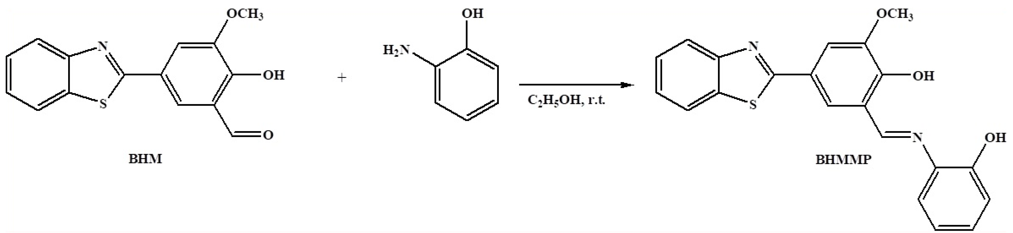
4.6. Synthesis of BHMMP

According to the reported literature, 5-(benzo[d]thiazol-2-yl)-2-hydroxy-3-methoxybenzaldehyde (BHM) was obtained via a condensation reaction between 2-aminothiophenol and vanillin, which was further used in the formylation through the Duff reaction [69].
The classic Schiff base reaction was shown in Scheme 1. Compounds BHM (100 mg, 0.35 mmol) and 2-aminophenol (38 mg, 0.35 mmol) were placed in a round bottom flask containing 15 mL ethanol, followed by adding two drops of acetic acid as a catalyst. The mixture was stirred for 8 h at room temperature, until the reactant was consumed. The precipitate was collected by filtration and washed by ethanol three times, and the desired product BHMMP (103 mg) was obtained after drying. Yield: 78 %. m.p. 250.9–251.7; 1H NMR (Figure S8) (600 MHz, DMSO-d6) δ (ppm) 15.24 (s, 1H), 10.23 (s, 1H), 9.23 (s, 1H), 8.11 (d, J = 7.8 Hz, 1H), 8.01 (d, J=8.0 Hz, 1H), 7.92 (s, 1H), 7.64 (s, 1H), 7.59 (d, J = 7.7 Hz, 1H), 7.52 (t, J = 7.5 Hz, 1H), 7.42 (t, J = 7.4 Hz, 1H), 7.18 (t, J = 7.5 Hz, 1H), 7.02 (d, J = 7.9 Hz, 1H), 6.95 (t, J = 7.5 Hz, 1H), and 3.93 (s, 3H); 13C NMR (Figure S9) (151 MHz, DMSO-d6) δ (ppm) 167.62, 161.98, 159.59, 154.10, 150.75, 134.60, 131.36, 128.89, 126.95, 125.42, 125.02, 122.71, 122.61, 121.45, 120.24, 119.95, 119.03, 117.71, 116.97, 111.67, and 56.24; HRMS: m/z (TOF MS ES) (Figure S10), calcd. for C21H16N2O3S: 375.0803 [BHMMP-H+], found: 375.0808.

5. Conclusions

In summary, a Schiff-based probe BHMMP, revealed recognition towards Al3+, with an obvious optical color change, as well as a fluorescence-enhanced behavior in the EtOH/H2O (2/3, v/v, 0.01 M HEPES, pH = 5) solution. The detection limit was obtained from fluorescence titration data as 0.70 µM. The binding mode and working mechanism between the probe BHMMP and Al3+ was inferred by various spectra, HRMS, and 1H NMR titration. The 1:1 complex, formed between Al3+ and O/N atoms in BHMMP, inhibited the C=N isomerization and PET processes, thus opening fluorescent response. Importantly, not only could BHMMP be suitable for quantitatively monitoring Al3+ in real water samples and test paper, but it was also successfully applied for turn-on fluorescently sensing Al3+ in HSC cells. This work will provide an effective sensor for detecting Al3+ both environmentally and biologically.

Supplementary Materials

The following supporting information can be downloaded at https://www.mdpi.com/article/10.3390/molecules27082569/s1. Figure S1. Fluorescence intensity probe BHMMP with varying concentration of Al3+. Figure S2. The absorbance of probe BHMMP with varying concentration of Al3+. Figure S3. Benesi–Hildebrand plot from fluorescence titration data of BHMMP (10 µM) with Al3+. Figure S4. Tauc plot of BHMMP and BHMMP–Al3+. Figure S5. Reversibility of BHMMP for Al3+ Figure S6. Fluorescent response of probe BHMMP in actual water samples. Figure S7. The cell viability of probe BHMMP. Figure S8. 1H NMR spectrum of probe BHMMP. Figure S9. 13C NMR spectrum of probe BHMMP. Figure S10. ESI-MS spectrum of probe BHMMP in DMF. Table S1 Comparison of previously reported Al3+ probes with functional groups similar to BHMMP. Table S2 The fluorimetric determination results for Al3+ in actual water samples by probe BHMMP.

Author Contributions

Conceptualization, Z.X.; methodology, Z.X., J.W. and J.H.; formal analysis, Z.Z., C.F. and X.C.; data curation, G.H.; writing—original draft preparation, Z.X.; writing—review and editing, Z.X. and J.W. All authors have read and agreed to the published version of the manuscript.

Funding

This research was funded by the National Natural Science Foundation of China (no. 82060293).

Institutional Review Board Statement

Not applicable.

Informed Consent Statement

Not applicable.

Data Availability Statement

The data are available from the corresponding author on reasonable request.

Acknowledgments

This work was supported by the National Natural Science Foundation of China (No. 82060293). We kindly thank Li S.J. and Qu B. of Northeast Agricultural University for the cell image experiments. We also thank Yue M.L. of Northeast Agricultural University and Cheng K.L. of Harbin Institute of Technology for the DFT study.

Conflicts of Interest

The authors declare no conflict of interest.

Sample Availability

Samples of the compounds are available from the authors.

References

  1. Zhao, G.; Wei, G.; Yan, Z.Q.; Guo, B.Y.; Guang, S.Y.; Wu, R.L.; Xu, H.Y. A multiple fluorescein-based turn-on fluorophore (FHCS) identified for simultaneous determination and living imaging of toxic Al3+ and Zn2+ by improved Stokes shift. Anal. Chim. Acta 2020, 1095, 185–196. [Google Scholar] [CrossRef] [PubMed]
  2. Sun, X.J.; Liu, T.T.; Li, N.N.; Zeng, S.; Xing, Z.Y. A novel dual-function probe for recognition of Zn2+ and Al3+ and its application in real samples. Spectrochim. Acta A Mol. Biomol. Spectrosc. 2020, 228, 117786. [Google Scholar] [CrossRef] [PubMed]
  3. Kashyap, K.S.; Kumara, A.; Hira, S.K.; Dey, S. Recognition of Al3+ through the off-on mechanism as a proficient driving force for the hydrolysis of BODIPY conjugated Schiff base and its application in bio-imaging. Inorg. Chim. Acta 2019, 498, 119157. [Google Scholar] [CrossRef]
  4. Aydin, D. A novel turn on fluorescent probe for the determination of Al3+ and Zn2+ ions and its cells applications. Talanta 2020, 210, 120615. [Google Scholar] [CrossRef] [PubMed]
  5. Xing, C.B.; Hao, L.B.; Zang, L.B.; Tang, X.D.; Zhao, Y.Y.; Lu, J.L. A highly selective fluorescent probe for Al3+ based on bis(2-hydroxy-1-naphthaldehyde) oxaloyldihydrazone with aggregation-induced emission enhancement and gel properties. Spectrochim. Acta A Mol. Biomol. Spectrosc. 2020, 224, 117406. [Google Scholar] [CrossRef] [PubMed]
  6. Xu, P.F.; Bao, Z.Y.; Yu, C.Y.; Qiu, Q.Q.; Wei, M.G.; Xi, W.B.; Qian, Z.S.; Feng, H. A water-soluble molecular probe with aggregation-induced emission for discriminative detection of Al3+ and Pb2+ and imaging in seedling root of Arabidopsis. Spectrochim. Acta A Mol. Biomol. Spectrosc. 2019, 223, 117335. [Google Scholar] [CrossRef]
  7. Niu, H.; Leng, Y.F.; Ran, S.M.; Amee, M.; Du, D.Y.; Sun, J.; Chen, K.; Hong, S. Toxicity of soil labile aluminum fractions and aluminum species in soil water extracts on the rhizosphere bacterial community of tall fescue. Ecotox. Environ. Saf. 2019, 187, 109828. [Google Scholar] [CrossRef]
  8. Chen, F.M.; Ai, H.L.; Wei, M.T.; Qin, C.L.; Feng, Y.; Ran, S.M.; Wei, Z.H.; Niu, H.; Zhu, Q.; Zhu, H.H.; et al. Distribution and phytotoxicity of soil labile aluminum fractions and aluminum species in soil water extracts and their effects on tall fescue. Ecotox. Environ. Saf. 2018, 163, 180–187. [Google Scholar] [CrossRef]
  9. Wang, W.L.; Jin, L.; Kuang, Y.; Yuan, Z.Z.; Wang, Q.M. Isonicotinoylhydrazide modified 3-acetylcoumarin scaffold as an efficient chemical reversible sensor to detect Al3+ selectively and its application in live cells imaging. Synth. Commun. 2019, 49, 2501–2522. [Google Scholar] [CrossRef]
  10. Zhu, Y.L.; Gong, X.S.; Li, Z.P.; Zhao, X.; Liu, Z.Q.; Cao, D.X.; Guan, R.F. A simple turn-on ESIPT and PET-based fluorescent probe for detection of Al3+ in real-water sample. Spectrochim. Acta A Mol. Biomol. Spectrosc. 2019, 219, 202–205. [Google Scholar] [CrossRef]
  11. Mirza, A.; King, A.; Troakes, C.; Exley, C. The Identification of Aluminum in Human Brain Tissue Using Lumogallion and Fluorescence Microscopy. J. Alzheimers Dis. 2016, 54, 1333–1338. [Google Scholar] [CrossRef] [PubMed] [Green Version]
  12. Mold, M.; Linhart, C.; Gómez-Ramírez, J.; Villegas-Lanau, A.; Exley, C. Aluminum and Amyloid-β in Familial Alzheimer’s Disease. J. Alzheimers Dis. 2020, 73, 1627–1635. [Google Scholar] [CrossRef] [PubMed] [Green Version]
  13. Mclachlan, D.R.; Bergeron, C.; Alexandrov, P.N.; Walsh, W.J.; Pogue, A.I.; Percy, M.E.; Kruck, T.P.A.; Fang, Z.; Sharfman, N.M.; Jaber, V.; et al. Aluminum in Neurological and Neurodegenerative Disease. Mol. Neurobiol. 2019, 56, 1531–1538. [Google Scholar] [CrossRef] [PubMed]
  14. Santos, A.S.; De Souza, C.T.; Leao, D.J.; Correia, F.O.; Almeida, T.S.; Ferreira, S.L.C. Simultaneous Determination of Chromium and Iron in Powdered Milk Using High-Resolution Continuum Source Graphite Furnace Atomic Absorption Spectrometry. Food Anal. Meth. 2020, 13, 284–290. [Google Scholar] [CrossRef]
  15. Panhwar, A.H.; Tuzen, M.; Kazi, T.G. Deep eutectic solvent based advance microextraction method for determination of aluminum in water and food samples: Multivariate study. Talanta 2018, 178, 588–593. [Google Scholar] [CrossRef] [PubMed]
  16. Son, S.H.; Lee, W.B.; Kim, D.; Lee, Y.; Nam, S.H. An alternative analytical method for determining arsenic species in rice by using ion chromatography and inductively coupled plasma-mass spectrometry. Food Chem. 2019, 27, 353–358. [Google Scholar] [CrossRef]
  17. Zhang, L.Y.; Luo, J.J.; Shen, X.Y.; Li, C.Y.; Wang, X.; Nie, B.; Fang, H.F. Quantitative Determining of Ultra-Trace Aluminum Ion in Environmental Samples by Liquid Phase Microextraction Assisted Anodic Stripping Voltammetry. Sensors 2018, 18, 1503. [Google Scholar] [CrossRef] [Green Version]
  18. Wang, H.Q.; Da, L.G.; Yang, L.; Chu, S.Y.; Yang, F.; Yu, S.M.; Jiang, C.L. Colorimetric Fluorescent Paper Strip with Smartphone Platform for Quantitative Detection of Cadmium Ions in Real Samples. J. Hazard. Mater. 2020, 392, 122506. [Google Scholar] [CrossRef]
  19. Li, M.S.; Xing, Y.L.; Zou, Y.X.; Chen, G.; You, J.M.; Yu, F.B. Imaging of the mutual regulation between zinc cation and nitrosyl via two-photon fluorescent probes in cells and in vivo. Sens. Actuators B Chem. 2020, 309, 127772. [Google Scholar] [CrossRef]
  20. Zeng, R.F.; Lan, J.S.; Wu, T.; Liu, L.; Liu, Y.; Ho, R.J.Y.; Ding, Y.; Zhang, T. A novel mitochondria-targetted near-infrared fluorescent probe for selective and colorimetric detection of sulfite and its application in vitro and vivo. Food Chem. 2020, 318, 126358. [Google Scholar] [CrossRef]
  21. Ling, C.; Cui, M.Y.; Chen, J.R.; Xia, L.L.; Deng, D.W.; Gu, Y.Q.; Wang, P. A novel highly selective fluorescent probe with new chalcone fluorophore for monitoring and imaging endogenous peroxynitrite in living cells and drug-damaged liver tissue. Talanta 2020, 215, 120934. [Google Scholar] [CrossRef] [PubMed]
  22. Jiang, W.L.; Wang, W.X.; Liu, J.; Li, Y.F.; Li, C.Y. A novel hepatocyte-targeting ratiometric fluorescent probe for imaging hydrogen peroxide in zebrafish. Sens. Actuators B Chem. 2020, 313, 128054. [Google Scholar] [CrossRef]
  23. Li, N.N.; Ma, Y.Q.; Sun, X.J.; Lia, M.Q.; Zeng, S.; Xing, Z.Y.; Li, J.L. A dual-function probe based on naphthalene for fluorescent turn-on recognition of Cu2+ and colorimetric detection of Fe3+ in neat H2O. Spectrochim. Acta A Mol. Biomol. Spectrosc. 2019, 210, 266–274. [Google Scholar] [CrossRef] [PubMed]
  24. Fu, Y.; Pang, X.X.; Wang, Z.Q.; Chai, Q.; Ye, F. A highly sensitive and selective fluorescent probe for determination of Cu (II) and application in live cell imaging. Spectrochim. Acta A Mol. Biomol. Spectrosc. 2019, 208, 198–205. [Google Scholar] [CrossRef]
  25. Ye, F.; Liang, X.M.; Xu, K.X.; Pang, X.X.; Chai, Q.; Fu, Y. A novel dithiourea- appended naphthalimide “on-off” fluorescent probe for detecting Hg2+ and Ag+ and its application in cell imaging. Talanta 2019, 200, 494–502. [Google Scholar] [CrossRef]
  26. Wu, N.; Zhao, L.X.; Jiang, C.Y.; Li, P.; Liu, Y.; Fu, Y.; Ye, F. A naked-eye visible colorimetric and fluorescent chemosensor for rapid detection of fluoride anions: Implication for toxic fluorine-containing pesticides detection. J. Mol. Liq. 2020, 302, 112549. [Google Scholar] [CrossRef]
  27. Liu, Y.; Yang, L.; Li, L.; Liang, X.; Li, S.; Fu, Y. A dual thiourea-appended perylenebisimide “turn-on” fluorescent chemosensor with high selectivity and sensitivity for Hg2+ in living cells. Spectrochim. Acta A Mol. Biomol. Spectrosc. 2020, 241, 118678. [Google Scholar] [CrossRef]
  28. Liu, Y.L.; Yang, L.; Li, P.; Li, S.J.; Li, L.; Pang, X.X.; Ye, F.; Fu, Y. A novel colorimetric and “turn-off” fluorescent probe based on catalyzed hydrolysis reaction for detection of Cu2+ in real water and in living cells. Spectrochim. Acta A Mol. Biomol. Spectrosc. 2020, 227, 117540. [Google Scholar] [CrossRef]
  29. Ye, F.; Wu, N.; Li, P.; Liu, Y.L.; Li, S.J.; Fu, Y. A lysosome-targetable fluorescent probe for imaging trivalent cations Fe3+, Al3+ and Cr3+ in living cells. Spectrochim. Acta A Mol. Biomol. Spectrosc. 2019, 222, 117242. [Google Scholar] [CrossRef]
  30. Dhineshkumar, E.; Iyappan, M.; Anbuselvan, C. A novel dual chemosensor for selective heavy metal ions Al3+, Cr3+ and its applicable cytotoxic activity, HepG2 living cell images and theoretical studies. J. Mol. Struct. 2020, 1210, 128033. [Google Scholar] [CrossRef]
  31. Ko, M.; Kurapati, S.; Jo, Y.; Cho, B.; Cho, D. Tuned Al3+ selectivity and π-extended properties of di-2-picolylamine-substituted quinoline-based tolan. Tetrahedron Lett. 2020, 61, 151808. [Google Scholar] [CrossRef]
  32. Li, Z.; Wang, Q.; Wang, J.; Jing, X.M.; Wang, S.J.; Xiao, L.W.; Li, L.L. A fluorescent light-up probe for selective detection of Al3+ and its application in living cell imaging. Inorg. Chim. Acta 2020, 500, 119231. [Google Scholar] [CrossRef]
  33. Li, Y.Y.; Xu, K.; Si, Y.; Yang, C.P.; Peng, Q.C.; He, J.; Hu, Q.G.; Li, K. An aggregation-induced emission (AIE) fluorescent chemosensor for the detection of Al(III) in aqueous solution. Dyes Pigments 2019, 171, 107682. [Google Scholar] [CrossRef]
  34. Zeng, S.; Li, S.J.; Sun, X.J.; Li, M.Q.; Ma, Y.Q.; Xing, Z.Y.; Li, J.L. A naphthalene-quinoline based chemosensor for fluorescent “turn-on” and absorbance-ratiometric detection of Al3+ and its application in cells imaging. Spectrochim. Acta A Mol. Biomol. Spectrosc. 2018, 205, 276–286. [Google Scholar] [CrossRef] [PubMed]
  35. Kang, L.; Xing, Z.Y.; Ma, X.Y.; Liu, Y.T.; Zhang, Y. A highly selective colorimetric and fluorescent turn-on chemosensor for Al3+ based on naphthalimide derivative. Spectrochim. Acta A Mol. Biomol. Spectrosc. 2016, 167, 59–65. [Google Scholar] [CrossRef]
  36. Hou, J.T.; Wang, B.Y.; Wang, S.; Wu, Y.Q.; Liao, Y.X.; Ren, W.X. Detection of hydrazine via a highly selective fluorescent probe: A case study on the reactivity of cyano-substituted C=C bond. Dyes Pigments 2020, 178, 108366. [Google Scholar] [CrossRef]
  37. Huang, Y.F.; Zhang, Y.B.; Huo, F.J.; Liu, Y.M.; Yin, C.X. Mitochondrial-targeted near-infrared “dual mode” fluorescent dyes with large Stokes shift for detection of hypochlorous acid and its bioimaging in cell and mice. Dyes Pigments 2020, 179, 108387. [Google Scholar] [CrossRef]
  38. Liu, Y.; Bi, A.Y.; Gao, T.; Cao, X.Z.; Gao, F.; Rong, P.F.; Wang, W.; Zeng, W.B. A novel self-assembled nanoprobe for the detection of aluminum ions in real water samples and living cells. Talanta 2019, 194, 38–45. [Google Scholar] [CrossRef]
  39. Lu, Z.N.; Wang, L.; Zhang, X.; Zhu, Z.J. A selective fluorescent chemosensor for Cd2+ based on 8-hydroxylquinoline-benzothiazole conjugate and imaging application. Spectrochim. Acta A Mol. Biomol. Spectrosc. 2019, 213, 57–63. [Google Scholar] [CrossRef]
  40. Shi, Z.C.; Zhao, Z.G. Microwave irradiation synthesis of novel indole triazole Schiff base fluorescent probe for Al3+ ion. Inorg. Chim. Acta 2019, 498, 119135. [Google Scholar] [CrossRef]
  41. Fu, J.X.; Yao, K.; Chang, Y.X.; Li, B.; Yang, L.; Xu, K.X. A novel colorimetric-fluorescent probe for Al3+ and the resultant complex for F and its applications in cell imaging. Spectrochim. Acta A Mol. Biomol. Spectrosc. 2019, 222, 117234. [Google Scholar] [CrossRef] [PubMed]
  42. Hu, T.; Cheng, J.P.; Li, L.J.; Zhan, Y.F.; Li, W.J.; Chang, Z.D.; Sun, C.Y. A new Schiff base fluorescent-colorimetric probe containing fluorene-naphthalene structure: Multifunction detection. Inorg. Chim. Acta 2019, 498, 119131. [Google Scholar] [CrossRef]
  43. Wang, J.H.; Feng, L.H.; Chao, J.B.; Wang, Y.; Shuang, S.M. A new ‘turn-on’ and reversible fluorescent sensor for Al3+ detection and live cell imaging. Anal. Methods 2019, 11, 5598–5606. [Google Scholar] [CrossRef]
  44. Leng, X.; Jia, X.; Qiao, C.F.; Xu, W.F.; Ren, C.T.; Long, Y.; Yang, B.Q. Synthesis, characterization and Al3+ sensing application of a new chromo-fluorogenic chemosensor. J. Mol. Struct. 2019, 1193, 69–75. [Google Scholar] [CrossRef]
  45. Mondal, A.; Ahmmed, E.; Chakraborty, S.; Sarkar, A.; Lohar, S.; Chattopadhyay, P. Aggregation Induced Emission Enhancement (AIEE) of Naphthalene-Appended Organic Moiety: An Al3+ Ion Selective Turn-On Fluorescent Probe. Chemistryselect 2020, 5, 147–155. [Google Scholar] [CrossRef] [Green Version]
  46. Berrones-Reyes, J.; Muñoz-Flores, B.M.; Gómez-Treviño, A.; Treto-Suárez, M.A.; Páez-Hernández, D.; Schott, E.; Zarate, X.; Jiménez-Pérez, V.M. Novel fluorescent Schiff bases as Al3+ sensors with high selectivity and sensitivity, and their bioimaging applications. Mater. Chem. Phys. 2019, 233, 89–101. [Google Scholar] [CrossRef]
  47. Sarma, S.; Bhowmik, A.; Sarma, M.J.; Banu, S.; Phukan, P.; Das, D.K. Condensation product of 2-hydroxy-1-napthaldehyde and 2-aminophenol: Selective fluorescent sensor for Al3+ ion and fabrication of paper strip sensor for Al3+ ion. Inorg. Chim. Acta 2018, 469, 202–208. [Google Scholar] [CrossRef]
  48. Ren, Y.P.; Han, J.; Wang, Y.; Tang, X.; Wang, L.; Ni, L. An OFF-ON-OFF type fluorescent probe based on a naphthalene derivative for Al3+ and F- ions and its biological application. Luminescence 2018, 33, 15–21. [Google Scholar] [CrossRef]
  49. Maity, D.; Dey, S.; Roy, P. A two-pocket Schiff-base molecule as a chemosensor for Al3+. New J. Chem. 2017, 41, 10677–10685. [Google Scholar] [CrossRef]
  50. Alici, O.; Erdemir, S. A cyanobiphenyl containing fluorescence ‘turn on’ sensor for Al3+ ion in CH3CN-water. Sens. Actuators B Chem. 2015, 208, 159–163. [Google Scholar] [CrossRef]
  51. Sheet, S.K.; Sen, B.; Thounaojam, R.; Aguan, K.; Khatua, S. Highly selective light-up Al3+ sensing by a coumarin based Schiff base probe: Subsequent phosphate sensing DNA binding and live cell imaging. J. Photochem. Photobiol. A-Chem. 2017, 332, 101–111. [Google Scholar] [CrossRef]
  52. Liang, C.S.; Jiang, S.M. Single sensor for multiple analytes in different optical channel: Applying for multi-ion response modulation. Spectrochim. Acta A Mol. Biomol. Spectrosc. 2017, 183, 267–274. [Google Scholar] [CrossRef] [PubMed]
  53. Liu, T.Q.; Wan, X.J.; Dong, Y.S.; Li, W.B.; Wu, L.S.; Pei, H.; Yao, Y.W. Facile synthesis of a water-soluble fluorescence sensor for Al3+ in aqueous solution and on paper substrate. Spectrochim. Acta A Mol. Biomol. Spectrosc. 2016, 173, 625–629. [Google Scholar] [CrossRef] [PubMed]
  54. Sen, B.; Sanjoy, S.K.; Thounaojam, R.; Jamatia, R.; Pal, A.K.; Aguan, K.; Khatua, S. A coumarin based Schiff base probe for selective fluorescence detection of Al3+ and its application in live cell imaging. Spectrochim. Acta A Mol. Biomol. Spectrosc. 2017, 173, 537–543. [Google Scholar] [CrossRef] [PubMed]
  55. Pan, X.; Jiang, J.M.; Li, J.F.; Wu, W.P.; Zhang, J.L. Theoretical Design of Near-Infrared Al3+ Fluorescent Probes Based on Salicylaldehyde Acylhydrazone Schiff Base Derivatives. Inorg. Chem. 2019, 58, 12618–12627. [Google Scholar] [CrossRef]
  56. Kumar, V.; Kumar, P.; Kumar, S.; Singhal, D.; Gupta, R. Turn-On Fluorescent Sensors for the Selective Detection of Al3+ (and Ga3+) and PPi Ions. Inorg. Chem. 2019, 58, 10364–10376. [Google Scholar] [CrossRef]
  57. Chandra, R.; Manna, A.K.; Sahu, M.; Rout, K.; Patra, G.K. Simple salicylaldimine-functionalized dipodal bis Schiff base chromogenic and fluorogenic chemosensors for selective and sensitive detection of Al3+ and Cr3+. Inorg. Chim. Acta 2020, 499, 119192. [Google Scholar] [CrossRef]
  58. Tian, L.M.; Xue, J.; Li, S.L.; Yang, Z.Y. A novel chromone derivative as dual probe for selective sensing of Al(III) by fluorescent and Cu(II) by colorimetric methods in aqueous solution. J. Photochem. Photobiol. A-Chem. 2019, 382, 111955. [Google Scholar] [CrossRef]
  59. Sun, X.J.; Liu, T.T.; Fu, H.; Li, N.N.; Xing, Z.Y.; Yang, F. A naphthalimide-based fluorescence “off-on-off” chemosensor for relay detection of Al3+ and ClO. Front. Chem. 2019, 7, 549. [Google Scholar] [CrossRef] [Green Version]
  60. Ma, Y.Q.; Sun, X.J.; Li, M.Q.; Zeng, S.; Xing, Z.Y.; Li, J.L. A quinoline-based schiff base for significant fluorescent “turn-on” and absorbance-ratiometric detection of Al3+. Chem. Pap. 2019, 73, 1469–1479. [Google Scholar] [CrossRef]
  61. Wang, J.X.; Xing, Z.Y.; Tian, Z.N.; Wu, D.Q.; Xiang, Y.Y.; Li, J.L. A dual-functional probe for sensing pH change and ratiometric detection of Cu2+. Spectrochim. Acta A Mol. Biomol. Spectrosc. 2020, 235, 118318. [Google Scholar] [CrossRef]
  62. Liu, T.T.; Li, S.J.; Fu, H.; Tian, Z.N.; Sun, X.J.; Xing, Z.Y. A fluorescence turn-on probe for the recognition of Al3+ and its application in cell image. J. Photochem. Photobiol. A-Chem. 2020, 403, 112865. [Google Scholar] [CrossRef]
  63. Zeng, S.; Li, S.J.; Sun, X.J.; Liu, T.T.; Xing, Z.Y. A dual-functional chemosensor for fluorescent on-off and ratiometric detection of Cu2+ and Hg2+ and its application in cell imaging. Dyes Pigments 2019, 170, 107642. [Google Scholar] [CrossRef]
  64. Liu, T.T.; Li, S.J.; Zhang, X.S.; Wang, J.; Deng, Y.X.; Sun, X.J.; Xing, Z.Y.; Wu, R.F. A Facile Probe for Fluorescence Turn-on and Simultaneous Naked-Eyes Discrimination of H2S and biothiols (Cys and GSH) and Its Application. J. Fluoresc. 2022, 32, 175–188. [Google Scholar] [CrossRef]
  65. Delley, B. An All-Electron Numerical Method for Solving the Local Density Functional for Polyatomic Molecules. J. Chem. Phys. 1990, 92, 508–517. [Google Scholar] [CrossRef]
  66. Perdew, J.P.; Burke, K.; Ernzerhof, M. Generalized Gradient Approximation Made Simple. Phys. Rev. Lett. 1996, 77, 3865–3868. [Google Scholar] [CrossRef] [Green Version]
  67. Grimme, S. Semiempirical GGA-Type Density Functional Constructed with a Long-Range Dispersion Correction. J. Comput. Chem. 2006, 27, 1787–1799. [Google Scholar] [CrossRef]
  68. Zeng, S.; Li, S.J.; Sun, X.J.; Li, M.Q.; Xing, Z.Y.; Li, J.L. A benzothiazole-based chemosensor for significant fluorescent turn-on and ratiometric detection of Al3+ and its application in cell imaging. Inorg. Chim. Acta 2019, 486, 654–662. [Google Scholar] [CrossRef]
  69. Tian, Z.N.; Wu, D.Q.; Sun, X.J.; Liu, T.T.; Xing, Z.Y. A Benzothiazole-Based Fluorescent Probe for Ratiometric Detection of Al3+ and Its Application in Water Samples and Cell Imaging. Int. J. Mol. Sci. 2019, 20, 5993. [Google Scholar] [CrossRef] [Green Version]
Scheme 1. Synthesis procedure of the probe BHMMP.
Molecules 27 02569 sch001
Figure 1. (a) Fluorescence spectrum changes of BHMMP (10 μM) in EtOH/H2O (2/3, v/v, 0.01 M HEPES, pH = 5) after adding different metal ions (50 μM), λex = 370 nm. Inset: visual fluorescence change of BHMMP solution upon addition of Al3+ under UV illumination at 365 nm. (b) The emission intensity of BHMMP (10 μM) solution containing Al3+ (50 μM), as well as the same amount of interfering ions at 522 nm.
Molecules 27 02569 g001
Figure 2. Fluorescence titration spectrum of BHMMP (10 µM), with varying ratios of Al3+ (0–5 eq.) in EtOH/H2O (2/3, v/v, 0.01 M HEPES, pH = 5) medium, λex = 370 nm. Inset: The variation trend of fluorescence intensity of BHMMP at 522 nm with increasing Al3+ concentration.
Molecules 27 02569 g002
Figure 3. (a) UV–Vis absorption spectral of BHMMP (10 µM), in the absence and presence of Al3+ (5 eq.). Inset: color change of BHMMP solution before and after addition of Al3+ in ambient light. (b) Absorption titration spectra of BHMMP (10 µM) with increasing Al3+ concentration (0–50 µM). Inset: the variation trend of absorbance of BHMMP at 425 nm, upon the gradual addition of Al3+.
Molecules 27 02569 g003
Figure 4. Job plot for evaluating the stoichiometry of BHMMP and Al3+ with fluorescence spectra (λex=370 nm, λem=522 nm).
Molecules 27 02569 g004
Figure 5. The ESI–MS spectrum of BHMMP–Al3+ complex in positive ion source mode.
Molecules 27 02569 g005
Figure 6. 1H NMR spectra of BHMMP in the presence of different equivalents of Al3+ in DMSO-d6.
Molecules 27 02569 g006
Figure 7. The FT-IR spectrum of BHMMP, compared to the BHMMP–Al3+ complex.
Molecules 27 02569 g007
Figure 8. (a) Energy optimized structure of probe BHMMP (left) and (BHMMP–Al3+) (right), and (b) the corresponding molecular orbital of BHMMP and BHMMP–Al3+.
Molecules 27 02569 g008
Scheme 2. Proposed recognition mechanism for the fluorescence enhancement of BHMMP to Al3+ and possible structure of BHMMP–Al3+ complex.
Molecules 27 02569 sch002
Figure 9. (a) Effect of pH on the fluorescence intensity of BHMMP and the BHMMP–Al3+ complex, λex = 370 nm. (b) Time-dependent fluorescence intensity changes of BHMMP (10 μM) to Al3+ (50 μM) at 522 nm.
Molecules 27 02569 g009
Figure 10. Photographic image of different concentrations of Al3+ by BHMMP (10 μM) on test strip by daylight and UV light.
Molecules 27 02569 g010
Figure 11. Images of HSC cells: cells themselves, (a) bright field, only incubated with 10 μM BHMMP; (b) fluorescence treated with 10 μM BHMMP, followed by addition of 50 μM Al3+; (c) fluorescence.
Molecules 27 02569 g011
Publisher’s Note: MDPI stays neutral with regard to jurisdictional claims in published maps and institutional affiliations.

