Review

Ergothioneine, Ovothiol A, and Selenoneine—Histidine-Derived, Biologically Significant, Trace Global Alkaloids

by
Geoffrey A. Cordell
1,2,* and
Sujeewa N. S. Lamahewage
3,4
1
Natural Products Inc., Evanston, IL 60202, USA
2
Department of Pharmaceutics, College of Pharmacy, University of Florida, Gainesville, FL 32610, USA
3
Department of Chemistry, Iowa State University, Ames, IA 50011, USA
4
Department of Chemistry, University of Ruhuna, Matara 81000, Sri Lanka
*
Author to whom correspondence should be addressed.
Molecules 2022, 27(9), 2673; https://doi.org/10.3390/molecules27092673
Submission received: 24 March 2022 / Revised: 15 April 2022 / Accepted: 16 April 2022 / Published: 21 April 2022

Abstract

:
The history, chemistry, biology, and biosynthesis of the globally occurring histidine-derived alkaloids ergothioneine (10), ovothiol A (11), and selenoneine (12) are reviewed comparatively and their significance to human well-being is discussed.

1. Introduction

Histidine-derived (1) alkaloids are rare in nature, and when 1 does serve as a precursor fragment, the products are invariably biologically and biosynthetically interesting, as illustrated in Figure 1. The most important plant-originating alkaloid derived from histidine (1) is pilocarpine (2), isolated from “jaborandi”, the leaves of Pilocarpus jaborandi Holmes (Rutaceae), native to Brasil [1]. It acts as a muscarinic agonist and is used as an antidote to atropine, for the treatment of glaucoma, and as a sialogogue. The biosynthetic origin of the scaffold beyond 1 remains to be established, with 2-oxobutyric acid (3) a biogenetic possibility in combination with a deaminated and oxidized histidine (1) side-chain [2]. Dolichotheline (4) originates from the cactus Dolichothele sphaerica Britton and Rose (Cactaceae) indigenous to Texas [3,4]. In biosynthetic studies, histidine (1) was the primary precursor, and the additional carbon atoms originated from L-leucine (5) following conversion to isovaleric acid (6) [5]. Cell cultures of the marine sponge Axinella corrugata (formerly Teichaxinella morchella) showed [6] that histidine (1), ornithine, and proline were precursors of the alkaloid stevensine (7) [7].
Opines are compounds present in plant crown gall or hairy root tumors produced by Agrobacterium and Rhizobium pathogenic bacteria, respectively. Two recent isolates with 1-derived fragments are cucumopine (8) from strains of A. tumefaciens and A. rhizogenes [8] and mikimopine (9) from hairy root cultures of A. rhizogenes NIAES 1724 [9,10]. These alkaloids are diastereomers at the C-4 position, as 4R,6S for 9 and 4S,6S for 8, respectively [10].
This review brings focus to an intriguing group of histidine-derived alkaloids in which sulfur was inserted at either the C-2 position on the imidazole ring, e.g., ergothioneine (10), or at the C-5 position, e.g., ovothiol A (11), or by selenium at the C-2 position, e.g., selenoneine (12). Methyl groups are found at the histidine Na or N-3 positions. As is be illustrated, their structural similarities belie their distinctive biosynthetic pathways.

2. Ergothioneine (10)

2.1. Occurrence

The oldest of the sulfur-containing alkaloids is ergothioneine. In 1909, Tanret isolated an alkaloid containing sulfur from ergot, Claviceps purpurea Tul. (Clavicipitaceae), established a molecular formula, C9H15N3S, and named the isolate ergothioneine [11]. Barger and Ewins, two years later, proposed the structure of a betaine, 10, also from ergot, based on reactivity and degradation reactions [12]. Subsequently, standardization issues related to the accurate determination of uric acid in pigs’ blood were traced to an interfering compound labeled “substance X”, which was isolated and explored chemically [13]. The name of the isolate was later changed to “sympectothion”; although no structure was proposed [14]. Other researchers pursuing the same analytical issue isolated a sulfur-containing alkaloid from corpuscles and named it thiasine [15]. Surprisingly, the person who had purified the metabolite in crystalline form five years earlier, Helen David Dugdale, was not a co-author. Subsequently, the same group identified the structure of “thiasine” to be the same as that of 10 from ergot based on degradation, color reactions, and direct comparison [16,17], and through correlation of reactions, deduced that “sympectothion” was also the same alkaloid. Although a name change to “thioneine” was suggested [17], the studies that followed have respectfully used the original name proposed by Tanret, ergothioneine.
The alkaloid gives a characteristic red color with p-diazobenzenesulfonate in the presence of NaOH [12] through a reaction that was later developed as a highly specific, quantitative test [18]. Analysis of various animal blood samples showed that pig blood had the highest level of 10 (~2–5 mg/mL), whereas, in the blood of cattle, sheep, rabbits, dogs, and cats, the level of 10 was significantly lower (0.02–0.09 mg/mL) and paralleled the range of 0.03–0.08 mg/mL found in humans [18]. More recently, the biosynthetic precursor of 10, hercynine (13), was also detected in human biological specimens, including saliva and urine, with the highest concentration in whole blood (1.3 μM/L) [19]. The level of 13 was markedly below that of 10 (~66 μM/L) in whole blood, a determination of physiological significance. High levels of 10 have been associated with patients diagnosed with autoimmune disorders, such as rheumatoid arthritis [20] and Crohn’s disease [21].
In the early research phase, it was determined that the 10 detected in mammals originated in the diet [22]. An analysis of the human diet in various European countries for the ingestion of 10 indicated that the Italian diet had the highest levels of 10 for both adults and children [23]. Another early study found 10 in oats (Avena sativa L. (Poaceae)) [24], although the overall distribution of 10 in foodstuffs is now established as very limited [25]. Analytical studies revealed the highest dietary levels of 10 to be in mushrooms, particularly Pleurotus osteratus (oyster), Lentinula edodes (shiitake), and Grifola frondosa (maitake) [26]. In a further analysis of foodstuffs, high levels of 10 were found in porcini (Boletus edulis) (526 mg/kg) and oyster mushrooms, and lower levels in chicken liver (10.76 mg/kg), as well as pork liver and kidney, black beans, red kidney beans, and garlic (3.11 mg/kg) [25]. All other foods tested had very low or non-detectable levels of 10. Subsequent studies confirmed the highest levels in porcini and yellow oyster mushrooms [27]. The important role of 10 in the American diet was discussed in the context of a “longevity vitamin” [28]. Reviews of the chemistry and biology of ergothioneine (10) are available [29,30,31,32].
For such an apparently simple and important alkaloid, it is surprising that there are few syntheses of ergothioneine (10) available [33,34,35], and only one of the L-(+)-isomer [36]. An improved synthesis was described more recently, affording 10 in 70% overall yield from N-benzyl protected histidine, and the route was used for the synthesis of 2H-labeled 13, which was then transformed into labeled 10 [37]. There are no published syntheses of the (-)-isomer, a compound of biological interest.

2.2. Biology

The earliest study of ergothioneine (10) [12] indicated that “Like other betaines it has no marked physiological action.” The intervening years have found that not to be the case, and summaries of the mammalian distribution and biological effects of ergothioneine (10) are available [29,30,31,32,38,39,40,41]. Ergothioneine (10) appears at high concentrations in bone marrow, liver, kidney, erythrocytes, seminal fluid, and the eye lens and cornea [42,43,44,45,46]. The distribution of 10 occurs through a highly specific transporter encoded by OCTN1 [46,47]. Evidence from ΔOCTN1 mutants indicates that the encoded enzyme is the only mediator for the transportation of the 10 obtained from dietary sources [48,49].
Despite quite extensive studies, the precise biological targets of 10 are still not apparent. The most important role is regarded as an antioxidant [40], controlling reactive oxygen species (ROS) and reactive nitrogen species (RNS), such as peroxy, hydroxy, and peroxynitrite [50,51,52]. Because 10 can pass through the blood–brain barrier (BBB), the antioxidant activity may serve to provide protection from neurodegeneration [53,54,55]. NMDA-induced neurotoxicity was ameliorated by the intraperitoneal administration of ergothioneine (10) [56]. Ergothioneine (10) levels in whole blood were significantly lower in Singaporean patients over 60 years old, and those with only mild cognitive impairment possessed even lower levels, signaling low levels of 10 as a possible correlative risk factor for neurodegenerative disease [57]. In a longitudinal clinical study in Sweden of 3236 participants monitoring 112 plasma metabolites, higher levels of ergothioneine (10) were correlated with a health-conscious food pattern which lowered the risk of cardiometabolic disease and death [58,59].

2.3. Biosynthesis

Curiosity regarding the biosynthetic origin of ergothioneine (10) began in earnest in 1955. Eagles and Vars [22] had suggested in 1928 that the origin of 10 was 2-sulfanylhistidine (14), a metabolite from corn. In this biogenesis, the only other elaborating feature to reach 10 would be triple N-methylation. However, 14 was never detected in biological specimens [60,61] and did not enhance blood levels of 10 in rats [62,63]. Intestinal bacteria in chickens were also ruled out as a site of biosynthesis [61], and radiolabeled histidine and methionine (S- or methyl group labeled) did not produce labeled 10, and it was concluded that the origin was dietary [63], based on feeding oats to rabbits and rats [64,65].
Ergothioneine (10) was detected in nine different fungi, where Neurospora crassa (85 mg/100 g dried cells) was the leading producer [66], while no production was detected in cultures in any of twelve diverse bacteria. The distribution of label from [2-14C]-acetate was the same in histidine (1) and 10, inferring that 1 was a precursor requiring methylation and thiolation [67]. Labeled histamine (15) was not a precursor [68]. The intact incorporation of 1 into 10 in C. purpurea was established with 1 labeled in the imidazole ring and the side chain [69,70]. Parallel results with 1 were obtained by Melville et al. in N. crassa [71], who also showed that [35S]methionine (16) was incorporated into 10; cysteine (17) was proposed as the source of sulfur (Figure 2). [methyl-14C]Methionine (16) was exclusively incorporated into the N-methyl groups. Based on the lack of both detection and incorporation of 14 into 10, it was postulated that N-methylation preceded thiolation [71]. Labeling studies established that the two nitrogen atoms of 1 were retained in the biosynthesis of 10 and that all three N-methyl groups were derived from methionine and not from other potential single carbon sources [72].
The postulated first biosynthetic pathway step of triple N-methylation affords the alkaloid hercynine (13) [73], previously characterized by Agaricus campestris [74]. Ergothioneine (10) was found in Mycobacterium tuberculosis [75], and subsequently, 10 and 13 were quantitated in every Mycobacterium species examined [76], including at least six human strains and eleven non-human organisms. Notably, high levels of 10 were found in M. kansasii, M. fortuitum, M. smegmatis, and M. avium.
A detailed taxonomic study of the distribution of 10 and 13 determined that these metabolites were present in fungi in the Zygomycetes, Ascomycetes, Deuteromycetes, Myxomycetes, and Actinomycetales [77]. However, the biosynthesis of 10 was not observed in bacteria in the genera Bacillus, Clostridium, Corynebacterium, Escherichia, Lactobacillus, Propionibacterium, Proteus, Pseudomonas, Staphylococcus, Streptococcus, and Vibrio. True yeasts did not produce 10 or 13, although some false yeasts in the Deuteromycetes did. The significance of their occurrence in the soil-based organisms in the Actinomycetales [77] supported the incorporation of 10 into plants through the roots [66] and rationalized the presence of 10 in the latex of Hevea brasiliensis (Willd. ex A. Juss.) Müll. Arg. (Euphorbiaceae) [78]. A trophic relationship was observed in the orchid Gastrodia elata Blume (Orchidaceae), which is dependent on the symbiotic fungus Armillaria mellea. The levels of 10 acquired by G. elata correlated with those in A. mellea [79]. In order to examine the organ-specific accumulation and the physiological effects of 10 in zebrafish, a reliable dietary source was needed [80]. Analysis of various options revealed oyster mushrooms, the alga Arthrospira platensis (spirulina), and the cyanobacterium Oscillatoria sp. to be leading sources, with other cyanobacteria also producing 10 and its precursor 13. Indeed, cyanobacteria, such as spirulina, are regarded as a promising source of 10 for the future [80,81].
Labeling studies with 1 and 16 indicated that the biosynthesis in mycobacteria (M. tuberculosis and M. smegmatis) paralleled the pathway in fungi [76], supported by the incorporation of 1 into 13. Histidine (1) added to cultures of M. smegmatis enhanced the level of 13, and the conversion of 13 to 10 was highly dependent on available sulfur. An intermediate between 13 and 10 [71] was proposed to be S-alanylergothioneine (18) [82], but this was not substantiated. A cell-free preparation from N. crassa converted 1 to 13 after 6 h [83], and a single methyltransferase was purified and established as responsible for all three N-methylation steps [84]. A sulfoxide derivative, S-(β-amino-β-carboxyethyl)ergothioneine sulfoxide (19), was characterized from N. crassa cultures supplied with [14C]hercynine (13) and 17 (Figure 3). The intermediate was converted to 10 by a pyridoxal-requiring enzyme [85].

2.4. Genetic Studies

It was 40 years before further progress on the biosynthesis of 10 was reported. Seventy-eight methyltransferases were identified in the genome of M. smegmatis. Through searching for one adjacent to a PLP-binding protein, a five gene cluster was identified, designated as egtABCDE, and shown to be responsible for the biosynthesis of 10 [86]. The suspected methyltransferase EgtD was cloned and expressed in E. coli, and L-histidine (1) and its N,N-dimethyl derivative were identified as the preferred substrates. EgtA, EgtC, and EgtD displayed sequence homology to a γ-glutamyl cysteine ligase [87], a class II glutamine amidotransferase, and a PLP-binding protein, respectively, leaving EgtB functionally unassigned with an N-terminal unit similar to a non-heme iron(II) motif. The capacity of EgtB to carry out oxidative desulfuration was evaluated with γ-glutamyl cysteine (20), which, in the presence of 13, produced 21. When EgtC was added, the sole product was the previously characterized sulfoxide 19. Neither 1, 17, nor glutathione (22) were substrates for EgtB, implying that 21 was a requisite intermediate in M. smegmatis [86]. Culturing 13, γ-glutamylcysteine (20), recombinant EgtB and EgtC, and a PLP-dependent β-lyase from Erwinia tasmaniensis afforded 10 exclusively (Figure 4).
EgtC, as an amidohydrolase, is a member of the superfamily of Ntn-hydrolases [88] which removes the glutamyl residue from the intermediate 20 to afford 19. Crystallographic examination of EgtC with the substrate identified the binding sites and allowed the stereochemistry of the sulfoxide group to be determined as S [89]. This conclusion was also reached [90] based on the crystallographic examination of the complex of EgtB from Mycobacterium thermoresistibile with 13, 20, and Mn2+. EgtB is a non-heme iron enzyme, and the X-ray structure indicated that the two substrates were bound with three histidine residues at an octahedral iron-binding site, with the thiyl radical attacking C-2 on the imidazole ring to create the C-S bond [90].
EgtD is a methyltransferase which conducts the three successive Na-methylation reactions on L-histidine (1) to yield 13 [91]. Crystal structures of EgtD alone and with Na,Na-dimethylhistidine (23) and S-adenosylhomocysteine (24) were determined, which identified the active sites for substrate specificity, including for L-tryptophan (25). In addition, a bioinformatics search revealed the presence of EgtD homologs in fungal genomes as a frequent occurrence in both ascomycetous and basidomycetous fungi [87]. Other searches indicated that EgtB and EgtD homologs are present in a wide range of proteobacteria and cyanobacteria [92].
Examination of the substrate binding in the methyltransferase EgtD from M. smegmatis for the conversion of 1 to 13 determined that the first methylation, conducted by a class I methyltransferase site [93], is rate-limiting as part of an ordered sequence [94]. In addition, the tri-N-methylated product of EgtD 13 is a competitive inhibitor since, when in place, it prevents the approach of the sulfonium center of methionine. Two amino acid residues, Asn166 and Gly161, are critical in the sequential methylation process, as the substrate for the final methylation step requires a re-orientation of the Na,Na-dimethylamino group to provide access to the nitrogen lone pair for methionine. Knowledge of the structural binding studies prompted the development of 3-(1H-imidazol-4-yl)isobutyric acid (26) and 3-(1H-imidazol-4-yl)-3-chloropropionic acid (27) as strong inhibitors of EgtD (Figure 5) [94]. In addition, when EgtD from M. tuberculosis was phosphorylated at Thr213 by the serine/threonine protein kinase PknD, the biosynthesis of 10 proliferated [95]. Interestingly, through an unknown mechanism, upregulation of the biosynthesis of 10 occurred in a gliotoxin-deficient strain of Aspergillus fumigatus [96] and a mycothiol-deficient strain of M. smegmatis [97].
The mycobacterial egtB gene encodes for the enzyme EgtB, which conducts the oxidative coupling of hercynine (13) and γ-glutamyl-cysteine (20) to form the sulfoxide 21. Homologs of EgtB were characterized by the fungi Collectotrichum graminicola and Neurospora crassa [98], Aspergillus fumigatus [99], and the yeast Schizosaccharomyces pombe [100]. The levels of 10 were significantly higher in the conidia than in the mycelia [98]. When the gene encoding for the trimodular EgtA in A. fumigatus was deleted, the mutant was unable to produce 10, showed reduced conidiation, and was more susceptible to stress from heavy metals and menadione [99]. The Δegt1 mutant from S. pombe did not produce 10 or its precursors, 13 and 21 [100], although the sulfoxide 21 accumulated in cultures of a Δegt2 mutant which was therefore encoding the enzyme responsible for conversion of the sulfoxide 21 to 10 [100].
A priori, there are two steps in the overall reaction enacted by EgtB to reach 21, regioselectively linking sulfur to the imidazole ring of 13 and oxidizing the sulfur to the sulfoxide; both mechanistic sequences were proposed based on the interpretation of ovothiol (11) biosynthesis (vide infra) [101,102]. Two other studies of the in silico assessment of the mechanism of EgtB were presented using quantum mechanics (QM) [103] and quantum mechanics/molecular mechanics (QM/MM) [104]. The proposed role of the critical Tyr377 residue and the rate-limiting steps resulting from the assessments were different; nucleophilic C-S bond formation in one instance [104] or the involvement of a sulfenic acid intermediate, with deprotonation as the rate-limiting step in the other case [103]. Through further QM/MM calculations and by examining the energy profile for alternative mechanisms, a new mechanistic proposal evolved [105]. Placing dioxygen in the structure of EgtB revealed two orientations (“side-on” and “end-on”) in relation to the Fe atom, of which the former was deemed not to be pathway relevant. It was proposed that after the Fe(IV)-S bond is formed in 28, and dioxygen adds to create a radical anion, the planar Fe-O-O-S ring is formed with the 17 S radical as in 29. Fragmentation of the O-O linkage forms the sulfoxide 30, the Fe(IV)-O radical, which then abstracts a proton from C-2 to generate the imidazole anion in 31. This was deduced to be the rate-limiting step, with an energy barrier of 21.7 kcal/mol. The imidazole anion then attacks the sulfoxide displacing Fe(IV) and creating the intermediate 32 [105]. The importance of the Tyr377 residue was explained through an alternative pathway involving 33 and 34 in which the C-2 proton is abstracted by a tyrosyl radical generated through proton abstraction by the Fe-O radical (Figure 6) [38]. The quintet surface pathway for the initial reactant 28 had the lowest energy profile [105].
The crystal structure of the EgtBCth from Candidatus Chloracidobacterium thermophilum exhibited both EgtB- and Egt1-type activities [106] and was compared in detail with the EgtBMth from M. thermoresistibile [90]. The egt operon of N. crassa eliminates two biosynthetic steps compared with the pathway in mycobacteria [107]. Two questions were addressed, why does this EgtB exhibit this substrate flexibility, and can the system be adapted to enhance the receptivity for 17 as a substrate rather than 20? The binding residues through iron for 13 between EgtBMth and EgtBCth were conserved as Gln137, Asn414, and Trp415 in EgtBMth and as Gln156, Asn414, and Phe415 in EgtBCth. The residues binding γ-Gly-Cys (20) in the two enzymes were identical, involving the two Arg residues, Arg 87 and Arg89, in EgtBMth and Arg103 and Arg 106 in EgtBCth, in which the 17 carboxy group is also involved. The overall binding site in EgtBCth was more open, and double mutation of two amino acids (Asp52 and Ala420) close to the active site significantly enhanced Egt1 activity when 17 was the substrate with 13 [106].
EgtE is a PLP-dependent C-S lyase whose activity was confirmed in vitro [108,109]. The preferred utilization of the sulfoxide 19 by the recombinant EgtE from M. smegmatis was established through a postulated π-cation interaction [108]. A labile sulfenic acid intermediate 35, proposed [108] as a part of the C-S lyase process in the biosynthesis of 10, was confirmed through trapping and mass spectrometry [109]. C-S lyases usually act on thioethers [110], whereas in the biosynthesis of 10 the sulfoxide derivatives are substrates [108,111]. Incubation of EgtE with the thioether 36 and the sulfoxide 19 yielded different products. The thiol ether gave 10, whereas the sulfoxide, in the absence of a reductant, gave 10 and the sulfenic acid derivative 35 in a 1:1 ratio (Figure 7) [108]. Based on the crystal structure of Egt2 from N. crassa with PLP covalently linked [109], a mechanistic model was proposed for the formation of 10 from 13 through 37 (Figure 8) [38]. The structural basis for the biosynthesis of 10 was summarized [112].
A BLAST search indicated that the genes for ergothioneine (10) biosynthesis were likely present in actinobacteria, pezizomycotina, cyanobacteria, basidomycota, bacteriodetes, and proteobacteria [86]. However, the physiological importance of this exceptionally broad distribution in nature remains to be discerned. Of 2509 prokaryote genomes surveyed for the five gene cluster for ergothioneine (10) biosynthesis, over 400 were deemed to have orthologs of egtB and egtD, some instances of which were considered to have occurred through horizontal gene transfer [110]. A parallel examination of more than 100 fungal genomes indicated a wide distribution of the erg1 gene across all phyla, except for the Saccharomycotina subphylum.

2.5. Ergothioneine Production

Attempts at expanding the potential sources of 10 for production purposes have included studies in E. coli [113,114], Aspergillus oryzae [115], and the yeast Saccharomyces cerevisiae [116,117]. Through the heterologous transfer of two genes for ergothioneine biosynthesis from the maitake mushroom Grifola frondosa into S. cerevisiae E1118, and with daily additions of 1% glycerol for 7 days, the yield of 10 was 20.6 mg/L [117]. More successful were efforts in S. cerevisiae through carefully defining the parameters for the medium and potentiating the influence of added amino acids, transporters, and enzymes from other pathways for ergothioneine (10), leading to a yield of 598.8 mg/L [116]. The introduction of multiple copies of egt1 from N. crassa into A. oryzae led to the accumulation of 13 [115]. When L-cysteine (17) was overproduced in E. coli, yields of 10 as high as 1.3 g/L were obtained with longer fermentation times [114].

3. Ovothiols

3.1. Occurrence

Other thiohistidine derivatives include 5-sulfanylhistidine (38) as a constituent of adenochromine [118] and 1-methyl-5-sulfanylhistidine (39) isolated from the unfertilized eggs of the sea urchin, Paracentrotus lividus [119]. The eggs of a different sea urchin, Strongylocentrotus purpuratus, afforded the ovothiols A (11), B (40), and C (41), with increasing levels of side-chain Na-methylation [120,121]. Ovothiol A (11) is 3-methyl-5-sulfanyl-L-histidine and was subsequently obtained from the trypanosomatid Crithidia fasciculata [122] and Leishmania donovani [123]. The biological significance of ovothiol A (11), and its challenging isolation due to oxidative dimerization, led to the development of several total syntheses [124,125,126]. As the result of a total synthesis [124], the structure of a previously isolated alkaloid from Paracentrotus lividus was revised to that of ovothiol A (11). The ovalothiol A moiety is also apparent in the structure of the starfish alkaloid imbricatine (42), which has biosynthetically interesting meta-locations of the two phenolic groups on the tetrahydroisoquinoline ring [127,128], possibly implying a polyketide derivation for those aspects of the scaffold (Figure 9). These early studies on the ovothiols were reviewed [129], and some of the more recent studies were also summarized [38,130].

3.2. Biology

Ovothiols are free radical scavengers [121,131,132] and are noted for their antioxidant activity, which allows them to self-protect from various forms of oxidative stress [122,123,133,134]. The kinetic aspects of developing an antioxidant capacity were explored [135] since it provides protection for other organisms, particularly at critical life cycle stages, for example, during fertilization and larval development in the sea urchin [134], and for the mollusk, Mytilus galloprovincialis, during gametogenesis [136]. The ovothiols serve to defend against the host cell following parasite infection [137,138] and are distributed in the glandular cells and other tissues of marine Polychaeta, where they may function as signaling agents [139]. Ovothiol A (11) provides antioxidant activity in the lenses of fish [140], presumably being acquired through a dietary source. Oviothiol B (40) levels in Skeletonema marinoi are modulated by light [141], and 40 is present with several other antioxidants in the alga Euglena gracilis [142]. It also serves as a pheromone for marine worms and cone snails [143] and as an egg-release pheromone in Polychaeta [144]. Anti-inflammatory activity was observed in an in vitro system where endothelial dysfunction was induced by hypoglycemia [145], and 11 was effective against liver fibrosis in vivo [146,147]. Weak cytotoxicity was observed for 11 against the human liver carcinoma cell line Hep-G2 by inducing autophagy [148], and 11 also shows γ-glutamyl transpeptidase activity [149,150].

3.3. Biosynthesis

Intrinsically, only two biosynthetic processes are required to convert L-histidine (1) to ovothiol A (11), namely, regiospecific 3-methylation and regiospecific 5-sulfanylation. Based on the incorporation of 35S- and methyl-14C-labeled methionine and 35S-cysteine (17) in Crithidia fasciculata, it was concluded that sulfanylation preceded N-methylation [151]. A crude enzyme preparation requiring oxygen converted 1 to a 5-sulfanylated derivative 38 in the presence of cysteine, iron, and pyridoxal pyrophosphate (PLP). When PLP was not present, the intermediate S-(4′-l-histidyl)-l-cysteine sulfoxide (21) resulted, indicating that the C-S lyase requires PLP as the catalyst [152]. Thus, a fundamental difference was illuminated between the first steps in the formation of 10, which involves triple N-methylation of the side chain amino group of 1 to 13, and 11, where the initial pathway step is 5-sulfanylation.
The insertion of a sulfur atom onto an aromatic nucleus with the apparent requirement for both oxygen and iron implied a different enzyme mechanism in the instances of both 11 and 10. A distinction was made between the EgtB, which inserts sulfur at C-2 in the biosynthesis of 10 and the postulated OvaA, which had a methyltransferase domain at the C-terminal [101]. An informatics search based on this distinction revealed 80 homologs of OvoA in proteobacteria, as well as in uni- and multicellular eukaryotes. Recombinant OvoA enzymes from E. tasmaniensis and Trypanosoma cruzi were generated, and the former was determined as the more active, with up to 140 turnovers per active site. Several thiol derivatives were explored as potential substrates, including γ-glutamylcysteine (20) and glutathione (22); L-cysteine (17) was strongly preferred. N-Methylated histidines were not accepted as substrates. OvoA, therefore, operates on L-histidine (1) and L-cysteine (17) to form the sulfoxide intermediate 43 (Figure 10) [101].
Selective point mutations of an iron recognition area towards the N-terminal (His170, His 174, and Glu176) in each instance reduced OvoA activity 100-fold [101]. It was proposed that the initial step in the formation of 43 was the oxidation of L-cysteine (17) to an iron-bound sulfoxide, which is attacked by C-5 of 1. Further study revealed that D-histidine (44), 2-fluoro-L-histidine (45), histidine amide, and histamine (15) could also serve as substrates and be 5-sulfanylated by OvoA (Figure 11) [102]. Assuming the mechanism is consistent, the observed utilization of the weakly nucleophilic 45 prompted a different mechanistic proposal (Figure 12) in which a Fe(III)-superoxide complex produces an L-cysteine thiyl radical and then attacks C-5 followed by aromatization in a manner analogous to a thiol-ene reaction [153]; sulfur oxidation occurs subsequently. OvoA also catalyzes three other reactions of L-cysteine (17): the oxidative coupling with hercynine (13) with the new bond generated at C-2 to afford 19, and with either cysteine sulfinic acid (46) or cystine (47) (Figure 13) [154].
Further kinetic and 2H-labeling, as well as quantum mechanics studies in which the Y417 site was modulated with a 3′-hydroxytyrosine implant, suggested that C-S bond formation precedes the sulfur oxidation step conducted by OvoA [155]. The same modulated OvoA also demonstrated significantly higher (10% to 30%) dioxygenase activity [156]. In summary, OvoA and EgtB are responsible for the sulfoxide-generating processes in the biosynthesis of ovothiol A (11) and ergothioneine (10), respectively. Their substrates and reactants are quite different, however, and as a result, the regioselectivity of substitution on the imidazole scaffold is also different; OvoA uses 1 with 17 attacking the C-5 position, while EgtB catalyzes the reaction between 13 and 20 inserting the added fragment at C-2 [38].
When the substrate specificities of EgtB and OvoA were examined, there was a surprising outcome. EgtB did not utilize either of the two substrates of OvoA. On the other hand, OvoA demonstrated broader substrate specificity than EgtB, for it could also catalyze the reaction between hercynine (13) and L-cysteine (17) [107]. The product was 19, in which substitution occurred at the C-2 position, not at C-5, effectively reorienting the original regioselectivity of the native substrates. OvoA could also catalyze the Na-methyl and the Na,Na-dimethylhistidines as substrates in reaction with 17. However, in the former instance, a 2:3 mixture of 2-substituted and 5-substituted products were formed, whereas with Na,Na-dimethylhistidine (14), the dominant product was the 2-substituted regioisomer [107]. The binding pocket of OvoA, therefore, has the capacity, depending on the substitution of the Na-amino group of histidine, to flip the regioselectivity from C-5 to C-2. It is also important to recapitulate that the direct formation of 19 between 13 and 17 eliminates the two pathway steps conducted by EgtA and EgtC in the formation of 10 and therefore eliminates the inherent competition between the pathways of 10 and glutathione (22) for the common substrate [107]. Bioinformatic and biochemical analyses identified the fungus Neurospora crassa as displaying this more efficient biosynthetic pathway [157]. The gene egt1 encodes for the non-heme iron enzyme Egt1 and uses γ-glutamylcysteine (19) and not L-cysteine (17) in reaction with 13. However, the observed side reactions already mentioned and the reduced rate of the reaction makes this a non-viable approach for ergothioneine (10) production.
A Cytoscape network [158] for the EgtB domains revealed that fungal genes with this capacity were distinct from other bacterial clusters. One of these from N. crassa was of special interest. Biochemical information [159] indicated that deletion of egt1 of N. crassa enhanced the sensitivity to oxidative stress, implying a loss of 10 formation [98]. The product of the cloned and expressed Egt1 with 13 and 17 was the sulfoxide 19 substituted at the C-2 position, together with a minor amount (12:1 ratio) of the cysteine sulfinic acid (46) [157]. The specificity of Egt1 for 17 compared with 19 was 62-fold. The important conclusion is that mycobacteria, and the fungus N. crassa, have different biosynthetic pathways to produce ergothioneine (10) involving the enzymes EgtA-EgtE in the former instance and Egt1-Egt2 in the latter [157].

3.4. Anaerobic Synthesis of Ergothioneine

More recently, it was disclosed that ergothioneine (10) is also produced under anaerobic conditions. Since in the aerobic pathway oxygen is central to the biosynthesis of the sulfoxide intermediate 19 (Figure 2), the requisite alternate mechanism became of interest. From the green bacterium Chlorobium limicola DSM 245, which functions under anoxic conditions, the homolog of EgtD was identified and designated as EanA [160]. The enzyme conducted the trimethylation of 1 to 13, and in the genome, eanA was proximate to a gene encoding for EanB, a rhodanese-like sulfur transferase. This combination of genes was identified in the genomes of over 20 anaerobic bacteria and archaea. In the presence of the cysteine desulfurase IscS, EanB affected the transfer of sulfur to 13 to produce 10, thereby identifying a third pathway for the biosynthesis of 10 [161]. The role of 10 in anaerobic organisms remains to be determined, given its established association with antioxidant activity and the removal of reactive oxygen species [50].
However, the role of IscS has now been re-interpreted [162]. One of the features of the reaction on 13 with EanB in the presence of ergothionase [163], which converts 10 to thiol-urocanic acid (48) (Figure 14), was a precipitate that was analyzed predominantly for S8 accompanied by other polysulfides, as observed previously in the absence of a sulfur acceptor [164,165]. Incubation studies with S8 and dithiothreitol produced 10, and the highest yield of 10 was obtained with potassium polysulfide (K2S4) [162]. Of the five cysteine residues in EanB only Cys412 was necessary for catalysis of the sulfur insertion reaction. Demonstration of this came through point mutation to a Ser residue which gave an inactive EanB, whereas a mutant in which the four other Cys residues were changed to Ala still produced 10. The Cys412 active site is buried at the end of a 13 Å tunnel which supports the concept that the IscS-Cys328 complex could not reach the site, whereas a slim polysulfide could.
Co-crystallization of EanB with polysulfide and 13 revealed a binary complex with the Cys persulfide in which the terminal sulfur atom of the Cys persulfide was only 3.2 Å from the imidazole C-2 [162]. Additionally, the Tyr353 residue in EanB was only 3.1 Å from the imidazole C-2. When Tyr353 was mutated, 10 formation was abrogated. QM/MM studies then suggested that Tyr353 provides the proton at N-1 of the imidazole unit in 13, leading to an intermediate 49. This was supported by free energy calculations in which Thr414 specifically assists in orienting 13 since mutation at the site also abrogated the production of 10. After 49, the Cys412 persulfide attacks at C-2 to form a tetrahedral intermediate 50. Two different pathways were proposed for the return of the proton from 50 to Tyr353, one of which was favored energetically (Figure 15a). An alternative route from 49 removes the hydrogen from C-2, leaving a carbene intermediate 51, which then attacks the terminal S atom of the polysulfide generating the thione at C-2 of 10 (Figure 15b) [162]. This was the first, and thus far only, reported instance of polysulfide as an unambiguous direct source of sulfur in biosynthesis. Other biosynthetic pathways also proposed a carbene intermediate [166,167].
Additional studies supported the intermediacy of an imidazole carbene [168]. In the presence of EanB and D2O, the C-2 proton in 13 is readily exchanged, and when Tyr353 was modified with the 3′,5′-difluoro analog, the exchange rate was increased 10-fold. It was proposed that Tyr353 acts as a Lewis acid in the activation of 13 and as a Lewis base in the deprotonation step at C-2. An attempt to prepare selenoneine (12) (Figure 16) (vide infra) using EanB, where the substitution at C-2 is by selenium rather than sulfur, was unsuccessful [168]. However, this alkaloid could be produced by Schizosaccharomyces pombe after the addition of sodium selenate [100,169], and EgtBCth can use selenocysteine (52) to produce the corresponding selenoxide from 13 [170].

3.5. Final Pathway Steps

Attention was also focused on the final pathway steps, the formation of 11 from 43, which involves regioselective N-methylation at N-3 and sulfoxide reduction. Bioinformatics identified a gene in E. tasmaniensis classified as an ergothioneine C-S lyase [86], even though the organism does not produce 10. When purified, the enzyme showed modest efficiency for the conversion of the cysteinyl-histidine sulfoxide derivative 19 and high efficiency for the conversion of 43 to 5-sulfanylhistidine (38), together with ammonia and pyruvate (1:1:1). The enzyme was designated as OvoB, and the crystal structure was used to examine the docking interactions of the substrates 43 and 19 (Figure 17) with both OvoB [171] and Egt2, the ergothioneine C-S lyase [109,172]. The observed interactions provided a rationalization for the substrate selectivity of the respective enzymes, Egt2 in the biosynthesis of 10 and OvoB in the formation of 11.

3.6. Additional Sources
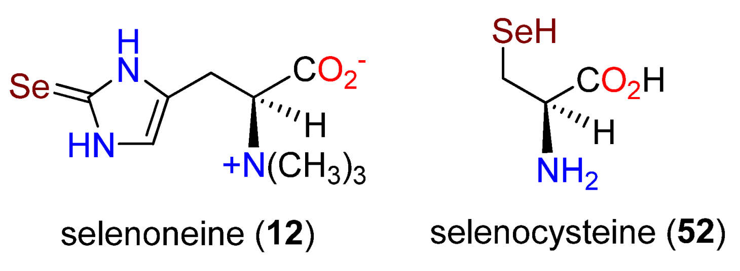
The search for an imidazole N-methyltransferase revealed 52 such enzymes in E. tasmaniensis, none of which was associated with the Ovo operon. However, a methyltransferase was one of the three domains in OvoA from T. cruzi [101] and was suggested to conduct the imidazole N-methylation [92]. When OvoA was cultured with 38 and SAM, the exclusive product was ovothiol A (11) through N-3 methylation [171].
Unicellular diatoms are microalgae found in the moist soils, waterways, and oceans and comprise a significant proportion of Earth’s biomass. They contribute to the ecosystem by producing much of the oxygen generated each year. OvoA homologs were identified in diatoms [101,173], and OvoA was indicated to be present in both centric and pennate diatoms [174]. From cultures of Skeletonema marinoi, a thiol fraction was identified that contained ovothiol B (40) [174], whose only previous isolation at the time was from the scallop Chlamys hastata [121].
The breadth of ovothiol distribution in nature is still being explored and continuously expanded based on searching for ovoA. A study of arthropods revealed a very surprising situation [175]. It was established that metazoans in the Porifera, Cnidaria, Echinodermata, and Hemicordata all carried the ovoA gene [130], whereas insects and fish do not carry the gene. A search relating to the Crustacea subphylum indicated at least eight insects (6 Hemiptera and 2 Diptera) had OvoA transcripts [175]. Marine arthropod studies indicated two clades for the OvoA sequences. One clade included twelve sequences from the copepods and three from the decapods, while the second clade had seven copepods, four decapods, and two amphipods. The expression of OvoA in generating an antioxidant was enhanced in the copepod Calanus finmarchicus when subjected to a toxic algal diet and when going through specific molting stages [175].
Bioinformatics analysis revealed the global distribution of OvoA and OvoB in aerobic Proteobacteria in numerous pelagic (open ocean) and other environments with high oxygen levels, such as surface and subsurface waters [176]. About 2% of the species were from deep-sea hydrothermal vents, and two species were known to be anaerobic [177,178]. This implies a possible alternative biosynthetic pathway for 11, as observed for ergothioneine (10) biosynthesis in Chlorobium limicola [179]. In analyzing the bacterial OvoA proteins, two other aspects were revealed. The first was that some of the OvoA-like proteins lacked a methyltransferase domain, and secondly, the N-terminal of OvoB may be fused with OvoA, as observed in hydrozoans [173]. About 36% of the bacteria with ovoA and ovoB genes are regarded as human or animal parasites, with the inherent antioxidant activity possibly to protect the host from other organisms or to protect themselves from the immune response of the host [175].

4. Selenoneine

4.1. Introduction

One of the most interesting and biologically significant human alkaloids to be discovered in the past few years is selenoneine (12), which at physiological pH exists in the selenol form [180,181]. Selenium (Se) is an essential element for human health [182,183] and is present in a wide variety of foods, including some beans, nuts, meats, and soy products [184,185,186,187,188]. The recommended dietary intake for an adult is 50–70 μg/day [181]. However, access to dietary selenium varies widely, from 7 μg/day to 5000 μg/day [189], depending on the presence of selenium in the local soil, local dietary characteristics, and accessibility. Selenium deficiency leads to bone disorders and stunted growth known as Kashin–Beck disease [183,190], while an excess of Se may lead to “garlic breath” [191] or, at higher intake levels, to alopecia and tooth darkening [192,193]. Some other sources of selenium in foods include derivatives of selenocysteine (52) and selenomethionine (53) in fruits and vegetables and the dimer selenocystine (54) in meats (Figure 18) [181]. These seleno amino acids can also be incorporated into proteins [194,195], providing alternative centers for redox activity [194,196].
Selenoneine was initially isolated and characterized spectroscopically as the dimer 55 from the blood and other tissues of the bluefin tuna, Thunnus orientalis; the monomer 12 was originally deemed too unstable to isolate [197,198]. Similar levels were found in mackerel, and 2- to 4-fold lower levels in tilapia blood, squid blood, chicken heart and liver, and porcine kidney. A reduction of 55 to the monomer was achieved with glutathione or dithiothreitol (DTT) [198]. Significantly stronger radical scavenging activity was demonstrated for 12 in the DPPH assay than 10 (1.9 μM vs. 1.7 mM, respectively) [197]. The alkaloid also reacted with methyl mercury and bound to heme proteins affording protection [198].
Clinical studies in 167 patients from four remote islands in Kagoshima Prefecture in Japan revealed high levels of 12 in their red blood cells, which were directly correlated with the frequency of fish consumption [199]. Another study examined 12 levels in red blood cells in the Inuit population from Nunavik in Northern Quebec, Canada, where high levels of 12 were also observed, together with the Se-methyl derivative, suggesting that 12 protects against the presence of MeHg in local fish [200,201]. In a more detailed study of 12 in fish muscles, the highest levels were observed in swordfish (2.8 nmol/g tissue), followed by bigeye, bluefin, and yellowfin tuna. Salmon, conger, saury, and sole had no 12 [202]. The Se:Hg molar ratio was also assessed, with swordfish having a ratio of 1 and sole a ratio of 217. An HPLC/MS-based system was developed to determine the concentrations of 12 and 10 in human red blood cells [203], and by using a pentabromobenzyl HPLC column, the monomers of 12 and 10 could be isolated from extracts of genetically modified yeast [204]. In 2014, a patent for the chiral total synthesis of 12 was described [181], and in 2019, an 11-step (2% overall yield) synthesis of 12 was presented, which allowed for biological activities to be identified, thereby distinguishing 10 and 12 [205]. Interestingly, and under physiological conditions, the reaction of the stable selenoneine diselenide occurred with several aromatic natural products, including resveratrol, vancomycin, and proansamitocin, to form derivatives through electrophilic aromatic substitution at positions ortho to phenolic groups [205]. This led to the suggestion that Se-derived adducts may be present at low levels in many natural matrices.
Selenoneine (12) has radical scavenging and strong antioxidant activity [181] in various models [206,207]. In addition, it ameliorates methylmercury accumulation in zebrafish in conjunction with the transporter OCTN1 [208], shows activity against the development of colorectal cancer in mice [209], inhibits tyrosinase in B16 melanoma cells and human melanocytes, probably by chelating copper at the active site of the enzyme [210], has ACE-inhibiting activity by binding to zinc [211], and crosses the blood–brain barrier only very slowly [212]. In order to account for the cytotoxicity in K562 human leukemic cells, it was proposed that 12 interacted with cysteine residues forming antioxidant, high molecular protein complexes [213]. Based on mammalian studies [198], 12 is regarded as probably non-toxic to humans. Further biological assessment of 12 appears to be limited by the absence of a commercial source [181].

4.2. Biosynthesis

Aspects of the biosynthesis of 12 were discussed previously [100,169,170]. The addition of 10 μM Na2SeO4 was used in an EMM2 medium, and each stress change (e.g., limiting access to nitrogen or glucose) induced significant 12 production due to an increase in the transcription of the egt1 gene. From the reaction of 10 and Na2SeO4, no 12 was produced, indicating a separation in the biosynthetic pathways and not an elemental replacement of Se for S in a pool of preformed 10 [100]. Overexpression of the egt1 gene in S. pombe cultures raised the level to 1606.3 μM in the mutant strain from 0.3 μM in the wild-type strain. In a Δegt1 strain with added 10, no 12 was formed. A double mutant of egt1+ and Δegt2 afforded a new intermediate, hercynylselenocysteine (56), and not the corresponding selenoxide, indicating a major difference in the function of Egt1 in the biosynthesis of 12. A comparison of the two pathways is summarized in Figure 19 [100].

5. Conclusions

The histidine-derived alkaloids ergothioneine (10), ovothiol A (11), and selenothioneine (12) are exceptionally widely distributed at low levels throughout numerous phyla, including humans. They exhibit a range of biological activities, although their specific biological functions in situ are not well characterized beyond as “anti-oxidants”. Biosynthetically, their simplicity belies varied biosynthetic pathways, with three distinct approaches established for the formation of 10 and likely two for 11. Much remains to be learned about these metabolites, particularly in terms of their dietary significance for humans.

Author Contributions

Conceptualization, G.A.C.; data curation, G.A.C.; writing-original draft preparation, G.A.C.; preparation of figures, S.N.S.L. and G.A.C.; writing-review and editing, S.N.S.L. and G.A.C. All authors have read and agreed to the published version of the manuscript.

Funding

This review received no external funding.

Conflicts of Interest

The authors declare no conflict of interest.

References

  1. Funayama, S.; Cordell, G.A. Alkaloids—A Treasury of Poisons and Medicines; Academic Press: New York, NY, USA, 2015; p. 157. [Google Scholar]
  2. Santos, A.P.; Moreno, P.R.H. Alkaloids derived from histidine: Imidazole (pilocarpine, pilosine). In Handbook of Natural Products; Ramawat, K.G., Mérillon, J.-M., Eds.; Springer: Berlin/Heidelberg, Germany, 2013; pp. 861–882. [Google Scholar]
  3. Rosenberg, H.; Paul, A.G. Dolichotheline, an imidazole alkaloid from Dolichothele sphaerica. Tetrahedron Lett. 1969, 10, 1039–1042. [Google Scholar] [CrossRef] [Green Version]
  4. Ferrigni, N.R.; Meyer, B.N.; McLaughlin, J.L. Cactus alkaloids. LVI. Carbon-13 and proton NMR of the imidazoles, Nα,Nα-dimethylhistamine and dolichotheline. Rev. Latinoamer. Quim. 1984, 14, 131–133. [Google Scholar]
  5. Rosenberg, H.; Paul, A.G. Biosynthesis of dolichotheline in Dolichothele sphaerica. Lloydia 1971, 34, 372–376. [Google Scholar]
  6. Andrade, P.; Willoughby, R.; Pomponi, S.A.; Kerr, R.G. Biosynthetic studies of the alkaloid, stevensine, in a cell culture of the marine sponge Teichaxinella morchella. Tetrahedron Lett. 1999, 40, 4775–4778. [Google Scholar] [CrossRef]
  7. Albizati, K.F.; Faulkner, D.J. Stevensine, a novel alkaloid of an unidentified marine sponge. J. Org. Chem. 1985, 50, 4163–4164. [Google Scholar] [CrossRef]
  8. Chilton, M.-D.; Drummond, M.H.; Merlo, D.J.; Sciaky, D.; Montoya, A.L.; Gordon, M.P.; Nester, E.W. Stable incorporation of plasmid DNA into higher plant cells: The molecular basis of crown gall tumorigenesis. Cell 1977, 11, 263–271. [Google Scholar] [CrossRef]
  9. Isogai, A.; Fukuchi, N.; Hayashi, M.; Kamada, H.; Harada, H.; Suzuki, A. Structure of a new opine, mikimopine, in hairy root induced by Agrobacterium rhizogenes. Agric. Biol. Chem. 1988, 52, 3235–3237. [Google Scholar] [CrossRef]
  10. Isogai, A.; Fukuchi, N.; Hayashi, M.; Kamada, H.; Harada, H.; Suzuki, A. Mikimopine, an opine in hairy roots of tobacco induced by Agrobacterium rhizogenes. Phytochemistry 1990, 29, 3131–3134. [Google Scholar] [CrossRef]
  11. Tanret, C. Sur une base nouvelle retirée du seigle ergote, l’ergothioneine. Compt. Rend. Acad. Sci. 1909, 149, 222–224. [Google Scholar]
  12. Barger, G.; Ewins, A.J. The constitution of ergothioneine: A betaine related to histidine. J. Chem. Soc. Trans. 1911, 99, 2336–2341. [Google Scholar] [CrossRef] [Green Version]
  13. Hunter, G.; Eagles, B.A. The isolation from blood of a hitherto unknown substance, and its bearing on present methods for the estimation of uric acid. J. Biol. Chem. 1925, 65, 623–641. [Google Scholar] [CrossRef]
  14. Hunter, G.; Eagles, B.A. Non-protein sulphur compounds of blood. I. Sympectothion. J. Biol. Chem. 1927, 72, 123–132. [Google Scholar] [CrossRef]
  15. Benedict, S.A.; Newton, E.B.; Behre, J.A. A new sulfur-containing compound (thiasine) in the blood. J. Biol. Chem. 1926, 67, 267–277. [Google Scholar] [CrossRef]
  16. Newton, E.B.; Benedict, S.R.; Dakin, H.D. The chemical constitution of thiasine. Science 1926, 64, 602. [Google Scholar] [CrossRef] [PubMed]
  17. Newton, E.B.; Benedict, S.R.; Dakin, H.D. On thiasine, its structure and identification with ergothioneine. J. Biol. Chem. 1927, 62, 367–373. [Google Scholar] [CrossRef]
  18. Hunter, G. A new test for ergothioneine upon which is based a method for its estimation in simple solution and blood-filtrates. Biochem. J. 1928, 22, 4–10. [Google Scholar] [CrossRef] [Green Version]
  19. Sotgia, S.; Mangoni, A.A.; Forteschi, M.; Murphy, R.B.; Elliot, D.; Sotgiu, E.; Pintus, G.; Carru, C.; Zinellu, A. Identification of the main intermediate precursor of L-ergothioneine biosynthesis in human biological specimens. Molecules 2016, 21, 1298. [Google Scholar] [CrossRef] [Green Version]
  20. Tokuhiro, S.; Yamada, R.; Chang, C.; Suzuki, A.; Kochi, Y.; Sawada, T.; Suzuki, M.; Nagasaki, M.; Ohtsuki, M.; Ono, M.; et al. An intronic SNP in a RUNX1 binding site of SLC22A4, encoding an organic cation transporter, is associated with rheumatoid arthritis. Nat. Genet. 2003, 35, 341–348. [Google Scholar] [CrossRef]
  21. Peltekova, V.D.; Wintle, R.F.; Rubin, L.A.; Amos, C.I.; Huang, Q.; Gu, X.; Newman, B.; Oene, M.V.; Cescon, D.; Greenberg, G.; et al. Functional variants of OCTN cation transporter genes are associated with Crohn disease. Nat. Genet. 2004, 36, 471–475. [Google Scholar] [CrossRef] [Green Version]
  22. Eagles, B.A.; Vars, H.M. The physiology of ergothioneine. J. Biol. Chem. 1928, 80, 615–622. [Google Scholar] [CrossRef]
  23. Ramirez-Martinez, A.; Wesolek, N.; Yadan, J.-C.; Moutet, M.; Roudot, A.-C. Intake assessment of L-ergothioneine in some European countries and in the United States. Hum. Ecol. Risk Assess. 2016, 22, 667–677. [Google Scholar] [CrossRef]
  24. Melville, D.B.; Eich, S. The occurrence of ergothioneine in plant material. J. Biol. Chem. 1956, 218, 647–651. [Google Scholar] [CrossRef]
  25. Ey, J.; Schömig, E.; Taubert, D. Dietary sources and antioxidant effects of ergothioneine. J. Agric. Food Chem. 2007, 55, 6466–6474. [Google Scholar] [CrossRef] [PubMed]
  26. Dubost, N.; Ou, B.; Beelman, R. Quantification of polyphenols and ergothioneine in cultivated mushrooms and correlation to total antioxidant capacity. Food Chem. 2007, 105, 727–735. [Google Scholar] [CrossRef]
  27. Kalaras, M.D.; Richie, J.P.; Calcagnotto, A.; Beelman, R.B. Mushrooms: A rich source of the antioxidants ergothioneine and glutathione. Food Chem. 2017, 233, 429–433. [Google Scholar] [CrossRef]
  28. Beelman, R.B.; Kalaras, M.D.; Phillips, A.T.; Richie, J.P. Is ergothioneine a ‘longevity vitamin’ limited in the American diet? J. Nutr. Sci. 2020, 9, e52. [Google Scholar] [CrossRef]
  29. Servillo, L.; D’Onofrio, N.; Balestrieri, M.L. Ergothioneine antioxidant function: From chemistry to cardiovascular therapeutic potential. J. Cardiovasc. Pharmacol. 2017, 69, 183–191. [Google Scholar] [CrossRef]
  30. Cumming, B.M.; Chinta, K.C.; Reddy, V.P.; Steyn, A.J.C. Role of ergothioneine in microbial physiology and pathogenesis. Antioxid. Redox Signal. 2018, 28, 431–444. [Google Scholar] [CrossRef] [Green Version]
  31. Halliwell, B.; Cheah, I.K.; Tang, R.M.Y. Ergothioneine—A diet-derived antioxidant with therapeutic potential. FEBS Letts. 2018, 592, 3357–3366. [Google Scholar] [CrossRef] [Green Version]
  32. Cheah, I.K.; Halliwell, B. Ergothioneine, recent developments. Redox Biol. 2021, 42, 101868. [Google Scholar] [CrossRef]
  33. Heath, H.; Lawson, A.; Rimington, C. Synthesis of ergothioneine. Nature 1950, 166, 106. [Google Scholar] [CrossRef] [PubMed]
  34. Heath, H.; Lawson, A.; Rimington, C. 2-Mercaptoglyoxalines. Part I. The synthesis of ergothioneine. J. Chem. Soc. 1951, 2215–2217. [Google Scholar] [CrossRef]
  35. Sunko, D.E.; Wolf, G. Synthesis of DL-2-mercaptohistidine-α-C14 and DL-ergothioneine-α-C14. J. Am. Chem. Soc. 1958, 80, 4405–4406. [Google Scholar] [CrossRef]
  36. Xu, J.; Yadan, J.C. Synthesis of L-(+)-ergothioneine. J. Org. Chem. 1995, 60, 6296–6301. [Google Scholar] [CrossRef]
  37. Khonde, P.L.; Jardine, A. Improved synthesis of the super antioxidant, ergothioneine, and its biosynthetic pathway intermediates. Org. Biomol. Chem. 2015, 13, 1415–1419. [Google Scholar] [CrossRef] [PubMed] [Green Version]
  38. Naowarojna, N.; Cheng, R.; Chen, L.; Quill, M.; Xu, M.; Zhao, C.; Liu, P. Mini-review: Ergothioneine and ovothiol biosyntheses, an unprecedented trans-sulfur strategy in natural product biosynthesis. Biochemistry 2018, 57, 3309–3325. [Google Scholar] [CrossRef] [Green Version]
  39. Borodina, I.; Kenny, L.C.; McCarthy, C.M.; Paramasivan, K.; Pretorius, E.; Roberts, T.J.; van der Hoek, S.A.; Kell, D.B. The biology of ergothioneine, an antioxidant nutraceutical. Nutr. Res. Rev. 2020, 33, 190–217. [Google Scholar] [CrossRef] [Green Version]
  40. Halliwell, B.; Cheah, I.K.; Drum, C.L. Ergothioneine, an adaptive antioxidant for the protection of injured tissues? A hypothesis. Biochem. Biophys. Res. Commun. 2016, 470, 245–250. [Google Scholar] [CrossRef]
  41. Lam-Sidun, D.; Peters, K.M.; Borradaile, N.M. Mushroom-derived medicine? Preclinical studies suggest potential benefits of ergothioneine for cardiometabolic health. J. Mol. Sci. 2021, 2, 32446. [Google Scholar] [CrossRef]
  42. Melville, D.B.; Horner, W.H.; Lubschez, R. Tissue ergothioneine. J. Biol. Chem. 1954, 206, 221–228. [Google Scholar] [CrossRef]
  43. Shires, T.K.; Brummel, M.C.; Pulido, J.S.; Stegink, L.D. Ergothioneine distribution in bovine and porcine ocular tissues. Comp. Biochem. Physiol. Part C Pharmacol. Toxicol. Endocrinol. 1997, 117, 117–120. [Google Scholar] [CrossRef]
  44. Leone, E.; Mann, T. Ergothioneine in the seminal vesicle secretion. Nature 1951, 168, 205–206. [Google Scholar] [CrossRef] [PubMed]
  45. Heath, H.; Rimington, C.; Mann, T. Further studies on seminal ergothioneine of the pig. Biochem. J. 1957, 65, 369–373. [Google Scholar] [CrossRef] [PubMed] [Green Version]
  46. Grundemann, D.; Harlfinger, S.; Golz, S.; Geerts, A.; Lazar, A.; Berkels, R.; Jung, N.; Rubbert, A.; Schomig, E. Discovery of the ergothioneine transporter. Proc. Natl. Acad. Sci. USA 2005, 102, 5256–5261. [Google Scholar] [CrossRef] [PubMed] [Green Version]
  47. Tschirka, J.; Kreisor, M.; Betz, J.; Gründemann, D. Substrate selectivity check of the ergothioneine transporter. Drug Metab. Disp. 2018, 46, 779–785. [Google Scholar] [CrossRef]
  48. Kato, Y.; Kubo, Y.; Iwata, D.; Kato, S.; Sudo, T.; Sugiura, T.; Kagaya, T.; Wakayama, T.; Hirayama, A.; Sugimoto, M.; et al. Gene knockout and metabolome analysis of carnitine/organic cation transporter OCTN1. Pharm. Res. 2010, 27, 832–840. [Google Scholar] [CrossRef] [Green Version]
  49. Paul, B.; Snyder, S. The unusual amino acid L-ergothioneine is a physiologic cytoprotectant. Cell Death Differ. 2010, 17, 1134. [Google Scholar] [CrossRef] [Green Version]
  50. Cheah, I.K.; Halliwell, B. Ergothioneine; antioxidant potential, physiological function and role in disease. Biochim. Biophys. Acta Mol. Basis Dis. 2012, 1822, 784–793. [Google Scholar] [CrossRef] [Green Version]
  51. Aruoma, O.I.; Whiteman, M.; England, T.G.; Halliwell, B. Antioxidant action of ergothioneine: Assessment of its ability to scavenge peroxynitrite. Biochem. Biophys. Res. Commun. 1997, 231, 389–391. [Google Scholar] [CrossRef]
  52. Aruoma, O.I.; Spencer, J.P.E.; Mahmood, N. Protection against oxidative damage and cell death by the natural antioxidant ergothioneine. Food Chem. Toxicol. 1999, 37, 1043–1053. [Google Scholar] [CrossRef]
  53. Kaneko, I.; Takeuchi, Y.; Yamaoka, Y.; Tanaka, Y.; Fukuda, T.; Fukumori, Y.; Mayumi, T.; Hama, T. Quantitative determination of ergothioneine in plasma and tissues by TLC densitometry. Chem. Pharm. Bull. 1980, 28, 3093–3097. [Google Scholar] [CrossRef] [PubMed] [Green Version]
  54. Briggs, I. Ergothioneine in the central nervous system. J. Neurochem. 1972, 19, 27–35. [Google Scholar] [CrossRef] [PubMed]
  55. Crossland, J.; Mitchell, J.; Woodruff, G.N. The presence of ergothioneine in the central nervous system and its probable identity with the cerebellar factor. J. Physiol. 1966, 182, 427–438. [Google Scholar] [CrossRef] [PubMed] [Green Version]
  56. Moncaster, J.A.; Walsh, D.T.; Gentleman, S.M.; Jen, L.-S.; Aruoma, O.I. Ergothioneine treatment protects neurons against N-methyl-D-aspartate excitotoxicity in an in vivo rat retinal model. Neurosci. Lett. 2002, 328, 55–59. [Google Scholar] [CrossRef]
  57. Cheah, I.K.; Feng, L.; Tang, R.M.Y.; Lim, K.H.C.; Halliwell, B. Ergothioneine levels in an elderly population decrease with age and incidence of cognitive decline; a risk factor for neurodegeneration? Biochem. Biophys. Res. Commun. 2016, 478, 162–167. [Google Scholar] [CrossRef]
  58. Libby, P.; Ridker, P.M.; Hansson, G.K. Progress and challenges in translating the biology of atherosclerosis. Nature 2011, 473, 317–325. [Google Scholar] [CrossRef]
  59. Smith, E.; Ottosson, F.; Hellstrand, S.; Ericson, U.; Orho-Melander, M.; Fernandez, C.; Melander, O. Ergothioneine is associated with reduced mortality and decreased risk of cardiovascular disease. Heart 2020, 106, 691–697. [Google Scholar] [CrossRef] [Green Version]
  60. Hunter, G. On ergothioneine in blood and diazo-reacting substances in maize. Biochem. J. 1951, 48, 265–270. [Google Scholar] [CrossRef] [Green Version]
  61. Melville, D.B.; Homer, W.H. Blood ergothioneine in the germ-free chicken. J. Biol. Chem. 1953, 202, 187–191. [Google Scholar] [CrossRef]
  62. Heath, H. The metabolism of 35S-labelled 2-thiolhistidine and ergothioneine in the rat. Biochem. J. 1953, 54, 689–694. [Google Scholar] [CrossRef] [Green Version]
  63. Melville, D.B.; Horner, W.H.; Otken, C.C.; Ludwig, M.L. Studies on the origin of ergothioneine in animals. J. Biol. Chem. 1955, 213, 61–68. [Google Scholar] [CrossRef]
  64. Baldridge, R.C.; Lewis, H.B. Diet and the ergothioneine content of blood. J. Biol. Chem. 1953, 202, 169–176. [Google Scholar] [CrossRef]
  65. Baldridge, R.C. Blood ergothioneine and dietary oats. J. Nutr. 1955, 56, 107–113. [Google Scholar] [CrossRef] [PubMed]
  66. Melville, D.B.; Genghof, D.S.; Inamine, E.; Kovalenko, V. Ergothioneine in microorganisms. J. Biol. Chem. 1956, 223, 9–17. [Google Scholar] [CrossRef]
  67. Heath, H.; Wildy, J. The biosynthesis of ergothioneine and histidine by Claviceps purpurea. I. The incorporation of [2-14C]-acetate. Biochem. J. 1956, 64, 612–620. [Google Scholar] [CrossRef] [Green Version]
  68. Wildy, J.; Heath, H. Biosynthesis of ergothioneine by Claviceps purpurea. II. Incorporation of [35S]methionine and the non-utilization of [2(ring)-14C]histamine. Biochem. J. 1957, 65, 220–222. [Google Scholar] [CrossRef]
  69. Heath, H.; Wildy, J. Biosynthesis of ergothioneine. Nature 1957, 179, 196–197. [Google Scholar] [CrossRef]
  70. Heath, H.; Wildy, J. Biosynthesis of ergothioneine by Claviceps purpurea. III. The incorporation of labelled histidine. Biochem. J. 1958, 68, 407–410. [Google Scholar] [CrossRef] [Green Version]
  71. Melville, D.B.; Eich, S.; Ludwig, M.L. The biosynthesis of ergothioneine. J. Biol. Chem. 1957, 224, 871–877. [Google Scholar] [CrossRef]
  72. Melville, D.B.; Ludwig, M.L.; Inamine, E.; Rachele, J.R. Transmethylation in the biosynthesis of ergothioneine. J. Biol. Chem. 1959, 234, 1195–1198. [Google Scholar] [CrossRef]
  73. Askari, A.; Melville, D.B. The reaction sequence in ergothioneine biosynthesis: Hercynine as an intermediate. J. Biol. Chem. 1962, 237, 1615–1618. [Google Scholar] [CrossRef]
  74. Kutscher, F. Die basischen Extraktstoffe des Champignons (Agaricus campestris). Z. Für Unters. Der Nahr.-Und Genußmittel Sowie Der Gebrauchsgegenstände 1911, 21, 535–540. [Google Scholar]
  75. Genghof, D.S. The production of ergothioneine by Mycobacterium tuberculosis. Bacteriol. Proc. 1960, 190. [Google Scholar]
  76. Genghof, D.S.; Van Damme, O. Biosynthesis of ergothioneine and hercynine by mycobacteria. J. Bacteriol. 1964, 87, 852–862. [Google Scholar] [CrossRef] [PubMed] [Green Version]
  77. Genghof, D.S. Biosynthesis of ergothioneine and hercynine by fungi and Actinomycetales. J. Bacteriol. 1970, 103, 475–478. [Google Scholar] [CrossRef] [PubMed] [Green Version]
  78. Tan, C.H.; Audley, B.G. Ergothioneine and hercynine in Hevea brasiliensis latex. Phytochemistry 1968, 7, 109–118. [Google Scholar] [CrossRef]
  79. Park, E.-J.; Lee, W.Y.; Kim, S.T.; Ahn, J.K.; Bae, E.K. Ergothioneine accumulation in a medicinal plant Gastrodia elata. J. Med. Plants Res. 2010, 4, 1141–1147. [Google Scholar]
  80. Pfeiffer, C.; Bauer, T.; Surek, B.; Schömig, E.; Gründemann, D. Cyanobacteria produce high levels of ergothioneine. Food Chem. 2011, 129, 1766–1769. [Google Scholar] [CrossRef]
  81. Ferrazzano, G.F.; Papa, C.; Pollio, A.; Ingenito, A.; Sangianantoni, G.; Cantile, T. Cyanobacteria and microalgae as sources of functional foods to improve human general and oral health. Molecules 2020, 25, 5164. [Google Scholar] [CrossRef]
  82. Genghof, D.S.; Van Damme, O. Biosynthesis of ergothioneine from endogenous hercynine in Mycobacterium smegmatis. J. Bacteriol. 1968, 95, 340–344. [Google Scholar] [CrossRef] [Green Version]
  83. Reinhold, V.N.; Ishikawa, Y.; Melville, D.B. Conversion of histidine to hercynine by Neurospora crassa. J. Bacteriol. 1970, 101, 881–884. [Google Scholar] [CrossRef] [PubMed] [Green Version]
  84. Ishikawa, Y.; Melville, D.B. The enzymatic α-N-methylation of histidine. J. Biol. Chem. 1970, 245, 5967–5973. [Google Scholar] [CrossRef]
  85. Ishikawa, Y.; Israel, S.E.; Melville, D.B. Participation of an intermediate sulfoxide in the enzymatic thiolation of the imidazole ring of hercynine to form ergothioneine. J. Biol. Chem. 1974, 249, 4420–4427. [Google Scholar] [CrossRef]
  86. Seebeck, F.P. In Vitro reconstitution of mycobacterial ergothioneine biosynthesis. J. Am. Chem. Soc. 2010, 132, 6632–6633. [Google Scholar] [CrossRef]
  87. Harth, G.; Masleša-Galić, S.; Tullius, M.V.; Horwitz, M.A. All four Mycobacterium tuberculosis glnA genes encode glutamine synthetase activities but only GlnA1 is abundantly expressed and essential for bacterial homeostasis. Mol. Microbiol. 2005, 58, 1157–1172. [Google Scholar] [CrossRef] [PubMed]
  88. Brannigan, J.A.; Dodson, G.; Duggleby, H.J.; Moody, P.C.; Smith, J.L.; Tomchick, D.R.; Murzin, A.G. A protein catalytic framework with an N-terminal nucleophile is capable of self-activation. Nature 1995, 378, 416–419. [Google Scholar] [CrossRef] [PubMed]
  89. Vit, A.; Mashabela, G.; Blankenfeldt, W.; Seebeck, F.P. Structure of the ergothioneine-biosynthesis amidohydrolase EgtC. ChemBioChem 2015, 16, 1490–1496. [Google Scholar] [CrossRef] [Green Version]
  90. Goncharenko, K.V.; Vit, A.; Blankenfeldt, W.; Seebeck, F.P. Structure of the sulfoxide synthase EgtB from the ergothioneine biosynthetic pathway. Angew. Chem. Int Ed. Engl. 2015, 54, 2821–2824. [Google Scholar] [CrossRef] [Green Version]
  91. Vit, A.; Misson, L.; Blankenfeldt, W.; Seebeck, F.P. Ergothioneine biosynthetic methyltransferase EgtD reveals the structural basis of aromatic amino acid betaine biosynthesis. ChemBioChem 2015, 16, 119–125. [Google Scholar] [CrossRef]
  92. Liao, C.; Seebeck, F.P. Convergent evolution of ergothioneine biosynthesis in cyanobacteria. ChemBioChem 2017, 18, 2115–2118. [Google Scholar] [CrossRef]
  93. Liscombe, D.K.; Louie, G.V.; Noel, J.P. Architectures, mechanisms and molecular evolution of natural product methyltransferases. Nat. Prod. Rep. 2012, 29, 1238–1250. [Google Scholar] [CrossRef] [PubMed]
  94. Misson, L.; Burn, R.; Vit, A.; Hildesheim, J.; Beliaeva, M.A.; Blankenfeldt, W.; Seebeck, F.P. Inhibition and regulation of the ergothioneine biosynthetic methyltransferase EgtD. ACS Chem. Biol. 2018, 13, 1333–1342. [Google Scholar] [CrossRef] [PubMed]
  95. Richard-Greenblatt, M.; Bach, H.; Adamson, J.; Peña-Diaz, S.; Li, W.; Steyn, A.J.C.; Av-Gay, Y. Regulation of ergothioneine biosynthesis and its effect on Mycobacterium tuberculosis growth and infectivity. J. Biol. Chem. 2015, 290, 23064–23076. [Google Scholar] [CrossRef] [PubMed] [Green Version]
  96. Gallagher, L.; Owens, R.A.; Dolan, S.K.; O’Keeffe, G.; Schrettl, M.; Kavanagh, K.; Jones, G.W.; Doyle, S. The Aspergillus fumigatus protein GliK protects against oxidative stress and is essential for gliotoxin biosynthesis. Eukaryot. Cell 2012, 11, 1226–1238. [Google Scholar] [CrossRef] [PubMed] [Green Version]
  97. Ta, P.; Buchmeier, N.; Newton, G.L.; Rawat, M.; Fahey, R.C. Organic hydroperoxide resistance protein and ergothioneine compensate for loss of mycothiol in Mycobacterium smegmatis mutants. J. Bacteriol. 2011, 193, 1981–1990. [Google Scholar] [CrossRef] [Green Version]
  98. Bello, M.H.; Barrera-Perez, V.; Morin, D.; Epstein, L. The Neurospora crassa mutant NcΔEgt-1 identifies an ergothioneine biosynthetic gene and demonstrates that ergothioneine enhances conidial survival and protects against peroxide toxicity during conidial germination. Fungal Genet. Biol. 2012, 49, 160–172. [Google Scholar] [CrossRef]
  99. Sheridan, K.J.; Lechner, B.E.; O’Keeffe, G.; Keller, M.A.; Werner, E.R.; Lindner, H.; Jones, G.W.; Haas, H.; Doyle, S. Ergothioneine biosynthesis and functionality in the opportunistic fungal pathogen, Aspergillus fumigatus. Sci. Rep. 2016, 6, 35306. [Google Scholar] [CrossRef] [Green Version]
  100. Pluskal, T.; Ueno, M.; Yanagida, M. Genetic and metabolomic dissection of the ergothioneine and selenoneine biosynthetic pathway in the fission yeast, S. pombe, and construction of an overproduction system. PLoS ONE 2014, 9, e97774. [Google Scholar] [CrossRef] [Green Version]
  101. Braunshausen, A.; Seebeck, F.P. Identification and characterization of the first ovothiol biosynthetic enzyme. J. Am. Chem. Soc. 2011, 133, 1757–1759. [Google Scholar] [CrossRef]
  102. Mashabela, G.T.M.; Seebeck, F.P. Substrate specificity of an oxygen dependent sulfoxide synthase in ovothiol biosynthesis. Chem. Commun. 2013, 49, 7714–7716. [Google Scholar] [CrossRef] [PubMed] [Green Version]
  103. Wei, W.J.; Siegbahn, P.E.; Liao, R.Z. Theoretical study of the mechanism of the non-heme iron enzyme EgtB. Inorg. Chem. 2017, 56, 3589–3599. [Google Scholar] [CrossRef] [PubMed]
  104. Faponle, A.S.; Seebeck, F.P.; de Visser, S.P. Sulfoxide synthase versus cysteine dioxygenase reactivity in a nonheme iron enzyme. J. Am. Chem. Soc. 2017, 139, 9259–9270. [Google Scholar] [CrossRef] [PubMed] [Green Version]
  105. Tian, G.; Su, H.; Liu, Y. Mechanism of sulfoxidation and C–S bond formation involved in the biosynthesis of ergothioneine catalyzed by ergothioneine synthase (EgtB). ACS Catal. 2018, 8, 5875–5889. [Google Scholar] [CrossRef]
  106. Naowarojna, N.; Irani, S.; Hu, W.; Cheng, R.; Zhang, L.; Li, X.; Chen, J.; Zhang, Y.J.; Liu, P. Crystal structure of the ergothioneine sulfoxide synthase from Candidatus Chloracidobacterium thermophilum and structure-guided engineering to modulate its substrate selectivity. ACS Catal. 2019, 9, 6955–6961. [Google Scholar] [CrossRef] [PubMed]
  107. Song, H.; Leninger, M.; Lee, N.; Liu, P. Regioselectivity of the oxidative C–S bond formation in ergothioneine and ovothiol biosyntheses. Org. Lett. 2013, 15, 4854–4857. [Google Scholar] [CrossRef] [PubMed] [Green Version]
  108. Song, H.; Hu, W.; Naowarojna, N.; Her, A.S.; Wang, S.; Desai, R.; Qin, L.; Chen, X.; Liu, P. Mechanistic studies of a novel C-S lyase in ergothioneine biosynthesis: The involvement of a sulfenic acid intermediate. Sci. Rep. 2015, 5, 11870. [Google Scholar] [CrossRef] [PubMed] [Green Version]
  109. Irani, S.; Naowarojna, N.; Tang, Y.; Kathuria, K.R.; Wang, S.; Dhembi, A.; Lee, N.; Yan, W.; Lyu, H.; Costello, C.E.; et al. Snapshots of CS cleavage in Egt2 reveals substrate specificity and reaction mechanism. Cell Chem. Biol. 2018, 25, 519–529. [Google Scholar] [CrossRef] [Green Version]
  110. Jones, G.W.; Doyle, S.; Fitzpatrick, D.A. The evolutionary history of the genes involved in the biosynthesis of the antioxidant ergothioneine. Gene 2014, 549, 161–170. [Google Scholar] [CrossRef]
  111. Kuettner, E.B.; Hilgenfeld, R.; Weiss, M.S. The active principle of garlic at atomic resolution. J. Biol. Chem. 2002, 277, 46402–46407. [Google Scholar] [CrossRef] [Green Version]
  112. Stampfli, A.R.; Blankenfeldt, W.; Seebeck, F.P. Structural basis of ergothioneine biosynthesis. Curr. Opin. Struct. Biol. 2020, 65, 1–8. [Google Scholar] [CrossRef]
  113. Osawa, T.; Kamide, T.; Satoh, Y.; Kawano, Y.; Ohtsu, I.; Dairi, T. Heterologous and high production of ergothioneine in Escherichia coli. J. Agric. Food Chem. 2018, 66, 1191–1196. [Google Scholar] [CrossRef] [PubMed] [Green Version]
  114. Tanaka, N.; Kawano, Y.; Satoh, Y.; Dairi, T.; Ohtsu, I. Gramscale fermentative production of ergothioneine driven by overproduction of cysteine in Escherichia coli. Sci. Rep. 2019, 9, 1895. [Google Scholar] [CrossRef] [PubMed]
  115. Takusagawa, S.; Satoh, Y.; Ohtsu, I.; Dairi, T. Ergothioneine production with Aspergillus oryzae. Biosci. Biotechnol. Biochem. 2019, 83, 181–184. [Google Scholar] [CrossRef] [PubMed] [Green Version]
  116. van der Hoek, S.A.; Darbani, B.; Zugaj, K.E.; Prabhala, B.K.; Biron, M.B.; Randelovic, M.; Medina, J.B.; Kell, D.B.; Borodina, I. Engineering the yeast Saccharomyces cerevisiae for the production of L-(+)-ergothioneine. Front. Bioeng. Biotechnol. 2019, 7, 262. [Google Scholar] [CrossRef] [PubMed] [Green Version]
  117. Yu, Y.-Y.; Pan, H.-Y.; Guo, L.-Q.; Lin, J.-F.; Liao, H.-L.; Li, H.-Y. Successful biosynthesis of natural antioxidant ergothioneine in Saccharomyces cerevisiae required only two genes from Grifola frondosa. Microb. Cell Fact. 2020, 19, 164. [Google Scholar] [CrossRef]
  118. Nardi, G.; Palumbo, A.; Prota, G. The role of the white bodies in the biosynthesis of adenochrome. Comp. Biochem. Phvsiol. 1982, 71B, 297–300. [Google Scholar] [CrossRef]
  119. Palumbo, A.; d’Ischia, M.; Misuraca, G.; Prota, G. Isolation and structure of a new sulphur-containing aminoacid from sea urchin eggs. Tetrahedron Lett. 1982, 23, 3207–3208. [Google Scholar] [CrossRef]
  120. Turner, E.; Klevit, R.; Hopkins, P.B.; Shapiro, B.M. Ovothiol: A novel thiohistidine compound from sea urchin eggs that confers NAD(P)H-O2 oxidoreductase activity on ovoperoxidase. J. Biol. Chem. 1986, 261, 13056–13063. [Google Scholar] [CrossRef]
  121. Turner, E.; Klevit, R.; Hager, L.J.; Shapiro, B.M. Ovothiols, a family of redox-active mercaptohistidine compounds from marine invertebrate eggs. Biochemistry 1987, 26, 4028–4036. [Google Scholar] [CrossRef]
  122. Steenkamp, D.J.; Spies, H.S. Identification of a major low-molecular-mass thiol of the trypanosomatid Crithidia fasciculata as ovothiol A. Facile isolation and structural analysis of the bimane derivative. Eur. J. Biochem. 1994, 223, 43–50. [Google Scholar] [CrossRef]
  123. Spies, H.S.; Steenkamp, D.J. Thiols of intracellular pathogens. Identification of ovothiol A in Leishmania donovani and structural analysis of a novel thiol from Mycobacterium bovis. Eur. J. Biochem. 1994, 224, 203–213. [Google Scholar] [CrossRef] [PubMed]
  124. Holler, T.P.; Salpenstein, A.; Turner, E.; Klevit, R.E.; Shapiro, B.M.; Hopkins, P.B. Synthesis and structure reassignment of mercaptohistidines of marine origin. Syntheses of L-ovothiols A and C. J. Org. Chem. 1987, 52, 4420–4421. [Google Scholar] [CrossRef]
  125. Holler, T.P.; Ruan, F.; Salpenstein, A.; Hopkins, P.B. Total synthesis of marine mercaptohistidines: Ovothiols A, B, and C. J. Org. Chem. 1989, 54, 4570–4575. [Google Scholar] [CrossRef]
  126. Mirzahosseini, A.; Hosztafi, S.; Tóth, G.; Noszál, B. A cost-effective synthesis of enantiopure ovothiol A from L-histidine, its natural precursor. Arkivoc 2014, vi, 1–9. [Google Scholar] [CrossRef] [Green Version]
  127. Pathirana, C.; Andersen, R.J. Imbricatine, an unusual benzyltetrahydro-isoquinoline alkaloid isolated from the starfish Dermasterias imbricata. J. Am. Chem. Soc. 1986, 108, 8288–8289. [Google Scholar] [CrossRef]
  128. Burgoyne, D.L.; Miao, S.; Pathirana, C.; Andersen, R.J.; Ayer, W.A.; Singer, P.P.; Kokke, W.C.M.C.; Ross, D.M. The structure and partial synthesis of imbricatine, a benzyltetrahydroisoquinoline alkaloid from the starfish Dermasterias imbricata. Can. J. Chem. 1991, 69, 20–27. [Google Scholar] [CrossRef]
  129. Shapiro, B.M.; Hopkins, P.B. Ovothiols: Biological and chemical perspectives. Adv. Enzymol. Relat. Areas Mol. Biol. 1991, 64, 291–316. [Google Scholar]
  130. Castellano, I.; Seebeck, F.P. On ovothiol biosynthesis and biological roles: From life in the ocean to therapeutic potential. Nat. Prod. Rep. 2018, 35, 1241–1250. [Google Scholar] [CrossRef] [Green Version]
  131. Turner, E.; Hager, L.J.; Shapiro, B.M. Ovothiol replaces glutathione peroxidase as a hydrogen peroxide scavenger in sea urchin eggs. Science 1988, 242, 939–941. [Google Scholar] [CrossRef]
  132. Holler, T.P.; Hopkins, P.B. Ovothiols as free-radical scavengers and the mechanism of ovothiol-promoted NAD(P)H-O2 oxidoreductase activity. Biochemistry 1990, 29, 1953–1961. [Google Scholar] [CrossRef]
  133. Marjanovic, B.; Simic, M.G.; Jovanovic, S.V. Heterocyclic thiols as antioxidants: Why ovothiol C is a better antioxidant than ergothioneine. Free Radic. Biol. Med. 1995, 18, 679–685. [Google Scholar] [CrossRef]
  134. Castellano, I.; Migliaccio, O.; D’Aniello, S.; Merlino, A.; Napolitano, A.; Palumbo, A. Shedding light on ovothiol biosynthesis in marine metazoans. Sci. Rep. 2016, 6, 21506. [Google Scholar] [CrossRef] [PubMed] [Green Version]
  135. Osik, N.A.; Zelentsova, E.A.; Tsentalovich, Y.P. Kinetic studies of antioxidant properties of ovothiol A. Antioxidants 2021, 10, 1470. [Google Scholar] [CrossRef] [PubMed]
  136. Diaz de Cerio, O.; Reina, L.; Squatrito, V.; Etxebarria, N.; Gonzalez-Gaya, B.; Cancio, I. Gametogenesis-related fluctuations in ovothiol levels in the mantle of mussels from different estuaries: Fighting oxidative stress for spawning in polluted waters. Biomolecules 2020, 10, 373. [Google Scholar] [CrossRef] [PubMed] [Green Version]
  137. Krauth-Siegel, R.L.; Leroux, A.E. Low-molecular-mass antioxidants in parasites. Antioxid. Redox Signal. 2012, 17, 583–607. [Google Scholar] [CrossRef] [PubMed]
  138. Ariyanayagam, M.R.; Fairlamb, A.H. Ovothiol and trypanothione as antioxidants in trypanosomatids. Mol. Biochem. Parasitol. 2001, 115, 189–198. [Google Scholar] [CrossRef]
  139. Gonçalves, C.; Costa, P.M. Histochemical detection of free thiols in glandular cells and tissues of different marine Polychaeta. Histochem. Cell Biol. 2020, 154, 315–325. [Google Scholar] [CrossRef]
  140. Yanshole, V.V.; Yanshole, L.V.; Zelentsova, E.A.; Tsentalovich, Y.P. Ovothiol A is the main antioxidant in fish lens. Metabolites 2019, 9, 95. [Google Scholar] [CrossRef] [Green Version]
  141. Milito, A.; Orefice, I.; Smerilli, A.; Castellano, I.; Napolitano, A.; Brunet, C.; Palumbo, A. Insights into the light response of Skeletonema marinoi: Involvement of ovothiol. Mar. Drugs 2020, 18, 477. [Google Scholar] [CrossRef]
  142. O’Neill, E.C.; Trick, M.; Hill, L.; Rejzek, M.; Dusi, R.G.; Hamilton, C.J.; Zimba, P.V.; Henrissat, B.; Field, R.A. The transcriptome of Euglena gracilis reveals unexpected metabolic capabilities for carbohydrate and natural product biochemistry. Mol. BioSyst. 2015, 11, 2808–2820. [Google Scholar] [CrossRef] [Green Version]
  143. Torres, J.P.; Lin, Z.; Watkins, M.; Salcedo, P.F.; Baskin, R.P.; Elhabian, S.; Safavi-Hemami, H.; Taylor, D.; Tun, J.; Concepcion, G.P. Small-molecule mimicry hunting strategy in the imperial cone snail, Conus imperialis. Sci. Adv. 2021, 7, eabf2704. [Google Scholar] [CrossRef] [PubMed]
  144. Röhl, I.; Schneider, B.; Schmidt, B.; Zeeck, E. L-Ovothiol A: The egg release pheromone of the marine polychaete Platynereis dumerilii: Annelida: Polychaeta. Z. Nat. C 1999, 54, 1145–1174. [Google Scholar] [CrossRef]
  145. Castellano, I.; Di Tomo, P.; Di Pietro, N.; Mandatori, D.; Pipino, C.; Formoso, G.; Napolitano, A.; Palumbo, A. Anti-inflammatory activity of marine ovothiol A in an in vitro model of endothelial dysfunction induced by hyperglycemia. Oxid. Med. Cell. Longev. 2018, 2018, 2087373. [Google Scholar] [CrossRef] [PubMed] [Green Version]
  146. Brancaccio, M.; D’Argenio, G.; Lembo, V.; Palumbo, A.; Castellano, I. Antifibrotic effect of marine ovothiol in an in vivo model of liver fibrosis. Oxid. Med. Cell. Longev. 2018, 2018, 5045734. [Google Scholar] [CrossRef] [PubMed] [Green Version]
  147. Milito, A.; Brancaccio, M.; D’Argenio, G.; Castellano, I. Natural sulfur-containing compounds: An alternative therapeutic strategy against liver fibrosis. Cells 2019, 8, 1356. [Google Scholar] [CrossRef] [Green Version]
  148. Russo, G.L.; Russo, M.; Castellano, I.; Napolitano, A.; Palumbo, A. Ovothiol isolated from sea urchin oocytes induces autophagy in the Hep-G2 cell line. Mar. Drugs 2014, 12, 4069–4085. [Google Scholar] [CrossRef] [Green Version]
  149. Brancaccio, M.; Russo, M.; Masullo, M.; Palumbo, A.; Russo, G.L.; Castellano, I. Sulfur-containing histidine compounds inhibit γ-glutamyl transpeptidase activity in human cancer cells. J. Biol. Chem. 2019, 294, 14603–14614. [Google Scholar] [CrossRef]
  150. Milito, A.; Brancaccio, M.; Lisurek, M.; Masullo, M.; Palumbo, A.; Castellano, I. Probing the interactions of sulfur-containing histidine compounds with human gamma-glutamyl transpeptidase. Mar. Drugs 2019, 17, 650. [Google Scholar] [CrossRef] [Green Version]
  151. Steenkamp, D.J.; Weldrick, D.; Spies, H.S. Studies on the biosynthesis of ovothiol A. Identification of 4-mercaptohistidine as an intermediate. Eur. J. Biochem. 1996, 242, 557–566. [Google Scholar] [CrossRef]
  152. Vogt, R.N.; Spies, H.S.; Steenkamp, D.S. The biosynthesis of ovothiol A (N-methyl-4-mercaptohistidine). Identification of S-(4′-L-histidyl)-L-cysteine sulfoxide as an intermediate and the products of the sulfoxide lyase reaction. Eur. J. Biochem. 2001, 268, 5229–5241. [Google Scholar] [CrossRef]
  153. Hoyle, C.E.; Bowman, C.N. Thiol-ene click chemistry. Angew. Chem. Int. Ed. 2010, 49, 1540–1573. [Google Scholar] [CrossRef] [PubMed]
  154. Song, H.; Her, A.S.; Raso, F.; Zhen, Z.; Huo, Y.; Liu, P. Cysteine oxidation reactions catalyzed by a mononuclear non-heme iron enzyme (OvoA) in ovothiol biosynthesis. Org. Lett. 2014, 16, 2122–2125. [Google Scholar] [CrossRef] [PubMed]
  155. Chen, L.; Naowarojna, N.; Chen, B.; Xu, M.; Quill, M.; Wang, J.; Deng, Z.; Zhao, C.; Liu, P. Mechanistic studies of a nonheme iron enzyme OvoA in ovothiol biosynthesis using a tyrosine analogue, 2-amino-3-(4-hydroxy-3-(methoxyl)phenyl) propanoic acid (MeOTyr). ACS Catal. 2018, 9, 253–258. [Google Scholar] [CrossRef]
  156. Chen, L.; Naowarojna, N.; Song, H.; Wang, S.; Wang, J.; Deng, Z.; Zhao, C.; Liu, P. Use of a tyrosine analogue to modulate the two activities of a nonheme iron enzyme OvoA in ovothiol biosynthesis, cysteine oxidation versus oxidative C–S bond formation. J. Am. Chem. Soc. 2018, 140, 4604–4612. [Google Scholar] [CrossRef] [Green Version]
  157. Hu, W.; Song, H.; Her, A.S.; Bak, D.W.; Naowarojna, N.; Elliott, S.J.; Qin, L.; Chen, X.; Liu, P. Bioinformatic and biochemical characterizations of C-S bond formation and cleavage enzymes in the fungus Neurospora crassa ergothioneine biosynthetic pathway. Org. Lett. 2014, 16, 5382–5385. [Google Scholar] [CrossRef] [Green Version]
  158. Atkinson, H.J.; Morris, J.H.; Ferrin, T.E.; Babbitt, P.C. Using sequence similarity networks for visualization of relationships across diverse protein superfamilies. PLoS ONE 2009, 4, e4345. [Google Scholar] [CrossRef]
  159. Galagan, J.E.; Calvo, S.E.; Borkovich, K.A.; Selker, E.U.; Read, N.D.; Jaffe, D.; FitzHugh, W.; Ma, L.-J.; Smirnov, S.; Purcell, S.; et al. The genome sequence of the filamentous fungus Neurospora crassa. Nature 2003, 422, 859–868. [Google Scholar] [CrossRef]
  160. Burn, R.; Misson, L.; Meury, M.; Seebeck, F.P. Anaerobic origin of ergothioneine. Angew. Chem. Int. Ed. 2017, 56, 12508–12511. [Google Scholar] [CrossRef]
  161. Ruszczycky, M.W.; Liu, H.W. The surprising history of an antioxidant. Nature 2017, 551, 37–38. [Google Scholar] [CrossRef]
  162. Cheng, R.; Wu, L.; Lai, R.; Peng, C.; Naowarojna, N.; Hu, W.; Li, X.; Whelan, S.A.; Lee, N.; Lopez, J.; et al. Single-step replacement of an unreactive C–H bond by a C–S bond using polysulfide as the direct sulfur source in the anaerobic ergothioneine biosynthesis. ACS Catal. 2020, 10, 8981–8994. [Google Scholar] [CrossRef]
  163. Muramatsu, H.; Matsuo, H.; Okada, N.; Ueda, M.; Yamamoto, H.; Kato, S.-I.; Nagata, S. Characterization of ergothionase from Burkholderia sp. HME13 and its application to enzymatic quantification of ergothioneine. Appl. Microbiol. Biotechnol. 2013, 97, 5389–5400. [Google Scholar] [CrossRef] [PubMed]
  164. Black, K.A.; Dos Santos, P.C. Shared-intermediates in the biosynthesis of thio-cofactors: Mechanism and functions of cysteine desulfurases and sulfur acceptors. Biochim. Biophys. Acta Mol. Cell Res. 2015, 1853, 1470–1480. [Google Scholar] [CrossRef] [PubMed] [Green Version]
  165. Zheng, L.M.; White, R.H.; Cash, V.L.; Jack, R.F.; Dean, D.R. Cysteine desulfurase activity indicates a role for NIFS in metallocluster biosynthesis. Proc. Natl. Acad. Sci. USA 1993, 90, 2754–2758. [Google Scholar] [CrossRef] [PubMed] [Green Version]
  166. Meyer, D.; Neumann, P.; Ficner, R.; Tittmann, K. Observation of a stable carbene at the active site of a thiamin enzyme. Nat. Chem. Biol. 2013, 9, 488–490. [Google Scholar] [CrossRef]
  167. Miller, B.G.; Wolfenden, R. Catalytic proficiency: The unusual case of OMP decarboxylase. Annu. Rev. Biochem. 2002, 71, 847–885. [Google Scholar] [CrossRef] [Green Version]
  168. Cheng, R.; Lai, R.; Peng, C.; Lopez, J.; Li, Z.; Naowarojna, N.; Li, K.; Wong, C.; Lee, N.; Whelan, S.A.; et al. Implications for an imidazole-2-yl carbene intermediate in the rhodanase-catalyzed C-S bond formation reaction of anaerobic ergothioneine biosynthesis. ACS Catal. 2021, 11, 3319–3334. [Google Scholar] [CrossRef] [PubMed]
  169. Turrini, N.G.; Kroepfl, N.; Jensen, K.B.; Reiter, T.C.; Francesconi, K.A.; Schwerdtle, T.; Kroutil, W.; Kuehnelt, D. Biosynthesis and isolation of selenoneine from genetically modified fission yeast. Metallomics 2018, 10, 1532–1538. [Google Scholar] [CrossRef]
  170. Goncharenko, K.V.; Flückiger, S.; Liao, C.; Lim, D.; Stampfli, A.R.; Seebeck, F.P. Selenocysteine as a substrate, an inhibitor and a mechanistic probe for bacterial and fungal iron-dependent sulfoxide synthases. Chem.-Eur. J. 2020, 26, 1328–1334. [Google Scholar] [CrossRef]
  171. Naowarojna, N.; Huang, P.; Cai, Y.; Song, H.; Wu, L.; Cheng, R.; Li, Y.; Wang, S.; Lyu, H.; Zhang, L.; et al. In Vitro reconstitution of the remaining steps in ovothiol A biosynthesis: C–S lyase and methyltransferase reactions. Org. Lett. 2018, 20, 5427–5430. [Google Scholar] [CrossRef]
  172. Kathuria, K.R.; Irani, S.; Liu, P.; Zhang, Y. Examining the mechanism of Egt2 in ergothioneine biosynthesis. FASEB J. 2017, 31, 606–608. [Google Scholar]
  173. Gerdol, M.; Sollitto, M.; Pallavicini, A.; Castellano, I. The complex evolutionary history of sulfoxide synthase in ovothiol biosynthesis. Proc. R. Soc. B 2019, 286, 1812. [Google Scholar] [CrossRef] [PubMed]
  174. Milito, A.; Castellano, I.; Burn, R.; Seebeck, F.P.; Brunet, C.; Palumbo, A. First evidence of ovothiol biosynthesis in marine diatoms. Free Rad. Biol. Med. 2020, 152, 680–688. [Google Scholar] [CrossRef] [PubMed]
  175. Roncalli, V.; Lauritano, C.; Carotenuto, Y. First report of OvoA gene in marine arthropods: A new candidate stress biomarker in copepods. Marine Drugs 2021, 19, 647. [Google Scholar] [CrossRef]
  176. Brancaccio, M.; Tangherlini, M.; Danovaro, R.; Castellano, I. Metabolic adaptations to marine environments: Molecular diversity and evolution of ovothiol biosynthesis in bacteria. Genome Biol. Evol. 2021, 13, evab169. [Google Scholar] [CrossRef] [PubMed]
  177. Chen, S.; Dong, X. Proteiniphilum acetatigenes gen. nov., sp. nov., from a UASB reactor treating brewery wastewater. Int. J. Syst. Evol. Microbiol. 2005, 55, 2257–2261. [Google Scholar] [CrossRef] [Green Version]
  178. Cai, L.; Shao, M.F.; Zhang, T. Non-contiguous finished genome sequence and description of Sulfurimonas hongkongensis sp. nov., a strictly anaerobic denitrifying, hydrogen- and sulfur-oxidizing chemolithoautotrophy isolated from marine sediment. Stand Genom. Sci. 2014, 9, 1302–1310. [Google Scholar] [CrossRef] [Green Version]
  179. Leisinger, F.; Burn, R.; Meury, M.; Lukat, P.; Seebeck, F.P. Structural and mechanistic basis for anaerobic ergothioneine biosynthesis. J. Am. Chem. Soc. 2019, 141, 6906–6914. [Google Scholar] [CrossRef]
  180. Yamashita, M.; Imamura, S.; Yabu, T.; Ishihara, K.; Yamashita, Y. Selenium in seafood: Potential nutritional and physiological functions of selenoneine. Biomed. Res. Trace Elem. 2013, 24, 176–184. [Google Scholar]
  181. Alhasan, R.; Nasim, M.J.; Jacob, C.; Gaucher, C. Selenoneine: A unique reactive selenium species from the blood of tuna with implications for human diseases. Curr. Pharmacol. Rep. 2019, 5, 163–173. [Google Scholar] [CrossRef]
  182. Kurokawa, S.; Berry, M.J. Selenium. Role of the essential metalloid in health. Metal Ions Life Sci. 2013, 13, 499–534. [Google Scholar]
  183. Reich, H.J.; Hondal, R.J. Why nature chose selenium. ACS Chem Biol. 2016, 11, 821–841. [Google Scholar] [CrossRef] [PubMed]
  184. Schrauzer, G.N. Nutritional selenium supplements: Product types, quality, and safety. J. Am. Coll. Nutr. 2001, 20, 1–4. [Google Scholar] [CrossRef] [PubMed]
  185. Ellis, D.R.; Salt, D.E. Plants, selenium and human health. Curr. Opin. Plant Biol. 2003, 6, 273–279. [Google Scholar] [CrossRef]
  186. Tinggi, U. Selenium: Its role as antioxidant in human health. Environ. Health Prev. Med. 2008, 13, 102–108. [Google Scholar] [CrossRef] [Green Version]
  187. Rayman, M.P. Selenium and human health. Lancet 2012, 379, 1256–1268. [Google Scholar] [CrossRef]
  188. Vinceti, M.; Filippini, T.; Wise, L.A. Environmental selenium and human health: An update. Curr. Environ. Health Rep. 2018, 5, 464–485. [Google Scholar] [CrossRef]
  189. Fairweather-Tait, S.J.; Bao, Y.; Broadley, M.R.; Collings, R.; Ford, D.; Hesketh, J.E.; Hurst, R. Selenium in human health and disease. Antioxid. Redox Signal. 2011, 14, 1337–1383. [Google Scholar] [CrossRef]
  190. Zou, K.; Liu, G.; Wu, T.; Du, L. Selenium for preventing Kashin-Beck osteoarthropathy in children: A meta-analysis. Osteoarthr. Cartilage 2009, 17, 144–151. [Google Scholar] [CrossRef] [Green Version]
  191. Papp, L.V.; Holmgren, A.; Khanna, K.K. Selenium and selenoproteins in health and disease. Antioxid. Redox Signal. 2010, 12, 793–795. [Google Scholar] [CrossRef]
  192. Hira, C.K.; Partal, K.; Dhillon, K. Dietary selenium intake by men and women in high and low selenium areas of Punjab. Public Health Nutr. 2004, 7, 39–43. [Google Scholar] [CrossRef] [Green Version]
  193. Rayman, M.P. Selenium intake, status, and health: A complex relationship. Hormones 2020, 19, 9–14. [Google Scholar] [CrossRef] [PubMed] [Green Version]
  194. Plateau, P.; Saveanu, C.; Lestini, R.; Dauplais, M.; Decourty, L.; Jacquier, A.; Blanquet, S.; Lazard, M. Exposure to selenomethionine causes selenocysteine misincorporation and protein aggregation in Saccharomyces cerevisiae. Sci. Rep. 2017, 7, 44761. [Google Scholar] [CrossRef] [PubMed] [Green Version]
  195. Lazard, M.; Dauplais, M.; Blanquet, S.; Plateau, P. Recent advances in the mechanism of selenoamino acids toxicity in eukaryotic cells. Biomol. Concepts 2017, 8, 93–104. [Google Scholar] [CrossRef] [PubMed]
  196. Avery, J.C.; Hoffmann, P.R. Selenium, selenoproteins, and immunity. Nutrients 2018, 10, 1203. [Google Scholar] [CrossRef] [PubMed] [Green Version]
  197. Yamashita, Y.; Yamashita, M. Identification of a novel selenium-containing compound, selenoneine, as the predominant chemical form of organic selenium in the blood of bluefin tuna. J. Biol. Chem. 2010, 285, 18134–18138. [Google Scholar] [CrossRef] [Green Version]
  198. Yamashita, Y.; Yabu, T.; Yamashita, M. Discovery of the strong antioxidant selenoneine in tuna and selenium redox metabolism. World J. Biol. Chem. 2010, 1, 144–150. [Google Scholar] [CrossRef]
  199. Yamashita, M.; Yamashita, Y.; Ando, T.; Wakamiya, J.; Akiba, S. Identification and determination of selenoneine, 2-selenyl-Nα,Nα,Nα-trimethyl-L-histidine, as the major organic selenium in blood cells in a fish-eating population on remote Japanese islands. Biol. Trace Elem. Res. 2013, 156, 36–44. [Google Scholar] [CrossRef] [Green Version]
  200. Little, M.; Achouba, A.; Dumas, P.; Ouellet, N.; Ayotte, P.; Lemire, M. Determinants of selenoneine concentration in red blood cells of Inuit from Nunavik (Northern Québec, Canada). Environ. Int. 2019, 127, 243–252. [Google Scholar] [CrossRef]
  201. Achouba, A.; Dumas, P.; Ouellet, N.; Little, M.; Lemire, M.; Ayotte, P. Selenoneine is a major selenium species in beluga skin and red blood cells of Inuit from Nunavik. Chemosphere 2019, 229, 549–558. [Google Scholar] [CrossRef]
  202. Yamashita, Y.; Amlund, H.; Suzuki, T.; Hara, T.; Hossain, M.A.; Yabu, T.; Touhata, K.; Yamashita, M. Selenoneine, total selenium, and total mercury content in the muscle of fishes. Fish. Sci. 2011, 77, 679–686. [Google Scholar] [CrossRef]
  203. Kroepfl, N.; Francesconi, K.A.; Schwerdtle, T.; Kuehnelt, D. Selenoneine and ergothioneine in human blood cells determined simultaneously by HPLC/ICP-QQQ-MS. J. Anal. Atom. Spectrom. 2019, 34, 127–134. [Google Scholar] [CrossRef]
  204. Seko, T.; Uchida, H.; Yamashita, Y.; Yamashita, M. Novel method for separating selenoneine reduced monomer and ergothioneine from fission yeast extracts. Sep. Purif. Technol. 2021, 254, 117607. [Google Scholar] [CrossRef]
  205. Lim, D.; Gründemann, D.; Seebeck, F.P. Total synthesis and functional characterization of selenoneine. Angew. Chem. Int. Ed. 2019, 58, 15026–15030. [Google Scholar] [CrossRef] [PubMed]
  206. Rohn, I.; Kroepfl, N.; Aschner, M.; Bornhorst, J.; Kuehnelt, D.; Schwerdtle, T. Selenoneine ameliorates peroxide-induced oxidative stress in C. elegans. J. Trace Elem. Med. Biol. 2019, 55, 78–81. [Google Scholar] [CrossRef] [PubMed]
  207. Tohfuku, T.; Ando, H.; Morishita, N.; Yamashita, M.; Kondo, M. Dietary intake of selenoneine enhances antioxidant activity in the muscles of the amberjack Seriola dumerili grown in aquaculture. Mar. Biotechnol. 2021, 23, 847–853. [Google Scholar] [CrossRef]
  208. Yamashita, M.; Yamashita, Y.; Suzuki, T.; Kani, Y.; Mizusawa, N.; Imamura, S.; Takemoto, K.; Hara, T.; Hossain, M.A.; Yaki, T.; et al. Selenoneine, a novel selenium-containing compound, mediates detoxification mechanisms against methylmercury accumulation and toxicity in zebrafish embryo. Mar. Biotechnol. 2013, 15, 559–570. [Google Scholar] [CrossRef] [Green Version]
  209. Masuda, J.; Umemura, C.; Yokozawa, M.; Yamauchi, K.; Seko, T.; Yamashita, M.; Yamashita, Y. Dietary supplementation of selenoneine-containing tuna dark muscle extract effectively reduces pathology of experimental colorectal cancers in mice. Nutrients 2018, 10, 1380. [Google Scholar] [CrossRef] [Green Version]
  210. Seko, T.; Imamura, S.; Ishihara, K.; Yamashita, Y.; Yamashita, M. Selenoneine suppresses melanin synthesis by inhibiting tyrosinase in murine B16 melanoma cells and 3D-cultured human melanocytes. Fish. Sci. 2020, 86, 171–179. [Google Scholar] [CrossRef]
  211. Seko, T.; Imamura, S.; Ishihara, K.; Yamashita, Y.; Yamashita, M. Inhibition of angiotensin-converting enzyme by selenoneine. Fish. Sci. 2019, 85, 731–736. [Google Scholar] [CrossRef] [Green Version]
  212. Drobyshev, E.; Raschke, S.; Glabonjat, R.A.; Bornhorst, J.; Ebert, F.; Kuehnelt, D.; Schwerdtle, T. Capabilities of selenoneine to cross the in vitro blood-brain barrier model. Metallomics 2021, 13, mfaa007. [Google Scholar] [CrossRef]
  213. Seko, T.; Imamura, S.; Ishihara, K.; Yamashita, Y.; Yamashita, M. Antioxidative effects of selenium containing imidazole compound, selenoneine, on human leukemic K562 cells. FEBS Open Bio 2018, 8, 358. [Google Scholar]
Figure 1. Representative histidine-derived alkaloids.
Molecules 27 02673 g001
Figure 2. Structures of 2-sulfanylhistidine (14), histidine (1), histamine (15), methionine (16), and L-cysteine (17).
Molecules 27 02673 g002
Figure 3. Structures of S-alanylergothioneine (18) and 19.
Molecules 27 02673 g003
Figure 4. Structures of γ-glutamylcysteine (20), 21, glutathione (22), and 19.
Molecules 27 02673 g004
Figure 5. Structures of N,N-dimethyl-L-histidine (23), S-adenosylhomocysteine (24), L-tryptophan (25), 26, and 27.
Molecules 27 02673 g005
Figure 6. Postulated mechanism and transition states for EgtB based on QM/MM calculations (adapted from ref. [105]).
Molecules 27 02673 g006
Figure 7. Incubation of EgtE with the thioether 36 and the sulfoxide 19 (adapted from ref. [108]).
Molecules 27 02673 g007
Figure 8. Postulated pathway from hercynine (13) to ergothioneine (10) involving PLP (adapted from ref. [38]).
Molecules 27 02673 g008
Figure 9. Structures of 5-sulfanylhistidine (38), 1-methyl-5-sulfanylhistidine (39), ovothiol A (11), ovothiol B (40), ovothiol C (41), and imbricatine (42).
Molecules 27 02673 g009
Figure 10. Formation of the sulfoxide intermediate 43 from L-histidine (1) and L-cysteine (17) by OvoA (adapted from ref. [101]).
Molecules 27 02673 g010
Figure 11. Structures of D-histidine (44) and 2-fluoro-L-histidine (45).
Molecules 27 02673 g011
Figure 12. Reaction of L-cysteine (17) in the presence of Fe(III) (adapted from ref. [102]).
Molecules 27 02673 g012
Figure 13. Structures of cysteine sulfinic acid (46), cytisine (47), and N,N-dimethyl-L-histidine (14).
Molecules 27 02673 g013
Figure 14. Structure of thiolurocanic acid (48).
Molecules 27 02673 g014
Figure 15. Proposed alternative mechanistic pathways a and b for the interaction of hercynine (13) and polysulfur in the presence of EanB to form ergothioneine (10) (adapted from ref. [162]).
Molecules 27 02673 g015
Figure 16. Structures of selenoneine (12) and selenocysteine (52).
Molecules 27 02673 g016
Figure 17. Structures of 43 and 19.
Molecules 27 02673 g017
Figure 18. Structures of selenocysteine (52), selenomethionine (53), selenocystine (54), and the Se-Se-linked dimer of selenoneine (55).
Molecules 27 02673 g018
Figure 19. Divergent biosynthetic pathways from histidine (1) to ergothioneine (10) and selenoneine (12) through 19 and 56 (adapted from ref. [100]).
Molecules 27 02673 g019
Publisher’s Note: MDPI stays neutral with regard to jurisdictional claims in published maps and institutional affiliations.

