Article

Determination of Hg(II) and Methylmercury by Electrothermal Atomic Absorption Spectrometry after Dispersive Solid-Phase Microextraction with a Graphene Oxide Magnetic Material

by
Yesica Vicente-Martínez
,
María Jose Muñoz-Sandoval
,
Manuel Hernandez-Cordoba
and
Ignacio Lopez-Garcia
*
Department of Analytical Chemistry, Faculty of Chemistry, Regional Campus of International Excellence “Campus Mare Nostrum”, University of Murcia, 30100 Murcia, Spain
*
Author to whom correspondence should be addressed.
Molecules 2023, 28(1), 14; https://doi.org/10.3390/molecules28010014
Submission received: 5 December 2022 / Revised: 17 December 2022 / Accepted: 18 December 2022 / Published: 20 December 2022
(This article belongs to the Special Issue Nanomaterials Applied to Analytical Chemistry)

Abstract

:
The toxicity of all species of mercury makes it necessary to implement analytical procedures capable of quantifying the different forms this element presents in the environment, even at very low concentrations. In addition, due to the assorted environmental and health consequences caused by each mercury species, it is desirable that the procedures are able to distinguish these forms. In nature, mercury is mainly found as Hg0, Hg2+ and methylmercury (MeHg), with the latter being rapidly assimilated by living organisms in the aquatic environment and biomagnified through the food chain. In this work, a dispersive solid-phase microextraction of Hg2+ and MeHg is proposed using as the adsorbent a magnetic hybrid material formed by graphene oxide and ferrite (Fe3O4@GO), along with a subsequent determination by electrothermal atomic absorption spectrometry (ETAAS). On the one hand, when dithizone at a pH = 5 is used as an auxiliary agent, both Hg(II) and MeHg are retained on the adsorbent. Next, for the determination of both species, the solid collected by the means of a magnet is suspended in a mixture of 50 µL of HNO3 (8% v/v) and 50 µL of H2O2 at 30% v/v by heating for 10 min in an ultrasound thermostatic bath at 80 °C. On the other hand, when the sample is set at a pH = 9, Hg(II) and MeHg are also retained, but if the solid collected is washed with N-acetyl-L-cysteine only, then the Hg(II) remains on the adsorbent, and can be determined as indicated above. The proposed procedure exhibits an enrichment factor of 49 and the determination presents a linear range between 0.1 and 10 µg L−1 of mercury. The procedure has been applied to the determination of mercury in water samples from different sources.

Graphical Abstract

1. Introduction

Mercury (Hg) is present in nature as a pure elemental substance (liquid mercury) and as inorganic mercury in the form of salts such as HgS (cinnabar), Hg2Cl2 (calomel), HgCl2 and mercury acetate. It is also found in the form of organometallic cations such as methyl-, dimethyl-, and ethyl-mercury. In addition, mercury is also released into the air, water, and soil through anthropogenic activities [1].
It is well known that all forms of Hg are toxic. Acute exposure to Hg0 causes lung, kidney, and brain damage, while prolonged exposure to low levels causes neurological and psychiatric syndromes. Mercury is preferentially released into the environment as Hg0 vapor. In the atmosphere, it exhibits several transformations, predominantly its oxidation to mercury ions (mainly Hg2+), whereas in nature, mercury is mainly found as three different chemical species: elemental (Hg0), ionic (Hg2+) and organic (MeHg). The Hg released into the environment by both natural and anthropogenic sources reaches the marine ecosystem, where it is methylated by the action of microorganisms. The methylated form, MeHg, is rapidly taken up by living organisms in the aquatic environment and biomagnified through the food chain, reaching humans through the consumption of fish [2].
Human exposure to MeHg is global, as it is present in different concentrations in virtually all freshwater and marine organisms. Until the 1970s, organomercurials, especially MeHg and ethylmercury (EtHg), were widely used in agriculture as antifungal agents in cereal seeds [3]. Accordingly, the study and development of simple analysis techniques capable of speciating and quantifying trace amounts of mercury is a priority for many researchers [4,5,6].
Mercury determination involves two steps: a sample preparation and quantification. The sample preparation step is complicated due to the volatile character of mercury and its species [7]; however, the determination step is relatively simple and has often been carried out using the technique of vapor generation coupled to atomic absorption spectrometry (CVAAS), fluorescence spectrometry (CVAFS), inductively coupled plasma optical emission spectrometry (CVICP-OES) and coupled plasma mass spectrometry (CVICP-MS) [8,9,10].
Mercury has also been determined by electrothermal atomic absorption spectrometry (ETAAS) [11,12,13]. Due to the extraordinary volatility of the element, which has a significant vapor pressure even at room temperature, the direct use of ETAAS does not achieve a suitable sensitivity for this species because the concentration is usually very low. However, if a preconcentration procedure is used, the necessary sensitivity can be achieved taking advantage of the relative simplicity and convenience of this analytical technique which is present in most laboratories and is often underused [14].
In recent years, different microextraction techniques coupled with atomic absorption spectrometry have been used for the determination of Hg(II) [15,16,17]; however, very few works are found in the literature in which the speciation of mercury and its subsequent quantification are carried out [18]. Moreover, the analysis of MeHg in water samples is usually carried out using chromatographic techniques, although a derivatizing treatment of the sample is required [19,20]. Recently, the use of novel, hybrid sorption nanomaterials such as graphene oxide functionalized with ionic liquids (IL), and graphene-nickel functionalized with IL, have allowed the determination of Hg(II), MeHg and phenyl mercury (PhHg) without using chromatographic systems [21], although the separation of these materials from the analysis medium is difficult to complete. Efficient recoveries can be achieved by employing adsorbent materials exhibiting magnetic properties, such as graphitic carbon nitride [22].
In this work, an adsorbent with a large surface area and excellent characteristics, such as graphene oxide (GO) has been employed. The GO suspensions are so stable that its separation by a centrifugation step after the adsorption process is difficult; however, when GO forms a hybrid material with a magnetic compound such as Fe3O4, the hybrid product, a Fe3O4@GO composite, is easily separated from the solution by the application of a magnetic field. In the literature there are publications on the use of magnetic-reduced graphene oxide for Hg(II) retention and water purification, either without modification [23,24], or functionalized with EDTA [25]. Moreover, the development of these and other functionalized hybrid materials for the determination of Hg(II), both in water and food, has been recently reviewed [26]. While the analysis of Hg(II) has been extensively studied using these hybrid materials [27,28,29,30,31], however, the determination of MeHg has been scarcely implemented [32].
The main species of mercury in water are Hg(II) and mercury-organic species, in particular MeHg [33]. The latter has a strong bioaccumulation in living organisms. Moreover, the concentration of mercury in aquatic organisms is determined by its presence in the water, which in turn is related to the methylation and demethylation processes [34].
In this paper, a novel procedure is proposed based on a dispersive solid-phase microextraction using Fe3O4@GO as the adsorbent material and ETAAS as the technique for the determination of both Hg2+ and MeHg. The reliability of the approach has been verified by means of certified reference materials and then applied to the determination of mercury in water from different sources.

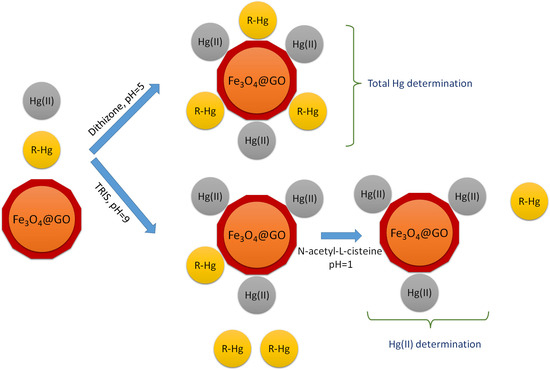
2. Results and Discussion

2.1. Effect of pH

The effect of pH on the adsorption of Hg(II) on Fe3O4@GO was studied by preparing suspensions with different acidity obtained by adding diluted HNO3, sodium hydroxide or buffer solutions. The results showed that the extent of the retention depended not only on the pH, but also on the main species present in the usual buffer solutions. The ability of Hg(II) to form complexes with common species such as chloride, hydroxyl, carbonate, sulfate and phosphate [35], ammonium [36] and dissolved organic matter [37] is known; consequently, the presence of these chemicals affects the determination of Hg(II) [38]. Due to the fact that in the development of the preliminary experiments the maximum retention was obtained at a pH slightly higher than 7, it was decided to use compounds that form buffer solutions lying in the range from 7 to 9 without interfering in the measurement of mercury, such as 2-amino-2-hydroxymethyl-propane-1,3-diol (TRIS), N-(2-hydroxyethyl) piperazine-N′-(2-ethanesulfonic acid) (HEPES), 2-[(2-hydroxy-1,1-bis(hydroxymethyl)ethyl)amino]ethanesulfonic acid (TES) and 2-(N-morpholino)ethanesulfonic acid (MES). The best outcomes were obtained with the use of TRIS as the main component of the buffer solution. Consequently, several suspensions were prepared, and the pH adjusted in the 3–10 range by adding sodium acetate-acetic acid or TRIS-HNO3 buffer solutions, as can be seen in Figure 1 where the results are summarized.

2.2. Retention of the Different Forms of Mercury

The retention of other mercury species that could eventually exist in the samples to be analyzed, namely, MeHg, EtHg, PhHg and diphenylmercury (Ph2Hg) was considered. A retention study of all of them on Fe3O4@GO was carried out at different pH values in the 5–11 range using the buffer solutions above commented. Figure 2 shows the percentage of retention of these chemicals in the different pH media. As can be seen, the greatest retention of mercury species occurred at a pH close to 9; however, EtHg and MeHg were hardly retained on Fe3O4@GO at any pH value.
In order to achieve a reliable discrimination between Hg(II) and MeHg, a large number of experiments were carried out using other composites with magnetic properties. As Figure 3 shows, none of the sorbents assayed proved suitable for our purpose of speciation; therefore, a different strategy based in the use of auxiliary complexing agents was tried.

2.3. Effect of the Presence of Complexing Agents of Mercury Species

Chelating agents with thiol groups, such as L-Cysteine (L-Cys), sodium mercapto ethane sulfonate (MESNA) and 2,3-dimercaptopropanol (BAL) at different pH values, were assayed for the purpose. In the presence of L-Cys, the Hg(II) retention decreased and it was not even observed in the presence of MESNA and BAL.
However, when studying the effect of dithizone (1,5-diphenylthiocarbazone), a quantitative retention of all the mercury forms on the adsorbent material was observed. The amounts of dithizone and Fe3O4@GO to be used to achieve the total retention of Hg(II) and MeHg on the adsorbent were studied. The results showed that when 1 mL of a 0.05% w/v dithizone solution was employed, the retention of both forms of mercury was complete. A 100 µL amount of Fe3O4@GO suspension led to a maximum and constant retention of the mercury forms. In order to avoid that in the case of real samples, the presence of other species that can be adsorbed to saturate the active points of the adsorbent, the use of 300 µL of the Fe3O4@GO suspension is recommended. This volume involves 7 mg of adsorbent material per determination.

2.4. Study of Desorption Conditions

As mentioned above, all forms of mercury are retained in the presence of dithizone. To achieve the speciation of Hg(II) and MeHg, selective desorption media were studied. The study showed that if the magnetic particles were washed with N-acetyl-L-cysteine, MeHg was desorbed, while Hg(II) remained retained on the adsorbent material; however, this procedure was not considered reliable because when the concentration of N-acetyl-L-cysteine exceeded 0.05 M, the Hg(II) was also partially desorbed.
The strategy for the differentiation was then focused on the medium used for the adsorption process, since in the presence of dithizone both analytes were retained. Meanwhile, when a TRIS medium at pH = 9 was employed in the absence of dithizone, the Hg(II) was completely adsorbed together with a small fraction of the MeHg that could be easily released by the treatment with N-acetyl-L-cysteine.
In both adsorption procedures, the desorption of the species to be quantified by ETAAS was achieved in an acid medium. Hydrochloric acid is not recommended for use in atomic absorption spectrometry due to the formation of premature volatile species; therefore, HNO3 was employed for the purpose. Excellent results were obtained with the use of a mixture of 50 µL of 8% v/v HNO3 and 50 µL of 30% H2O2 and heating at 80 °C in an ultrasonic bath. After the treatment, the mixture was vortexed for 1 min to obtain a suspension, then, 10 µL aliquots were taken and injected into the atomizer, before applying the heating program shown in Table 1. For the Hg (II) determination, before adding the mixture of 8% v/v HNO3 and 50 µL of 30% H2O2, the adsorbent was washed with N-acetyl-L-cysteine to desorb the small amount of MeHg that might have been retained in the sorbent under those adsorption conditions.

2.5. Optimization of Analysis Conditions by ETAAS

At a first glance, taking into account the thermal and atomic characteristics of mercury, ETAAS does not appear to be a suitable quantification technique; however, a review of the literature revealed that by using the appropriate chemical modification, good sensitivity could be obtained. For example, it has been reported that the use of the mixture of Ag+ and MnO4- led to excellent results in the determination of mercury in edible oils [18], waters [39], soils and sediments [40], sludges [41], infant foods [42] and food colorants [43]. Moreover, an interesting alternative is the use of metals such as palladium [13] or iridium [44] as permanent modifiers that are deposited on the walls of the graphite tube, either by electrolysis or by previously injecting several aliquots of the modifier [45].
It should be noted that in the procedure here studied, the suspension medium recommended caused the Pd(0) coating generated by electrolysis to be exhausted relatively quickly. A 20% loss of sensitivity in just 50 injections of 10 µL of the final suspensions was verified. An effective alternative was found in the prior injection of palladium and heating to a calcination temperature to achieve a deposition of the modifier on the surface of the L’Vov platform. It was found that the use of 20 µL of a 125 mg L−1 Pd(II) solution was sufficient to achieve mercury stabilization at 300 °C and its atomization at 1500 °C. Higher concentrations caused an excessive broadening of the signal that required too long integration times.

2.6. Calibration, Validation and Application to Real Samples

Applying the heating program recommended in Table 1, the analytical signal of Hg(II) and MeHg in the samples that were no submitted to the microextraction showed a linear behavior between 0.05 and 1 mg L−1 with a slope of 0.5814 s mg−1 L. The signal obtained after the application of the proposed procedure for the determination of the total mercury content showed a linear behavior between 0.1 and 10 µg L−1 with a calibration slope of 0.0284 s µg−1 L. The limit of detection (LOD), calculated as three times the standard error of the estimate, was 0.02 µg L−1, the limit of quantification (LOQ) was 0.06 µg L−1 and the average relative standard deviation for concentrations in the calibration interval was found to be 5.2%. The enrichment factor, calculated as the ratio of the slopes of a calibration line obtained in the procedure and the calibration line for the direct measurement of mercury was found to be 49. Extraction recovery (ER) is defined as the percentage of the total mercury amount which is extracted into the acceptor phase:
E R ,   % = E F × V e V a c × 100
where Ve and Vac are the volumes of the acceptor phase and the aqueous sample, respectively. The ERs for the mercury ranged from 97–99%.
When the recommended procedure for the Hg(II) determination was applied, the calibration slope was 0.0281 s µg−1 L. The LOD and LOQ were 0.02 and 0.06 µg L−1, respectively, and the average relative standard deviation for concentrations in the calibration interval was found to be 4.7%, while the enrichment factor did not change.
The quantification was carried out by a direct calibration applying the proposed procedure to standard solutions of the analytes. No matrix effect was detected.
The reliability of the procedure was verified by analyzing the Hg(II) content in three reference materials: NCS DC 73347 (human hair), DORM-2 (fish muscle) and DORM-4 (fish protein) that were subjected to a microwave digestion process. In the NCS DC 73347, only the total mercury content was certified. Table 2 shows the results obtained. As can be seen, the concentrations found were consistent with the certified values.
In addition, another four standard reference samples, namely, NIST SRM 1640a (natural water with a low salt content), SPS-SW2 (surface water reference material), ERM CAO11b (reference material), and NASS-6 (sea water) were also submitted to the recommended procedure. It is of note that these water samples did not have a certified mercury content. In this case, the standard addition method was applied. The results are summarized in Table 3.
The procedure proposed for the determination of the total Hg and Hg(II) was also applied to the determination of these species in the water samples from different sources. As shown in Table 4, a recovery test was applied because the signal obtained in the samples was below the detection limit.
Table 5 shows a comparison of the proposed procedure with others that, using different microextraction techniques, have in common the determination of mercury by ETAAS. As can be seen, the calibration interval and LOD were comparable to, or even better than many of the procedures listed in the table. The sample consumption in the determination was very low, the adsorbent material was easy to prepare and the magnetic separation facilitates the experimental procedure and the determination of the two main mercury species in water.

3. Materials and Methods

3.1. Materials and Instrumentation

All experiments were carried out using high purity water (resistivity 18 MΩ cm) obtained with a Millipore system (Millipore, Bedford, MA, USA). The Hg(II) standard solution (1000 mg L−1 in 1% HNO3, 250 mL) was purchased from Fluka (Buchs, Switzerland). The standard solution of MeHg 1000 mg L−1 was prepared by dissolving the appropriate amount of its chloride (Merck, Darmstadt, Germany) in the smallest possible volume of methanol and diluting to volume with water. Standard solutions of 100 mg mL−1 of EtHg, PhHg and Ph2Hg were prepared in the minimum amount of acetone or hexane from the products also supplied by Sigma. Working standard solutions were obtained by an appropriate dilution and were prepared daily by dilution with ultrapure water. Caution: all forms of mercury are toxic and suitable precautions must be taken when handling them [54,55,56]. Mercury poisoning can result from inhalation, ingestion, and injection or absorption through the skin. All forms of mercury penetrate the placental barrier and should be considered teratogenic and reproductive effectors. The effects may not be noticeable for months or years. Inorganic mercury salts are toxic (LD50 from 6–200 mg kg−1). Dimethyl mercury is even more toxic (LD50: 50 µg kg−1) and extreme caution is required when working with this material.
The solutions of dithizone, L-cysteine, N-acetyl-L-cysteine, 2-Amino-2-hydroxymethyl-propane-1,3-diol (TRIS), tetramethyl ammonium hydroxide (TMAH), and other reagents were prepared from reagents supplied by Sigma-Aldrich. The dithizone solution was prepared by dissolving, with the help of ultrasound, 0.05 g of the product in 100 mL of a 4:1 (v/v) mixture of acetone and an aqueous solution adjusted to a pH = 9 with ammonium hydroxide.
The Fe(II) and Fe(III) salts used were FeCl2·4H2O and FeCl3·6H2O, respectively, which were obtained from Sigma. The graphene oxide was provided by Timesnano (Chengdu Organic Chemicals Co. Ltd., Chinese Academy of Sciences, Chengdu, China). The manufacturer’s specifications indicate a purity > 98%, ash < 1.5%, 1–2 layers and a 1–5 µm of diameter. The atomic percentage of C and O are 64.71 and 35.29, respectively.
The materials used as the water reference samples were SRM 1640a (trace elements in natural water) from the National Institute of Standards and Technology (Gaithersburg, MD, USA), SRMs SPS-SW2 (surface water) and ERM CAO11b (natural water) from LGC Limited (Teddington, UK), and the NASS-6 material (seawater) from the National Research Council of Canada, Ottawa, Canada (NRC-CNRC).
To verify the reliability of procedures, the results obtained in the determination of the total mercury, Hg(II) and MeHg were compared with certified reference materials: DORM-4 (fish protein), DORM-2 (fish muscle) and DOLT-2 (fish liver), supplied by the NRC-CNRC, as well as the material DC 73347 (human hair) that was supplied by the China National Analysis Center for Iron and Steel, Beijing, P. R. China.
An Analytik Jena ContrAA 700 high-resolution atomic absorption spectrometer, equipped with a 300 W xenon short-arc lamp as a continuous radiation source (Analytik Jena, Germany) was used to carry out all the measurements. A transversely heated graphite atomizer with pyrolytically coated graphite tubes and a L’Vov platform was used for the analyte atomization. The data were evaluated using the summed integrated absorbance for 3, 5 or 7 pixels using the software (ASPECT CS v5.1) provided by Analytik Jena.
For the ultrasonic treatment, a 50 W thermostatted ultrasound bath (ATU, Valencia, Spain) was employed. In the desorption species process, a vortex, Reax model from Heidolph (Schwabach, Germany) was used. The digestion of the reference materials was carried out with a microwave oven (Multiwave 3000 Anton-Paar, Perkin Elmer, Shelton, CT, USA) equipped with pressure and temperature control.

3.2. Preparation of Fe3O4@GO

The Fe3O4@GO was prepared by the precipitation of Fe(II) and Fe(III) salts in the presence of graphene oxide and ammonium hydroxide. Firstly, a suspension of 0.1 g of graphene oxide in 50 mL of water was prepared and sonicated for one hour. Then, under a nitrogen atmosphere, 10 mL of FeCl3·6H2O (0.1475 g mL−1) and FeCl2·4H2O (0.046 g mL−1) solutions were added dropwise. The mixture was heated at 80 °C while being stirred for 10 min. Concentrated ammonium hydroxide was then added slowly until reaching a pH = 11. In this way, a black precipitate of Fe3O4 particles was obtained. The magnetic particles were separated with the help of an external magnet. The supernatant was decanted and discarded. The magnetic particles were washed 3 times with 25 mL aliquots of ethanol in order to remove the excess salts and ions. Finally, they were suspended in 30 mL of water. The estimated concentration of Fe3O4@GO obtained was 24 mg mL−1.

3.3. Sample Treatment

For the determination of the total mercury in the reference materials, 0.5 g of this material was placed into a digestion vial. Then, 4.0 mL of HNO3 acid and 4 mL of 30% v/v H2O2 were added. The vial was placed into the microwave oven and a power of 1400 W was applied for 20 min. At the end of the digestion program, the mixture was cooled down before being removed from the microwave unit (about 15 min). Finally, the pH was adjusted to 5.0 by the addition of a concentrated NaOH solution and the liquid was diluted to 25 mL with pure water in a volumetric flask.
For the determination of Hg(II) and MeHg, a procedure based on previous studies was carried out [57,58]. To achieve the solubilization of these mercury species, 0.5 g amounts of the certified reference materials were accurately weighed into a conical tube. Then, 2 mL of a 25% (m/v) TMAH solution were added to the sample and the mixture was first kept at room temperature for three hours and then heated in a water bath at 80 °C for 30 min. Finally, the mixture was cooled and diluted to 25 mL with pure water.
The water samples were filtered immediately after collection using a cellulose nitrate filter membrane (with a 0.45 µm pore size). Subsequently, they were stored in low-density polypropylene bottles at 4 °C until analysis within 3 days.

3.4. Procedure for the Determination of Total Mercury

A 10 mL sample was placed in a 15 mL tube, and 400 µL of a 0.1 M acetate/acetic buffer (pH = 5) and 1 mL of a 0.05% w/v dithizone solution were added. The solution was manually homogenized and 300 µL of the Fe3O4@GO suspension was added. The mixture was stirred for 10 min. Afterwards, the magnetic particles were separated by action of the magnet and the supernatant liquid was discarded. The solid residue was suspended in 50 µL of HNO3 at 8% v/v and 50 µL of H2O2 at 30% v/v using ultrasound into a thermostatic bath at 80 °C for 10 min.
An amount of 10 µL of this suspension was injected into the atomizer where 20 µL of a Pd(II) solution at 125 mg L−1 was previously injected. The heating program shown in Table 1 was applied, thus obtaining an analytical signal corresponding to the total mercury content in the sample.
Figure 4 shows a scheme of the extraction process. Complete and incomplete retentions of the analytes are represented by (1) and (2), respectively.

3.5. Procedure for the Determination of Hg(II)

A 300 µL volume of a 0.1 mol L−1 buffer solution (TRIS/HNO3 with a pH = 9) was added to 10 mL of water. The solution was mixed and 300 µL of the Fe3O4@GO suspension was added then the mixture was stirred for 10 min. Next, the magnetic nanoparticles were separated by a magnet and the supernatant liquid was discarded. Afterwards, the particles were washed with 1 mL of 0.01 M N-acetyl-L-cysteine. Finally, the magnetic particles were suspended in 50 µL of HNO3 at 8% v/v and 50 µL of H2O2 at 30% v/v using ultrasound in a thermostatic bath at 80 °C for 10 min.
A 10 µL volume of this suspension was injected with 20 µL of the Pd(II) solution at 125 mg L−1 and the heating program shown in Table 1 was applied. The measured signal corresponded to the content of Hg(II) in the sample.

3.6. Procedure for the Determination of MeHg

Since the level of organomercurials different to MeHg was considered negligible for the samples considered, the content of MeHg was estimated as the difference between the total content of total mercury and the content of Hg(II).

4. Conclusions

A novel procedure based on a dispersive solid-phase microextraction with Fe3O4@GO as the adsorbent material for the determination of different forms of mercury is proposed. For this approach, it is not necessary to resort to sophisticated and relatively expensive analysis techniques, instead it employs a robust and economical methodology accessible to many laboratories such as ETAAS. The coupling of an adequate microextraction technique allowed for reaching the necessary detection limits for mercury and the validation of the method of analysis of this species. The proposed procedure presents an enrichment factor of 49 and the determination presents a linear interval between 0.1 and 10 µg L−1 of mercury. Furthermore, the method was successfully applied to the determination of mercury in waters of different origins and reference materials.

Author Contributions

Conceptualization, I.L.-G.; methodology, I.L.-G. and M.J.M.-S.; investigation, M.J.M.-S. and Y.V.-M.; data curation, I.L.-G., M.J.M.-S. and M.H.-C.; writing—original draft preparation, Y.V.-M.; writing—review and editing, M.H.-C. and I.L.-G.; supervision, M.J.M.-S.; funding acquisition, M.H.-C. All authors have read and agreed to the published version of the manuscript.

Funding

The authors acknowledge the financial support of the Spanish MCIN (Project PID2021-123201NB-I00 financed by MCIN/AEI/10.13039/501100011033/FEDER, UE).

Institutional Review Board Statement

Not applicable.

Informed Consent Statement

Not applicable.

Data Availability Statement

Data are available from the corresponding authors upon reasonable request.

Acknowledgments

M.J.M.-S. acknowledges a fellowship from the University of Murcia.

Conflicts of Interest

The authors declare no conflict of interest. The funders had no role in the design of the study; in the collection, analyses, or interpretation of data; in the writing of the manuscript; or in the decision to publish the results.

References

  1. Zhao, M.; Li, Y.; Wang, Z. Mercury and Mercury-Containing Preparations: History of Use, Clinical Applications, Pharmacology, Toxicology, and Pharmacokinetics in Traditional Chinese Medicine. Front. Pharmacol. 2022, 13, 807807. [Google Scholar] [CrossRef] [PubMed]
  2. da Conceicao Nascimento Pinheiro, M.; do Nascimento, J.L.M.; de Lima Silveira, L.C.; da Rocha, J.B.T.; Aschner, M. Mercury and Selenium—A Review on Aspects Related to the Health of Human Populations in the Amazon. Environ. Bioindic. 2009, 4, 222–245. [Google Scholar] [CrossRef] [PubMed]
  3. Clarkson, T.W.; Magos, L. The toxicology of mercury and its chemical compounds. Crit. Rev. Toxicol. 2006, 36, 609–662. [Google Scholar] [CrossRef] [PubMed]
  4. Enrico, M.; Balcom, P.; Johnston, D.T.; Foriel, J.; Sunderland, E.M. Simultaneous combustion preparation for mercury isotope analysis and detection of total mercury using a direct mercury analyzer. Anal. Chim. Acta 2021, 1154, 338327. [Google Scholar] [CrossRef] [PubMed]
  5. Report, Utilizing Direct Mercury Analysis for Mercury Detection in Botanical Extracts, Vitamins, Minerals and Dietary Supplements. Braz. J. Anal. Chem. 2021, 8, 237–240.
  6. Lee, S.; Oh, S.; Shin, S.; Kim, D.; Lee, J.; Kang, H.; Jo, G. Quantitative analysis and risk assessment on total mercury and methyl mercury in seafood distributed in Daejeon. J. Biomed. Transl. Res. 2022, 23, 7–16. [Google Scholar] [CrossRef]
  7. Matusiewicz, H.; Sturgeon, R.E. Chemical Vapor Generation with Slurry Sampling: A Review of Applications to Atomic and Mass Spectrometry. Appl. Spectros. Rev. 2012, 47, 41–82. [Google Scholar] [CrossRef]
  8. Ferreira, S.L.C.; Lemos, V.A.; Silva, L.O.B.; Queiroz, A.F.S.; Souza, A.S.; da Silva, E.G.P.; dos Santos, W.N.L.; das Virgens, C.F. Analytical strategies of sample preparation for the determination of mercury in food matrices—A review. Microchem. J. 2015, 121, 227–236. [Google Scholar] [CrossRef]
  9. Suvarapu, L.N.; Baek, S.-O. Recent Developments in the Speciation and Determination of Mercury Using Various Analytical Techniques. J. Anal. Methods Chem. 2015, 2015, 372459. [Google Scholar] [CrossRef] [Green Version]
  10. Kallithrakas-Kontos, N.; Foteinis, S. Recent Advances in the Analysis of Mercury in Water—Review. Curr. Anal. Chem. 2016, 12, 22–36. [Google Scholar] [CrossRef]
  11. Volynkin, S.S.; Demakov, P.A.; Shuvaeva, O.V.; Kovalenko, K.A. Metal-organic framework application for mercury speciation using solid phase extraction followed by direct thermal release-electrothermal atomization atomic absorption spectrophotometric detection (ETA AAS). Anal. Chim. Acta 2021, 1177, 338795. [Google Scholar] [CrossRef]
  12. Burylin, M.Y.; Romanovskiy, K.A.; Temerdashev, Z.A.; Galai, E.F. Determination of Mercury in Sediments by Slurry Sampling Electrothermal Atomic Absorption Spectrometry. J. Anal. Chem. 2019, 74, 1184–1191. [Google Scholar] [CrossRef]
  13. Lopez-Garcia, I.; Rivas, R.E.; Hernandez-Cordoba, M. Hollow fiber based liquid-phase microextraction for the determination of mercury traces in water samples by electrothermal atomic absorption spectrometry. Anal. Chim. Acta 2012, 743, 69–74. [Google Scholar] [CrossRef]
  14. Amico, D.; Tassone, A.; Pirrone, N.; Sprovieri, F.; Naccarato, A. Recent applications and novel strategies for mercury determination in environmental samples using microextraction-based approaches: A review. J. Hazard Mater. 2022, 433, 128823. [Google Scholar] [CrossRef]
  15. Ahmad, H.; Koo, B.H.; Khan, R.A. Preconcentration and determination of trace Hg(II) using ultrasound-assisted dispersive solid phase microextraction. RSC Adv. 2021, 12, 53–61. [Google Scholar] [CrossRef]
  16. Bahar, S.; Zakerian, R. Determination of Hg(II) in Environmental Water Samples by Dispersive Liquid Phase Microextraction Combined with Flame Atomic Absorption Spectrometry. J. Chin. Chem. Soc.-Taip. 2013, 60, 179–184. [Google Scholar] [CrossRef]
  17. Martinis, E.M.; Escudero, L.B.; Salvarezza, R.; Calderon, M.F.; Ibanez, F.J.; Wuilloud, R.G. Liquid-liquid microextraction based on a dispersion of Pd nanoparticles combined with ETAAS for sensitive Hg determination in water samples. Talanta 2013, 108, 46–52. [Google Scholar] [CrossRef]
  18. Lopez-Garcia, I.; Vicente-Martinez, Y.; Hernandez-Cordoba, M. Determination of ultratraces of mercury species using separation with magnetic core-modified silver nanoparticles and electrothermal atomic absorption spectrometry. J. Anal. Atom. Spectrom. 2015, 30, 1980–1987. [Google Scholar] [CrossRef]
  19. Ito, R.; Kawaguchi, M.; Sakui, N.; Okanouchi, N.; Saito, K.; Seto, Y.; Nakazawa, H. Stir bar sorptive extraction with in situ derivatization and thermal desorption-gas chromatography-mass spectrometry for trace analysis of methylmercury and mercury(II) in water sample. Talanta 2009, 77, 1295–1298. [Google Scholar] [CrossRef]
  20. Fabbri, D.; Lombardo, M.; Trombini, C.; Vassura, I. A new procedure for the speciation of mercury in water based on the transformation of mercury(II) and methylmercury(II) into stable acetylides followed by HPLC analysis. Appl. Organomet. Chem. 1995, 9, 713–718. [Google Scholar] [CrossRef]
  21. Cruz Sotolongo, A.; Messina, M.M.; Ibanez, F.J.; Wuilloud, R.G. Hybrid ionic liquid-3D graphene-Ni foam for on-line preconcentration and separation of Hg species in water with atomic fluorescence spectrometry detection. Talanta 2020, 210, 120614. [Google Scholar] [CrossRef] [PubMed]
  22. Shi, M.-T.; Yang, X.-A.; Zhang, W.-B. Magnetic graphitic carbon nitride nano-composite for ultrasound-assisted dispersive micro-solid-phase extraction of Hg(II) prior to quantitation by atomic fluorescence spectroscopy. Anal. Chim. Acta 2019, 1074, 33–42. [Google Scholar] [CrossRef] [PubMed]
  23. Zhang, Y.; Yan, L.; Xu, W.; Guo, X.; Cui, L.; Gao, L.; Wei, Q.; Du, B. Adsorption of Pb(II) and Hg(II) from aqueous solution using magnetic CoFe2O4-reduced graphene oxide. J. Mol. Liq. 2014, 191, 177–182. [Google Scholar] [CrossRef]
  24. Sreeprasad, T.S.; Maliyekkal, S.M.; Lisha, K.P.; Pradeep, T. Reduced graphene oxide-metal/metal oxide composites: Facile synthesis and application in water purification. J. Hazard Mater. 2011, 186, 921–931. [Google Scholar] [CrossRef] [PubMed]
  25. Cui, L.; Wang, Y.; Gao, L.; Hu, L.; Yan, L.; Wei, Q.; Du, B. EDTA functionalized magnetic graphene oxide for removal of Pb(II), Hg(II) and Cu(II) in water treatment: Adsorption mechanism and separation property. Chem. Eng. J. 2015, 281, 1–10. [Google Scholar] [CrossRef]
  26. Jinadasa, K.K.; Pena-Vazquez, E.; Bermejo-Barrera, P.; Moreda-Pineiro, A. Smart materials for mercury and arsenic determination in food and beverages. Microchem. J. 2022, 179, 107472. [Google Scholar] [CrossRef]
  27. Jamshidi, S.; Rofouei, M.K.; Seidi, S.; Emmer, Å. Applicability of a magnetic bucky gel for microextraction of mercury from complicated matrices followed by cold vapor atomic absorption spectroscopy. Sep. Sci. Technol. 2020, 55, 1505–1514. [Google Scholar] [CrossRef] [Green Version]
  28. Seidi, S.; Fotouhi, M. Magnetic dispersive solid phase extraction based on polythiophene modified magnetic graphene oxide for mercury determination in seafood followed by flow-injection cold vapor atomic absorption spectrometry. Anal. Methods 2017, 9, 803–813. [Google Scholar] [CrossRef]
  29. Xu, Y.; Zhang, W.; Shi, J.; Zou, X.; Li, Y.; Tahir, H.E.; Huang, X.; Li, Z.; Zhai, X.; Hu, X. Electrodeposition of gold nanoparticles and reduced graphene oxide on an electrode for fast and sensitive determination of methylmercury in fish. Food Chem. 2017, 237, 423–430. [Google Scholar] [CrossRef]
  30. Keramat, A.; Zare-Dorabei, R. Ultrasound-assisted dispersive magnetic solid phase extraction for preconcentration and determination of trace amount of Hg (II) ions from food samples and aqueous solution by magnetic graphene oxide (Fe3O4@ GO/2-PTSC): Central composite design optimization. Ultrason. Sonochem. 2017, 38, 421–429. [Google Scholar]
  31. Akbar, M.; Manoochehri, M. An efficient 2-mercapto-5-phenylamino-1,3,4-thiadiazole functionalized magnetic graphene oxide nanocomposite for preconcentrative determination of mercury in water and seafood samples. Inorg. Chem. Commun. 2019, 103, 37–42. [Google Scholar] [CrossRef]
  32. Li, L.; Bi, R.; Wang, Z.; Xu, C.; Li, B.; Luan, L.; Chen, X.; Xue, F.; Zhang, S.; Zhao, N. Speciation of mercury using high-performance liquid chromatography-inductively coupled plasma mass spectrometry following enrichment by dithizone functionalized magnetite-reduced graphene oxide. Spectrochim. Acta B 2019, 159, 105653. [Google Scholar] [CrossRef]
  33. Boszke, L.; Głosińska, G.; Siepak, J. Some Aspects of Speciation of Mercury in Water Environment. Pol. J. Environ. Stud. 2002, 11, 285–298. [Google Scholar]
  34. Si, L.; Branfireun, B.A.; Fierro, J. Chemical Oxidation and Reduction Pathways of Mercury Relevant to Natural Waters: A Review. Water 2022, 14, 1891. [Google Scholar] [CrossRef]
  35. Powell, K.J.; Brown, P.L.; Byrne, R.H.; Gajda, T.; Hefter, G.; Sjoberg, S.; Wanner, H. Chemical speciation of environmentally significant heavy metals with inorganic ligands—Part 1: The Hg2+-Cl, OH, CO32−, SO42−, and PO43− aqueous systems—(IUPAC technical report). Pure Appl. Chem. 2005, 77, 739–800. [Google Scholar] [CrossRef] [Green Version]
  36. Golding, G.R.; Sparling, R.; Kelly, C.A. Effect of pH on intracellular accumulation of trace concentrations of Hg(II) in Escherichia coli under anaerobic conditions, as measured using a mer-lux bioreporter. Appl. Environ, Microb. 2008, 74, 667–675. [Google Scholar] [CrossRef] [Green Version]
  37. Dong, W.; Liang, L.; Brooks, S.; Southworth, G.; Gu, B. Roles of dissolved organic matter in the speciation of mercury and methylmercury in a contaminated ecosystem in Oak Ridge, Tennessee. Environ. Chem. 2010, 7, 94–102. [Google Scholar] [CrossRef] [Green Version]
  38. Bittrich, D.R.; Chadwick, S.P.; Babiarz, C.L.; Manolopoulos, H.; Rutter, A.P.; Schauer, J.J.; Armstrong, D.E.; Collett, J.; Herckes, P. Speciation of Mercury (II) and Methylmercury in Cloud and Fog Water. Aerosol Air Qual. Res. 2011, 11, 161–169. [Google Scholar] [CrossRef]
  39. Lopez-Garcia, I.; Rivas, R.E.; Hernandez-Cordoba, M. Liquid-phase microextraction with solidification of the organic floating drop for the preconcentration and determination of mercury traces by electrothermal atomic absorption spectrometry. Anal. Bioanal. Chem. 2010, 396, 3097–3102. [Google Scholar] [CrossRef]
  40. Lopez-Garcia, I.; Sanchez-Merlos, M.; Hernandez-Cordoba, M. Determination of mercury in soils and sediments by graphite furnace atomic absorption spectrometry with slurry sampling. Spectrochim. Acta B 1997, 52, 2085–2092. [Google Scholar] [CrossRef]
  41. Lopez-Garcia, I.; Sanchez-Merlos, M.; Arroyo-Cortez, J.; Hernandez-Cordoba, M. Determination of mercury in sewage sludges by slurry sampling electrothermal atomic absorption spectrometry. J. AOAC Int. 2002, 85, 25–30. [Google Scholar] [CrossRef] [PubMed] [Green Version]
  42. Vinas, P.; Pardo-Martinez, M.; Lopez-Garcia, I.; Hernandez-Cordoba, M. Determination of mercury in baby food and seafood samples using electrothermal atomic absorption spectrometry and slurry atomization. J. Anal. Atom. Spectrom. 2001, 16, 633–637. [Google Scholar] [CrossRef]
  43. Vinas, P.; Pardo-Martinez, M.; Lopez-Garcia, I.; Hernandez-Cordoba, M. Rapid determination of mercury in food colorants using electrothermal atomic absorption spectrometry with slurry sample introduction. J. Agr. Food Chem. 2002, 50, 949–954. [Google Scholar] [CrossRef] [PubMed]
  44. Vereda Alonso, E.; del Mar Guerrero, M.; Colorado Cueto, P.; Barreno Benitez, J.; Cano Pavon, J.M.; Garcia de Torres, A. Development of an on-line solid phase extraction method based on new functionalized magnetic nanoparticles. Use in the determination of mercury in biological and sea-water samples. Talanta 2016, 153, 228–239. [Google Scholar] [CrossRef] [PubMed]
  45. Moreno, R.G.M.; de Oliveira, E.; Pedrotti, J.J.; Oliveira, P.V. An electrochemical flow-cell for permanent modification of graphite tube with palladium for mercury determination by electrothermal atomic absorption spectrometry. Spectrochim. Acta B 2002, 57, 769–778. [Google Scholar] [CrossRef]
  46. Thongsaw, A.; Sananmuang, R.; Udnan, Y.; Ross, G.M.; Chaiyasith, W.C. Dual-cloud point extraction for speciation of mercury in water and fish samples by electrothermal atomic absorption spectrometry. Spectrochim. Acta B 2019, 160, 105685. [Google Scholar] [CrossRef]
  47. Thongsaw, A.; Sananmuang, R.; Udnan, Y.; Ross, G.M.; Chaiyasith, W.C. Speciation of mercury in water and freshwater fish samples using two-step hollow fiber liquid phase microextraction with electrothermal atomic absorption spectrometry. Spectrochim. Acta B 2019, 152, 102–108. [Google Scholar] [CrossRef]
  48. Sakanupongkul, A.; Sananmuang, R.; Udnan, Y.; Ampiah-Bonney, R.J.; Chaiyasith, W.C. Speciation of mercury in water and freshwater fish samples by a two-step solidified floating organic drop microextraction with electrothermal atomic absorption spectrometry. Food Chem. 2019, 277, 496–503. [Google Scholar] [CrossRef]
  49. Akramipour, R.; Golpayegani, M.R.; Ghasemi, M.; Noori, N.; Fattahi, N. Optimization of a methodology for speciation of arsenic, selenium and mercury in blood samples based on the deep eutectic solvent. Methodsx 2019, 6, 2141–2147. [Google Scholar] [CrossRef]
  50. Cruz Sotolongo, A.; Martinis, E.M.; Wuilloud, R.G. An easily prepared graphene oxide-ionic liquid hybrid nanomaterial for micro-solid phase extraction and preconcentration of Hg in water samples. Anal. Methods 2018, 10, 338–346. [Google Scholar] [CrossRef] [Green Version]
  51. Krawczyk, M.; Stanisz, E. Silver nanoparticles as a solid sorbent in ultrasound-assisted dispersive micro solid-phase extraction for the atomic absorption spectrometric determination of mercury in water samples. J. Anal. Atom. Spectrom. 2015, 30, 2353–2358. [Google Scholar] [CrossRef]
  52. Mitani, C.; Kotzamanidou, A.; Anthemidis, A.N. Automated headspace single-drop microextraction via a lab-in-syringe platform for mercury electrothermal atomic absorption spectrometric determination after in situ vapor generation. J. Anal. Atom. Spectrom. 2014, 29, 1491–1498. [Google Scholar] [CrossRef]
  53. Sarica, D.Y.; Turker, A.R. Speciation and Determination of Inorganic Mercury and Methylmercury by Headspace Single Drop Microextraction and Electrothermal Atomic Absorption Spectrometry in Water and Fish. Clean-Soil Air Water 2012, 40, 523–530. [Google Scholar] [CrossRef]
  54. United States Enronmental Protection Agency (EPA). Health Effects of Exposures to Mercury. Available online: https://www.epa.gov/mercury/health-effects-exposures-mercury (accessed on 1 December 2022).
  55. Bernhoft, R.A. Mercury Toxicity and Treatment: A Review of the Literature. J. Environ. Public Health 2012, 2012, 460508. [Google Scholar] [CrossRef]
  56. De Palma, G.; Mariotti, O.; Lonati, D.; Goldoni, M.; Catalani, S.; Mutti, A.; Locatelli, C. Toxicokinetics and toxicodynamics of elemental mercury following self-administration. Clin. Toxicol. 2008, 46, 869–876. [Google Scholar] [CrossRef] [Green Version]
  57. Wu, Y.; Lee, Y.-I.; Wu, L.; Hou, X. Simple mercury speciation analysis by CVG-ICP-MS following TMAH pre-treatment and microwave-assisted digestion. Microchem. J. 2012, 103, 105–109. [Google Scholar] [CrossRef]
  58. Vieira, M.A.; Ribeiro, A.S.; Curtius, A.J.; Sturgeon, R.E. Determination of total mercury and methylmercury in biological samples by photochemical vapor generation. Anal. Bioanal. Chem. 2007, 388, 837–847. [Google Scholar] [CrossRef]
Figure 1. Effect of pH on Hg(II) retention. The error bars correspond to the standard deviation of three experiments.
Molecules 28 00014 g001
Figure 2. Retention of different forms of mercury in Fe3O4@GO at different pH values.
Molecules 28 00014 g002
Figure 3. Retention of different forms of mercury in various adsorbent materials using TRIS as the buffer solution at pH = 9. Error bars correspond to the standard deviation of three experiments.
Molecules 28 00014 g003
Figure 4. Scheme of the process from extraction to analysis. Complete and incomplete retentions of the analytes are represented by (1) and (2), respectively.
Molecules 28 00014 g004
Table 1. Instrumental parameters and heating program for mercury determination.
Parameter
Wavelength, nm253.6519
Slit, nm0.7
AtomizerTransversal with L’Vov platform
Background correctionZeeman effect
Injected sample volume, µL10
Chemical modifier20 µL of 125 mg L−1 Pd(II) solution
Sample volume, mL10
Heating program
StepTemperature, °CRamp, °C s−1Hold, s
1: Dry1101030
2: Pyrolysis3005020
4: Atomization a,b150022005
5: Clean150004
Sequence: Inject chemical modifier and run steps 1 to 2. Stop heating and inject sample. Then run the heating program.
a Internal argon flow stopped 5 s before. b Reading step.
Table 2. Total mercury, Hg(II) and MeHg contents in hair and fish reference materials (SRM).
Total Hg, µg g−1MeHg a, µg g−1Hg(II) a, µg g−1
SRMCertifiedFoundCertifiedFoundCertifiedFound
NCS DC733470.360 ± 0.0500.37 ± 0.07--0.37 ± 0.07--<LOD
DORM-40.410 ± 0.0550.408 ± 0.060.355 ± 0.0280.348 ± 0.04--0.06 ± 0.03
DORM-24.64 ± 0.264.67 ± 0.124.47 ± 0.324.42 ± 0.06--0.26 ± 0.06
DOLT-22.14 ± 0.182.18 ± 0.09--1.49 ± 0.09--0.693 ± 0.08
a mean value in Hg ± standard deviation of three digestions.
Table 3. Total mercury, Hg(II) and MeHg in water reference materials (SRM).
Hg Total, µg L−1MeHg a, µg L−1Hg(II) a, µg L−1
SRMAddedFoundRec. %AddedFoundRec. %AddedFoundRec. %
NIST SRM 1640a0<LOD--0<LOD--0<LOD--
76.9 ± 0.198 ± 321.9 ± 0.196 ± 255.0 ± 0.199 ± 3
77.3 ± 0.1105 ± 355.1 ± 0.2103 ± 322.2 ± 0.1110 ± 5
SPS-SW20<LOD--0<LOD--0<LOD--
76.9 ± 0.198 ± 321.9 ± 0.196 ± 354.9 ± 0.199 ± 3
76.8 ± 0.297 ± 254.9 ± 0.198 ± 321.9 ± 0.195 ± 4
ERM CAO11b0<LOD--0<LOD--0<LOD--
77.1 ± 0.1102 ± 322.1 ± 0.1104 ± 455.0 ± 0.2101 ± 3
77.2 ± 0.1103 ± 255.1 ± 0.2103 ± 322.0 ± 0.2101 ± 4
NASS-60<LOD--0<LOD--0<LOD--
76.9 ± 0.299 ± 321.9 ± 0.195 ± 355.0 ± 0.2101 ± 3
76.9 ± 0.199 ± 255.0 ± 0.1100 ± 321.9 ± 0.196 ± 3
a mean value in Hg ± standard deviation of three determinations.
Table 4. Total mercury, Hg(II) and MeHg in real water samples (RWS).
Hg Total. µg L−1MeHg a. µg L−1Hg(II) a. µg L−1
RWSAddedFoundRec. %AddedFoundRec. %AddedFoundRec. %
M10<LOD--0<LOD--0<LOD--
43.92 ± 0.0997.9 ± 5.321.97 ± 0.0498.5 ± 2.321.95 ± 0.0397.3 ± 1.8
M20<LOD--0<LOD--0<LOD--
43.80 ± 0.0395.1 ± 2.321.91 ± 0.0795.5 ± 4.121.89 ± 0.0794.5 ± 4.1
M30<LOD--0<LOD--0<LOD--
44.01 ± 0.06100.3 ± 4.222.00 ± 0.06100.0 ± 3.222.01 ± 0.02100.6 ± 1.7
M40<LOD--0<LOD--0<LOD--
44.02 ± 0.05100.6 ± 4.022.02 ± 0.05100.9 ± 2.622.01 ± 0.05100.3 ± 2.6
M50<LOD--0<LOD--0<LOD--
43.89 ± 0.0197.2 ± 2.121.89 ± 0.0694.5 ± 3.122.00 ± 0.0499.9 ± 2.4
M60<LOD--0<LOD--0<LOD--
43.67 ± 0.0791.8 ± 3.921.88 ± 0.0793.9 ± 4.121.79 ± 0.0189.7 ± 1.1
M70<LOD--0<LOD--0<LOD--
43.99 ± 0.0699.7 ± 4.222.07 ± 0.03103.5 ± 1.921.92 ± 0.0395.8 ± 1.7
M80<LOD--0<LOD--0<LOD--
44.11 ± 0.02102.6 ± 3.722.08 ± 0.07103.8 ± 4.022.03 ± 0.09101.4 ± 5.1
M1: drinking water from the supply network; influent water (M2) and effluent (M3) from a wastewater treatment plant; M4: water from a natural source; M5: snow water; M6: water from the Segura river; M7: water from the La Cierva reservoir (Mula, Murcia, Spain); M8: water from the Mar Menor. a mean value in Hg ± standard deviation of three digestions.
Table 5. Some recent procedures for ETAAS determination and/or speciation of mercury using microextraction techniques.
SpecieMicroextraction
Technique
RemarksCMT, °CL, µg L−1LOD, µg L−1EFRef.
Hg(II), MeHg, PhHgSPE with MOF Thermal release. None135–275/8000.001–0.5 a0.06--[11]
Hg(II), MeHgDouble CPE 1: dithizone; 2: thioureaPd(II)200/18000.4–150.2317.8[46]
MeHg; Hg(II)Double HF-LPME1: MeHg (S2O3=); 2: Hg(II) (DDTC).Pd(II)200;120/
1800; 1300
0.5–8; 0.2–120.143;
0.063
103; 95[47]
MeHg; Hg(II)SFODME in two steps1: MeHg (1-undecanol); 2: MeHg (4-NODP).Pd(II)250/13000.8–80.24;
0.25
32.2;
25.7
[48]
Hg(II)LPME with eutectic solventDDTP.Ir(IV) 210/11000.36–600.198[49]
Hg(II)SPME with GO + (C4C12Im)BrMethodology in flow and elution with HNO3. Pd(II) 250/13000.02–80.014100[50]
Hg(II)SPME with Fe3O4@SiO2@DPTHMethodology in flow and elution in thiourea.Ir(0)20/12000.1–100.00785.4[44]
Hg(II); MeHg, Me2Hg, EtHg, PhHg,Ph2HgDSPME with Fe3O4@Ag@MESNA
or Fe3O4@Ag@CYS
1: Fe3O4@Ag@MESNA for Hg(II); 2: Fe3O4@Ag@CYS for others.Ag(I) + KMnO4300/13000.03–3.50.01200[18]
Hg(II)AgNPs and DSPMEThe amalgamated Hg is dissolved in HNO3. Pd(II) + Mg(II)300/13000.1–200.00515[51]
Hg(II)HS-SDMEHg(0) with SnCl2 and drop of Pd(0).Pd(II)200/13001.5–400.4875[52]
Hg(II)DSPME with PdNPs functionalized with dodecanethiolateThe nanoparticles are in a mixture of toluene and chloroform.Pd(II)250/13000.1–100.007595[17]
Hg(II), MeHgHS-SDME with thiourea or APDCGeneration of Hg(0) and MeHg hydride with NaBH4.--250/110017–355535[53]
Hg(II)HF-LPME three-phase1: complexation of Hg(II) with PAN; 2: toluene extraction; 3: iodide back extraction.Pd(0)300/11000.2–30.06270[13]
Hg(II), MeHgDSPME with Fe3O4@GO1: Dithizone for total Hg; 2: NAC for Hg(II).Pd(II)300/15000.1–100.0249TW
CM: chemical modifier; T: ash and atomization temperatures; EF: enrichment factor; SPE: solid-phase extraction; MOF: metal organic framework; CPE: cloud point extraction; HF-LPME: hollow fiber liquid phase microextraction; DDTC: diethyldithiocarbamate; 4-NODP: 4-nitro-o-fenilen diamine; DDTP: diethyldithiophosphate; (C4C12im)Br: 1-butil 3-dodecilimidazolium bromide; Fe3O4@SiO2@DPTH: ferrite-silica-1,5-bis(di-2pyridyl)methylene thiocarbohydrazide nanocomposite; MESNA: sodium 2-mercapto ethane sulfonate; CYS: cysteine; HS-SDME: head space single drop microextraction; DSPME: dispersive solid-phase microextraction; NAC: N-acetyl-L-cysteine; TW: this work. a mean value in Hg ± standard deviation of three digestions.
Disclaimer/Publisher’s Note: The statements, opinions and data contained in all publications are solely those of the individual author(s) and contributor(s) and not of MDPI and/or the editor(s). MDPI and/or the editor(s) disclaim responsibility for any injury to people or property resulting from any ideas, methods, instructions or products referred to in the content.

