Article

Ecofriendly Green Synthesis of Copper (II) Oxide Nanoparticles Using Corchorus olitorus Leaves (Molokhaia) Extract and Their Application for the Environmental Remediation of Direct Violet Dye via Advanced Oxidation Process

Department of Chemistry, Faculty of Science, King Abdulaziz University, P.O. Box 80200, Jeddah 21589, Saudi Arabia
*
Author to whom correspondence should be addressed.
Molecules 2023, 28(1), 16; https://doi.org/10.3390/molecules28010016
Submission received: 15 October 2022 / Revised: 30 November 2022 / Accepted: 2 December 2022 / Published: 20 December 2022
(This article belongs to the Special Issue Advanced Nanoscale Materials for Energy and Environment Applications)

Abstract

:
In this research, copper (II) oxide nanoparticles were prepared by an ecofriendly green method using the extract of corchorus olitorus leaves (Molokhaia) as a surfactant, capping and anti-agglomeration agent. The ecofriendly green CuO NPs were characterized using different chemical and physical techniques and the results confirmed the formation of monoclinic tenorite CuO nanoparticles with an average particle size of 12 nm and BET surface area of 11.1 m2/g. The eco-friendly green CuO NPs were used in environmental remediation for the efficient catalytic degradation of direct violet dye via advanced oxidation process (AOP) in presence of H2O2. The impact of AOP environmental parameters affecting the degradation process was investigated. Moreover, the catalytic degradation of the direct violet dye using the ecofriendly green CuO NPs was studied kinetically and thermodynamically and the results showed that the catalytic degradation process agreed well with the pseudo-second-order kinetic model and the process was spontaneous and endothermic in nature. Finally, high catalytic degradation of the direct violet dye was observed when the eco-friendly prepared green CuO NPs were placed in real water samples.

1. Introduction

Water pollution has been a recurrent environmental issue and global challenge, particularly due to the exceptionally increasing rate of anthropogenic activities, such as population expansion, industrialization and agricultural practices, which consequently make the issue of water quantity, quality and availability a main concern in many parts of the world [1]. The efforts of research scientists, decision-makers and legislators focus on the introduction of more rigorous methods for effective environmental remediation, as well as the introduction of new laws to control environmental pollution. Conversely, the application of synthetic dyes became more common in the production of clothes, furniture and plastic products instead of natural dyes, but unfortunately these dyes are toxic and carcinogenic, affecting aquatic and human life [2]. Discouragingly, conventional water treatment methods such as coagulation, sedimentation, filtration, disinfection, decontamination and desalination are chemically and operationally intensive, which mainly require large systems, infrastructure and engineering expertise, making the treatment time-consuming, costly and ineffective when immediate demand arises. Currently, the search for and development of new robust procedures with high efficiency for the remediation of polluted water are mandatory and urgent. In this scenario, nanotechnology is found to be a promising route for the development of an effective procedure for the treatment of polluted water, water pollution prevention, detection and water quality monitoring [3,4]. This is a result of the unique and exceptional properties of nanomaterials, as the nanoscale level particles have different chemical, physical, biological, optical, magnetic and electrical properties compared to macro-particles, providing a wide range of applications [5]. Metal oxide nanoparticles such as copper oxides have become increasingly important in a wide range of nanotechnology and environmental remediation [1], environmental monitoring [6], catalysis [7,8], biological activities [9,10,11], energy [12], organic reactions [13] and nanofluid [14]. CuO NPs were synthesized using different chemical or physical procedures such as chemical vapor deposition [15], sol-gel [16], liquid phase [17], electrodeposition [18] and colloidal [19] methods. Unfortunately, the synthesis of CuO NPs using physical and chemical methods involves laborious steps with long reaction times and the use of various hazardous chemicals compounds, in addition to the generation of huge amounts of dangerous byproducts, as well as hazardous chemical waste [20]. During the last few years, more eco-friendly, biosynthesis and green procedures have been applied based on natural-based extracts of different plants, algae, fungi and bacteria as capping and anti-agglomeration agents to facilitate the formation of the CuO NPs [21,22]. Comparing plants and microorganisms, plants are more suitable as they are easily available, can be directly employed without following any complex procedure and contain bioactive substances such as terpenoids, proteins, sugar, phenolic compounds, flavonoids and alkaloids, which can easily reduce the metal ions into stable nanoparticles [23]. More importantly, the biosynthesized CuO NPs are characterized by higher activities compared with the conventionally synthesized ones [21,22,24]. Understanding this, new methods are required for the biosynthesis of CuO NPs, as it is a challenging task to find a more convenient method.
Advanced oxidation processes (AOPs) are commonly used to remove/eliminate/degrade different pollutants in contaminated water by an oxidation process in the presence of ozone (O3), hydrogen peroxide (H2O2) and/or UV light through reactions with hydroxyl radicals (·OH) [25]; therefore, AOPs are promising technologies for the elimination of dyes in solution because they are cheap, more efficient, eco-friendly and can completely degrade the pollutants to water and carbon dioxide [26,27]. Much research focuses on the application of metal/metal oxides nanoparticles and AOPs for water treatment. Metal/metal oxide nanoparticles are mainly composed of one, two, or three metals (such as Ag, Au and Pd nanoparticles) and/or metal oxides (such as ZnO, V2O5, Ta2O5, CeO2, MgO, CuO and TiO2) used for the degradation of organic pollutants in water, due to their high surface contact area and improved photolytic properties, thus being considered the best candidate for water remediation [28,29,30]. Among these, CuO is considered as a cheap, non-toxic and effective environmentally friendly catalyst and was used as an effective catalyst for AOPs in the environmental remediation of water polluted with different pollutants [31,32,33,34].
In this research, a new, simple and efficient strategy for the eco-friendly, green and robust biosynthesis of CuO nanoparticles was explored using corchorus olitorius leaves extract (Molokhia), one of the most common plant leaves in the region. The morphological and structural characteristics of the eco-friendly green CuO NPs were investigated and applied as a catalyst for the catalytic degradation of direct violet Dye 31 (DV31) via advanced oxidation process AOPs in presence of hydrogen peroxide.

2. Experimental Section

2.1. Materials

All chemicals were obtained from SIGMA-ALDRICH, were general-purpose reagents and were used in their original form without further purification. The leaves of corchorus olitorus (Molokhia) were obtained from Jeddah local market.

2.1.1. Corchorus Olitorus Leaves Extract (Molokhia) Preparation

Fresh and healthy leaves of corchorus olitorus were collected from the local market in Jeddah, Saudi Arabia. The leaves were washed very well 3 times with tap water, then 100.0 g of the cleaned leaves were added to 100 mL of deionized water in a beaker. The mixture was boiled for 20.0 min, then cooled to room temperature and filtered; a clear filtrate was obtained and was used to prepare the solutions of NaOH and CuSO4.

2.1.2. Conventional CuO NPs (CuO NPs) Preparation

An aqueous solution of copper sulfate pentahydrate (CuSO4.5H2O) (50.0 mL, 1.0 M) and the solution of NaOH (100 mL, 1.0 M) were prepared with deionized water. The NaOH solution was added drop by drop using a burette to the copper sulfate solution at 70 °C temperature under 200 rpm stirring. The black precipitate of the colloidal copper oxide was separated by centrifugation at 3500 rpm for 30 min and washed with deionized water three times, followed by acetone. The obtained product was dried at 110 °C in air atmosphere for 24 h in a regular drying oven and then calcinated in a muffle furnace at 500 °C for 60 min.

2.1.3. Green CuO NPs (G CuO NPs) Preparation

Green CuO NPs were prepared according to the literature with little modification [35]. An aqueous solution of copper sulfate pentahydrate (CuSO4.5H2O) (50.0 mL, 1.0 M) and the solution of NaOH (100 mL, 1.0 M) were prepared using the corchorus olitorus extract. The NaOH solution was added drop by drop using a burette to the copper sulfate solution at 70 °C temperature under 200 rpm stirring. The black precipitate of the colloidal copper oxide was separated by centrifugation at 3500 rpm for 30 min and washed with deionized water three times, followed by acetone. The obtained product was dried at 110 °C in air atmosphere for 24 h in a regular drying oven and then calcinated in a muffle furnace at 500 °C for 60 min.

2.2. Characterization Techniques

X-ray diffraction (XRD) patterns were recorded for phase analysis and the measurement of crystallite size on an X-ray diffraction device (XRD; type Ultima-IV; Rigaku, Tokyo, Japan), which was operated at 40 mA and 40 kV by using CuK αradiation and a nickel filter in the 2θ range from 20 to 80° in steps of 0.02°, with a sampling time of one second per step. Scherrer equation was used for the calculation of crystal size of the CuO NPs. The morphology and size of the CuO NPs were examined using a scanning electron microscope (SEM; type JEOL JSM-7600F) and transmission electron microscope (TEM; type JEOL JEM-2100 operating at 200 kV, attached to a CCD camera). The XPS experiments were performed on a Kratos Axis Ultra DLD instrument equipped with a monochromatic Al Kα X-ray source (hν = 1486.6 eV) operated at a power of 75 W and under UHV conditions in the range of ∼10−9 mbar. All spectra were recorded in hybrid mode using electrostatic and magnetic lenses and an aperture slot of 300 μm × 700 μm. The survey and high-resolution spectra were acquired at fixed analyzer pass energies of 160 and 20 eV, respectively. The samples were mounted in floating mode in order to avoid differential charging. Thereafter, XPS spectra were acquired using charge neutralization. The specific surface area of the CuO NPs were estimated using the nitrogen adsorption/desorption isotherm at 77 K, by NOVA3200e (Quantachrome, Boynton Beach, FL, USA).

2.3. Removal Study

The advanced oxidation processes for the removal of the DV31 dye from water were performed using CuO NPs in presence of H2O2 as follows: a certain amount of CuO NPs was added to 100 mL containing DV31 dye (25.0 mg/L) in the presence of H2O2, followed by magnetic stirring, and the CuO NPs were separated from the solution using a centrifuge and at the offset time. The clear filtrate was collected for the analysis of the remaining DV31 dye using ultraviolet-visible (UV–vis) spectrophotometer (UV-1650 PC, CPS-240A SHIMADZU) at 553 nm. The removal percentage (%R) and the removal capacity; qt (mg/g), were calculated using the following equations:
% R = C 0 C t C 0 × 100
q t = ( C 0 C t ) × V m
where C0 and Ct are the concentrations of the DV31 dye in solution (mg/L) at time t = 0 and t, respectively, V is the volume of the solution (L) and m is the mass of CuO NPs (g).

3. Results & Discussion

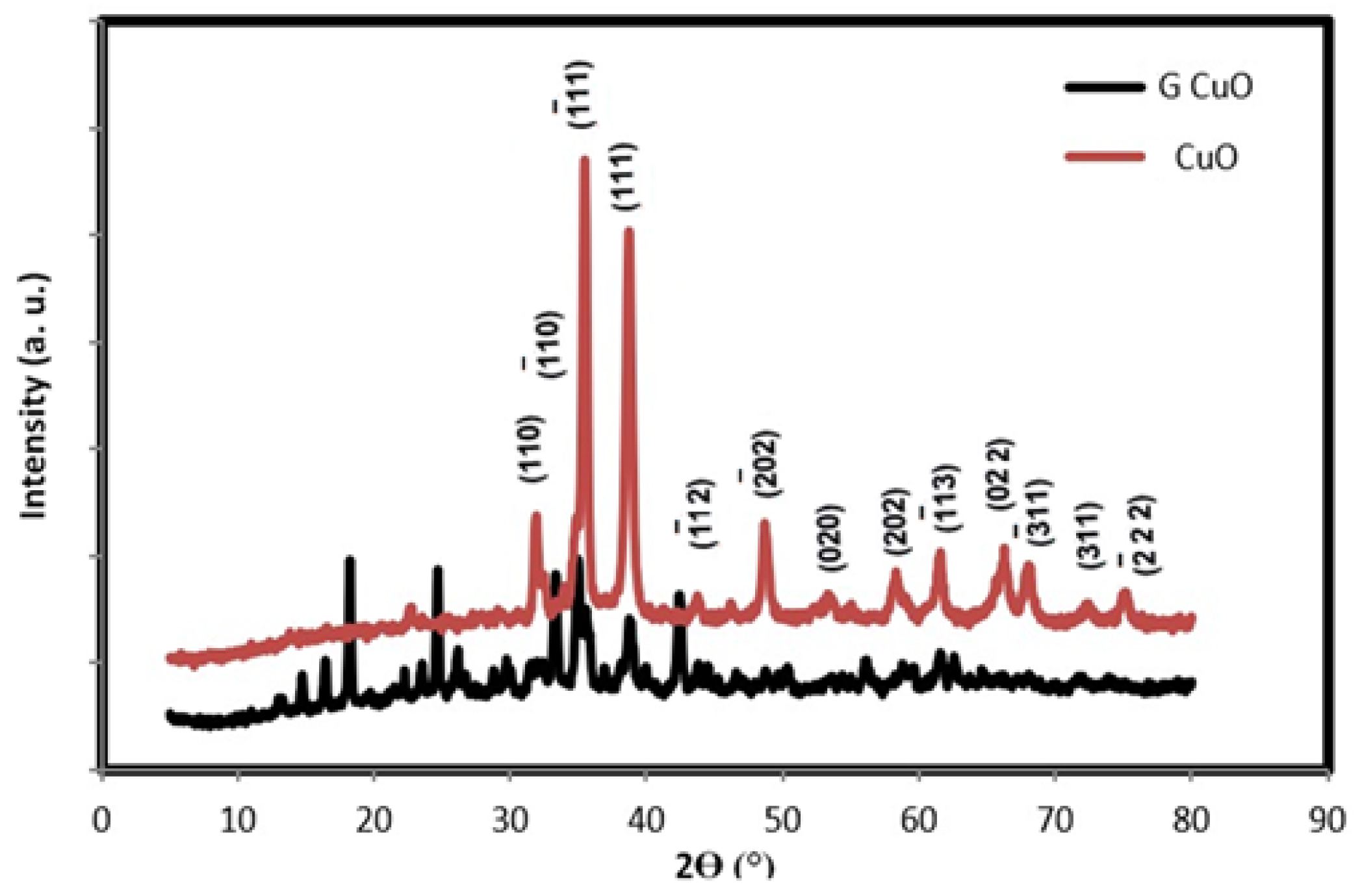
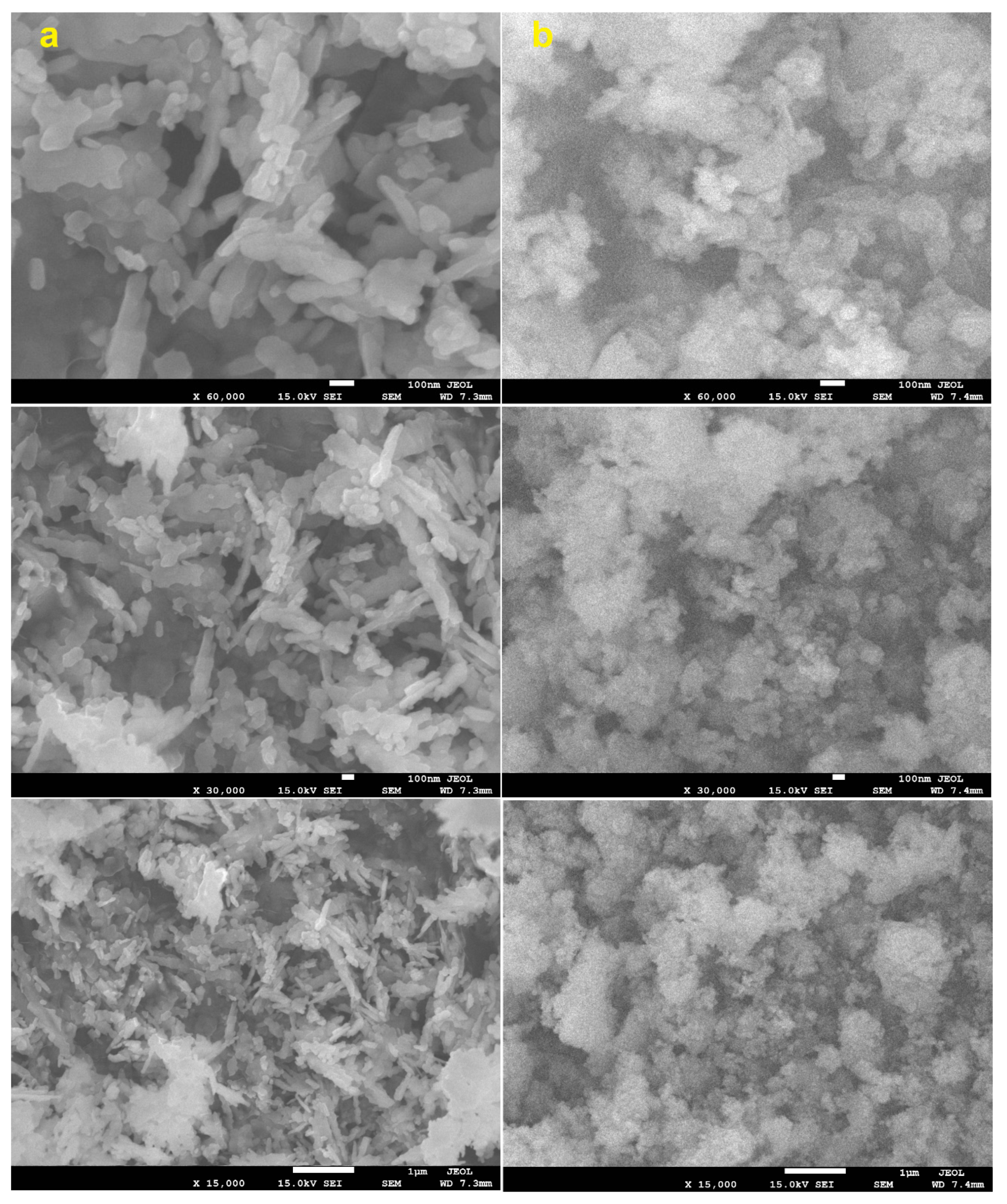
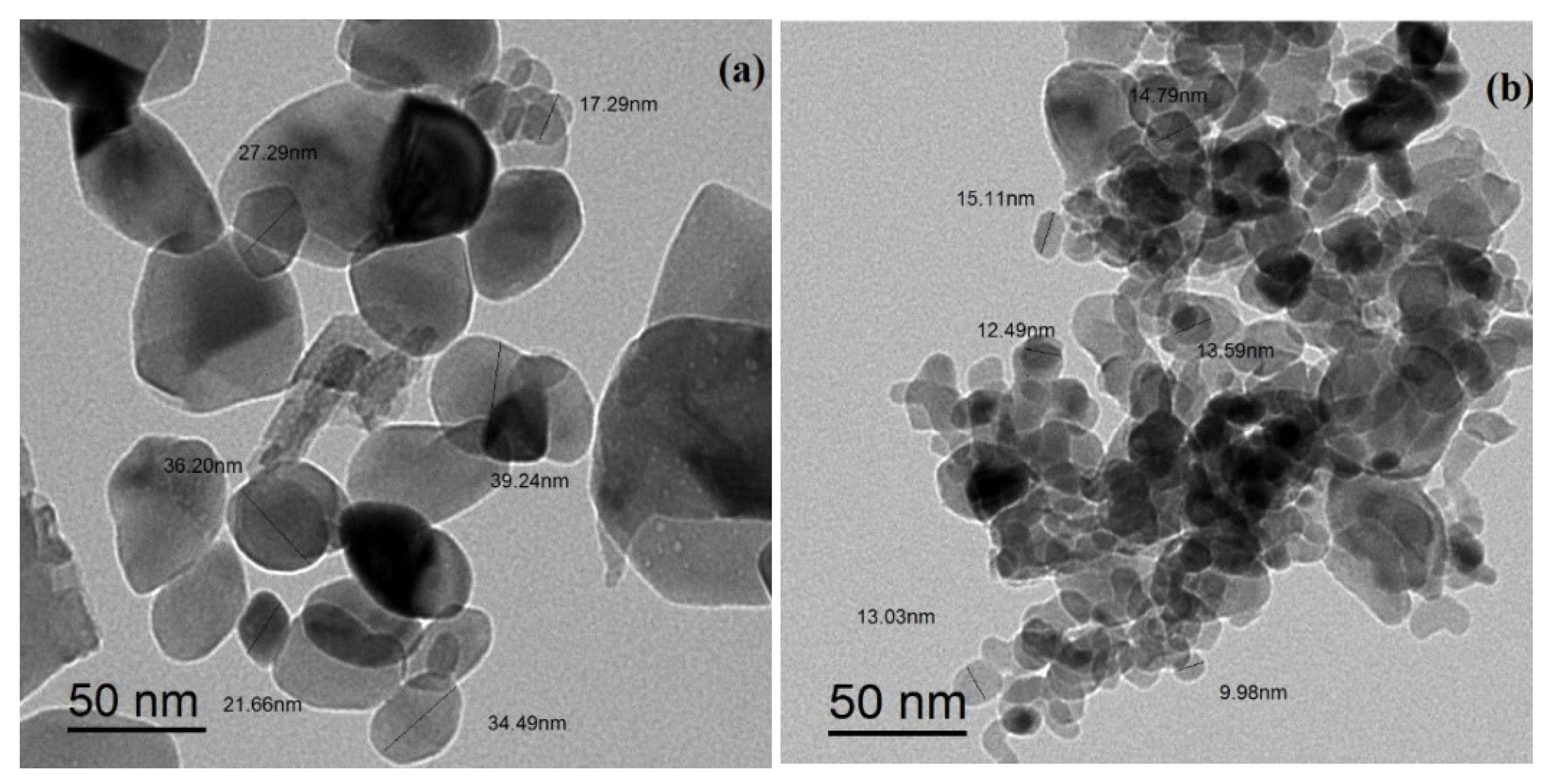
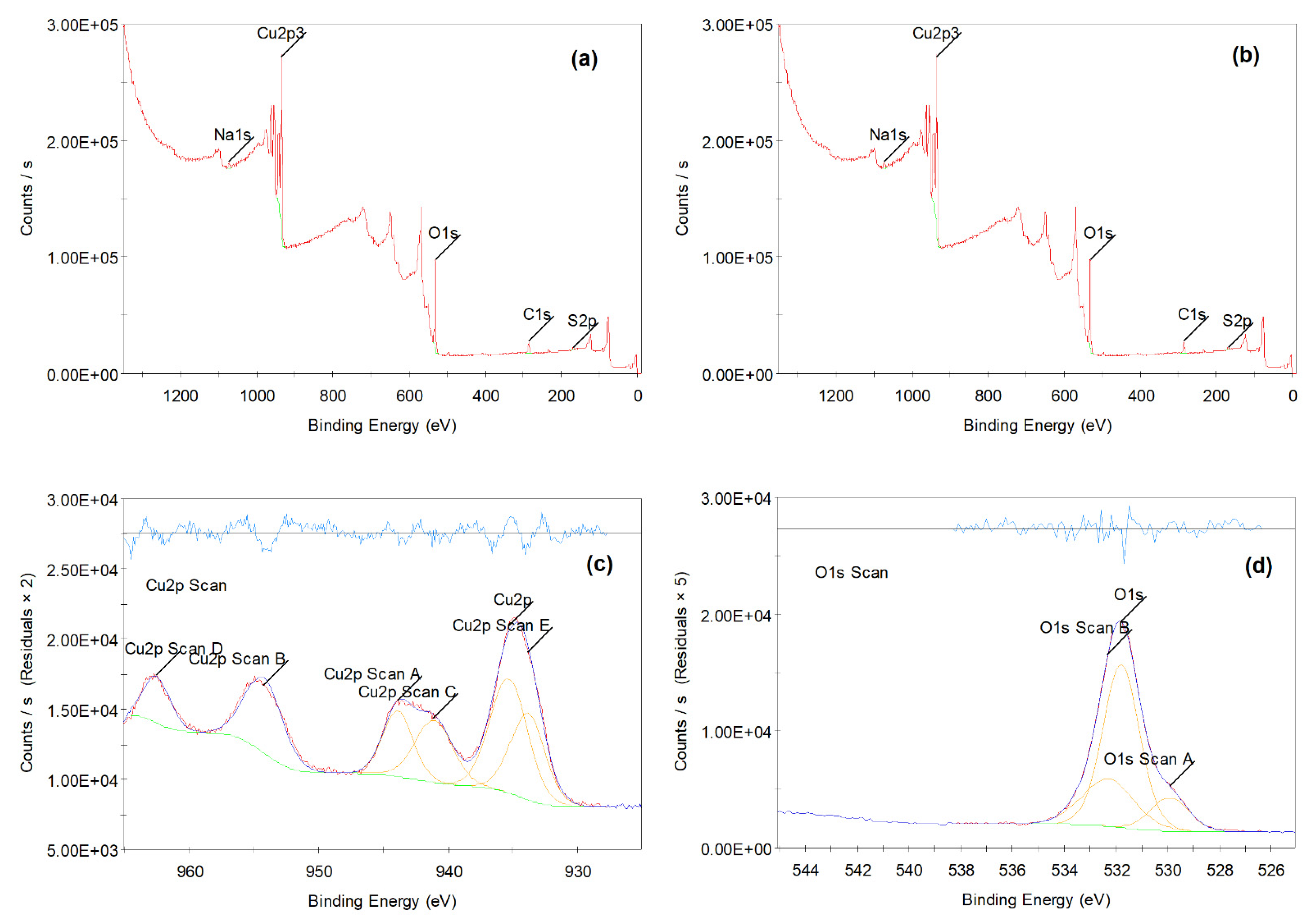
3.1. Characterization of CuO NPs
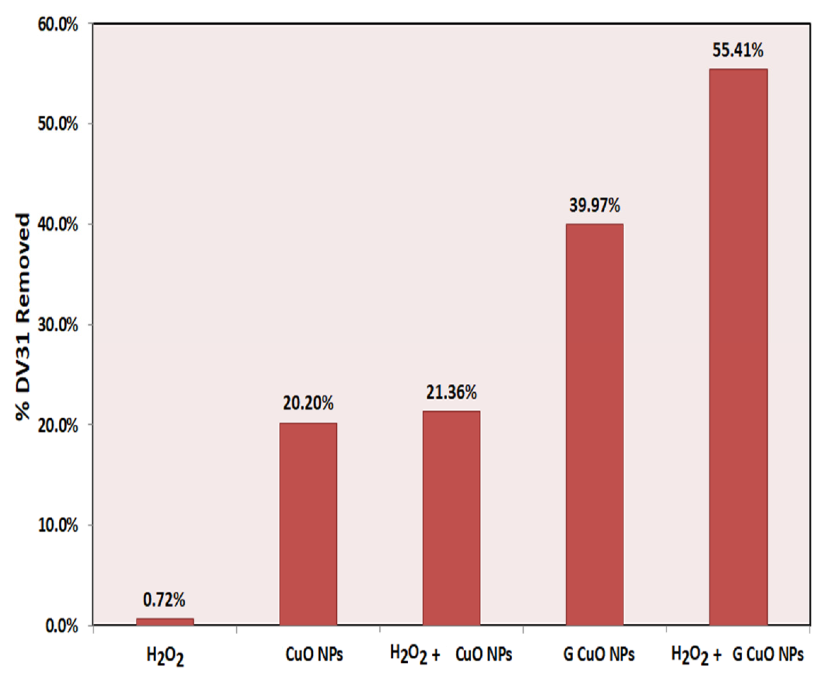
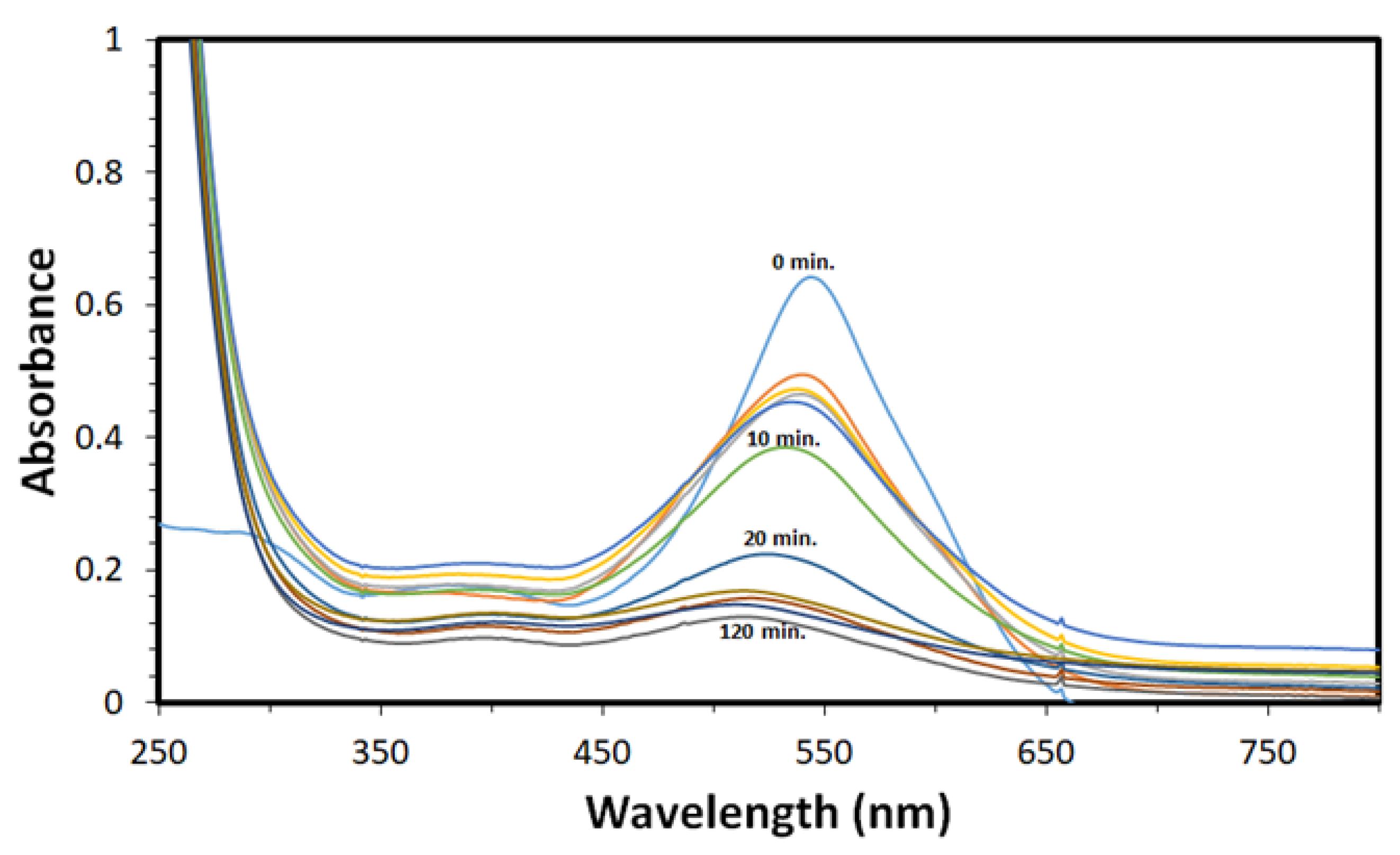
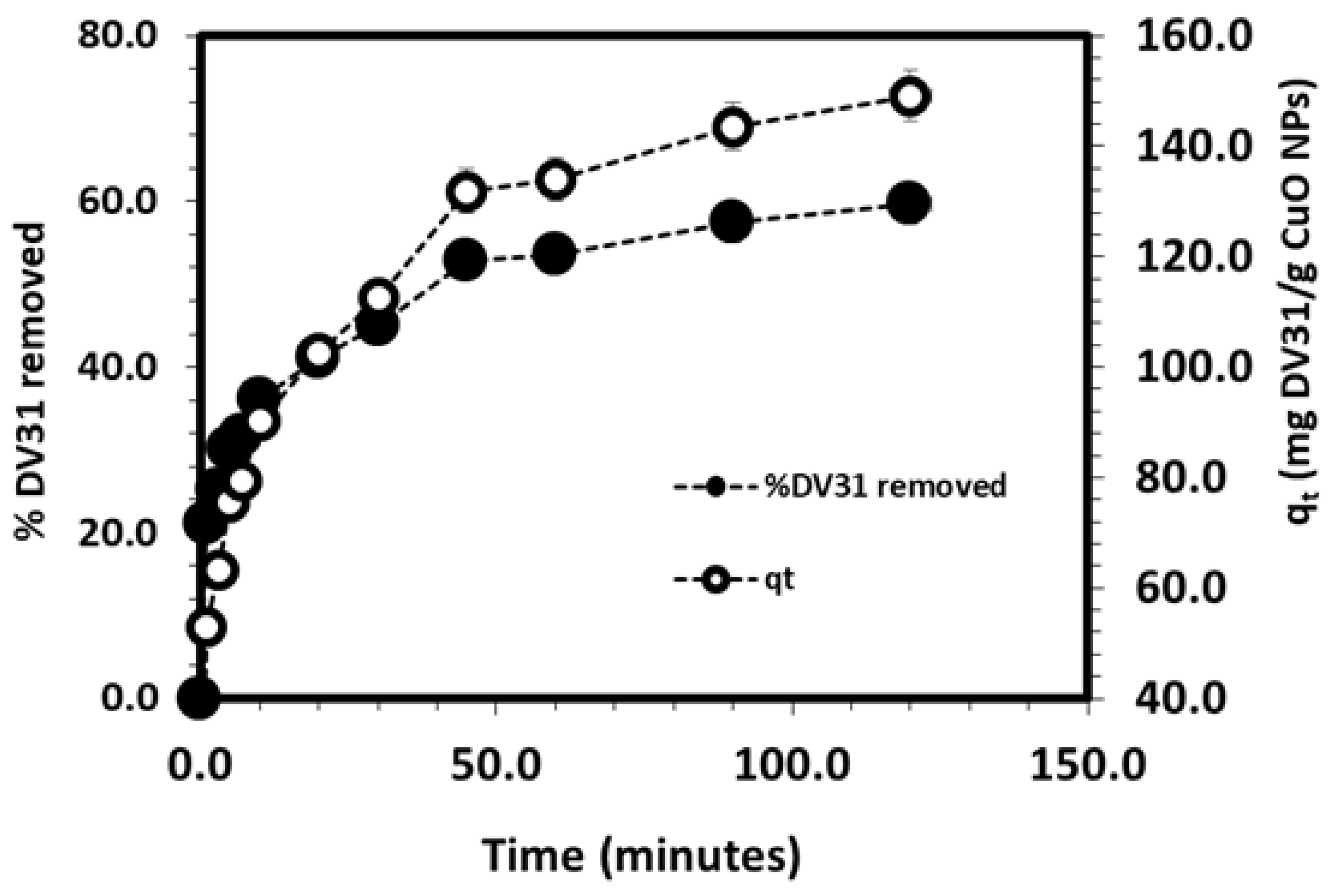
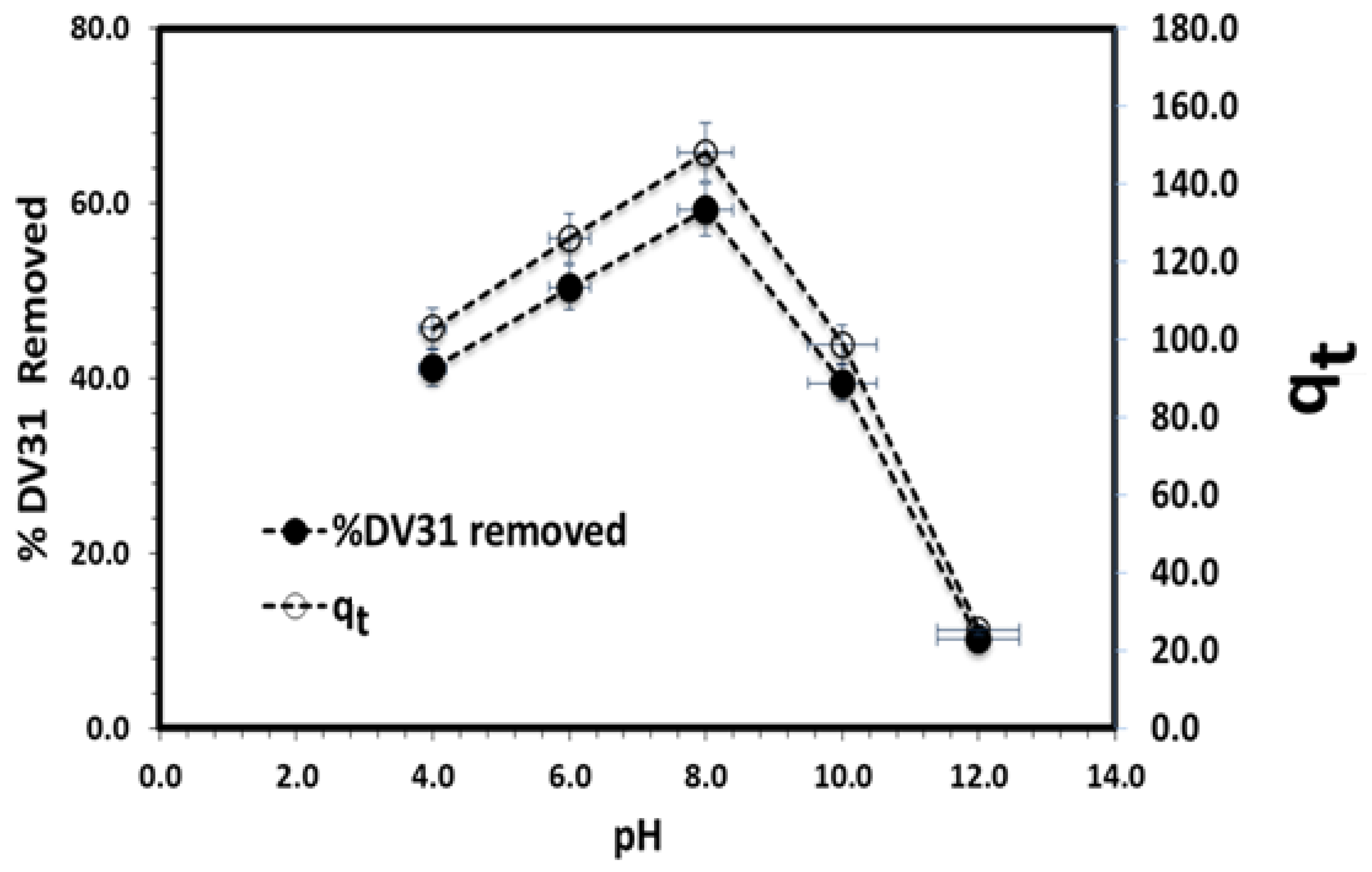
The morphological structure and composition of the prepared CuO NPs, conventional CuO NPs (CuO NPs) and conventional green CuO NPs (G CuO NPs), were explored using XRD, SEM, TEM, XPS and BET. The crystal structure and purity of the different CuO NPs were examined by XRD. Figure 1 shows the XRD pattern of CuO NPs, which showed the presence of characteristic peaks at 2θ values of 32.9°, 33.5°, 35.9°, 39.1°, 42.3°, 49.1°, 54.9°, 58.7°, 61.9°, 66.5°, 68.4°, 72.8° and 75.5°, related to (110), ( 1 ¯ 10), ( 1 ¯ 11), (111), ( 1 ¯ 12), ( 2 ¯ 02), (020), (202), ( 1 ¯ 13), (022), ( 3 ¯ 11), (311) and ( 2 ¯ 22) planes for both the conventional and green CuO, respectively, which can be indexed on the basis of monoclinic tenorite CuO with the C2/c space group (JCPDs file No. 01-080-1916) [36], indicating the good crystallinity of the prepared CuO NPs and the absence of any characteristics peaks of any other phase of CuO. Moreover, there were few unidentified diffractions peaks at 2θ values less than 20°; especially for the conventional CuO, which could not be assigned to any copper-based compound and were considered as impurities. It is also observed that the intensities of the peaks were different, indicating the different degrees of crystallinity as well as the different particle sizes depending on the preparation method. The size of the crystals (D) has been calculated from the FWHM of the main peak with the Debye Scherrer formula using Equation (3):
D = K λ/β COS θ
where K is constant, λ is the wavelength of X-rays employed radiation (1.54056 Å), β is corrected full width at half maximum and θ is Bragg angle, and the crystallite sizes of the CuO NPs were 14.7 nm and 14.4 nm for the T CuO NPs and G CuO NPs, respectively. Figure 2 and Figure 3 show the SEM and TEM images of the different CuO NPs. It is clear that the preparation method greatly affects the shape and dimensions of the CuO NPs as all of them featured different shapes, lengths and widths. The conventional and green CuO NPs were spherical in shape with average particle size of 36 nm and 12 nm, respectively.
The surface binding state and elemental speciation of the different CuO NPs were analyzed by XPS and the wide-scan XPS survey spectra are presented in Figure 4, which confirmed the presence mainly of copper (Cu) and oxygen (O). The XPS survey spectrum of CuO NPs showed sub peaks at binding energies 934.24 eV corresponding to the Cu 2p3 sub peaks corresponding to Cu+2 in CuO NPs; these sub peaks are also consistent with the oxygen bonds indicated as O1s at binding energies and 531 eV. Additionally, the presence of an insignificant amount of carbon and sodium within the CuO NPs sample could be attributed to the preparation process. Figure 4c shows the high-resolution spectra (core XPS spectra) of Cu 2p, which presents the main shake-up peak at the higher binding BE side of the Cu 2p3/2 and the increase in the binding energy of the man peak, suggesting the existence of an unfilled Cu3d9 shell, confirming the presence of Cu2+ in the prepared samples of G CuO NPs. Figure 4d shows the high-resolution spectra (core XPS spectra) of O1s, which shows three components at 529.1 eV, 530.8 eV and 532.2 eV. The first peak at 529.1 eV could be assigned to the BE for lattice oxygen (OL)2− in the G CuO NPs lattice, which is in good agreement with the BE of O2− ion in the metal oxide sites (Cu2+-O2−). The second peak at 530.8 eV could be assigned to the BE for oxygen defects/vacancies (OV)2− in the G CuO NPs matrix, whereas the last peak at 532.2 eV could be assigned to the BE for the adsorbed residual carbon or other surface oxygen species, which easily reacts with the G CuO NPs surface [37].
The specific surface areas of the different CuO NPs were calculated from the nitrogen gas adsorption/desorption isotherms at 77 K applying the BET equation. The BET specific surface areas were 9.4 and 11.1 m2/g for the T CuO NPs and TG CuO NPs, respectively. The calculated average pore diameters were 60.3 nm and 1.93 nm and the pore volume were 0.232 cm3/g and 0.160 cm3/g for the T CuO NPs and TG CuO NPs, respectively.
The above characterization results demonstrate the major effect and the significant enhancement in the CuO NPs upon the application of the corchorus olitorus extract which acts as a capping and stabilizing agent and prevents the agglomeration of the CuO NPs due to the presence of long-chain natural products in the plant extract [38].

3.2. Removal Study

In this comparative study, the removal of DV31 dye from aqueous solution was performed using the different CuO NPs synthesized through a different route (conventional and green) in the presence and absence of the hydrogen peroxide in order to select the most effective NPs, and the results are presented in Figure 5. According to the figure, hydrogen peroxide showed no effect on the catalytic degradation of the DV31 dye in the absence of CuO NPs as the removal percentage was 0.72%, whereas the CuO NPs in the absence of the hydrogen peroxide have low removal efficacies: 20.20% and 39.97%, for T CuO NPs and G CuO NPs, respectively. These removal efficacies were enhanced greatly upon the combination of both CuO NPs and hydrogen peroxide, as the removal percentage reached 21.36% and 55.41% for the hydrogen peroxide and T CuO NPs and G CuO NPs, respectively. This may indicate that CuO NPs may activate the hydrogen peroxide and produce free radicals which greatly participate in the degradation of the DV31 dye [39]. The experimental results revealed that the removal of DV31 dye was higher in the case of G CuO NPs prepared using corchorus olitorus as the capping agent by precipitation method, compared with the conventional CuO NPs. This may be attributed to the small particle size of the G CuO NPs as well as the good dispersion compared with the T CuO NPs. Accordingly, the G CuO NPs were selected for the rest of the experiments to study the removal of DV31 dye in the presence of hydrogen peroxide via AOP.
The effect of G CuO NPs mass on the removal of DV31 dye from aqueous solution was studied and the results are presented in Figure 6. The experimental results revealed that increasing the G CuO NPs mass greatly enhanced the removal efficiency due to the increase in the number of active sites available for the binding of the DV31 dye molecules, till it reached its complete removal of the DV31 dye using 30 mg of G CuO NPs, whereas the removal capacity was decreased linearly with increasing the G CuO NPs mass and the maximum capacity obtained was 150.25 mg DV31/g CuO NPs using 10 mg of the CuO NPs in the presence of 0.5 mL of hydrogen peroxide.
The effect of the H2O2 volume on the degradation of DV31 dye in the presence of the G CuO NPs was optimized (Figure 7). It is clear that increasing H2O2 volume significantly decreased the removal efficiency of the DV31 dye, which may indicate that H2O2 is not the main reagent for the catalytic degradation of the DV31 dye molecules in water, the degradation mainly depends on the presence of CuO NPs and the H2O2 as the CuO NPs may activate the hydrogen peroxide and produce free radicals, which greatly participate in the degradation of the DV31 dye.
The contact, interaction and removal time between the pollutant such as DV31 dye and the solid catalyst such as CuO NPs is an important factor greatly affecting the removal of any pollutants from the environment. Figure 8 shows the UV-Vis spectra of DV31 dye and the variation of the absorbance with time for the removal of DV31 dye from aqueous solution using G CuO NPs.
The effect of the contact time on DV31 dye removal from model solution by CuO NPs was explored and the results are shown in Figure 9. It is noted that the percentage removal of DV31 increased within the first 30.0 min with 45% efficacy and removal capacity of 112.48 mg DV31/g CuO NPs. Increasing the reaction time was associated with an insignificant increase of the percentage removal to 60% and removal capacity of 149.15 mg DV31 dye/g CuO NPs after 120 min. According to the results, the equilibration time was assigned as 30.0 min for the rest of the experiments.
The variation of the catalytic degradation of DV31 dye using G CuO NPs by solution temperature was studied in the presence of 0.5 mL hydrogen peroxide. According to Figure 10, raising the solution temperature correlated with a significant increase in the % DV31 dye degradation; 22.07% at 281 K to 100% at 333 K, indicating the endothermic nature of the removal process. The observed enhancement of the removal efficacy may be attributed to two main factors; the first factor may be the improvement in the diffusion of the DV31 dye molecules from the bulk solution to the G CuO NPs surface associated with rising the solution temperature owing to the decrease in the viscosity of the solution [40] and the second factor is the fact that increasing the solution temperature enhanced the degradation of the hydrogen peroxide and the production of the hydroxyl free radicals [41], which reacted with CuO NPs and degraded the DV31 dye molecules.
The solution pH is another principal parameter which controls the removal process, especially for DV31 dye, because they contain different functional groups, amide group (HN-CO-R) and amino group (HN-R), which may ionize depending on the pH of the solution. The effect of solution pH on DV31 dye degradation by G CuO NPs was therefore explored in the pH range from 4.0 to 12.0. The results are presented in Figure 11 and it was observed that the removal percentage increased from 41.15% to 59.27% as the pH of the solution changed from 4.0 to 8.0. Further increase in the solution pH from 8.0 to 12.0 was accompanied by a sharp decrease in the removal percentage from 59.27% to 10.11%. This phenomenon could be due to many factors. Firstly, the point-of-zero-charge of CuO NPs is equal to 9.0 [42,43]; which means that CuO NPs are positively charged below a pH value of 9.0 and negatively charged above a pH value of 9.0; this is because at pH less than 7.0 the major abundant species is [H+], while at pH more than 7.0 the major abundant species reacts with [OH-], which adsorbed at the CuO surface. At the same time, DV31 is characterized by a pKa value of 4.0. As the pH value increased from 4.0 to 9.0, the removal of DV31 increased due to the attraction forces between the positively charged G CuO NPs surface and the negatively charged DV31 dye molecules, which agreed well with a previous study [44]. Further increase in the solution pH from 8.0 to 11.55 was associated with a sharp decrease in the removal percentage of DV31, due to the electrostatic repulsion between the negatively charged G CuO NPs and the negatively charged DV31 molecules. This may indicate that the adsorption and further degradation of DV31 on G CuO NPs surface could be electrostatic-in-nature and mainly depends on the charge of both DV31 dye molecules and the G CuO NPs surface.

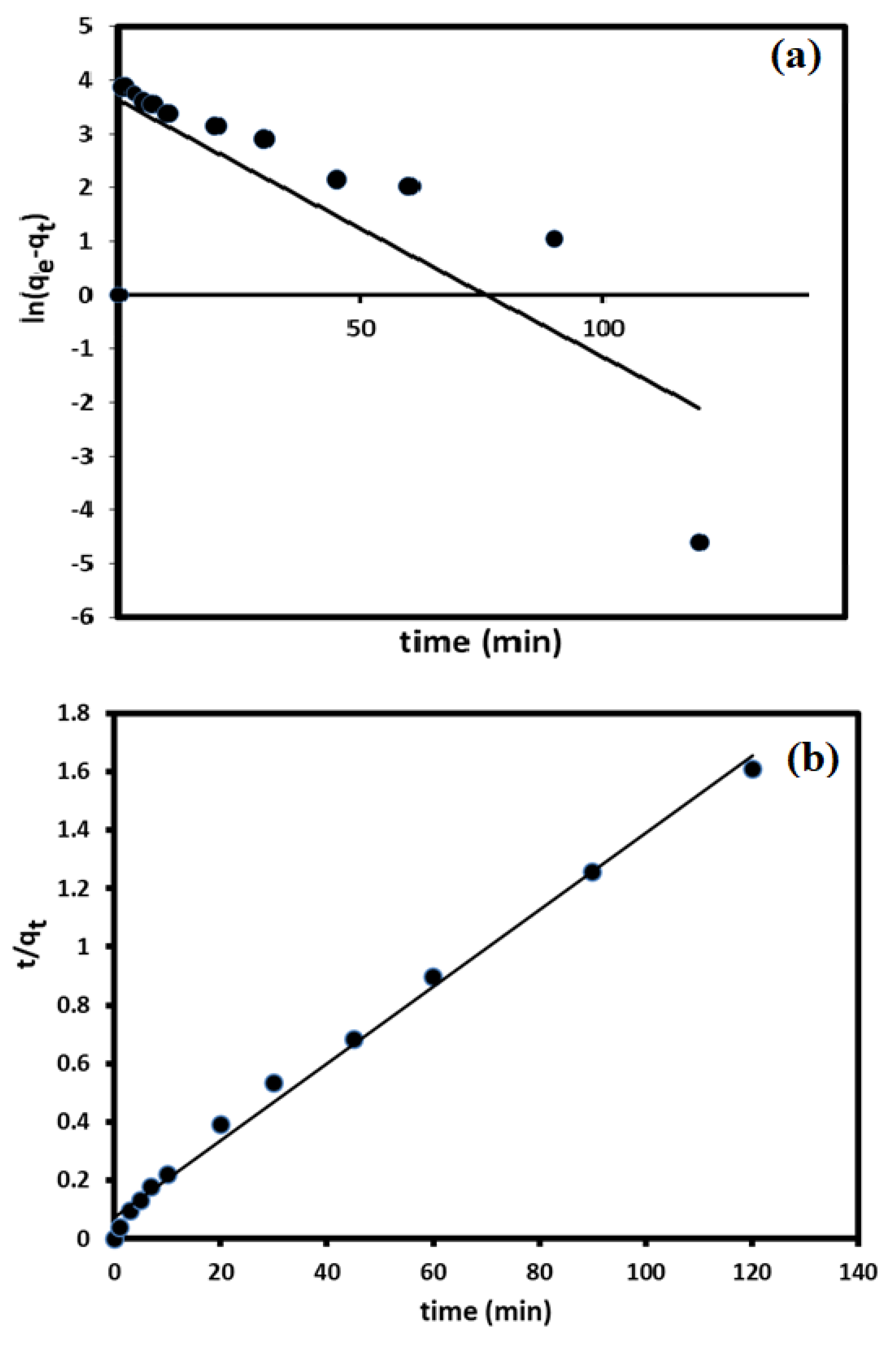
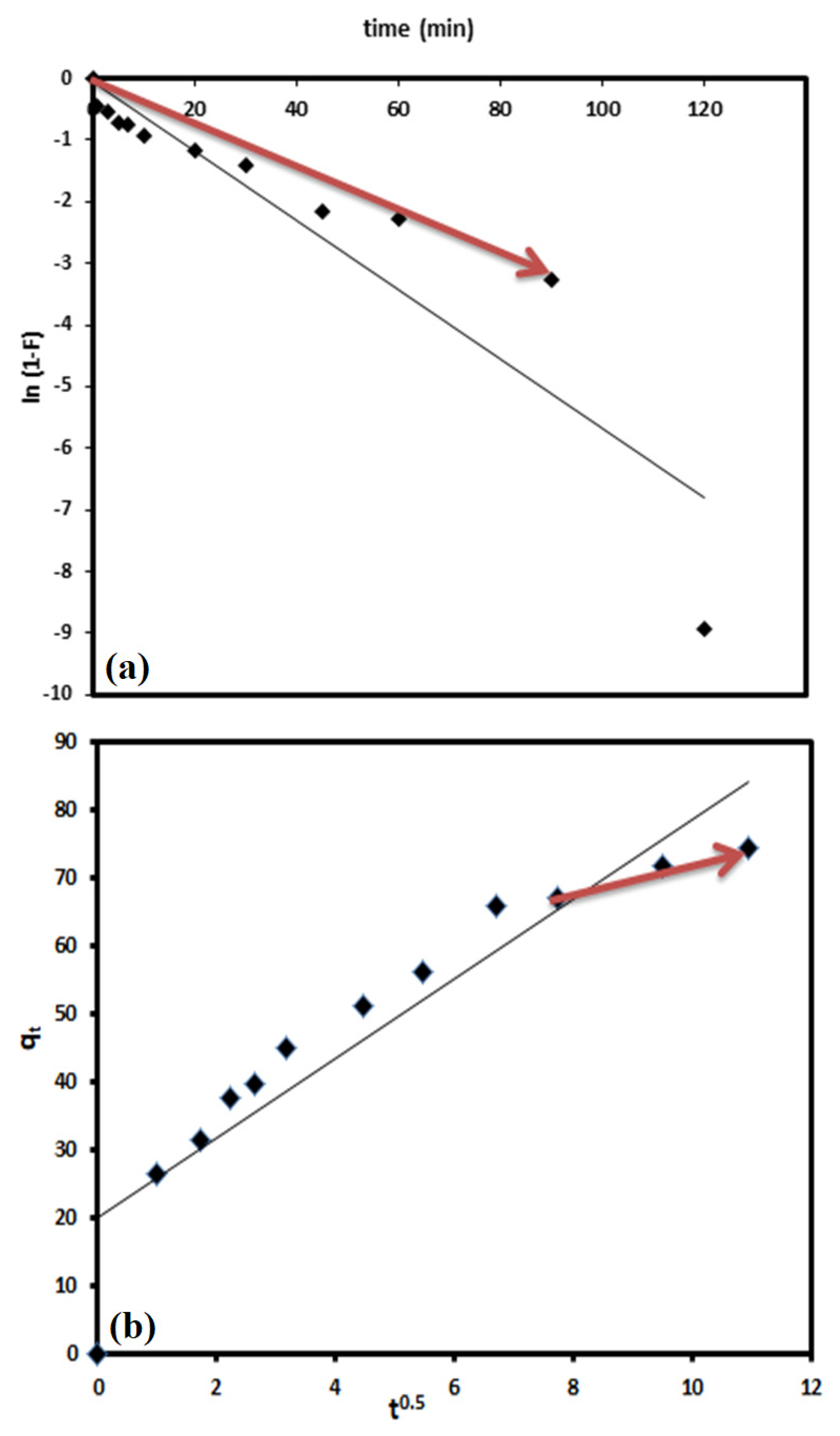
3.3. Kinetic Studies

To understand the adsorption/removal of pollutants such as DV31 dye from an aqueous solution by a solid catalyst/adsorbent such as G CuO NPs, it must be studied kinetically in order to obtain the appropriate mathematical models to describe the interactions between the pollutant and the solid adsorbent, in order to design suitable adsorbent materials for environmental and industrial applications. Figure 9 shows the adsorption/removal experimental data representing the variation of the amount DV31 dye adsorbed/removed from aqueous solution by G CuO NPs (qt) with time and Figure 8 shows that the adsorption/removal of DV31 by G CuO NPs reached equilibrium within 45 min. Because all chemical reactions involving solid reactants such as the CuO NPs are usually based on the adsorption of the reactants in aqueous solution on the surface of the solid reactants, it is very important to apply the adsorption kinetic models to the removal experimental data. The adsorption experimental data were treated kinetically using the most common and well-known kinetic models to understand the nature of the adsorption process; Lagergren pseudo-first-order kinetic model [45] and pseudo-second-order kinetic model [46].
Lagergren pseudo-first-order kinetic model is one of the most frequent kinetic models used to describe the adsorption of different adsorbates from an aqueous solution by solid adsorbent [45,47] and it is linearized for as follows:
ln ( q e q t ) = ln q e k 1 t
where k1 (min−1) is the pseudo-first-order rate coefficient and qe and qt are the values of the amount adsorbed/removed per unit mass at equilibrium and at any time t, respectively. Applying Equation (4) to the experimental adsorption data in Figure 12a and plotting ln (qe − qt) vs. t did not give a straight line, and low convergence and an unacceptable R2 value were obtained, as is shown in Figure 12a and Table 1, demonstrating the unsuitability of the Lagergren pseudo-first-order kinetic model for describing the adsorption/removal of DV31 dye by G CuO NPs from aqueous solution.
The linearized equation of the pseudo-second-order kinetic model equation is given as:
t q t = 1 k 2 q e 2 + t q t
where k2 (g/(mg·min)) is the pseudo-second-order rate coefficient. Applying the pseudo-second-order kinetic model equation to the adsorption/removal experimental data in Figure 8, by plotting of t/qt and t of Equation (5), converges well, with excellent R2 value, and a straight line was obtained from which qe and k2 were estimated from the slope and intercept of the plot, respectively, as presented in Figure 11b and Table 1, confirming the appropriateness of the pseudo-second-order kinetic model for the description of DV31 dye adsorption/removal by the G CuO NPs from aqueous solution.
Another test was performed to confirm the suitability of the pseudo-second-order kinetic is by calculating the Chi-square test using the following mathematical Equation (6) [48] and Sum of the squares of errors (SSE) at Equation (7) [49]:
χ 2 = ( q e , calc q e , exp ) 2 q e , calc
SSE = i = 1 n ( q e , calc i q e , exp i ) 2
where qe,calc and qe,exp are the calculated and experimental removed amount of DV31 dye molecules per unit mass of the G CuO NPs at equilibrium. The χ2 values were 35.84 and 0.02284 and the achieved SSE values were 1354 and 1.734, for the pseudo-first-order kinetic model and the pseudo-second-order kinetic model applied, respectively, indicating the appropriateness of the pseudo-second-order kinetic model for describing the removal process.
Based on the above results, it could be concluded here that the pseudo-second-order kinetic model was the best for describing the adsorption of DV31 dye from aqueous solution by CuO NPs.
The adsorption usually occurs in four distinctive steps: bulk diffusion, mass action, liquid film diffusion and finally intra-particle diffusion, which is classified as a pore and surface diffusion. This part focuses on the determination of the rate-determining step for the adsorption/diffusion process.
The linearized form of the liquid film diffusion equation is as follows [50]:
ln ( 1 F ) = k fd t  
where F is the fractional attainment of equilibrium and equals to q t q e and kfd (min−1) is the film diffusion rate coefficient. By applying Equation (8) to the experimental data in Figure 9 and by plotting ln (1 − F) versus t, a straight line was obtained with zero intercept, but only for the first 90.0 min of the interaction, from which kfd was estimated from the slope of the plot, as shown in Figure 13a, for the removal of DV31 dye by G CuO NPs, with acceptable R2 value of 0.965 for the first 90.0 min of the adsorption/removal process.
The linearized intraparticle diffusion equation is as follows [50]:
q t = k id t 0.5 + C i
where kid (mg/(g.min0.5)) is the intraparticle diffusion coefficient and Ci (mg/g) is related to the boundary layer thickness. Applying Equation (9) to the experimental data by plotting qt versus t0.5, as presented in Figure 12b, a straight line was obtained for the removal of DV31 dye by CuO NPs after the first 60.0 min of the interaction with a good R2 value of 0.99. This may indicate that the removal of DV31 dye by the G CuO NPs was controlled by both liquid film and intraparticle diffusion kinetics during the time of adsorption.

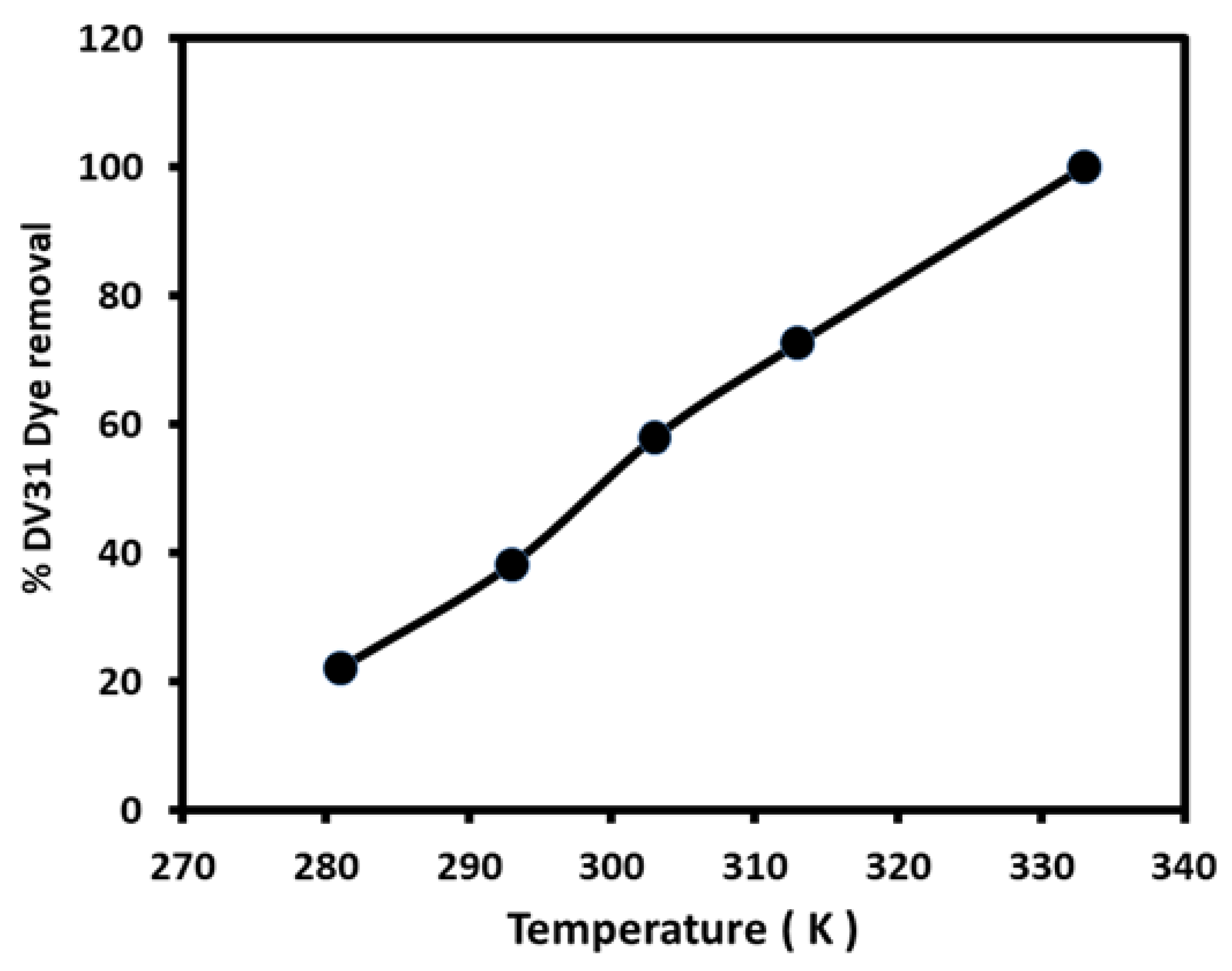
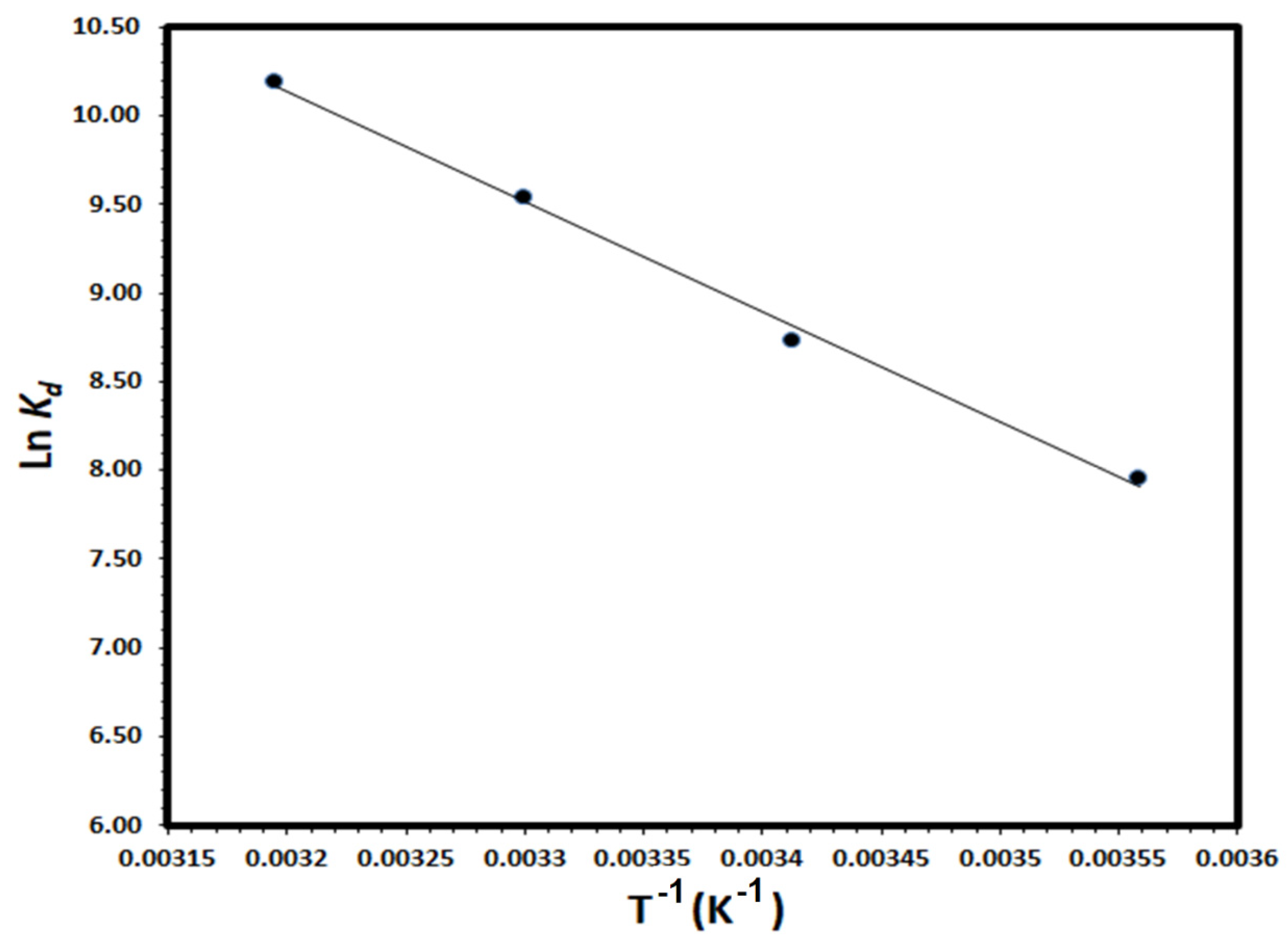
3.4. Thermodynamic Studies

Thermodynamic parameters; enthalpy change (ΔH), entropy change (ΔS) and Gibbs free energy change (ΔG), were calculated to evaluate the thermodynamic feasibility and the spontaneous nature of the removal of DV31 dye molecules by G CuO NPs from aqueous solution. Thermodynamic parameters were calculated using the variation of the thermodynamic distribution coefficient D with a change in temperature according to the equation [51]:
K d = q e C e × 1000
where qe is the amount of DV31 molecules adsorbed/removed by G CuO NPs (mg/g) at equilibrium and Ce is the equilibrium concentration of DV31 in solution (mg/L). The ΔH and ΔS could be calculated according to the following equation [52]:
ln K d = Δ S R Δ H R T
A straight line was obtained by plotting ln Kd vs. 1/T; as shown in Figure 14 and the ΔH and ΔS values were calculated from the slope and the intercept of the straight line, respectively. The ΔG values were calculated at 298K from the relation:
ΔG = ΔH − TΔS
Thermodynamic parameters for the adsorption/removal of DV31 molecules degraded by CuO NPs were calculated and ∆H, ∆S and ∆G values were +51.64 kJ/mole, +249.5 J/mole.K and −22.71 kJ/mole, respectively. The positive value of the enthalpy change indicated the endothermic nature of DV31 by G CuO NPs and the positive value of the entropy change indicated the increase in the degree of freedom at the solid-liquid interface and apparently, the negative value of the free energy change as would be expected for a product favored and spontaneous degradation process. Accordingly, the negative value of ∆G and positive values of ∆H and ∆S suggested that the adsorption of DV31 by CuO NPs from an aqueous solution is an entropy-driven process.

3.5. The Removal Mechanism

The reaction between G CuO NPs and H2O2 is well known and studied as an AOPs mechanism, CuO NPs react with H2O2 and produce OH•, which attacks nearly all organic complexes. Therefore, the reaction of the organic pollutant with the HO• leads to a complete breakdown of the organic compound and, as a result, AOPs diminish the concentration of the pollutant from a few hundred mg/L to less than 5 ng/L. Many studies have shown that, with AOP, organic chemicals disintegrate and become smaller and easily biodegradable [53,54]. The degradation of DV31 dye molecules through AOPs was monitored through TOC analysis. The %TOC, total organic carbon, in the solution with time were measured during the catalytic degradation process of the DV31 dye by G CuO NPs as shown in Figure 15. It is observed that the decrease in the % DV31 dye in solution was associated with the same decrease trend of the %TOC in solution. The DV31 dye in solution decreased to around 88% within 10 min associated with %TOC in the water of 78% and this percentage was decreased to 9% after 90 min with a %DV31 dye in the solution of 55%. This may indicate the successful removal of the DV31 dye by G CuO NPs in presence of H2O2.

3.6. Environmental Applications

Application of the proposed heterogeneous catalyst; G CuO NPs, for the removal of DV31 dye from real samples is a crucial point to validate the applicability of the method. Therefore, the removal of DV31 dye by CuO NPs from three different samples collected from different environments was investigated, namely, well water sample collected from a well, a seawater sample collected from Red Sea in Jeddah city, wastewater sample collected from King Abdulaziz University Wastewater (KAUWW). The % removed was calculated by comparing DV31 dye concentrations before and after the addition of CuO NPs in presence of hydrogen peroxide. The % DV31 dye removed from the real sample was calculated and was found to be 100% in all real samples. This may indicate that G CuO NPs are a promising and potential adsorbent for the remediation of a polluted environment.

4. Conclusions

This research work explored the preparation of CuO NPs via eco-friendly and green method using the extract of corchorus olitorus leaves (Molokhaia) and their characterization using XRD, TEM, XPS and textural properties and the results demonstrated the efficacious formation of monoclinic tenorite CuO nanoparticles with average particle size of 12 nm and surface area of 11.1 m2/g. The eco-friendly green CuO NPs were used for the environmental remediation for the efficient catalytic degradation of direct violet dye (DV31) via advanced oxidation process (AOP) in presence of H2O2. The impact of AOP environmental parameters affecting the degradation process was investigated and the results revealed that most of the DV31 were removed from the solution using G CuO NPs within 45 min at pH 8.0 and 298 K. Moreover, the catalytic degradation of the direct violet dye using the ecofriendly green CuO NPs was studied kinetically and thermodynamically and the results showed that the catalytic degradation process agreed well with the pseudo-second order kinetic model with removal capacity of 74.59 mg/g. Additionally, the process was spontaneous; (∆G = −22.71 kJ/mole), endothermic in-nature (∆H = +51.64 kJ/mole), with increase in the degree of freedom at the solid-liquid interface (∆S = +249.5 J/mole.K), suggesting that the adsorption/removal of DV31 by G CuO NPs from an aqueous solution is an entropy-driven process. Finally, the catalytic degradation of the direct violet dye using G CuO NPs was studied in real water samples and the results revealed the outstanding performance of the G CuO NPs and their possible application for the remediation of a polluted environment.

Author Contributions

Conceptualization, S.A.K. and M.A.S.; methodology, M.A.S.; validation, M.A.S. and S.A.K.; formal analysis, R.O.A.; investigation, R.O.A. and S.A.K.; resources, M.A.S.; data curation, R.O.A..; writing—original draft preparation, R.O.A.; writing—review and editing, M.A.S.; visualization, M.A.S.; supervision M.A.S. and S.A.K.; project administration, M.A.S.; funding acquisition, M.A.S. All authors have read and agreed to the published version of the manuscript.

Funding

This research was funded by the Ministry of Education and Deanship of Scientific Research (DSR), King Abdulaziz University, grant number [IFPIP-606-130-1442].

Institutional Review Board Statement

Not applicable.

Informed Consent Statement

Not applicable.

Data Availability Statement

Not applicable.

Acknowledgments

This research work was funded by Institutional Fund Projects under grant no. (IFPIP-606-130-1442). Therefore, the authors gratefully acknowledge technical and financial support from the Ministry of Education, and the Deanship of Scientific Research (DSR), King Abdulaziz University, Jeddah, Saudi Arabia.

Conflicts of Interest

The authors declare no conflict of interest.

References

  1. Ighalo, J.O.; Sagboye, P.A.; Umenweke, G.; Ajala, O.J.; Omoarukhe, F.O.; Adeyanju, C.A.; Adeniyi, A.G. CuO nanoparticles (CuO NPs) for water treatment: A review of recent advances. Environ. Nanotechnol. Monit. Manag. 2021, 15, 100443. [Google Scholar] [CrossRef]
  2. Pavithra, N.; Manukumar, K.; Viswanatha, R.; Nagaraju, G. Combustion-derived CuO nanoparticles: Application studies on lithium-ion battery and photocatalytic activities. Inorg. Chem. Commun. 2021, 130, 108689. [Google Scholar] [CrossRef]
  3. Tom, A.P. Nanotechnology for sustainable water treatment—A review. Mater. Today Proc. 2021; in press. [Google Scholar] [CrossRef]
  4. Bhawana, P.; Fulekar, M.H. Nanotechnology: Remediation technologies to clean up the environmental pollutants. Res. J. Chem. Sci. 2012, 2, 90–96. [Google Scholar]
  5. Saglam, N.; Korkusuz, F.; Prasad, R. Nanotechnology Applications in Health and Environmental Sciences; Springer International Publishing: Cham, Switzerland, 2021. [Google Scholar] [CrossRef]
  6. Karimi-Maleh, H.; Sheikhshoaie, M.; Sheikhshoaie, I.; Ranjbar, M.; Alizadeh, J.; Maxakato, N.W.; Abbaspourrad, A. A novel electrochemical epinine sensor using amplified CuO nanoparticles and an-hexyl-3-methylimidazolium hexafluorophosphate electrode. New J. Chem. 2019, 43, 2362–2367. [Google Scholar] [CrossRef]
  7. Cosma, D.; Urda, A.; Radu, T.; Rosu, M.C.; Mihet, M.; Socaci, C. Evaluation of the Photocatalytic Properties of Copper Oxides/Graphene/TiO2 Nanoparticles Composites. Molecules 2022, 27, 5803. [Google Scholar] [CrossRef]
  8. Momeni, M.M.; Ghayeb, Y.; Menati, M. Facile and green synthesis of CuO nanoneedles with high photo catalytic activity. J. Mater. Sci. Mater. Electron. 2016, 27, 9454–9460. [Google Scholar] [CrossRef]
  9. Sulaiman, S.; Ahmad, S.; Naz, S.S.; Qaisar, S.; Muhammad, S.; Alotaibi, A.; Ullah, R. Synthesis of Copper Oxide-Based Nanoformulations of Etoricoxib and Montelukast and Their Evaluation through Analgesic, Anti-Inflammatory, Anti-Pyretic, and Acute Toxicity Activities. Molecules 2022, 27, 1433. [Google Scholar] [CrossRef]
  10. Jadhav, M.S.; Kulkarni, S.; Raikar, P.; Barretto, D.A.; Vootla, S.K.; Raikar, U.S. Green biosynthesis of CuO & Ag–CuO nanoparticles from Malus domestica leaf extract and evaluation of antibacterial, antioxidant and DNA cleavage activities. New J. Chem. 2017, 42, 204–213. [Google Scholar] [CrossRef]
  11. Gnanavel, V.; Palanichamy, V.; Roopan, S.M. Biosynthesis and characterization of copper oxide nanoparticles and its anticancer activity on human colon cancer cell lines (HCT-116). J. Photochem. Photobiol. B Biol. 2017, 171, 133–138. [Google Scholar] [CrossRef]
  12. Nwanya, A.C.; Ndipingwi, M.M.; Mayedwa, N.; Razanamahandry, L.; Ikpo, C.O.; Waryo, T.; Ntwampe, S.; Malenga, E.; Fosso-Kankeu, E.; Ezema, F.I.; et al. Maize (Zea mays L.) fresh husk mediated biosynthesis of copper oxides: Potentials for pseudo capacitive energy storage. Electrochim. Acta 2019, 301, 436–448. [Google Scholar] [CrossRef]
  13. Nasrollahzadeh, M.; Sajadi, S.M.; Rostami-Vartooni, A. Green synthesis of CuO nanoparticles by aqueous extract of Anthemis nobilis flowers and their catalytic activity for the A3 coupling reaction. J. Colloid Interface Sci. 2015, 459, 183–188. [Google Scholar] [CrossRef] [PubMed]
  14. Sone, B.T.; Diallo, A.; Fuku, X.G.; Gurib-Fakim, A.; Maaza, M. Biosynthesized CuO nano-platelets: Physical properties & enhanced thermal conductivity nanofluidics. Arab. J. Chem. 2020, 13, 160–170. [Google Scholar] [CrossRef]
  15. Shi, G.; Liu, J.; Chen, B.; Bao, Y.; Xu, J. Phase-controlled growth of cubic phase CuO nanoparticles by chemical vapor deposition. Phys. Status Solidi 2017, 214, 1700041. [Google Scholar] [CrossRef]
  16. Arunkumar, B.; Jeyakumar, S.J.; Jothibas, M. A sol-gel approach to the synthesis of CuO nanoparticles using Lantana camara leaf extract and their photo catalytic activity. Optik 2019, 183, 698–705. [Google Scholar] [CrossRef]
  17. Meng, J.; Yang, Z.; Chen, L.; Qin, H.; Cui, F.; Jiang, Y.; Zeng, X. Energy storage performance of CuO as a cathode material for aqueous zinc ion battery. Mater. Today Energy 2019, 15, 100370. [Google Scholar] [CrossRef]
  18. Mahmood, A.; Tezcan, F.; Kardaş, G. Photoelectrochemical characteristics of CuO films with different electrodeposition time. Int. J. Hydrog. Energy 2017, 42, 23268–23275. [Google Scholar] [CrossRef]
  19. Ashok, A.; Kumar, A.; Tarlochan, F. Highly efficient nonenzymatic glucose sensors based on CuO nanoparticles. Appl. Surf. Sci. 2019, 481, 712–722. [Google Scholar] [CrossRef]
  20. Velusamy, T.; Liguori, A.; Macias-Montero, M.; Padmanaban, D.B.; Carolan, D.; Gherardi, M.; Colombo, V.; Maguire, P.; Svrcek, V.; Mariotti, D. Ultra-small CuO nanoparticles with tailored energy-band diagram synthesized by a hybrid plasma-liquid process. Plasma Process. Polym. 2017, 14, 1600224. [Google Scholar] [CrossRef] [Green Version]
  21. Cuong, H.N.; Pansambal, S.; Ghotekar, S.; Oza, R.; Hai, N.T.T.; Viet, N.M.; Nguyen, V.H. New frontiers in the plant ex-tract mediated biosynthesis of copper oxide (CuO) nanoparticles and their potential applications: A review. Environ. Res. 2022, 203, 111858. [Google Scholar] [CrossRef]
  22. Adeyemi, J.O.; Onwudiwe, D.C.; Oyedeji, A.O. Biogenic Synthesis of CuO, ZnO, and CuO–ZnO Nanoparticles Using Leaf Extracts of Dovyalis caffra and Their Biological Properties. Molecules 2022, 27, 3206. [Google Scholar] [CrossRef] [PubMed]
  23. Sharma, S.; Kumar, K.; Thakur, N.; Chauhan, S.; Chauhan, M.S. Eco-friendly Ocimum tenuiflorum green route synthesis of CuO nanoparticles: Characterizations on photocatalytic and antibacterial activities. J. Environ. Chem. Eng. 2021, 9, 105395. [Google Scholar] [CrossRef]
  24. Chakraborty, N.; Banerjee, J.; Chakraborty, P.; Banerjee, A.; Chanda, S.; Ray, K.; Acharya, K.; Sarkar, J. Green synthesis of copper/copper oxide nanoparticles and their applications: A review. Green Chem. Lett. Rev. 2022, 15, 187–215. [Google Scholar] [CrossRef]
  25. Glaze, W.H.; Kang, J.-W.; Chapin, D.H. The Chemistry of Water Treatment Processes Involving Ozone, Hydrogen Peroxide and Ultraviolet Radiation. Ozone Sci. Eng. 1987, 9, 335–352. [Google Scholar] [CrossRef]
  26. O’Shea, K.E.; Dionysiou, D.D. Advanced Oxidation Processes for Water Treatment. J. Phys. Chem. Lett. 2012, 3, 2112–2113. [Google Scholar] [CrossRef]
  27. Li, Y.; Dong, H.; Li, L.; Tang, L.; Tian, R.; Li, R.; Chen, J.; Xie, Q.; Jin, Z.; Xiao, J.; et al. Recent advances in waste wa-ter treatment through transition metal sulfides-based advanced oxidation processes. Water Res. 2021, 192, 116850. [Google Scholar] [CrossRef]
  28. Adeleye, A.S.; Conway, J.R.; Garner, K.; Huang, Y.; Su, Y.; Keller, A.A. Engineered nanomaterials for water treatment and remediation: Costs, benefits, and applicability. Chem. Eng. J. 2016, 286, 640–662. [Google Scholar] [CrossRef] [Green Version]
  29. Cardoso, I.M.F.; Cardoso, R.M.F.; da Silva, J.C.G.E. Advanced Oxidation Processes Coupled with Nanomaterials for Water Treatment. Nanomaterials 2021, 11, 2045. [Google Scholar] [CrossRef]
  30. Bethi, B.; Sonawane, S.H.; Bhanvase, B.A.; Gumfekar, S.P. Nanomaterials-based advanced oxidation processes for wastewater treatment: A review. Chem. Eng. Process. Process. Intensif. 2016, 109, 178–189. [Google Scholar] [CrossRef]
  31. Kiani, R.; Mirzaei, F.; Ghanbari, F.; Feizi, R.; Mehdipour, F. Real textile wastewater treatment by a sulfate radicals-Advanced Oxidation Process: Peroxydisulfate decomposition using copper oxide (CuO) supported onto activated carbon. J. Water Process. Eng. 2020, 38, 101623. [Google Scholar] [CrossRef]
  32. Zou, J.P.; Chen, Y.; Liu, S.S.; Xing, Q.J.; Dong, W.H.; Luo, X.B.; Dai, W.L.; Xiao, X.; Luo, J.M.; Crittenden, J. Electrochemical oxida-tion and advanced oxidation processes using a 3D hexagonal Co3O4 array anode for 4-nitrophenol decomposition coupled with simultaneous CO2 conversion to liquid fuels via a flower-like CuO cathode. Water Res. 2019, 150, 330–339. [Google Scholar] [CrossRef] [PubMed]
  33. Li, Y.; Hu, Y.; You, W.; Zhou, G.; Peng, G. A facile method to fabricate AC/CuO for efficient removal of organic pollutants by adsorption and persulfate-based advanced oxidation processes. J. Water Supply Res. Technol. 2020, 70, 20–29. [Google Scholar] [CrossRef]
  34. Fard, N.E.; Fazaeli, R.; Yousefi, M.; Abdolmohammadi, S. Morphology-Controlled Synthesis of CuO, CuO Rod/MWW Com-posite for Advanced Oxidation of Indole and Benzothiophene. ChemistrySelect 2019, 4, 9529–9539. [Google Scholar] [CrossRef]
  35. Mthana, M.S.; Mthiyane, M.N.; Ekennia, A.C.; Singh, M.; Onwudiwe, D.C. Cytotoxicity and antibacterial effects of silver doped zinc oxide nanoparticles prepared using fruit extract of Capsicum Chinense. Sci. Afr. 2022, 17, e01365. [Google Scholar] [CrossRef]
  36. Lakhotiya, G.; Bajaj, S.; Nayak, A.K.; Pradhan, D.; Tekade, P.; Rana, A. Enhanced catalytic activity without the use of an external light source using microwave-synthesized CuO nanopetals. Beilstein J. Nanotechnol. 2017, 8, 1167–1173. [Google Scholar] [CrossRef] [Green Version]
  37. Yuan, J.; Zhang, J.-J.; Yang, M.-P.; Meng, W.-J.; Wang, H.; Lu, J.-X. CuO Nanoparticles Supported on TiO2 with High Efficiency for CO2 Electrochemical Reduction to Ethanol. Catalysts 2018, 8, 171. [Google Scholar] [CrossRef] [Green Version]
  38. Zeid, A.H.S.A. Stress metabolites from Corchorus olitorius L. leaves in response to certain stress agents. Food Chem. 2002, 76, 187–195. [Google Scholar] [CrossRef]
  39. Ji, F.; Li, C.; Deng, L. Performance of CuO/Oxone system: Heterogeneous catalytic oxidation of phenol at ambient condi-tions. Chem. Eng. J. 2011, 178, 239–243. [Google Scholar] [CrossRef]
  40. Tan, I.A.W.; Ahmad, A.L.; Hameed, B.H. Adsorption isotherms, kinetics, thermodynamics and desorption studies of 2,4,6-trichlorophenol on oil palm empty fruit bunch-based activated carbon. J. Hazard. Mater. 2009, 164, 473–482. [Google Scholar] [CrossRef]
  41. Sun, J.-H.; Sun, S.-P.; Wang, G.-L.; Qiao, L.-P. Degradation of azo dye Amido black 10B in aqueous solution by Fenton oxidation process. Dyes Pigments 2007, 74, 647–652. [Google Scholar] [CrossRef]
  42. Kosmulski, M. pH-dependent surface charging and points of zero charge. IV. Update and new approach. J. Colloid Interface Sci. 2009, 337, 439–448. [Google Scholar] [CrossRef] [PubMed]
  43. Khan, R.; Inam, M.A.; Park, D.R.; Khan, S.; Akram, M.; Yeom, I.T. The removal of CuO nanoparticles from water by con-ventional treatment C/F/S: The effect of pH and natural organic matter. Molecules 2019, 24, 914. [Google Scholar] [CrossRef] [PubMed] [Green Version]
  44. El-Rahim, W.M.A.; El-Ardy, O.A.M.; Mohammad, F.H. The effect of pH on bioremediation potential for the removal of direct violet textile dye by Aspergillus niger. Desalination 2009, 249, 1206–1211. [Google Scholar] [CrossRef]
  45. Lagergren, S.K. About the theory of so-called adsorption of soluble substances. Sven. Vetenskapsakad. Handingarl 1898, 24, 1–39. [Google Scholar]
  46. Rudzinski, W.; Plazinski, W. On the applicability of the pseudo-second order equation to represent the kinetics of adsorption at solid/solution interfaces: A theoretical analysis based on the statistical rate theory. Adsorption 2009, 15, 181–192. [Google Scholar] [CrossRef]
  47. Radini, I.A.; Hasan, N.; Malik, M.A.; Khan, Z. Biosynthesis of iron nanoparticles using Trigonella foenum-graecum seed extract for photocatalytic methyl orange dye degradation and antibacterial applications. J. Photochem. Photobiol. B Biol. 2018, 183, 154–163. [Google Scholar] [CrossRef] [PubMed]
  48. Bagdonavicius, V.; Nikulin, M. Chi-squared goodness-of-fit test for right censored data. Int. J. Appl. Math. Stat. 2011, 24, 1–11. [Google Scholar]
  49. Karri, R.R.; Sahu, J.; Jayakumar, N. Optimal isotherm parameters for phenol adsorption from aqueous solutions onto coconut shell based activated carbon: Error analysis of linear and non-linear methods. J. Taiwan Inst. Chem. Eng. 2017, 80, 472–487. [Google Scholar] [CrossRef]
  50. Boyd, G.E.; Adamson, A.W.; Myers, L.S. The Exchange Adsorption of Ions from Aqueous Solutions by Organic Zeolites. II. Kinetics1. J. Am. Chem. Soc. 1947, 69, 2836–2848. [Google Scholar] [CrossRef]
  51. Owija, N.Y.; Kosa, S.A.; Salam, M.A. Removal of cadmium ions from aqueous solution by zero valent iron nanoparticles: Equilibrium and thermodynamic studies. J. Mol. Liq. 2021, 342, 117462. [Google Scholar] [CrossRef]
  52. Atkins, P.; de Paula, J. Physical Chemistry, 9th ed.; Chapter 23: Catalysis; W. H. Freeman: New York, NY, USA, 2009; pp. 876–908. [Google Scholar]
  53. Krishnan, S.; Rawindran, H.; Sinnathambi, C.M.; Lim, J.-W. Comparison of various advanced oxidation processes used in remediation of industrial wastewater laden with recalcitrant pollutants. IOP Conf. Ser. Mater. Sci. Eng. 2017, 206, 012089. [Google Scholar] [CrossRef]
  54. Fang, M.; Zheng, R.; Wu, Y.; Yue, D.; Qian, X.; Zhao, Y.; Bian, Z. CuO nanosheet as a recyclable Fenton-like catalyst prepared from simulated Cu(ii) waste effluents by alkaline H2O2 reaction. Environ. Sci. Nano 2018, 6, 105–114. [Google Scholar] [CrossRef]
Figure 1. XRD pattern of different for CuO NPs and G CuO NPs.
Molecules 28 00016 g001
Figure 2. Scanning electron microscope (SEM) images for (a) T CuO NPs and (b) G CuO NPs.
Molecules 28 00016 g002
Figure 3. Transmission electron microscope (TEM) images for (a) CuO NPs and (b) G CuO NPs.
Molecules 28 00016 g003
Figure 4. Survey or wide energy range X-ray photoelectron spectroscopy (XPS) spectra of CuO NPs (a), G CuO NPs (b) and high resolution (Core level) or narrow energy range spectra of Cu 2p (c) and O 1s (d), from the G CuO NPs.
Molecules 28 00016 g004
Figure 5. Selection of the suitable CuO NPs for the removal DV31 dye from aqueous solution. (Experimental conditions: DV31 dye concentration 25 mg/L; sample volume 100 mL; 10.0 mg CuO NPs; 1 mL H2O2; 30 min contact time; pH 8.6 and temperature at 25 °C).
Molecules 28 00016 g005
Figure 6. Effect of CuO NPs mass for the removal of DV31 dye from aqueous solution. (Experimental conditions: DV31 dye concentration 25 mg/L; sample volume 100 mL; 30 min contact time; 0.5 mL H2O2; pH 8.6 and temperature at 25 °C).
Molecules 28 00016 g006
Figure 7. Effect of H2O2 volume on the removal of DV31 dye from aqueous solution using G CuO NPs. (Experimental conditions: DV31 dye concentration 25 mg/L; sample volume 100 mL; 10.0 mg CuO NPs; pH 8.6 and temperature at 25 °C).
Molecules 28 00016 g007
Figure 8. The variation of the uv-vis absorbance with time for the removal of DV31 dye from aqueous solution using G CuO NPs. (Experimental conditions: DV31 dye concentration 25 mg/L; sample volume 200 mL; 20.0 mg CuO NPs; 1 mL H2O2; pH 8.6 and temperature at 25 °C).
Molecules 28 00016 g008
Figure 9. Effect of contact time on the removal of DV31 dye from aqueous solution using G CuO NPs. (Experimental conditions: DV31 dye concentration 25 mg/L; sample volume 200 mL; 20.0 mg CuO NPs; 1 mL H2O2; pH 8.6 and temperature at 25 °C).
Molecules 28 00016 g009
Figure 10. Effect of solution temperature on the removal of DV31 dye from aqueous solution using G CuO NPs. (Experimental conditions: DV31 dye concentration 25 mg/L; sample volume 100 mL; 10.0 mg CuO NPs;0.5 mL H2O2; 10 min contact time and pH 8.6).
Molecules 28 00016 g010
Figure 11. Effect of solution pH on DV31 dye removal from an aqueous solution by CuO NPs. (Experimental conditions: DV31 concentration 25 mg/L; 0.5 mL H2O2; sample volume 100 mL; 10.0 mg CuO NPs; 30 min contact time and temperature at 25 °C).
Molecules 28 00016 g011
Figure 12. Pseudo-first-order kinetic model plot (a) and pseudo-second-order kinetic model plot (b) for DV31 dye removal from an aqueous solution by G CuO NPs. (Experimental conditions: DV31 concentration 25 mg/L; 1 mL H2O2; sample volume 200 mL; 20.0 mg CuO NPs; pH 8.6 and temperature at 25 °C).
Molecules 28 00016 g012
Figure 13. Liquid film diffusion plot (a) and intraparticle diffusion plot (b) for the removal of DV31 dye from an aqueous solution by G CuO NPs. (Experimental conditions: DV31 concentration 25 mg/L; 1 mL H2O2; sample volume 200 mL; 20.0 mg CuO NPs; pH 8.6 and temperature at 25 °C).
Molecules 28 00016 g013
Figure 14. Plot of ln Kd vs. T−1 for the thermodynamic parameters calculations for DV31 adsorption from an aqueous solution by G CuO NPs. (Experimental conditions: DV31 concentration 25 mg/L; 0.5 mL H2O2; sample volume 100 mL; 10.0 mg CuO NPs; 10 min contact time and pH 8.6).
Molecules 28 00016 g014
Figure 15. Variation of the %DV31 dye and %TOC in water with time for the removal of DV31 dye by CuO NPs from aqueous solution. (Experimental conditions: DV31 concentration 25 mg/L; 2 mL H2O2; sample volume 400 mL; 40.0 mg CuO NPs; pH 8.6 and temperature at 25 °C).
Molecules 28 00016 g015
Table 1. Different kinetic models’ parameters for DV31 removal from an aqueous solution by G CuO NPs.
ParameterKinetic Model
PFOPSO
K0.04790.0024
qe,exp (mg/g)74.5974.59
qe,calc (mg/g)37.7975.90
R20.6030.994
χ235.840.02284
SSE13541.734
Disclaimer/Publisher’s Note: The statements, opinions and data contained in all publications are solely those of the individual author(s) and contributor(s) and not of MDPI and/or the editor(s). MDPI and/or the editor(s) disclaim responsibility for any injury to people or property resulting from any ideas, methods, instructions or products referred to in the content.

