Article

In Vitro and In Vivo Antibiofilm Activity of Red Onion Scales: An Agro-Food Waste

1
Department of Pharmacognosy, Faculty of Pharmacy, Cairo University, Cairo 11562, Egypt
2
Department of Pharmacognosy, Faculty of Pharmacy, 6th October University, Cairo 12585, Egypt
3
Department of Microbiology and Immunology, Faculty of Pharmacy, Cairo University, Cairo 11562, Egypt
4
Department of Microbiology and Immunology, Faculty of Pharmacy, Galala University, New Galala City, Suez 43511, Egypt
*
Author to whom correspondence should be addressed.
Molecules 2023, 28(1), 355; https://doi.org/10.3390/molecules28010355
Submission received: 30 November 2022 / Revised: 21 December 2022 / Accepted: 23 December 2022 / Published: 1 January 2023

Abstract

:
Red onion wastes (ROW) are valuable sources of bioactive metabolites with promising antimicrobial effects. Methicillin-resistant Staphylococcus aureus (MRSA) infections are a growing risk in hospitals and communities. This study aims to investigate the in vitro and in vivo antibiofilm activities of the acidified ethanolic extract of red onion scales (RO-T) and its fractions against an MRSA vaginal colonization model. The RO-T extract, as well as its anthocyanin-rich fraction (RO-P) and flavonoid-rich fraction (RO-S), recorded a promising antibacterial activity against highly virulent strains of bacteria (MRSA, Acinetobacter baumannii, Escherichia coli and Pseudomonas aeruginosa). RO-S showed the highest antibacterial activity (MBC of 0.33 ± 0.11 mg/mL) against MRSA USA300 and significantly eradicated its biofilm formation with an IC50 of 0.003. Using a rat model, in vivo assessment on all samples, which were formulated as a hydrogel, revealed a significant reduction of MRSA bacterial load recovered from an infected vagina compared to that of the negative control group (NCG). RO-T extract and vancomycin groups recorded the highest antibacterial activity with a bacterial load 2.998 and 3.358 logs lower than the NCG, respectively. The histopathological investigation confirmed our findings. RO-T and RO-S were standardized for their quercetin content. Finally, ROW offers a new potent antibiofilm agent mostly due to its high quercetin content.

1. Introduction

Onion (Allium cepa L.) is one of the most cultivated vegetables in the world. It is rich in abundant phytoconstituents which aid in the cure and prevention of a high number of illnesses [1]. Onions of red, yellow, and white colors are known for their bioactive phenolic compounds, such as organosulphur compounds, flavonoids, phenolics, and anthocyanins [2]. The non-edible outer layers of the onion are a widely available waste product from the onion’s processing and utilization either at the household, kitchen, or industrial level [3]. As consumption expands, the generation of onion wastes increased. Thus, efficient environmental disposal and usage of those waste products is required [4].
Red onion outer dry scales are rich sources of natural phytoconstituents such as anthocyanins and flavonoids. Several studies confirmed their higher flavonoid content compared to the edible part of the plant; these flavonoids include quercetin, kaempferol, luteolin, and other quercetin derivatives [5]. Quercetin and its derivatives have potential antioxidant, antiallergic, anti-inflammatory, and antimicrobial activities and a broad-spectrum antibiofilm effect against various bacterial pathogens such as Enterococcus faecalis, Staphylococcus aureus, and Pseudomonas aeruginosa [6]. Throughout the years, human beings have suffered from many severe and complex diseases caused by various microorganisms. Nevertheless, several microorganisms have recently acquired resistance against antibiotics and different synthetic and natural antibacterial agents due to the abuse of these drugs.
According to the WHO (World Health Organization), antimicrobial resistant bacteria are considered to be clinically most impactful to human health and have been assigned for observation [7]. Staphylococcus aureus is a common opportunistic pathogenic bacterium that causes nosocomial and community-acquired infections in humans. Methicillin is one of the antibiotics that act against beta-lactamase-producing Staphylococci. The misuse of methicillin led to the development of the methicillin-resistant Staphylococcus aureus (MRSA) strain that is associated with an increase in serious human illnesses. MRSA causes a wide variety of infections ranging from skin and soft tissue infections to serious illnesses such as septicemia, infective endocarditis, and pneumonia [8,9]. Thus, the need to discover new, natural, potent antimicrobial agents has been increased [10]. If these bacteria are carried in the female genital tract, they could be transmitted to the neonate, resulting in local or systemic neonatal infections that will be difficult to treat with commonly available antimicrobials. In order to develop new effective treatment strategies, there is demand for updated data about the prevalence of colonization with significant antimicrobial-resistant pathogens. The present study aims to evaluate the antibiofilm activity of the total extract of red onion dry outer scales and its fractions against MRSA vaginal colonization and, finally, to support the market with formulated standardized products as valuable tools for multidrug resistant infections.

2. Results

2.1. Screening of the Antibacterial Activity

The antibacterial activity of the tested extract/fractions was determined using minimum bactericidal concentration (MBC). All the tested extract/fractions showed bactericidal activity against all the tested pathogens, except RO-P fraction did not show any bactericidal activity against Pseudomonas aeruginosa PAO1 (Table 1). The highest antibacterial activity was recorded against MRSA USA300 with a minimum bactericidal activity of 0.65 ±0.23, 4.17 ±1.8, and 0.33 ±0.11 mg/mL for RO-T, RO-P, and RO-S samples, respectively.

2.2. Effect of the Tested Extracts on the Biofilm Activity

The tested extract/fractions significantly inhibited MRSA USA300 and A. baumannii AB5057 biofilm formation at all the tested concentrations. RO-T extract also showed significantly greater antibiofilm activity against A. baumannii AB5057 compared to MRSA USA300 (Two-way ANOVA, Sidak’s multiple comparisons test, p < 0.0001) at all the tested concentration levels (Figure 1). Concerning the RO-P fraction, it showed significantly higher antibiofilm activity against A. baumannii AB5057 than against MRSA USA300 (Two-way ANOVA, Sidak’s multiple comparisons test, p < 0.0001) at a concentration of 2 mg/mL (Figure 1). All the tested extract/fractions showed significantly higher biofilm eradication activity against A. baumannii AB5057 than against MRSA USA300 (Two-way ANOVA, Sidak’s multiple comparisons test, p < 0.0001) at all the tested concentrations (Figure 1). The recorded mean IC50 (half maximal biofilm inhibitory concentration) and EC50 (half maximal biofilm eradication concentration) against the A. baumannii AB5057 biofilm for the RO-T, RO-P, and RO-S samples were higher than that against the MRSA USA300 biofilm (Table 2). The RO-P extract recorded the highest IC50 against the biofilms of both MRSA USA300 and A. baumannii AB5075 (Table 2).

2.3. In Vivo MRSA Vaginal Colonization Model

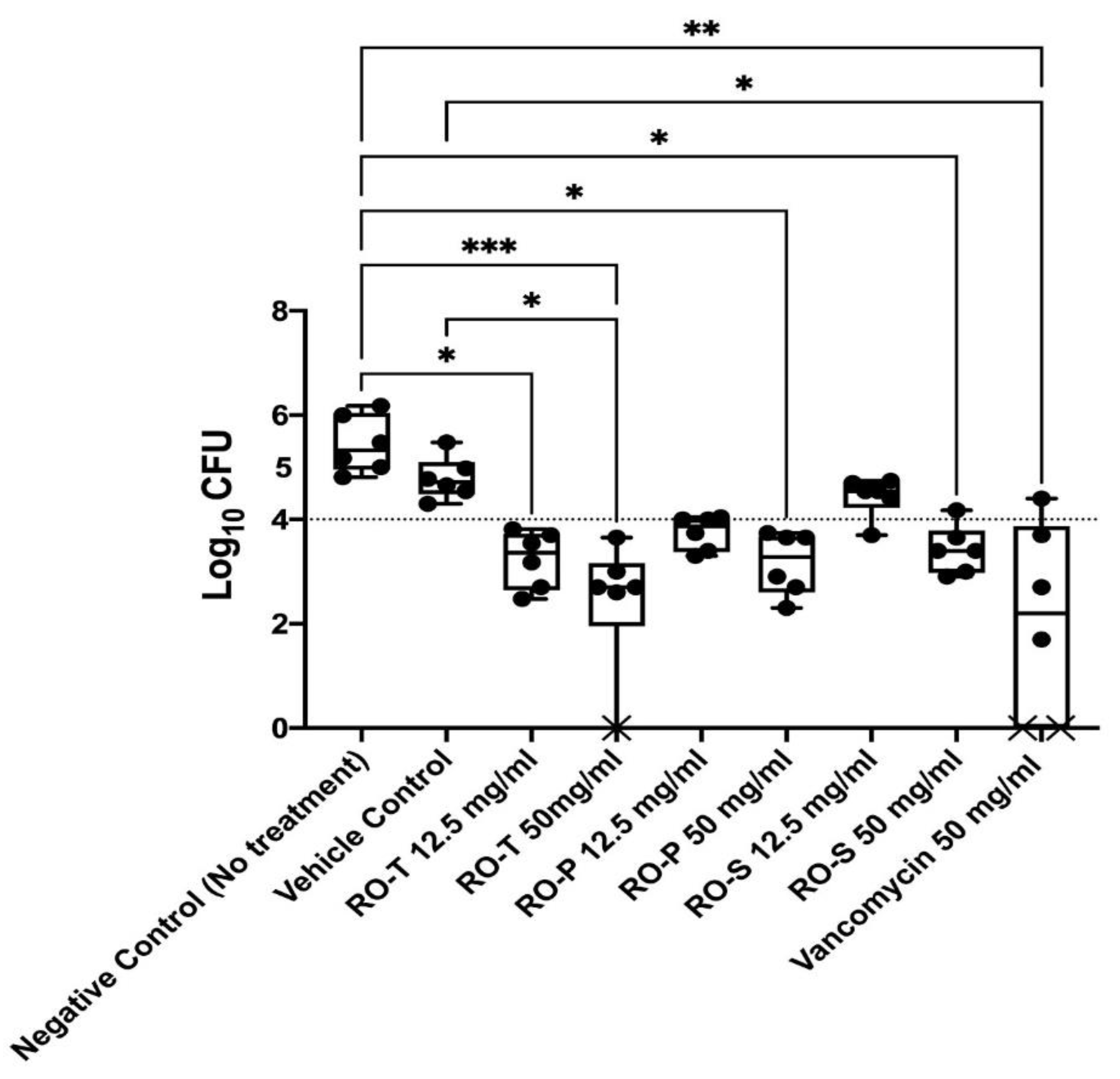
The promising in vitro antibacterial and anti-virulence activity of the tested extract/fractions was validated by investigating the in vivo activity through a rat model of MRSA vaginal infection (Figure 2). All the tested extract/fractions significantly reduced the MRSA bacterial load recovered from the infected vagina compared to that of the negative control (no treatment) group (Kruskal–Wallis Test, Dunn’s post-hoc test, p < 0.05). The RO-T extract and vancomycin groups recorded the highest antibacterial activity in vivo with a bacterial load 2.998 and 3.358 logs lower than that of the negative control group, respectively. There was no significant difference between the bacterial loads recovered from all the tested-extract-/fractions-treated groups and the vancomycin-treated group (positive control) (Kruskal–Wallis Test, Dunn’s post-hoc test, p < 0.05) (Figure 2). In addition, there was no significant difference between the bacterial loads recovered from the high concentration (50 mg/mL) or the low concentration (12.5 mg/mL) of the tested-extract-/fractions-treated groups (Kruskal–Wallis Test, Dunn’s post-hoc test, p < 0.05) (Figure 2).

2.4. Histopathological Analysis

Microscopic examination of group 1 (Healthy) revealed normal structure of the vaginal wall without any detectable alterations. Group 2 (Infection+ vehicle) showed mild vacuolation in the epithelial lining of the vagina, with inflammatory cell infiltration in the lamina propria. Group 3 (Bacterial infected) exhibited mild edema with inflammatory cell infiltration in the lamina propria. Group 4 (Vancomycin) showed apparently normal vaginal mucosa except for a few focal aggregations of inflammatory cells seen in the lamina propria. Group 5 (RO-T, 12.5) exhibited a moderate inflammatory reaction in the lamina propria of the vaginal wall. Group 6 (RO-T, 50) showed apparently normal vaginal wall with the presence of few congested blood vessels. Group 7 (RO-P, 12.5) exhibited congested blood vessels with mild perivascular edema and subsided inflammation. Group 8 (RO-P, 50) showed similar histological picture regarding mild edema and congestion. Group 9 (RO-S, 12.5) showed mild inflammatory edema. Group 10 (RO-S, 50) exhibited apparently normal mucosa with congested blood vessels (Figure S1).

2.5. HPLC Standardization

The HPLC chromatograms of the (1% HCL) acidified ethanolic extract of the red onion scales RO-T and its flavonoid-rich fraction RO-S (the most active) demonstrated a major peak for quercetin detected at 13.2 and 13.1 min, respectively. The peak of quercetin was identified and confirmed by comparison with HPLC chromatogram of the reference quercetin standard (Figure 3). According to the previously established HPLC calibration curve, it can be concluded that each 1 mg of acidified ethanolic extract contains 0.0478 ± 0.0009 mg quercetin, and each 1 mg of flavonoid-rich fraction contains 0.2188 ± 0.0036 mg quercetin.

3. Discussion

The development of multidrug resistance between clinical pathogens has been reported to be coupled with biofilm formation [11]. The predominance of Staphylococcus resistant bacteria in hospital and community infections, especially in immunocompromised patients, has motivated many researchers to investigate these bacteria. Genus Staphylococcus bacteria are known to be able to develop resistance against many antibiotics, which could delay the patient’s cure and cause high morbidity and mortality rates. Staphylococcus aureus is the major cause of bacterial infections in the genus Staphylococcus. The vancomycin antibiotic is the only one that can be used against infections of methicillin-resistant Staphylococcus strains. Recently, it showed inefficiency in some cases, which caused fear among healthcare professionals around the globe [12]. Hence, novel antimicrobial agents that cannot be countered by such pathogens are essentially needed [11]. Utilization of naturally occurring constituents that are derived from plant sources for the cure of different human disorders such as resistant bacterial infections is a promising safe alternative to using harmful synthetic drugs.
Onion is the most important horticultural crop cultivated globally. Onion processing produces numerous wastes, mainly the outer scales. Many researchers have explored onion outer scales as a rich source of antioxidants, dietary fibers, fructo-oligosaccharides, and polyphenols [13]. It has been reported that onion outer scales contain a significantly higher flavonoid content as compared to the edible part [14]. There are two main flavonoid subclasses present in red onion: anthocyanins and flavonol aglycone (quercetin type) and their derivatives, which all contribute to the yellow to purple color of onion scales [15]. On the way to relating the assessed activity with active metabolites, we extensively separated the flavonoids from the anthocyanins via the ether precipitation method and investigated both fractions alongside with the total extract. This is the first study concerning the inhibitory potential of red onion outer scales against MRSA vaginal colonization.
The evaluation of antibacterial effect against different pathogens revealed that the flavonoid-rich fraction (RO-S) has the highest bactericidal activity, especially against MRSA USA300. This is in accordance with the research previously reported about quercetin (the major flavonoid in the RO-S fraction) from different medicinal plants, which showed its potent antibacterial activity against a wide range of bacterial strains. This action has been linked to its solubility and its interaction with the bacterial cell membrane. In addition, quercetin has been well documented to have broad-spectrum antibacterial activities against Gram-positive bacteria such as methicillin-resistant Staphylococcus aureus (MRSA) [16].
It is noteworthy that the antimicrobial agents commonly used to treat vaginal colonization are usually ineffective, causing high failure rates, and this has often been associated with biofilm formation [17]. Furthermore, the in vitro and in vivo experiments revealed that the total extract and its flavonoid-rich fraction (RO-S) are powerful antibiofilm agents in comparison to the anthocyanin-rich fraction (RO-P), which also showed significant activity. In addition, the study revealed that red-onion outer-scales ethanolic extract and its fractions have a dose-dependent antibiofilm effect. The larger doses showed higher biofilm inhibition and biofilm eradication activities in vitro. Moreover, there was a great reduction of the bacterial load recovered from the infected vaginal tissue in vivo, which was confirmed by the histopathological study. The potential activity of the total ethanolic extract may be attributed to the synergistic effect of all its bioactive metabolites, viz, anthocyanins, phenolic acids, and flavonoids, especially quercetin.
Several studies investigated the chemical profile of red onion peels, and the authors documented that the polyphenols dominated metabolites in the extract as ferulic acid; 2-(3,4-dihydroxybenzoyl)-2,4,6-trihydroxy-3(2H)-benzofuranone; protocatechuic acid; quercetin; quercetin 4′-O-α-D-glucopyranoside; quercetin dimer; quercetin trimer, with its glycosidic forms; and kaempferol. They were present in large amounts, and this related to the strong biological properties of onion peels [18,19,20].
In my previous publication [20], the fragmentation pattern of the detected metabolites using HPLC/MS to confirm the proposed structures belonged to the classes of hydroxybenzoic acids, anthocyanins, and flavonols. Moreover, isomers of dihydroxybenzoic acid conjugated with hexose and condensation products of quercetin to dihydroxybenzoic acid were also detected. An in-depth analysis of tandem MS data made it possible to preliminarily predict novel phenolic constituents, the presence of which in onion peels could contribute to its bioactivity. The most prominent compounds were protocatechuic acid, cyanidin 3-O-glucoside, (2-(3,4-dihydroxyben zoic)-2,4,6-trihydroxy-3(2H)-benzofuranone, and a condensation product of quercetin to dihydroxybenzoic acid.
In this regard, the significant potent effect of RO-S fraction was mostly assigned to its high quercetin content. These results are in conformity with research previously reported about quercetin, namely, that it exerts a considerable anti-biofilm effect via bacterial-growth or motility inhibition, pathogenicity, and biofilm formation suppression [21]. In conclusion, quercetin is the key flavonoid behind all the health benefits and the significant antibiofilm activity [11,22,23,24]. The presence of quercetin aglycone in higher amounts in the most active tested samples drove us to quantify its content via HPLC analysis.
Finally, the powerful anti-biofilm activity of the red onion waste suggests its use as a natural antibiofilm agent and prompts further investigation into its antimicrobial properties in combination with other antibiotics.

4. Materials and Methods

4.1. Plant Material and Chemicals

The dry outer red onions scales (Allium cepa L.) were collected and purchased from a local market (Giza, Egypt). Quercetin standard solution (95% purity), diethyl ether, trifluoroacetic acid, HPLC-grade methanol, and HPLC-grade acetonitrile were purchased from Sigma–Aldrich Chemical Co. (St. Louis, MO, USA). Hydrochloric acid (HCL) and absolute ethanol (95%) were purchased from the PioChem company (Cairo, Egypt). Vancomycin® vials (500,000 IU) were purchased from Sigma-Tec (SIGMA Pharmaceutical Industries, St. Louis, MO, USA). Carbopol 934 P was provided by Chemical Industries Development Company (Giza, Egypt). Triethanolamine was supplied by Morgan Chemicals Ind. Co. (Cairo, Egypt).

4.2. Extraction and Fractionation Procedure

Infection-free red onion outer dry scales were separated from the onion’s bulbs. After grinding, the powdered red onion scales were exhaustively extracted by 1% hydrochloric acidified 70% ethanol by homogenization. Then, the solvent was evaporated under vacuum at a temperature not exceeding 40 °C. The obtained ethanolic extract residue was fractionated to obtain anthocyanin- and flavonoid-rich fraction using the diethyl ether precipitation method. The acidified ethanolic residue (20 g) was dissolved in 200 mL 95% ethanol and then a 10-fold volume of diethyl ether was added in a tightly sealed flask. After 24 h of standing in a refrigerator, the supernatant was decanted, and the red sediment was dried. This sediment was once more dissolved in 100 mL 95% ethanol, and a further 10-fold volume of diethyl ether was added. The supernatant was again decanted and added to the previous volume then evaporated under vacuum, and the red residue was dried in a desiccator for 24 h. The supernatant residue represents the flavonoid-rich fraction, while the red sediment represents the anthocyanin-rich fraction [25]. All samples were stored in a freezer (at −30 °C) until used.

4.3. Screening of the Antibacterial Activity

The antibacterial activity of the tested extract/fractions was determined against the following highly virulent bacterial strains: methicillin-resistant Staphylococcus aureus (MRSA USA300), Acinetobacter baumannii AB5075, Escherichia coli ATCC8, and Pseudomonas aeruginosa PAO1 [26,27,28]. The broth microdilution method was used according to the guidelines of the Clinical and Laboratory Standards Institute to determine the minimum bactericidal concentration (MBC) [29,30]. The experiment was done three independent times and the MBC was recorded as mean ± standard deviation.

4.4. Effect of the Tested Extracts on the Biofilm Activity

The anti-biofilm activity of the tested extract/fractions was investigated. They were tested at concentrations below the determined MBC. The biofilm-forming bacterial strains methicillin-resistant Staphylococcus aureus (MRSA USA300) and Acinetobacter baumannii AB5075 were used for this investigation [31].

4.5. Biofilm Inhibition Assay

The biofilm inhibition assay was performed using flat-bottom 96-well plates as reported before [31,32]. Different concentrations (sub-MBC) of the tested extract/fractions were tested. Untreated wells (no extract was added) were used as control (100% reference values). The experiment was repeated three independent times. The biofilm inhibition % was calculated using the following equation [31]:
Biofilm   inhibition   % = OD   Control OD   Test OD   Control × 100

4.6. Biofilm Eradication Assay

The ability of the tested extract/fractions to eradicate the previously established biofilm was investigated. The experiment was performed as reported before [31,32]. Different concentrations (sub-MBC) of the tested extract/fractions were prepared in fresh media and were added to the biofilm plate. Nothing was added to the biofilm control wells (untreated biofilm, 100% reference value). The experiment was repeated three independent times. The biofilm eradication % was calculated using the following equation [31]:
Biofilm   eradication   % = OD   Control OD   Test OD   Control × 100

4.7. In Vivo MRSA Vaginal Colonization Model

The tested extract/fractions were formulated into Carbopol hydrogel to assess their in vivo antibacterial activity [33]. Each extract was tested at two concentrations: 50 mg/mL and 12.5 mg/mL. All the animal-related procedures were done following the guidelines of the Research Ethics Committee of the Faculty of Pharmacy, Cairo University (Ethical Approval# MP (3121)). The rat MRSA vaginal-colonization model was performed as reported before [34,35]. Fifty-four Wistar rats (females/144 ± 20 g) were used in this model. The rats were injected intraperitoneally with 0.5 mg estradiol and 0.4 mg dexamethasone 24 h before colonization. Rats were vaginally inoculated with MRSA USA300 (8 × 10⁸ CFU) suspended in phosphate-buffered saline (PBS) for two successive days. The rats were randomly distributed into nine groups (six rats per group, n = 6) 24 h after the second inoculation, and treatments were administered inter-vaginally (200 µL of the hydrogel using a soft plastic tip). Six groups were treated with any of the tested extract/fractions in Carbopol hydrogel either at the high concentration (50 mg/mL) or the low concentration (12.5 mg/mL). The seventh group was treated with the empty Carbopol hydrogel to serve as the vehicle control. The eighth group was treated with vancomycin in Carbopol hydrogel (50 mg/mL) to serve as the positive control. The ninth group served as the negative control group and did not receive any treatment. An additional group (group 10, n = 6) was used as the healthy control group; this group was not infected and did not receive any treatment. One day post-treatment, the vaginal lumen of each rat was swabbed with a sterile swab. The recovered MRSA bacteria in the swabs were serially diluted in PBS and spotted on mannitol salt agar for the viable count, as described before [34]. The results of the tested groups were analyzed. At the end of the experiment, three rats from each group were used for histopathological examination. The whole excised vaginal tissues were preserved in a 10% formalin-saline solution before preparation of the histological sections using the paraffin method technique [34]. The histological sections of the colonized vaginal tissues were compared to that of healthy uncolonized rats.

4.8. HPLC Standardization Method

The total ethanolic extract and its flavonoid-rich fraction were dissolved in HPLC-grade methanol (5 mg/mL), filtered through a sterile 0.45 µm membrane filter, and transferred into sealed vials to be subjected to injection for qualitative and quantitative analysis using the Agilent Technologies 1100 Infinity series HPLC system chromatograph equipped with a diode-array detector (DAD). The analysis was performed on an Eclipse XDBC18 column (150 × 4.6 mm, particle size 5 μm) with a C18 guard column (Phenomenex, Torrance, CA), and the column was operated at room temperature. Separation was carried out with a 0.1% trifluoroacetic acid acidified acetonitrile and 0.1% trifluoroacetic acid acidified water gradient elution program at a flow rate of 1 mL/min. It started with 5% acidified acetonitrile, changed to 35% in 7 min, increased to 50% in 10 min, and at last increased to 100% in 2 min. Chromatograms were recorded at 280, 325, and 520 nm.
Establishment of the standard calibration curve of quercetin was achieved by preparing a standard stock solution by dissolving 10 mg of reference-standard quercetin in 10 mL HPLC-grade methanol. The stock solution was then serially diluted with methanol to obtain concentrations equivalent to 31.25–1000 µg/mL. An aliquot (20 µL) of each concentration was subjected to the HPLC system injection under the same conditions adopted for the extract and fraction in triplicates.
AUC (Area under curve) for each quercetin concentration was recorded, and subsequently, the average was plotted against its corresponding concentration.
Occurrence of quercetin in the total ethanolic extract and its flavonoid-rich fraction was ensured via comparison with the quercetin peak and retention time of the standard quercetin chromatogram. Afterwards, by using the following equation that was obtained from the constructed plot, the quercetin amount was calculated:
Y   = 21.486 x   + 424.41 , R 2 = 0.996

5. Conclusions

In brief, red onion outer scales showed efficient in vitro antibacterial effect against four contagious bacterial strains: methicillin-resistant Staphylococcus aureus (MRSA USA300), Acinetobacter baumannii AB5075, Escherichia coli ATCC8, and Pseudomonas aeruginosa PAO1. Furthermore, it showed a promising antibiofilm activity against MRSA. The flavonoid-rich fraction and total extract showed the most potent in vitro and in vivo antibacterial and antibiofilm effects, and that is due to the high content of quercetin. Finally, agro-food wastes could be a source of potential candidates used to reduce the burden of multidrug-resistant strains and defeat biofilm-associated infections.

Supplementary Materials

The following are available online at https://www.mdpi.com/article/10.3390/molecules28010355/s1, Figure S1. Photomicrograph of vagina sections of different experimental groups (H&E). (a) Gp1 showing normal stratified epithelium and propria of vaginal wall. (b) Gp2 showing inflammatory cells infiltration in lamina propria. (c) Gp3 showing mild edema in lamina propria with inflammatory cells infiltration (arrow). (d) Gp4 showing focal mononuclear inflammatory cells infiltration in lamina propria (arrow). (e) Gp5 showing moderate inflammatory cells infiltration in lamina propria (arrow). (f) Gp6 showing normal mucosa with few congested blood vessels (arrow). (g) Gp7 showing severely congested blood vessels with mild edema. (h) Gp8 showing mild edema. (i) Gp9 showing mild inflammatory edema in lamina propria (arrow). (j) Gp10 showing apparently normal mucosa.

Author Contributions

Conceptualization, N.B.A., R.A.E.-S. and M.H.; methodology, N.B.A., R.A.E.-S., R.M.A. and M.H; investigation, N.B.A., R.A.E.-S., R.M.A. and M.H.; writing—original draft preparation, R.M.A., S.H.E.-G. and E.A.-S.; su-pervision, R.A.E.-S., R.M.A., S.H.E.-G. and E.A.-S. All authors have read and agreed to the published version of the manuscript.

Funding

This research received no external funding.

Institutional Review Board Statement

Not applicable.

Informed Consent Statement

Not applicable.

Data Availability Statement

Not applicable.

Conflicts of Interest

The authors declare no conflict of interest.

Sample Availability

Samples of the red onion peels available from the authors.

References

  1. Škerget, M.; Majhenič, L.; Bezjak, M.; Knez, Ž. Antioxidant, radical scavenging and antimicrobial activities of red onion (Allium cepa L.) skin and edible part extracts. Chem. Biochem. Eng. Q. 2009, 23, 435–444. [Google Scholar]
  2. Nile, A.; Nile, S.H.; Cespedes-Acuña, C.L.; Oh, J.-W. Spiraeoside extracted from red onion skin ameliorates apoptosis and exerts potent antitumor, antioxidant and enzyme inhibitory effects. Food Chem. Toxicol. 2021, 154, 112327. [Google Scholar] [CrossRef] [PubMed]
  3. Pagano, C.; Marinozzi, M.; Baiocchi, C.; Beccari, T.; Calarco, P.; Ceccarini, M.R.; Chielli, M.; Orabona, C.; Orecchini, E.; Ortenzi, R. Bioadhesive polymeric films based on red onion skins extract for wound treatment: An innovative and eco-friendly formulation. Molecules 2020, 25, 318. [Google Scholar] [CrossRef] [Green Version]
  4. Fredotović, Ž.; Puizina, J.; Nazlić, M.; Maravić, A.; Ljubenkov, I.; Soldo, B.; Vuko, E.; Bajić, D. Phytochemical characterization and screening of antioxidant, antimicrobial and antiproliferative properties of Allium× cornutum clementi and two varieties of Allium cepa L. peel extracts. Plants 2021, 10, 832. [Google Scholar] [CrossRef] [PubMed]
  5. Sagar, N.A.; Pareek, S. Antimicrobial assessment of polyphenolic extracts from onion (Allium cepa L.) skin of fifteen cultivars by sonication-assisted extraction method. Heliyon 2020, 6, e05478. [Google Scholar] [CrossRef] [PubMed]
  6. Memariani, H.; Memariani, M.; Ghasemian, A. An overview on anti-biofilm properties of quercetin against bacterial pathogens. World J. Microbiol. Biotechnol. 2019, 35, 143. [Google Scholar] [CrossRef]
  7. Tumuhamye, J.; Steinsland, H.; Bwanga, F.; Tumwine, J.K.; Ndeezi, G.; Mukunya, D.; Namugga, O.; Kasede, A.N.; Sommerfelt, H.; Nankabirwa, V. Vaginal colonization with antimicrobial-resistant bacteria among women in labor in central Uganda: Prevalence and associated factors. Antimicrob. Resist. Infect. Control 2021, 10, 1–11. [Google Scholar] [CrossRef]
  8. Coolborn, A.F.; Charity, E.-B.C.; Ogbaji, O.S. Inhibition of methicillin resistant Staphylococcus aureus (MRSA) with underutilized medicinal plants extracts. J Int Med Res. 2021, 28, 4953–4964. [Google Scholar]
  9. Wang, B.; Wei, P.-W.; Wan, S.; Yao, Y.; Song, C.-R.; Song, P.-P.; Xu, G.-B.; Hu, Z.-Q.; Zeng, Z.; Wang, C. Ginkgo biloba exocarp extracts inhibit S. aureus and MRSA by disrupting biofilms and affecting gene expression. J. Ethnopharmacol. 2021, 271, 113895. [Google Scholar] [CrossRef]
  10. Chiew, S.P.; Thong, O.M.; Yin, K.B. Phytochemical composition, antimicrobial and cytotoxic activities of red onion peel extracts prepared using different methods. Int. J. Integr. Biol. 2014, 15, 49. [Google Scholar]
  11. Mu, Y.; Zeng, H.; Chen, W. Quercetin inhibits biofilm formation by decreasing the production of EPS and altering the composition of EPS in Staphylococcus epidermidis. Front. Microbiol. 2021, 12, 631058. [Google Scholar] [CrossRef] [PubMed]
  12. da Costa Júnior, S.D.; de Oliveira Santos, J.V.; de Almeida Campos, L.A.; Pereira, M.A.; Magalhães, N.S.S.; Cavalcanti, I.M.F. Antibacterial and antibiofilm activities of quercetin against clinical isolates of Staphyloccocus aureus and Staphylococcus saprophyticus with resistance profile. Int. J. Environ. Agric. Biotechnol. 2018, 3, 266213. [Google Scholar]
  13. Sagar, N.A.; Pareek, S.; Gonzalez-Aguilar, G.A. Quantification of flavonoids, total phenols and antioxidant properties of onion skin: A comparative study of fifteen Indian cultivars. J. Food Sci. Technol. 2020, 57, 2423–2432. [Google Scholar] [CrossRef] [PubMed]
  14. Downes, K.; Chope, G.A.; Terry, L.A. Effect of curing at different temperatures on biochemical composition of onion (Allium cepa L.) skin from three freshly cured and cold stored UK-grown onion cultivars. Postharvest Biol. Technol. 2009, 54, 80–86. [Google Scholar] [CrossRef]
  15. Benítez, V.; Mollá, E.; Martín-Cabrejas, M.; López-Andréu, F.J.; Downes, K.; Terry, L.; Esteban, R. Study of bioactive compound content in different onion sections. Plant Foods Hum. Nutr. 2011, 66, 48–57. [Google Scholar] [CrossRef] [Green Version]
  16. Nguyen, T.L.A.; Bhattacharya, D. Antimicrobial Activity of Quercetin: An Approach to Its Mechanistic Principle. Molecules 2022, 27, 2494. [Google Scholar] [CrossRef]
  17. Sousa, L.G.; Castro, J.; Cerca, N. The Utilization of Essential Oils to Treat Biofilm-Associated Vaginal Infections. In Microbial Biofilms; CRC Press: Boca Raton, FL, USA, 2020; pp. 395–420. [Google Scholar]
  18. Ly, T.N.; Hazama, C.; Shimoyamada, M.; Ando, H.; Kato, K.; Yamauchi, R. Antioxidative compounds from the outer scales of onion. J. Agric. Food Chem. 2005, 53, 8183–8189. [Google Scholar] [CrossRef]
  19. Singh, B.N.; Singh, B.; Singh, R.; Prakash, D.; Singh, D.; Sarma, B.; Upadhyay, G.; Singh, H. Polyphenolics from various extracts/fractions of red onion (Allium cepa) peel with potent antioxidant and antimutagenic activities. Food Chem. Toxicol. 2009, 47, 1161–1167. [Google Scholar] [CrossRef]
  20. Abouzed, T.K.; del Mar Contreras, M.; Sadek, K.M.; Shukry, M.; Abdelhady, D.H.; Gouda, W.M.; Abdo, W.; Nasr, N.E.; Mekky, R.H.; Segura-Carretero, A. Red onion scales ameliorated streptozotocin-induced diabetes and diabetic nephropathy in Wistar rats in relation to their metabolite fingerprint. Diabetes Res. Clin. Pract. 2018, 140, 253–264. [Google Scholar] [CrossRef]
  21. Roy, P.K.; Song, M.G.; Park, S.Y. The inhibitory effect of quercetin on biofilm formation of Listeria monocytogenes mixed culture and repression of virulence. Antioxidants 2022, 11, 1733. [Google Scholar] [CrossRef]
  22. Matilla-Cuenca, L.; Gil, C.; Cuesta, S.; Rapún-Araiz, B.; Žiemytė, M.; Mira, A.; Lasa, I.; Valle, J. Antibiofilm activity of flavonoids on staphylococcal biofilms through targeting BAP amyloids. Sci. Rep. 2020, 10, 1–12. [Google Scholar] [CrossRef] [PubMed]
  23. Roy, P.K.; Park, S.-H.; Song, M.G.; Park, S.Y. Antimicrobial Efficacy of Quercetin against Vibrio parahaemolyticus biofilm on food surfaces and downregulation of virulence genes. Polymers 2022, 14, 3847. [Google Scholar] [CrossRef] [PubMed]
  24. Bonaccorsi, P.; Caristi, C.; Gargiulli, C.; Leuzzi, U. Flavonol glucosides in Allium species: A comparative study by means of HPLC–DAD–ESI-MS–MS. Food Chem. 2008, 107, 1668–1673. [Google Scholar] [CrossRef]
  25. Cahlíková, L.; Ali, B.H.; Havlíková, L.; Ločárek, M.; Siatka, T.; Opletal, L.; Blunden, G. Anthocyanins of Hibiscus sabdiffera calyces from Sudan. Nat. Prod. Commun. 2015, 10, 1934578X1501000120. [Google Scholar] [CrossRef]
  26. Sedeek, M.S.; Afifi, S.M.; Mansour, M.K.; Hassan, M.; Mehaya, F.M.; Naguib, I.A.; Abourehab, M.A.S.; Farag, M.A. Unveiling Antimicrobial and Antioxidant Compositional Differences between Dukkah and Za&rsquo;atar via SPME-GCMS and HPLC-DAD. Molecules 2022, 27, 6471. [Google Scholar] [CrossRef]
  27. Ibrahim, N.M.; Fahim, S.H.; Hassan, M.; Farag, A.E.; Georgey, H.H. Design and synthesis of ciprofloxacin-sulfonamide hybrids to manipulate ciprofloxacin pharmacological qualities: Potency and side effects. Eur. J. Med. Chem. 2022, 228, 114021. [Google Scholar] [CrossRef]
  28. Ismail, M.M.; Hassan, M.; Moawad, S.S.; Okba, M.M.; Ashour, R.M.; Fayek, N.M.; Saber, F.R. Exploring the Antivirulence Activity of Pulverulentone A, a Phloroglucinol-Derivative from Callistemon citrinus Leaf Extract, against Multi-Drug Resistant Pseudomonas aeruginosa. Antibiotics 2021, 10, 907. [Google Scholar] [CrossRef]
  29. El-Shiekh, R.A.; Hassan, M.; Hashem, R.A.; Abdel-Sattar, E. Bioguided Isolation of Antibiofilm and Antibacterial Pregnane Glycosides from Caralluma quadrangula: Disarming Multidrug-Resistant Pathogens. Antibiotics 2021, 10, 811. [Google Scholar] [CrossRef]
  30. Humphries, R.M.; Ambler, J.; Mitchell, S.L.; Castanheira, M.; Dingle, T.; Hindler, J.A.; Koeth, L.; Sei, K.; Development, C.M.; Standardization Working Group of the Subcommittee on Antimicrobial Susceptibility Tests. CLSI Methods Development and Standardization Working Group Best Practices for Evaluation of Antimicrobial Susceptibility Tests. J. Clin. Microbiol. 2018, 56, e01934-17. [Google Scholar] [CrossRef] [Green Version]
  31. Salem, M.A.; El-Shiekh, R.A.; Hashem, R.A.; Hassan, M. In vivo Antibacterial Activity of Star Anise (Illicium verum Hook.) Extract Using Murine MRSA Skin Infection Model in Relation to Its Metabolite Profile. Infect. Drug Resist. 2021, 14, 33–48. [Google Scholar] [CrossRef]
  32. Albash, R.; Abdellatif, M.M.; Hassan, M.; Badawi, N.M. Tailoring Terpesomes and Leciplex for the Effective Ocular Conveyance of Moxifloxacin Hydrochloride (Comparative Assessment): In-vitro, Ex-vivo, and In-vivo Evaluation. Int. J. Nanomed. 2021, 16, 5247–5263. [Google Scholar] [CrossRef] [PubMed]
  33. Ramadan, A.A.; Eladawy, S.A.; El-Enin, A.S.M.A.; Hussein, Z.M. Development and investigation of timolol maleate niosomal formulations for the treatment of glaucoma. J. Pharm. Investig. 2020, 50, 59–70. [Google Scholar] [CrossRef]
  34. Deng, L.; Schilcher, K.; Burcham Lindsey, R.; Kwiecinski Jakub, M.; Johnson Paige, M.; Head Steven, R.; Heinrichs David, E.; Horswill Alexander, R.; Doran Kelly, S. Identification of Key Determinants of Staphylococcus aureus Vaginal Colonization. mBio 2019, 10, e02321-19. [Google Scholar] [CrossRef] [Green Version]
  35. Au-Patras, K.A.; Au-Doran, K.S. A Murine Model of Group B Streptococcus Vaginal Colonization. JoVE 2016, 117, e54708. [Google Scholar] [CrossRef]
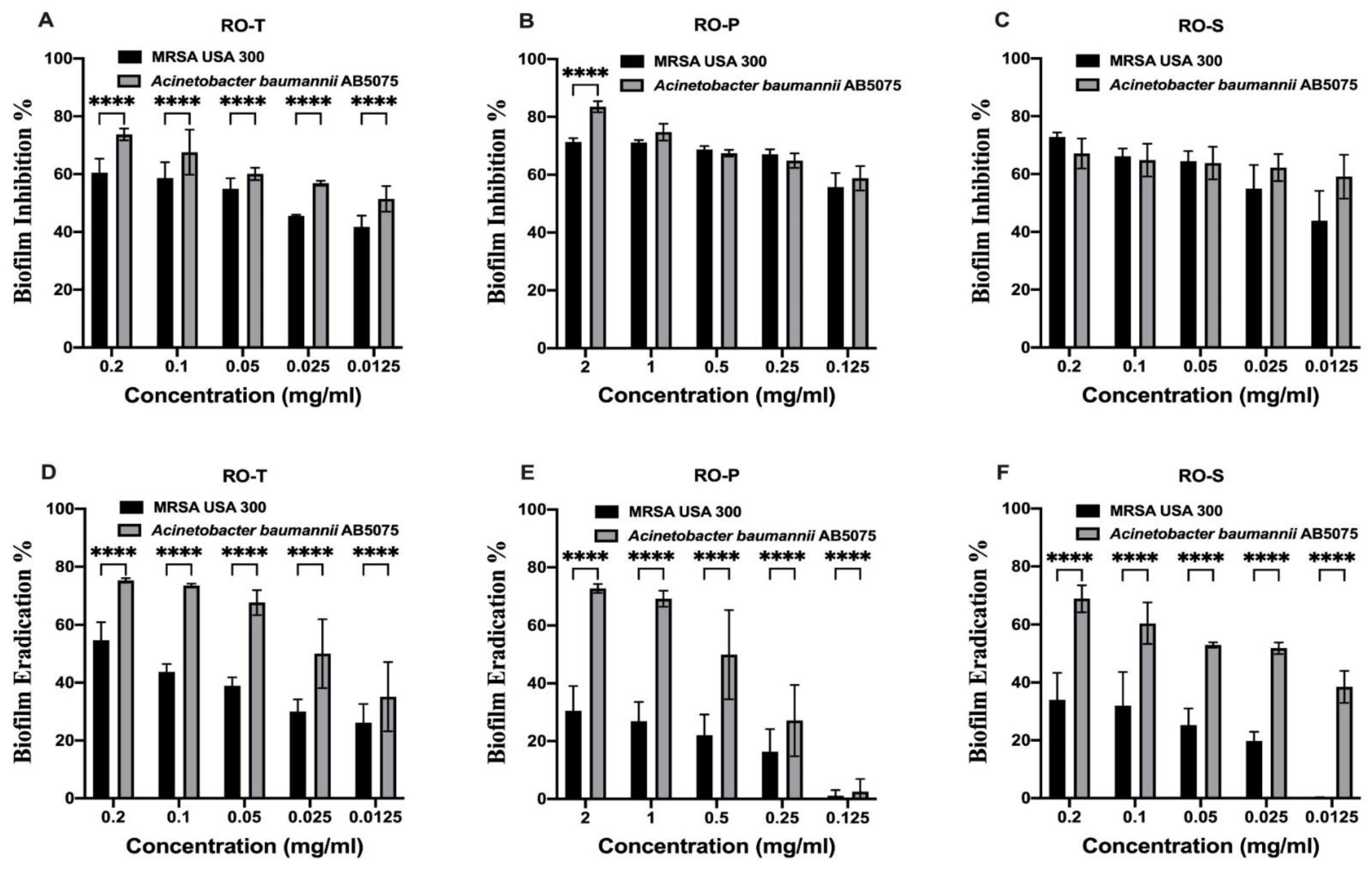
Figure 1. Antibiofilm activity. Effect of various concentrations of extracts RO-T, RO-P, and RO-S on MRSA USA300 and A. baumannii AB5057 biofilm formation. Results are expressed as mean biofilm inhibition % ± standard deviation (AC) and mean biofilm eradication % ± standard deviation (DF). **** Indicates that the difference is significant at p < 0.0001 (Two-way ANOVA, Sidak’s post-hoc test). Methicillin-resistant Staphylococcus aureus (MRSA), the total ethanolic extract of red onion scales (RO-T), anthocyanin-rich fraction (RO-P) and flavonoid-rich fraction (RO-S).
Molecules 28 00355 g001
Figure 2. Efficacy of extracts RO-T, RO-P, and RO-S in an in vivo rat model of MRSA vaginal infection. Fifty-four Wistar rats were divided into nine groups (n = 6). Six groups were treated with any of the tested extracts in Carpobol hydrogel, either at the high concertation (50 mg/mL) or the low concertation (12.5 mg/mL). One group was treated with the empty Carbopol hydrogel to serve as the vehicle control. Another group was treated with vancomycin in Carbopol hydrogel (50 mg/mL) to serve as the positive control. Another group served as the negative control group and did not receive any treatment. Each data point in the figure represents a rat. Results are expressed as box plots of bacterial loads recovered from the vaginal lumen. The whiskers span the difference between the minimum and maximum readings, and the horizontal bar represents the median. The symbols *, **, and *** indicate that the difference is significant at p < 0.05, < 0.01, and < 0.001, respectively (Kruskal–Wallis Test, Dunn’s post-hoc test). X means no colonies were detected in the sample.
Molecules 28 00355 g002
Figure 3. HPLC chromatograms of total extract (A), flavonoid-rich fraction (B), and quercetin std (C) at 280 nm.
Molecules 28 00355 g003
Table 1. Antibacterial activity of the tested extracts.
Minimum Bactericidal Concentration (MBC) mg/mL *
MRSA USA300Acinetobacter baumannii AB5075Escherichia coli ATCC87Pseudomonas aeruginosa PAO1
RO-T0.651 ± 0.2264.688 ± 2.7069.375 ± 5.41316.667 ± 7.217
RO-P4.167 ± 1.8046.25 ± 025 ± 0#
RO-S0.325 ± 0.1130.651 ± 0.2267.292 ± 4.77412.5 ± 0
* Data are presented as mean ± standard deviation. # No antibacterial activity was detected against the tested organism.
Table 2. The half maximal inhibitory/eradication concentration (IC50/EC50) of the tested extracts against biofilm of methicillin-resistant Staphylococcus aureus (MRSA USA300) and Acinetobacter baumannii AB5075.
Biofilm InhibitionBiofilm Eradication
Log IC50IC50 (mg/mL)Log EC50EC50 (mg/mL)
meanSEmeanmeanSEmean
RO-TMRSA USA300−1.1950.1640.064−1.7970.1870.016
Acinetobacter baumannii AB5075−0.8180.1730.152−0.7610.2240.174
RO-PMRSA USA3000.3110.1532.048−1.5730.3050.027
Acinetobacter baumannii AB50750.4760.1892.990−0.8180.3030.152
RO-SMRSA USA300−0.8410.1850.144−2.4920.3090.003
Acinetobacter baumannii AB5075−0.8330.1390.147−1.0920.1800.081
Disclaimer/Publisher’s Note: The statements, opinions and data contained in all publications are solely those of the individual author(s) and contributor(s) and not of MDPI and/or the editor(s). MDPI and/or the editor(s) disclaim responsibility for any injury to people or property resulting from any ideas, methods, instructions or products referred to in the content.