Communication

Enzymatic Protein Immobilization on Amino-Functionalized Nanoparticles

Key Laboratory of Coordination Chemistry, Chemistry and Biomedicine Innovation Center (ChemBIC), School of Chemistry and Chemical Engineering, Nanjing University, Nanjing 210023, China
*
Author to whom correspondence should be addressed.
Molecules 2023, 28(1), 379; https://doi.org/10.3390/molecules28010379
Submission received: 27 November 2022 / Revised: 23 December 2022 / Accepted: 28 December 2022 / Published: 2 January 2023

Abstract

:
The immobilization of proteins on nanoparticles has received much attention in recent years. Among different approaches, enzymatic protein immobilization shows unique advantages because of its site-specific connection. OaAEP1 is a recently engineered peptide ligase which can specifically recognize an N-terminal GL residue (NH2–Gly–Leu) and a C-terminal NGL amino acid residue (Asn–Gly–Leu–COOH) and ligates them efficiently. Herein, we report OaAEP1-mediated protein immobilization on synthetic magnetic nanoparticles. Our work showed that OaAEP1 could mediate C-terminal site-specific protein immobilization on the amino-functionalized Fe3O4 nanoparticles. Our work demonstrates a new method for site-specific protein immobilization on nanoparticles.

1. Introduction

Nanoparticle-protein complexes have a wide range of applications, such as magnetic separation of proteins [1,2], enzyme-catalyzed proteolysis [3], drug delivery [4], and disease diagnosis [5]. Thus, efficient immobilization of proteins on nanoparticles has received increasing interest in recent years [6,7,8,9,10]. Many interactions, such as electrostatic interaction and covalent binding, were used [11,12,13]. However, these methods suffer from the disadvantages of non-specific binding and uncontrolled orientation [14]. Recently, the enzyme-mediated method could realize N- or C-terminal site-specific protein immobilization on the nanoparticle, and many related works were reported [14,15]. In 2020, the Francis group reported that proteins containing proline, thiol, or aniline functional groups were coupled to phenols-functionalized Au nanoparticles by enzyme tyrosinase using an oxidative coupling reaction [16]. The enzyme sortase, one of the transpeptidases, has often been used for versatile enzymatic protein immobilization with the requirements of the two substrates with a C-terminal LPxTG tag and an N-terminal glycine repeat [1,17,18,19,20,21,22,23].
OaAEP1[C247A], cysteine 247 to alanine mutant of asparaginyl endopeptidedase 1 from Oldenlandia affinis, is a recently engineered protein ligase (abbreviated as OaAEP1). It recognizes an N-terminal GL residue (NH2–Gly–Leu) and a C-terminal NGL amino acid residue (Asn–Gly–Leu–COOH) explicitly and ligates them efficiently. Thus, it is extensively used in protein/peptide ligation studies [24,25,26,27,28,29,30,31]. It is an excellent choice for protein immobilization, which has been well demonstrated for AFM-based single molecule-force spectroscopy (SMFS) studies of the immobilized protein [32,33,34,35].
In 2021, David J. Craik and Thomas Durek reported that the OaAEP1-catalyzed peptide and protein were irreversibly labeled with various nonpeptidyl amine nucleophiles at a C-terminal asparagine of the peptide and protein. In their work, the protein eGFP with NGL at the C terminal could conjugate with the molecules containing primary amine nucleophiles, which was demonstrated by SDS-PAGE and ESI-MS [36]. Similar work reported on asparaginyl endopeptidase-mediated protein C-terminal ligation with amino-containing molecules [27]. Moreover, the Ploegh group reported OaAEP1 catalyzed one-step protein modification on the surface of red blood cells [37]. Thus, we believe that this powerful enzymatic ligation can be adopted for protein immobilization on nanoparticles, and thus we used this method to immobilize an enhanced green fluorescent protein (eGFP) with C-terminal NGL residues on the amino-functionalized magnetic nanoparticle, abbreviated as Fe3O4-NH2 nanoparticle (Scheme 1).

2. Results and Discussions

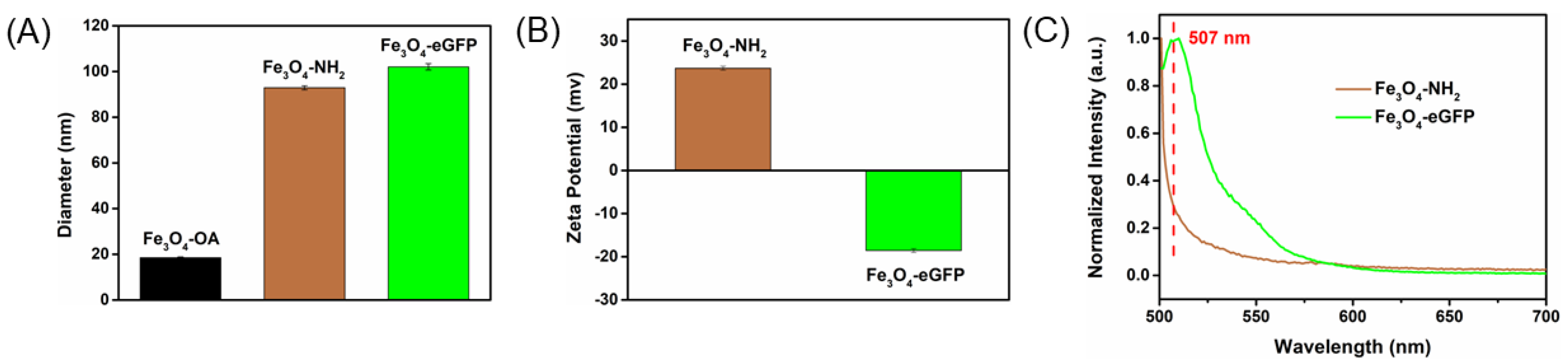
2.1. Synthesis and Characterization of Fe3O4 Nanoparticles

First, the Fe3O4–OA nanoparticles were synthesized by the high-temperature pyrolysis of the precursor iron trioleate (Figure 1A) [38]. In the XRD spectra, the peak of the obtained sample (Figure S1) was found to be well-matched to the standard diffraction peaks of Fe3O4, indicating the formation of Fe3O4 [38,39,40]. The picture of Fe3O4–OA nanoparticles dispersed in cyclohexane (Figure 1B, inset) showed that the sample was homogeneous without precipitation. The TEM images (Figure 1B) showed that the Fe3O4–OA nanoparticles were monodisperse in cyclohexane with an average diameter of 9.62 nm (Figure 1C).
Then, catechol–PEG5000–NH2 was introduced and replaced oleic acid on the surface of Fe3O4 nanoparticles by the classic ligand exchange method to change Fe3O4 nanoparticles from the hydrophobic Fe3O4–OA to hydrophilic Fe3O4–NH2 based on the strong coordination of phenolic hydroxy groups of catechol with iron [41] (Figure 1A). As illustrated in Figure 1D, the Fe3O4 nanoparticles were transferred from the organic phase to the aqueous phase, indicating the transformation from the hydrophobic Fe3O4–OA to hydrophilic Fe3O4–NH2. The changes in the average diameter of Fe3O4–OA and Fe3O4–NH2 measured by dynamic light scattering (DLS) indicated that the modification of catechol–PEG5000–NH2 on the Fe3O4 nanoparticle was successful (Figure S2). Besides, the TEM image (Figure 1E) of the product Fe3O4–NH2 revealed that the Fe3O4–NH2 nanoparticles were similar to the Fe3O4–OA nanoparticles (Figure 1B). According to the amount of the nanoparticles and catechol–PEG5000–NH2, the amount of catechol–PEG5000–NH2 on the surface of 1 Fe3O4 nanoparticle was estimated as about 1200.

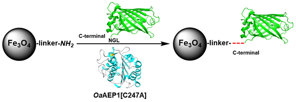
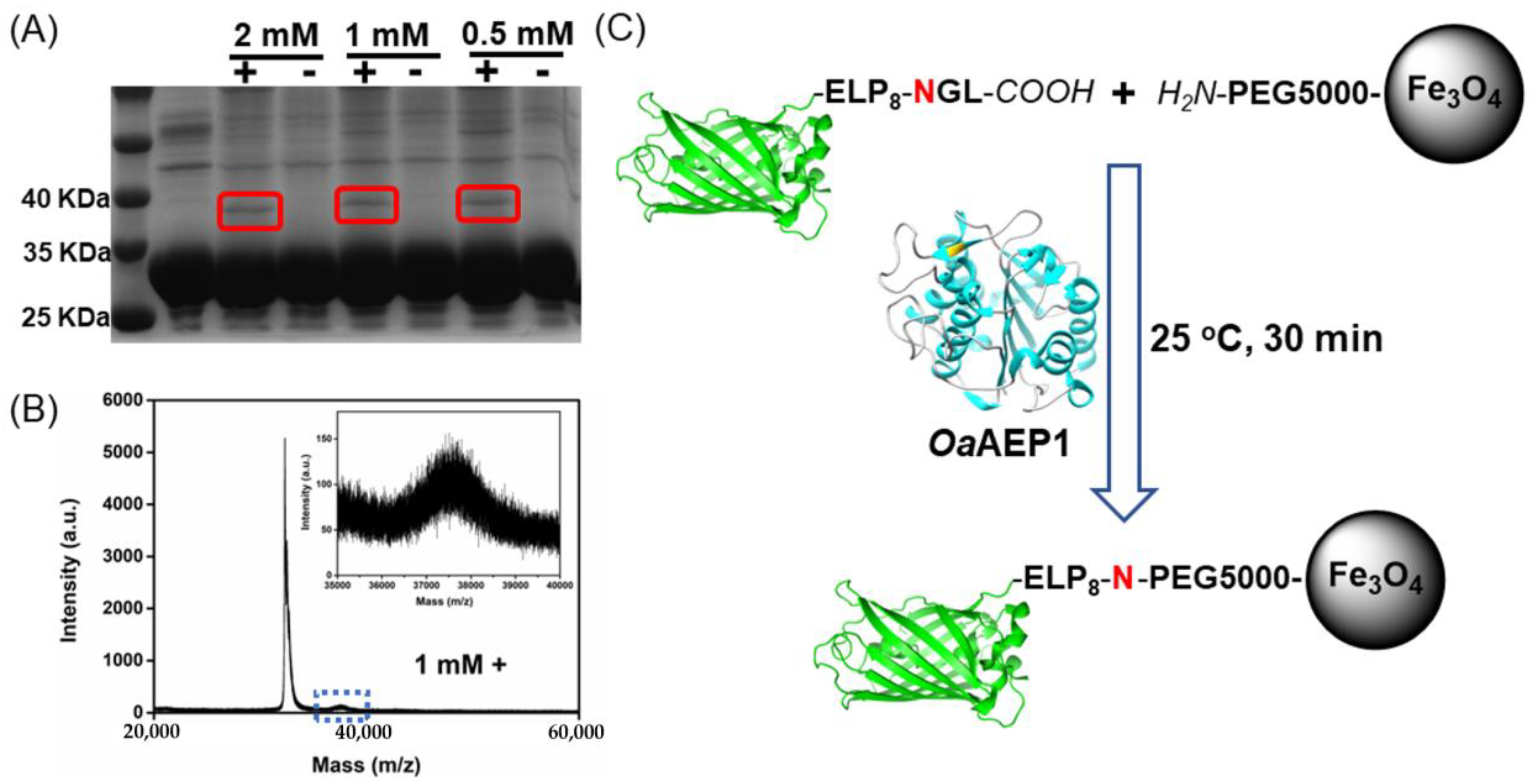
2.2. Immobilization of eGFP–ELP8–NGL on Fe3O4–NH2 Nanoparticle by OaAEP1

First, the target protein eGFP–ELP8–NGL was constructed, showing an expected molecular weight of ~33 kDa from the SDS-PAGE gel experiment [41,42]. Then, we confirmed its ability to ligate with the corresponding protein GL–eGFP in a solution by OaAEP1 using SDS-PAGE (Figure S3) [24,32]. Next, we examined the ability of eGFP–ELP8–NGL (50 μM) to conjugate with PEG5000–NH2 under various concentrations (2 mM; 1 mM; 0.5 mM). Notably, the eGFP–ELP8–NGL (50 μM), as the control group, is in the second lane. The SDS-PAGE gel result showed successful ligation, which is circled in red when OaAEP1 is present (Figure 2A) [36]. Moreover, MALDI-TOF MS results revealed that eGFP–ELP8–NGL could conjugate with PEG5000–NH2 under a very low concentration of PEG5000–NH2 (0.05 mM) (Figure S4) [36]. We then used a 1 mM concentration of PEG5000–NH2 for all following experiments (Figure 2B).
Finally, we immobilized eGFP–ELP8–NGL on a Fe3O4–NH2 nanoparticle by OaAEP1 (Figure 2C). Furthermore, a number of characterizations were carried out to prove eGFP–ELP8–NGL’s conjugation on the Fe3O4–NH2 nanoparticle. First, the changes in average diameter of Fe3O4–NH2 and Fe3O4–eGFP measured by dynamic light scattering (DLS) along with zeta potential indicated that the conjugation of eGFP–ELP8–NGL with the Fe3O4–NH2 nanoparticle was successful (Figure 3A,B and Figure S5). Furthermore, the fluorescence emission spectra of Fe3O4–NH2 and Fe3O4–eGFP are depicted in Figure 3C. The fluorescence emission spectra of Fe3O4–eGFP ranged from 500 to 700 nm, with the emission peak located at 507 nm when excited at 488 nm, while the Fe3O4–NH2 exhibited almost negligible emission at a wavelength of 507 nm (Figure 3C and Figure S7).
To confirm the immobilization was mediated by OaAEP1, not the non-specific interaction or electrostatic interaction, fluorescence emission spectroscopy measurement was performed. The group with OaAEP1 showed an apparent fluorescence intensity compared with the group when OaAEP1 was absent (Figure S6). It is worth noting that the two samples were washed with the dispersion solution three times and with washing buffer (50 mM Tris, 1 M NaCl, pH 7.0) five times, then dispersed in 0.5 mL dispersion buffer. In summary, all data above proved that the immobilization of protein eGFP–ELP8–NGL on Fe3O4–NH2 nanoparticles was successful. Moreover, considering the amount of protein eGFP–ELP8–NGL and the reaction efficiency, the number of proteins eGFP–ELP8–NGL on the Fe3O4–NH2 nanoparticle was estimated as about 20.

3. Materials and Methods

3.1. Materials

Iron trichloride hexahydrate, hexane, ethanol, and tetrahydrofuran (THF) were purchased from Sinopharm Chemical Reagent Co. Ltd. (Shanghai, China). Sodium oleate was purchased from Beijing InnoChem Science & Technology Co., Ltd. (Beijing, China). Oleic acid was purchased from Alfa Aesar (Shanghai, China). The 1-Octadecene was purchased from Acros Organics (Shanghai, China). Catechol–PEG5000–NH2 was purchased from Ponsure Biotechnology (Shanghai, China). Other reagents were purchased from Sangon Biotech Co. Ltd. (Shanghai, China). All reagents were used without further purification. Ultrapure water (18 MΩ cm−1) was obtained from a Millipore Milli-Q Advantage water purification system (Burlington, MA, USA). E. coil BL21 (DE3) and XL1-Blue cells were purchased from TransGen Biotech Co. Ltd. (Beijing, China).

3.2. Sample Characterizations

X-ray diffraction (XRD) spectra were recorded on an X’TRA Powder diffractometer (Waltham, MA, USA). The size and morphology of nanoparticles were observed using a JEOL JEM-2100 transmission electron microscope (Tokyo, Japan) at 200 kV. Dynamic light scattering (DLS) was recorded at a wavelength of 659 nm, and zeta potential experiments were conducted on 90 Plus/BI-MAS equipment (Brookhaven, NY, USA). The concentration of Fe was determined by Atomic Absorption Spectroscopy on analytik jena novAA350 (Jena, Germany). The fluorescence spectra were measured on a HORIBA Jobin Yvon Fluoromax-4 fluorescence spectrometer (Irvine, CA, USA). Mass spectrometric analyses were performed using an UltrafleXtreme MALDI-TOF mass spectrometer (Bruker Daltonics, Billerica, MA, USA) operating in linear positive ion mode with Sinapinic acid (SA) as the matrix.

3.3. Protein Engineering

All the plasmids, except OaAEP1, were based on the pET-28a vector, and all proteins were overexpressed in E. coli BL21(DE3) cells. OaAEP1 was kindly provided by Dr. Wu, Bin. ELP is an elastin-like polypeptide [43,44]. The expression and purification details of OaAEP1 can be found in our previous publications [45,46], and OaAEP1 was exchanged into water (A280 = 1.46) by ultrafiltration. The genes of eGFP were purchased from Genscript (Nanjing, China). Construction of eGFP–ELP8–NGL and GL–eGFP was in the expression vector pET-28a by standard molecular biology and PCR techniques. The plasmids were transformed and then overexpressed in E. coli BL21 (DE3) cells. The bacteria kept in the LB medium containing 50 µg mL−1 kanamycin were grown to an OD600 = 0.6 and then were induced by 0.4 mM isopropyl β–d–thiogalactoside (IPTG) overnight at 16 °C. The cells were obtained by centrifugation at 9000 rpm for 5 min at 4 °C (Avanti JXN series, Beckman Coulter) (Brea, CA, USA). The cells were redispersed in dispersion buffer (50 mM Tris, 100 mM NaCl, pH 7.0) and lysed via a high-pressure homogenization. The supernatants were mixed with Co–NTA affinity beads and then kept for 3 h after the centrifugation operation at 30,000 rpm for 15 min. The mixture was washed with buffer (20 mM Tris, 400 mM NaCl, 2 mM imidazole, pH 7.0) several times and eluted in buffer (20 mM Tris, 400 mM NaCl, 250 mM imidazole, pH 7.0) immediately. The buffer of the protein solutions was all exchanged into dispersion buffer (50 mM Tris, 100 mM NaCl, pH 7.0) by ultrafiltration. The concentrations of eGFP–ELP8–NGL and GL–eGFP were 4.1 mg/mL and 4.8 mg/mL, respectively, which were determined by Nanodrop 2000 (Waltham, MA, USA).

3.4. Synthesis of Fe3O4–OA Nanoparticles

First, the precursor iron trioleate was synthesized. A 5.4 g amount of iron trichloride hexahydrate and 18.3 g of sodium oleate were dissolved in a mixed solvent composed of 40 mL ethanol, 30 mL distilled water, and 70 mL hexane. The resulting solution was heated to 70 °C and kept at 70 °C for 4 hours. When the reaction was completed, the upper organic layer containing the iron trioleate was washed with distilled water four times in a separatory funnel. Then, hexane was evaporated off, and iron trioleate in a waxy solid form was obtained. The iron trioleate was dissolved in 100 mL 1-Octadecene after drying in the oven for 24 h.
Fe3O4–OA nanoparticles were synthesized according to a previously published method with modification. Briefly, 1 mL oleic acid was added into 20 mL of iron trioleate solution above a three-necked flask, and then the mixture was heated to 320 °C and kept at 320 °C for 30 min. After cooling to room temperature, ethanol was added to the solution to precipitate the Fe3O4–OA nanoparticles. The Fe3O4–OA nanoparticles were washed with hexane and ethanol mixture several times and then dispersed in 10 mL hexane (~20 mg/mL).

3.5. Synthesis of Fe3O4–NH2 Nanoparticles

From the above hexane solution 1 mL was isolated then centrifuged and transferred into 0.4 mL tetrahydrofuran (THF). A 60 mg portion of catechol–PEG5000–NH2 was dissolved in 0.4 mL THF. The two solutions were mixed and placed in a shaker at 37 °C for 3 h. When the reaction was finished, hexane was added to precipitate the Fe3O4–NH2 nanoparticles. The acquired Fe3O4–NH2 nanoparticles were washed with THF and water three times and then dispersed in 5 mL dispersion solution (50 mM Tris, 100 mM NaCl, pH 7.0) for use.

3.6. Immobilization of eGFP–ELP8–NGL on Fe3O4–NH2 Nanoparticle by OaAEP1

Briefly, 0.5 mL eGFP–ELP8–NGL solution, 0.5 mL Fe3O4–NH2 dispersion, and 75 μL OaAEP1 solution were mixed and placed in a shaker at 25 °C for 30 min. The reaction products were washed with the dispersion solution for three times and buffer (50 mM Tris, 1 M NaCl, pH 7.0) five times and then dispersed in 0.5 mL dispersion solution ([Fe] 31 mM).

4. Conclusions

In summary, we developed a new enzymatic method for protein immobilization on nanoparticles using OaAEP1. The target protein eGFP with a C-terminal NGL peptide tag was immobilized on the Fe3O4–NH2 nanoparticle. Considering the easy access of PEG–NH2 molecules or molecules with a primary amine, we believe this method can be useful for immobilizing other important proteins on nanoparticles and applied for further applications.

Supplementary Materials

The following supporting information can be downloaded at: https://www.mdpi.com/article/10.3390/molecules28010379/s1. Figure S1: XRD patterns of the obtained Fe3O4 and standard diffraction peaks of Fe3O4 (JCPDS NO.19-0629); Figure S2: Average diameter of Fe3O4-OA and Fe3O4-NH2 measured by DLS; Figure S3: SDS-PAGE gel result of the ligation of eGFP-ELP8-NGL with protein GL-eGFP by OaAEP1; Figure S4: MALDI-TOF MS result of the ligation of eGFP-ELP8-NGL with PEG5000-NH2 (A, E 1 mM; B, F 0.5 mM; C, G 0.1 mM; D, H 0.05 mM) when OaAEP1 was present (upper) and absent (bottom); Figure S5: Hydrodynamic diameter distribution of Fe3O4-OA, Fe3O4-NH2, Fe3O4-eGFP; Figure S6: The fluorescence emission spectra of Fe3O4-NH2 (black) and the obtained Fe3O4-eGFP complex when OaAEP1 was present (blue) and absent (red); Figure S7: The fluorescence emission spectrum of target protein eGFP-ELP8-NGL excited at 488 nm, which is same as free eGFP.

Author Contributions

Conceptualization, P.Z.; methodology, Q.M. and P.Z.; software, Q.M. and P.Z.; validation, Q.M., B.H. and G.T.; formal analysis, Q.M. and P.Z.; investigation, Q.M., B.H. and G.T.; resources, P.Z. and R.X.; data curation, Q.M.; writing—original draft preparation, Q.M.; writing—review and editing, P.Z.; visualization, P.Z.; supervision, P.Z.; project administration, P.Z.; funding acquisition, P.Z. All authors have read and agreed to the published version of the manuscript.

Funding

This research was funded by the Natural Science Foundation of Jiangsu Province (No. BK20200058).

Data Availability Statement

All data are presented in the manuscript.

Conflicts of Interest

The authors declare no competing interests.

Sample Availability

Samples of all protein-coated nanoparticles are available from the authors.

References

  1. Fauser, J.; Savitskiy, S.; Fottner, M.; Trauschke, V.; Gulen, B. Sortase-Mediated Quantifiable Enzyme Immobilization on Magnetic Nanoparticles. Bioconjugate Chem. 2020, 31, 1883–1892. [Google Scholar] [CrossRef] [PubMed]
  2. Shao, M.; Ning, F.; Zhao, J.; Wei, M.; Evans, D.G.; Duan, X. Preparation of Fe3O4@SiO2@Layered Double Hydroxide Core–Shell Microspheres for Magnetic Separation of Proteins. J. Am. Chem. Soc. 2012, 134, 1071–1077. [Google Scholar] [CrossRef] [PubMed]
  3. Li, L.; Shi, H.; Sheng, A.; Yang, Y.; Shi, L.; Li, C.; Li, G. A novel method to engineer proteases for selective enzyme inhibition. Chem. Commun. 2019, 55, 14039–14042. [Google Scholar] [CrossRef] [PubMed]
  4. Oh, J.Y.; Kim, H.S.; Palanikumar, L.; Go, E.M.; Jana, B.; Park, S.A.; Kim, H.Y.; Kim, K.; Seo, J.K.; Kwak, S.K.; et al. Cloaking nanoparticles with protein corona shield for targeted drug delivery. Nat. Commun. 2018, 9, 4548. [Google Scholar] [CrossRef] [PubMed] [Green Version]
  5. Loynachan, C.N.; Soleimany, A.P.; Dudani, J.S.; Lin, Y.; Najer, A.; Bekdemir, A.; Chen, Q.; Bhatia, S.N.; Stevens, M.M. Renal clearable catalytic gold nanoclusters for in vivo disease monitoring. Nat. Nanotechnol. 2019, 14, 883–890. [Google Scholar] [CrossRef] [PubMed] [Green Version]
  6. Lv, S. Silk Fibroin-Based Materials for Catalyst Immobilization. Molecules 2020, 25, 4929. [Google Scholar] [CrossRef] [PubMed]
  7. Cruz, G.; Saiz, L.P.; Bilal, M.; Eltoukhy, L.; Loderer, C.; Fernández-Lucas, J. Magnetic Multi-Enzymatic System for Cladribine Manufacturing. Int. J. Mol. Sci. 2022, 23, 13634. [Google Scholar] [CrossRef]
  8. Le, L.T.H.L.; Yoo, W.; Jeon, S.; Kim, K.K.; Kim, T.D. Characterization and Immobilization of a Novel SGNH Family Esterase (LaSGNH1) from Lactobacillus acidophilus NCFM. Int. J. Mol. Sci. 2020, 21, 91. [Google Scholar] [CrossRef] [Green Version]
  9. Popov, A.; Brasiunas, B.; Kausaite-Minkstimiene, A.; Ramanaviciene, A. Metal Nanoparticle and Quantum Dot Tags for Signal Amplification in Electrochemical Immunosensors for Biomarker Detection. Chemosensors 2021, 9, 85. [Google Scholar] [CrossRef]
  10. Makaraviciute, A.; Ruzgas, T.; Ramanavicius, A.; Ramanaviciene, A. Antibody fragment immobilization on planar gold and gold nanoparticle modified quartz crystal microbalance with dissipation sensor surfaces for immunosensor applications. Anal. Methods 2014, 6, 2134–2140. [Google Scholar] [CrossRef]
  11. Aubin-Tam, M.E.; Hamad-Schifferli, K. Structure and function of nanoparticle–protein conjugates. Biomed. Mater. 2008, 3, 034001. [Google Scholar] [CrossRef] [PubMed]
  12. Sapsford, K.E.; Algar, W.R.; Berti, L.; Gemmill, K.B.; Casey, B.J.; Oh, E.; Stewart, M.H.; Medintz, I.L. Functionalizing Nanoparticles with Biological Molecules: Developing Chemistries that Facilitate Nanotechnology. Chem. Rev. 2013, 113, 1904–2074. [Google Scholar] [CrossRef] [PubMed]
  13. Federsel, H.-J.; Moody, T.S.; Taylor, S.J.C. Recent Trends in Enzyme Immobilization—Concepts for Expanding the Biocatalysis Toolbox. Molecules 2021, 26, 2822. [Google Scholar] [CrossRef] [PubMed]
  14. Smith, A.D.; Walper, S.A.; Medintz, I.L. Enzymatic bioconjugation to nanoparticles. In Reference Module in Materials Science and Materials Engineering; Elsevier: Amsterdam, The Netherlands, 2021. [Google Scholar]
  15. Walper, S.A.; Turner, K.B.; Medintz, I.L. Enzymatic bioconjugation of nanoparticles: Developing specificity and control. Curr. Opin. Biotechnol. 2015, 34, 232–241. [Google Scholar] [CrossRef]
  16. Ramsey, A.V.; Bischoff, A.J.; Francis, M.B. Enzyme Activated Gold Nanoparticles for Versatile Site-Selective Bioconjugation. J. Am. Chem. Soc. 2021, 143, 7342–7350. [Google Scholar] [CrossRef]
  17. Matsumoto, T.; Tanaka, T.; Kondo, A. Sortase A-Catalyzed Site-Specific Coimmobilization on Microparticles via Streptavidin. Langmuir 2012, 28, 3553–3557. [Google Scholar] [CrossRef]
  18. Hata, Y.; Matsumoto, T.; Tanaka, T.; Kondo, A. C-Terminal-oriented Immobilization of Enzymes Using Sortase A-mediated Technique. Macromol. Biosci. 2015, 15, 1375–1380. [Google Scholar] [CrossRef]
  19. Raeeszadeh-Sarmazdeh, M.; Parthasarathy, R.; Boder, E.T. Site-specific immobilization of protein layers on gold surfaces via orthogonal sortases. Colloids Surf. Biointerfaces 2015, 128, 457–463. [Google Scholar] [CrossRef]
  20. Qafari, S.M.; Ahmadian, G.; Mohammadi, M. One-step purification and oriented attachment of protein A on silica and graphene oxide nanoparticles using sortase-mediated immobilization. RSC Adv. 2017, 7, 56006–56015. [Google Scholar] [CrossRef] [Green Version]
  21. Dai, X.; Mate, D.M.; Glebe, U.; Mirzaei Garakani, T.; Körner, A.; Schwaneberg, U.; Böker, A. Sortase-Mediated Ligation of Purely Artificial Building Blocks. Polymers 2018, 10, 151. [Google Scholar] [CrossRef]
  22. Liu, Y.; Tian, F.; Shi, S.; Deng, Y.; Zheng, P. Enzymatic Protein–Protein Conjugation through Internal Site Verified at the Single-Molecule Level. J. Phys. Chem. Lett. 2021, 12, 10914–10919. [Google Scholar] [CrossRef] [PubMed]
  23. Garg, S.; Singaraju, G.S.; Yengkhom, S.; Rakshit, S. Tailored Polyproteins Using Sequential Staple and Cut. Bioconjugate Chem. 2018, 29, 1714–1719. [Google Scholar] [CrossRef] [PubMed]
  24. Yang, R.; Wong, Y.H.; Nguyen, G.K.T.; Tam, J.P.; Lescar, J.; Wu, B. Engineering a Catalytically Efficient Recombinant Protein Ligase. J. Am. Chem. Soc. 2017, 139, 5351–5358. [Google Scholar] [CrossRef] [PubMed]
  25. Tang, T.M.S.; Cardella, D.; Lander, A.J.; Li, X.; Escudero, J.S.; Tsai, Y.-H.; Luk, L.Y.P. Use of an asparaginyl endopeptidase for chemo-enzymatic peptide and protein labeling. Chem. Sci. 2020, 11, 5881–5888. [Google Scholar] [CrossRef] [PubMed]
  26. Rehm, F.B.H.; Tyler, T.J.; Yap, K.; Durek, T.; Craik, D.J. Improved Asparaginyl-Ligase-Catalyzed Transpeptidation via Selective Nucleophile Quenching. Angew. Chem. Int. Ed. 2021, 60, 4004–4008. [Google Scholar] [CrossRef] [PubMed]
  27. Zhang, D.; Wang, Z.; Hu, S.; Chan, N.-Y.; Liew, H.T.; Lescar, J.; Tam, J.P.; Liu, C.-F. Asparaginyl Endopeptidase-Mediated Protein C-Terminal Hydrazinolysis for the Synthesis of Bioconjugates. Bioconjugate Chem. 2022, 33, 238–247. [Google Scholar] [CrossRef] [PubMed]
  28. Rehm, F.B.H.; Harmand, T.J.; Yap, K.; Durek, T.; Craik, D.J.; Ploegh, H.L. Site-Specific Sequential Protein Labeling Catalyzed by a Single Recombinant Ligase. J. Am. Chem. Soc. 2019, 141, 17388–17393. [Google Scholar] [CrossRef]
  29. Rehm, F.B.H.; Tyler, T.J.; Xie, J.; Yap, K.; Durek, T.; Craik, D.J. Asparaginyl Ligases: New Enzymes for the Protein Engineer’s Toolbox. ChemBioChem 2021, 22, 2079–2086. [Google Scholar] [CrossRef]
  30. Rehm, F.B.H.; Tyler, T.J.; de Veer, S.J.; Craik, D.J.; Durek, T. Enzymatic C-to-C Protein Ligation. Angew. Chem. Int. Ed. 2022, 61, e202116672. [Google Scholar] [CrossRef]
  31. Zhang, D.; Wang, Z.; Hu, S.; Balamkundu, S.; To, J.; Zhang, X.; Lescar, J.; Tam, J.P.; Liu, C.-F. pH-Controlled Protein Orthogonal Ligation Using Asparaginyl Peptide Ligases. J. Am. Chem. Soc. 2021, 143, 8704–8712. [Google Scholar] [CrossRef]
  32. Deng, Y.; Wu, T.; Wang, M.; Shi, S.; Yuan, G.; Li, X.; Chong, H.; Wu, B.; Zheng, P. Enzymatic biosynthesis and immobilization of polyprotein verified at the single-molecule level. Nat. Commun. 2019, 10, 2775. [Google Scholar] [CrossRef] [Green Version]
  33. Shi, S.; Wang, Z.; Deng, Y.; Tian, F.; Wu, Q.; Zheng, P. Combination of Click Chemistry and Enzymatic Ligation for Stable and Efficient Protein Immobilization for Single-Molecule Force Spectroscopy. CCS Chem. 2022, 4, 598–604. [Google Scholar] [CrossRef]
  34. Yuan, G.; Ma, Q.; Wu, T.; Wang, M.; Li, X.; Zuo, J.; Zheng, P. Multistep Protein Unfolding Scenarios from the Rupture of a Complex Metal Cluster Cd3S9. Sci. Rep. 2019, 9, 10518. [Google Scholar] [CrossRef] [PubMed] [Green Version]
  35. Deng, Y.; Zheng, B.; Liu, Y.; Shi, S.; Nie, J.; Wu, T.; Zheng, P. OaAEP1-Mediated Enzymatic Synthesis and Immobilization of Polymerized Protein for Single-Molecule Force Spectroscopy. JoVE 2020, 156, e60774. [Google Scholar] [CrossRef] [PubMed]
  36. Rehm, F.B.H.; Tyler, T.J.; Yap, K.; de Veer, S.J.; Craik, D.J.; Durek, T. Enzymatic C-Terminal Protein Engineering with Amines. J. Am. Chem. Soc. 2021, 143, 19498–19504. [Google Scholar] [CrossRef]
  37. Harmand, T.J.; Pishesha, N.; Rehm, F.B.H.; Ma, W.; Pinney, W.B.; Xie, Y.J.; Ploegh, H.L. Asparaginyl Ligase-Catalyzed One-Step Cell Surface Modification of Red Blood Cells. ACS Chem. Biol. 2021, 16, 1201–1207. [Google Scholar] [CrossRef] [PubMed]
  38. Park, J.; An, K.; Hwang, Y.; Park, J.-G.; Noh, H.-J.; Kim, J.-Y.; Park, J.-H.; Hwang, N.-M.; Hyeon, T. Ultra-large-scale syntheses of monodisperse nanocrystals. Nat. Mater. 2004, 3, 891–895. [Google Scholar] [CrossRef]
  39. Kim, B.H.; Lee, N.; Kim, H.; An, K.; Park, Y.I.; Choi, Y.; Shin, K.; Lee, Y.; Kwon, S.G.; Na, H.B.; et al. Large-Scale Synthesis of Uniform and Extremely Small-Sized Iron Oxide Nanoparticles for High-Resolution T1 Magnetic Resonance Imaging Contrast Agents. J. Am. Chem. Soc. 2011, 133, 12624–12631. [Google Scholar] [CrossRef]
  40. Liu, J.; Sun, Z.; Deng, Y.; Zou, Y.; Li, C.; Guo, X.; Xiong, L.; Gao, Y.; Li, F.; Zhao, D. Highly Water-Dispersible Biocompatible Magnetite Particles with Low Cytotoxicity Stabilized by Citrate Groups. Angew. Chem. Int. Ed. 2009, 48, 5875–5879. [Google Scholar] [CrossRef]
  41. Liu, Y.; Chen, T.; Wu, C.; Qiu, L.; Hu, R.; Li, J.; Cansiz, S.; Zhang, L.; Cui, C.; Zhu, G.; et al. Facile Surface Functionalization of Hydrophobic Magnetic Nanoparticles. J. Am. Chem. Soc. 2014, 136, 12552–12555. [Google Scholar] [CrossRef]
  42. Lu, Z.; Liu, Y.; Deng, Y.; Jia, B.; Ding, X.; Zheng, P.; Li, Z. OaAEP1-mediated PNA-protein conjugation enables erasable imaging of membrane proteins. Chem. Commun. 2022, 58, 8448–8451. [Google Scholar] [CrossRef]
  43. Wang, Z.; Nie, J.; Shi, S.; Li, G.; Zheng, P. Transforming de novo protein α3D into a mechanically stable protein by zinc binding. Chem. Commun. 2021, 57, 11489–11492. [Google Scholar] [CrossRef] [PubMed]
  44. Ott, W.; Jobst, M.A.; Bauer, M.S.; Durner, E.; Milles, L.F.; Nash, M.A.; Gaub, H.E. Elastin-like Polypeptide Linkers for Single-Molecule Force Spectroscopy. ACS Nano 2017, 11, 6346–6354. [Google Scholar] [CrossRef] [PubMed] [Green Version]
  45. Ding, X.; Wang, Z.; Zheng, B.; Shi, S.; Deng, Y.; Yu, H.; Zheng, P. One-step asparaginyl endopeptidase (OaAEP1)-based protein immobilization for single-molecule force spectroscopy. RSC Chem. Biol. 2022, 3, 1276–1281. [Google Scholar] [CrossRef] [PubMed]
  46. Tian, F.; Tong, B.; Sun, L.; Shi, S.; Zheng, B.; Wang, Z.; Dong, X.; Zheng, P. N501Y mutation of spike protein in SARS-CoV-2 strengthens its binding to receptor ACE2. eLife 2021, 10, e69091. [Google Scholar] [CrossRef]
Scheme 1. Schematic illustration of OaAEP1 mediated protein immobilization on Fe3O4 nanoparticle.
Molecules 28 00379 sch001
Figure 1. Characterizations of Fe3O4 nanoparticles. (A) Schematic description of the synthesis of oleic acid-capped hydrophobic Fe3O4–OA nanoparticles and catechol–PEG5000–NH2 modified hydrophilic Fe3O4–NH2 nanoparticles. (B) TEM image of Fe3O4–OA nanoparticles. Inset: photograph of the sample Fe3O4–OA nanoparticles dispersed in hexane. (C) The diameter distribution histogram of Fe3O4–OA nanoparticles. (D) Photograph of the Fe3O4 nanoparticles transferred from the organic phase to the aqueous phase. (E) TEM image of hydrophilic Fe3O4–NH2 nanoparticles.
Molecules 28 00379 g001
Figure 2. (A) SDS-PAGE gel result of the ligation of eGFP–ELP8–NGL with PEG5000–NH2 (2 mM; 1 mM; 0.5 mM) when OaAEP1 was present and absent. (B) MALDI-TOF MS result of the ligation of eGFP–ELP8–NGL with PEG5000–NH2 (1 mM) in solution. (C) Schematic illustration of the immobilization of protein eGFP–ELP8–NGL on Fe3O4–NH2 nanoparticle by OaAEP1.
Molecules 28 00379 g002
Figure 3. (A) Average diameter of Fe3O4–OA, Fe3O4–NH2, and Fe3O4–eGFP measured by DLS. (B) Zeta potential and (C) fluorescence emission spectra of Fe3O4–NH2 and Fe3O4–eGFP.
Molecules 28 00379 g003
Disclaimer/Publisher’s Note: The statements, opinions and data contained in all publications are solely those of the individual author(s) and contributor(s) and not of MDPI and/or the editor(s). MDPI and/or the editor(s) disclaim responsibility for any injury to people or property resulting from any ideas, methods, instructions or products referred to in the content.

