

Spectral Features Differentiate Aging-Induced Changes in Parchment—A Combined Approach of UV/VIS, µ-ATR/FTIR and µ-Raman Spectroscopy with Multivariate Data Analysis

 , , and
Abstract
:
1. Introduction
2. Results and Discussion
2.1. Changes observed by UV/VIS Spectroscopy
2.2. Changes of Lipids and Protein Hydration Observed in m-ATR/FTIR Spectroscopy
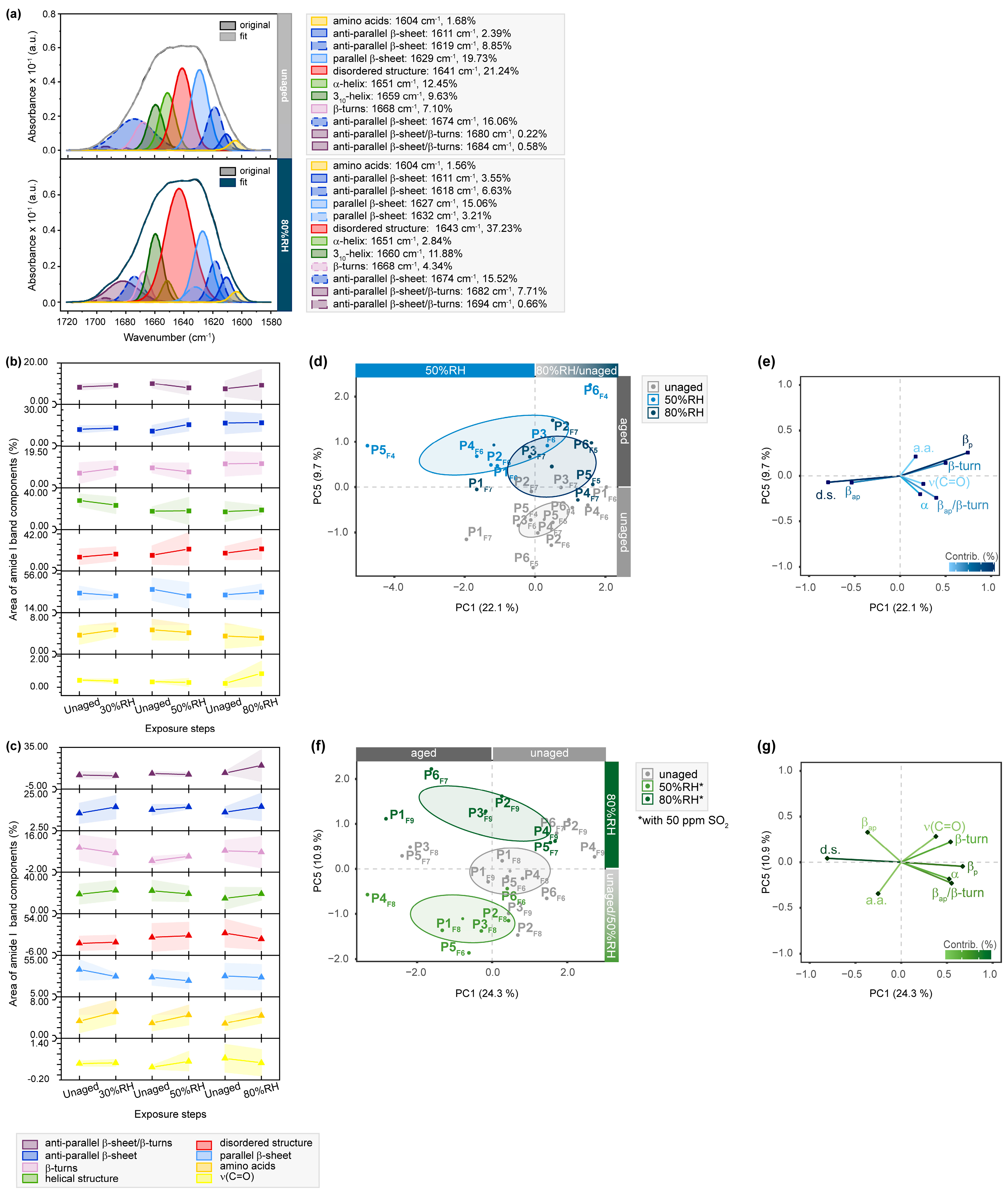
2.3. Conformational Protein Changes Observed by µ-ATR/FTIR and µ-Raman Analysis of the Vibrational Amide I, II and III Bands
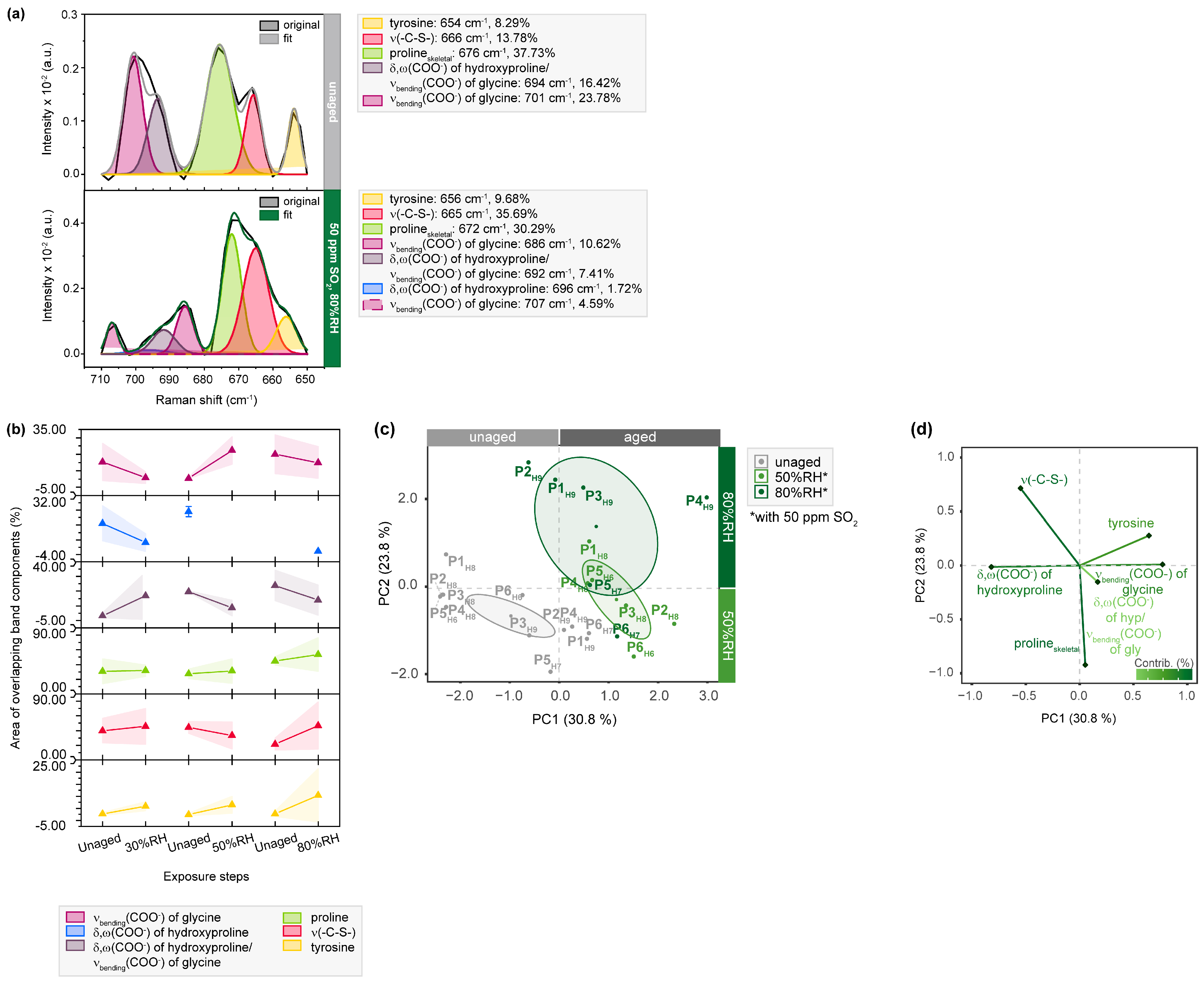
2.4. Changes of Single Amino Acids as Observed by µ-Raman Analysis
3. Materials and Methods
3.1. Parchment Samples
3.2. Artificial Aging with UV/VIS Light, Relative Humidity and SO2
3.3. Colorimetric Analysis by UV/VIS Spectroscopy
3.4. µ-FTIR Spectroscopy in ATR Mode (µ-ATR/FTIR)
3.5. µ-Raman Spectroscopy
3.6. Band Deconvolution
3.7. Multivariate Data Analysis
4. Conclusions
Supplementary Materials
Author Contributions
Funding
Institutional Review Board Statement
Informed Consent Statement
Data Availability Statement
Acknowledgments
Conflicts of Interest
Sample Availability
References
- Goggin, J.M. A History of Technology, Volume I: From Early Times to Fall of Ancient Empires. Am. Antiq. 1959, 25, 130–132. [Google Scholar] [CrossRef]
- Maor, Y.; Shor, P.; Aizenshtat, Z. Parchment Browning–part II: The Dead Sea Scrolls. Polym. Degrad. Stab. 2021, 183, 109414. [Google Scholar] [CrossRef]
- Fuchs, R. The history and biology of parchment. Karger Gaz. 2004, 67, 13–16. [Google Scholar]
- Thompson, D.V. Medieval Parchment-Making; Bibliographical Society: London, UK, 1935. [Google Scholar]
- Kadler, K.E.; Baldock, C.; Bella, J.; Boot-Handford, R.P. Collagens at a glance. J. Cell Sci. 2007, 120, 1955–1958. [Google Scholar] [CrossRef] [PubMed] [Green Version]
- Gordon, M.K.; Hahn, R.A. Collagens. Cell Tissue Res. 2010, 339, 247. [Google Scholar] [CrossRef] [PubMed]
- Shoulders, M.D.; Raines, R.T. Collagen structure and stability. Annu. Rev. Biochem. 2009, 78, 929–958. [Google Scholar] [CrossRef] [Green Version]
- Stinson, R.H.; Sweeny, P.R. Skin collagen has an unusual d-spacing. Biochim. Et Biophys. Acta (BBA)-Protein Struct. 1980, 621, 158–161. [Google Scholar] [CrossRef]
- Petruska, J.A.; Hodge, A.J. A subunit model for the tropocollagen macromolecule. Proc. Natl. Acad. Sci. USA 1964, 51, 871–876. [Google Scholar] [CrossRef] [Green Version]
- Bozec, L.; van der Heijden, G.; Horton, M. Collagen fibrils: Nanoscale ropes. Biophys. J. 2007, 92, 70–75. [Google Scholar] [CrossRef] [Green Version]
- Kennedy, C.; Wess, T. The Structure of Collagen within Parchment–A Review. Restaur.-Int. J. Preserv. Libr. Arch. Mater.-Restaur. 2003, 24, 61–80. [Google Scholar] [CrossRef]
- Guareschi, I. Della Pergamena, con Osservazioni ed Esperienze sul Ricupero e sul Restauro di Codici Danneggiati Negli Incendi e Notizie Storiche; Unione tipografico editrice: Torino, Italy, 1905. [Google Scholar]
- European Commission; Directorate-General for Research and Innovation. Improved Damage Assessment of Parchment–IDAP: Assessment, Data Collection and Sharing of Knowledge; Larsen, R., Ed.; Publications Office: Brussels, Belgium, 2007. [Google Scholar]
- Larsen, R. Microanalysis of Parchment; Archetype Publications: London, UK, 2002. [Google Scholar]
- Badea, E.; Usacheva, T.; Della Gatta, G. The use of differential scanning calorimetry to characterise collagen deterioration in parchment. Ross. Khimicheskij Zhurnal (Zhurnal Ross. Khimicheskogo Obs. Im. D.I. Mendeleeva) 2015, 59, 28. [Google Scholar]
- Badea, E.; Della Gatta, G.; Usacheva, T. Effects of temperature and relative humidity on fibrillar collagen in parchment: A micro differential scanning calorimetry (micro DSC) study. Polym. Degrad. Stab. 2012, 97, 346–353. [Google Scholar] [CrossRef]
- Melniciuc-Puica, N.; Dorohoi, D.O.; Melnig, V. Evaluation of parchment chemical degradation. Optoelectron. Adv. Mater.–Rapid Commun. 2008, 2, 383–386. [Google Scholar]
- Riccardi, A.; Mercuri, F.; Paoloni, S.; Zammit, U.; Marinelli, M.; Scudieri, F. Parchment ageing study: New methods based on thermal transport and shrinkage analysis. E-Preserv. Sci. 2010, 7, 87–95. [Google Scholar]
- Manfredi, M.; Bearman, G.; France, F.; Shor, P.; Marengo, E. Quantiative Multispectral Imaging for the Detection of Parchment Ageing Caused by Light: A Comparison with ATR-FTIR, CG-MS and TGA Analyses. Int. J. Conserv. Sci. 2015, 6, 3–14. [Google Scholar]
- Možir, A.; Strlič, M.; Trafela, T.; Cigić, I.K.; Kolar, J.; Deselnicu, V.; de Bruin, G. On oxidative degradation of parchment and its non-destructive characterisation and dating. Appl. Phys. A 2011, 104, 211–217. [Google Scholar] [CrossRef] [Green Version]
- Strlič, M.; Cigić, I.K.; Rabin, I.; Kolar, J.; Pihlar, B.; Cassar, M. Autoxidation of lipids in parchment. Polym. Degrad. Stab. 2009, 94, 886–890. [Google Scholar] [CrossRef] [Green Version]
- Badea, E.; Carşote, C.; Vetter, W.; Petroviciu, I.; Miu, L.; Schreiner, M.; della Gatta, G. How parchment responds to temperature and relative humidity: A combined micro DSC, MHT, SEM and FTIR study. In Proceedings of the ICAMS 2012, Bucharest, Romania, 27–29 September 2012; pp. 487–489. [Google Scholar]
- Edwards, H.G.; Perez, F.R. Application of Fourier transform Raman spectroscopy to the characterization of parchment and vellum. II—Effect of biodeterioration and chemical deterioration on spectral interpretation. J. Raman Spectrosc. 2004, 35, 754–760. [Google Scholar] [CrossRef]
- Maor, Y.; Shor, P.; Aizenshtat, Z. Parchment browning and the Dead Sea Scrolls–Part I: Artificial aging. Polym. Degrad. Stab. 2020, 176, 109109. [Google Scholar] [CrossRef]
- Della Gatta, G.; Badea, E.; Ceccarelli, R.; Usacheva, T.; Maši, A.; Coluccia, S. Assessment of damage in old parchments by DSC and SEM. J. Therm. Anal. Calorim. 2005, 82, 637–649. [Google Scholar] [CrossRef]
- Kennedy, C.J.; Wess, T.J. The use of X-ray scattering to analyse parchment structure and degradation. In Physical Techniques in the Study of Art, Archaeology and Cultural Heritage; Elsevier: Amsterdam, The Netherlands, 2006; Volume 1, pp. 151–172. [Google Scholar]
- De Groot, J.; Odlyha, M.; Bozec, L.; Horton, M.; Masic, A.; Coluccia, S. Damage assessment of parchment by micro-thermal analysis and scanning electron microscopy. In Proceedings of the Preprints of the ICOM-CC 14th Triennial Meeting, The Hague, The Netherlands, 12–16 September 2005; pp. 759–765. [Google Scholar]
- Axelsson, K.M.; Larsen, R.; Sommer, D.V.; Melin, R. Degradation of collagen in parchment under the influence of heat-induced oxidation: Preliminary study of changes at macroscopic, microscopic, and molecular levels. Stud. Conserv. 2016, 61, 46–57. [Google Scholar] [CrossRef]
- Cappa, F.; Paganoni, I.; Carsote, C.; Badea, E.; Schreiner, M. Studies on the effects of mixed light-thermal ageing on parchment by vibrational spectroscopy and micro hot table method. Herit. Sci. 2020, 8, 15. [Google Scholar] [CrossRef]
- Boyatzis, S.C.; Velivasaki, G.; Malea, E. A study of the deterioration of aged parchment marked with laboratory iron gall inks using FTIR-ATR spectroscopy and micro hot table. Herit. Sci. 2016, 4, 13. [Google Scholar] [CrossRef] [Green Version]
- Chadefaux, C.; Le Hô, A.-S.; Bellot-Gurlet, L.; Reiche, I. Curve-fitting Micro-ATR-FTIR studies of the amide I and II bands of type I collagen in archaeological bone materials. E-Preserv. Sci. 2009, 6, 129–137. [Google Scholar]
- Kern, M.; Pataki-Hundt, A.; Wouters, J.; Kirby, D. Accelerated Ageing of Parchment: Investigation of a Photo Catalysed, Low-Heat Approach. Restaurator. Int. J. Preserv. Libr. Arch. Mater. 2018, 39, 33–70. [Google Scholar] [CrossRef]
- Bretzendorfer, C.; Pataki-Hundt, A. Novel Approaches for Opaque Reconstituted Parchment. Restaurator. Int. J. Preserv. Libr. Arch. Mater. 2022, 43, 53–71. [Google Scholar] [CrossRef]
- Cappa, F.; Paganoni, I.; Carsote, C.; Schreiner, M.; Badea, E. Studies on the effect of dry-heat ageing on parchment deterioration by vibrational spectroscopy and micro hot table method. Polym. Degrad. Stab. 2020, 182, 109375. [Google Scholar] [CrossRef]
- Simonot, L.; Elias, M. Color change due to surface state modification. Color Res. Appl. 2003, 28, 45–49. [Google Scholar] [CrossRef]
- Pagnin, L.; Calvini, R.; Wiesinger, R.; Weber, J.; Schreiner, M. Photodegradation kinetics of alkyd paints: The influence of varying amounts of inorganic pigments on the stability of the synthetic binder. Front. Mater. 2020, 7, 600887. [Google Scholar] [CrossRef]
- Olsztyńska-Janus, S.; Pietruszka, A.; Kiełbowicz, Z.; Czarnecki, M.A. ATR-IR study of skin components: Lipids, proteins and water. Part I: Temperature effect. Spectrochim. Acta Part A Mol. Biomol. Spectrosc. 2018, 188, 37–49. [Google Scholar] [CrossRef]
- Boncheva, M.; Damien, F.; Normand, V. Molecular organization of the lipid matrix in intact Stratum corneum using ATR-FTIR spectroscopy. Biochim. Et Biophys. Acta (BBA)-Biomembr. 2008, 1778, 1344–1355. [Google Scholar] [CrossRef] [PubMed] [Green Version]
- Doherty, S.; Alexander, M.M.; Vnouček, J.; Newton, J.; Collins, M.J. Measuring the impact of parchment production on skin collagen stable isotope (δ13C and δ15N) values. STAR Sci. Technol. Archaeol. Res. 2021, 7, 1–12. [Google Scholar] [CrossRef]
- Možir, A.; Cigić, I.K.; Marinšek, M.; Strlič, M. Material properties of historic parchment: A reference collection survey. Stud. Conserv. 2014, 59, 136–149. [Google Scholar] [CrossRef]
- Fourneau, M.; Canon, C.; Van Vlaender, D.; Collins, M.J.; Fiddyment, S.; Poumay, Y.; Deparis, O. Histological study of sheep skin transformation during the recreation of historical parchment manufacture. Herit. Sci. 2020, 8, 78. [Google Scholar] [CrossRef]
- Rabotyagova, O.S.; Cebe, P.; Kaplan, D.L. Collagen Structural Hierarchy and Susceptibility to Degradation by Ultraviolet Radiation. Mater. Sci. Eng. C Mater. Biol. Appl. 2008, 28, 1420–1429. [Google Scholar] [CrossRef] [PubMed] [Green Version]
- Ye, H.; Rahul; Kruger, U.; Wang, T.; Shi, S.; Norfleet, J.; De, S. Burn-related Collagen Conformational Changes in ex vivo Porcine Skin using Raman Spectroscopy. Sci. Rep. 2019, 9, 19138. [Google Scholar] [CrossRef] [Green Version]
- Crawford-Manning, F.; Vardaki, M.Z.; Green, E.; Meakin, J.R.; Vergari, C.; Stone, N.; Winlove, C.P. Multiphoton imaging and Raman spectroscopy of the bovine vertebral endplate. Analyst 2021, 146, 4242–4253. [Google Scholar] [CrossRef]
- Hawkins, C.L.; Davies, M.J. Detection, identification, and quantification of oxidative protein modifications. J. Biol. Chem. 2019, 294, 19683–19708. [Google Scholar] [CrossRef] [Green Version]
- Wells-Knecht, M.C.; Lyons, T.J.; McCance, D.R.; Thorpe, S.R.; Baynes, J.W. Age-dependent increase in ortho-tyrosine and methionine sulfoxide in human skin collagen is not accelerated in diabetes. Evidence against a generalized increase in oxidative stress in diabetes. J. Clin. Investig. 1997, 100, 839–846. [Google Scholar] [CrossRef] [Green Version]
- Engel, B.; Suppan, J.; Nürnberger, S.; Power, A.M.; Marchetti-Deschmann, M. Revisiting amino acid analyses for bioadhesives including a direct comparison of tick attachment cement (Dermacentor marginatus) and barnacle cement (Lepas anatifera). Int. J. Adhes. Adhes. 2020, 105, 102798. [Google Scholar] [CrossRef]
- Simon, H.J.; Van Agthoven, M.A.; Lam, P.Y.; Floris, F.; Chiron, L.; Delsuc, M.-A.; Rolando, C.; Barrow, M.P.; O’Connor, P.B. Uncoiling collagen: A multidimensional mass spectrometry study. Analyst 2016, 141, 157–165. [Google Scholar] [CrossRef] [PubMed] [Green Version]
- David, C.; Foley, S.; Enescu, M. Mechanisms of Disulfide Bridges Reduction in Lysozyme Revealed by Raman Spectroscopy and Molecular Computing. AIP Conf. Proc. 2010, 1267, 418–419. [Google Scholar] [CrossRef]
- Šúri, M.; Huld, T.A.; Dunlop, E.D.; Ossenbrink, H.A. Potential of solar electricity generation in the European Union member states and candidate countries. Sol. Energy 2007, 81, 1295–1305. [Google Scholar] [CrossRef]
- Wiesinger, R.; Schreiner, M.; Kleber, C. Investigations of the interactions of CO2, O3 and UV light with silver surfaces by in situ IRRAS/QCM and ex situ TOF-SIMS. Appl. Surf. Sci. 2010, 256, 2735–2741. [Google Scholar] [CrossRef]
- Johnston-Feller, R. Color Science in the Examination of Museum Objects: Nondestructive Procedures; Getty Conservation Institute: Los Angeles, CA, USA, 2001. [Google Scholar]
- Lê, S.; Josse, J.; Husson, F. FactoMineR: An R Package for Multivariate Analysis. J. Stat. Softw. 2008, 25, 1–18. [Google Scholar] [CrossRef] [Green Version]
- Kassambara, A.; Mundt, F. Factoextra: Extract and Visualize the Results of Multivariate Data Analyses; R Package Version 1.0.7; R Core Team, 2021; Available online: https://rpkgs.datanovia.com/factoextra/index.html (accessed on 1 June 2023).
- Fowler, D.; Brimblecombe, P.; Burrows, J.; Heal, M.R.; Grennfelt, P.; Stevenson, D.S.; Jowett, A.; Nemitz, E.; Coyle, M.; Liu, X. A chronology of global air quality. Philos. Trans. R. Soc. A 2020, 378, 20190314. [Google Scholar] [CrossRef]
- Barique, M.A.; Tsuchida, E.; Ohira, A.; Tashiro, K. Effect of elevated temperatures on the states of water and their correlation with the proton conductivity of Nafion. ACS Omega 2018, 3, 349–360. [Google Scholar] [CrossRef] [Green Version]
- Lucassen, G.W.; Van Veen, G.N.; Jansen, J.A. Band analysis of hydrated human skin stratum corneum attenuated total reflectance Fourier transform infrared spectra in vivo. J. Biomed. Opt. 1998, 3, 267–280. [Google Scholar] [CrossRef]
- Wu, J.G.; Xu, Y.Z.; Sun, C.W.; Soloway, R.D.; Xu, D.F.; Wu, Q.G.; Sun, K.H.; Weng, S.F.; Xu, G.X. Distinguishing malignant from normal oral tissues using FTIR fiber-optic techniques. Biopolym. Orig. Res. Biomol. 2001, 62, 185–192. [Google Scholar] [CrossRef]
- Dovbeshko, G.I.; Gridina, N.Y.; Kruglova, E.B.; Pashchuk, O.P. FTIR spectroscopy studies of nucleic acid damage. Talanta 2000, 53, 233–246. [Google Scholar] [CrossRef]
- Schulz, H.; Baranska, M. Identification and quantification of valuable plant substances by IR and Raman spectroscopy. Vib. Spectrosc. 2007, 43, 13–25. [Google Scholar] [CrossRef]
- Smith, R.; Rehman, I. Fourier transform Raman spectroscopic studies of human bone. J. Mater. Sci. Mater. Med. 1994, 5, 775–778. [Google Scholar] [CrossRef]
- Movasaghi, Z.; Rehman, S.; ur Rehman, D.I. Fourier transform infrared (FTIR) spectroscopy of biological tissues. Appl. Spectrosc. Rev. 2008, 43, 134–179. [Google Scholar] [CrossRef]
- Eckel, R.; Huo, H.; Guan, H.-W.; Hu, X.; Che, X.; Huang, W.-D. Characteristic infrared spectroscopic patterns in the protein bands of human breast cancer tissue. Vib. Spectrosc. 2001, 27, 165–173. [Google Scholar] [CrossRef]
- Dovbeshko, G.; Chegel, V.; Gridina, N.Y.; Repnytska, O.; Shirshov, Y.; Tryndiak, V.; Todor, I.; Solyanik, G. Surface enhanced IR absorption of nucleic acids from tumor cells: FTIR reflectance study. Biopolym. Orig. Res. Biomol. 2002, 67, 470–486. [Google Scholar] [CrossRef]
- Yang, Y.; Sulé-Suso, J.; Sockalingum, G.D.; Kegelaer, G.; Manfait, M.; El Haj, A.J. Study of tumor cell invasion by Fourier transform infrared microspectroscopy. Biopolym. Orig. Res. Biomol. 2005, 78, 311–317. [Google Scholar] [CrossRef]
- Paluszkiewicz, C.; Kwiatek, W.M. Analysis of human cancer prostate tissues using FTIR microspectroscopy and SRIXE techniques. J. Mol. Struct. 2001, 565, 329–334. [Google Scholar] [CrossRef]
- Flach, C.R.; Moore, D.J. Infrared and Raman imaging spectroscopy of ex vivo skin. Int. J. Cosmet. Sci. 2013, 35, 125–135. [Google Scholar] [CrossRef]
- Saeed, A.; Raouf, G.A.; Nafee, S.S.; Shaheen, S.A.; Al-Hadeethi, Y. Effects of very low dose fast neutrons on cell membrane and secondary protein structure in rat erythrocytes. PLoS ONE 2015, 10, e0139854. [Google Scholar] [CrossRef]
- Arrondo, J.L.R.; Goni, F.M. Infrared studies of protein-induced perturbation of lipids in lipoproteins and membranes. Chem. Phys. Lipids 1998, 96, 53–68. [Google Scholar] [CrossRef] [Green Version]
- Mendelsohn, R.; Flach, C.R.; Moore, D.J. Determination of molecular conformation and permeation in skin via IR spectroscopy, microscopy, and imaging. Biochim. Et Biophys. Acta (BBA)-Biomembr. 2006, 1758, 923–933. [Google Scholar] [CrossRef] [PubMed] [Green Version]
- Coates, J. Interpretation of infrared spectra, a practical approach. In Encyclopedia of Analytical Chemistry; John Wiley & Sons Ltd.: Chichester, UK, 2000. [Google Scholar]
- Barth, A. Infrared spectroscopy of proteins. Biochim. Et Biophys. Acta (BBA)-Bioenerg. 2007, 1767, 1073–1101. [Google Scholar] [CrossRef] [PubMed] [Green Version]
- Kavanagh, G.M.; Clark, A.H.; Ross-Murphy, S.B. Heat-induced gelation of globular proteins: Part 3. Molecular studies on low pH β-lactoglobulin gels. Int. J. Biol. Macromol. 2000, 28, 41–50. [Google Scholar] [CrossRef] [PubMed]
- Kreuzer, M.; Dučić, T.; Hawlina, M.; Andjelic, S. Synchrotron-based FTIR microspectroscopy of protein aggregation and lipids peroxidation changes in human cataractous lens epithelial cells. Sci. Rep. 2020, 10, 15489. [Google Scholar] [CrossRef]
- Goormaghtigh, E.; Ruysschaert, J.-M.; Raussens, V. Evaluation of the Information Content in Infrared Spectra for Protein Secondary Structure Determination. Biophys. J. 2006, 90, 2946–2957. [Google Scholar] [CrossRef] [Green Version]
- Adochitei, A.; Drochioiu, G. Rapid Characterization of peptide secondary structure by FT-IR spectroscopy. Rev. Roum. Chim. 2011, 56, 783–791. [Google Scholar]
- Abu Teir, M.; Ghithan, J.; Darwish, S.; Abu-hadid, M. Multi-spectroscopic investigation of the interactions between cholesterol and human serum albumin. J. Appl. Biol. Sci. 2012, 6, 45–55. [Google Scholar]
- Téllez Soto, C.A.; Medeiros-Neto, L.P.; dos Santos, L.; Santos, A.B.; Ferreira, I.; Singh, P.; Canevari, R.A.; Martin, A.A. Infrared and confocal Raman spectroscopy to differentiate changes in the protein secondary structure in normal and abnormal thyroid tissues. J. Raman Spectrosc. 2018, 49, 1165–1173. [Google Scholar] [CrossRef]
- Azizova, L.R.; Kulik, T.V.; Palianytsia, B.B.; Zemlyakov, A.E.; Tsikalova, V.N.; Chirva, V.Y. Investigation of chemical transformations of thiophenylglycoside of muramyl dipeptide on the fumed silica surface using TPD-MS, FTIR spectroscopy and ES IT MS. Nanoscale Res. Lett. 2014, 9, 1–9. [Google Scholar] [CrossRef]
- Mirtič, A.; Grdadolnik, J. The structure of poly-L-lysine in different solvents. Biophys. Chem. 2013, 175, 47–53. [Google Scholar] [CrossRef]
- Sadat, A.; Joye, I.J. Peak fitting applied to fourier transform infrared and raman spectroscopic analysis of proteins. Appl. Sci. 2020, 10, 5918. [Google Scholar] [CrossRef]
- Byler, D.M.; Farrell Jr, H.M.; Susi, H. Raman spectroscopic study of casein structure. J. Dairy Sci. 1988, 71, 2622–2629. [Google Scholar] [CrossRef]
- Peters, J.; Park, E.; Kalyanaraman, R.; Luczak, A.; Ganesh, V. Protein secondary structure determination using drop coat deposition confocal raman spectroscopy. Spectroscopy 2016, 31, 31–39. [Google Scholar]
- Rivas-Arancibia, S.; Rodríguez-Martínez, E.; Badillo-Ramírez, I.; López-González, U.; Saniger, J.M. Structural changes of amyloid beta in hippocampus of rats exposed to ozone: A Raman spectroscopy study. Front. Mol. Neurosci. 2017, 10, 137. [Google Scholar] [CrossRef] [Green Version]
- Devitt, G.; Rice, W.; Crisford, A.; Nandhakumar, I.; Mudher, A.; Mahajan, S. Conformational evolution of molecular signatures during amyloidogenic protein aggregation. ACS Chem. Neurosci. 2019, 10, 4593–4611. [Google Scholar] [CrossRef] [PubMed]
- Martinez, M.G.; Bullock, A.J.; MacNeil, S.; Rehman, I.U. Characterisation of structural changes in collagen with Raman spectroscopy. Appl. Spectrosc. Rev. 2019, 54, 509–542. [Google Scholar] [CrossRef]
- Voicescu, M.; Ionescu, S.; Nistor, C.L. Spectroscopic study of 3-Hydroxyflavone-protein interaction in lipidic bi-layers immobilized on silver nanoparticles. Spectrochim. Acta Part A Mol. Biomol. Spectrosc. 2017, 170, 1–8. [Google Scholar] [CrossRef]
- Herrero, A.M. Raman spectroscopy for monitoring protein structure in muscle food systems. Crit. Rev. Food Sci. Nutr. 2008, 48, 512–523. [Google Scholar] [CrossRef]
- Cheng, W.T.; Liu, M.T.; Liu, H.N.; Lin, S.Y. Micro-Raman spectroscopy used to identify and grade human skin pilomatrixoma. Microsc. Res. Tech. 2005, 68, 75–79. [Google Scholar] [CrossRef]
- Pezzotti, G.; Boffelli, M.; Miyamori, D.; Uemura, T.; Marunaka, Y.; Zhu, W.; Ikegaya, H. Raman spectroscopy of human skin: Looking for a quantitative algorithm to reliably estimate human age. J. Biomed. Opt. 2015, 20, 065008. [Google Scholar] [CrossRef]
- Shetty, G.; Kendall, C.; Shepherd, N.; Stone, N.; Barr, H. Raman spectroscopy: Elucidation of biochemical changes in carcinogenesis of oesophagus. Br. J. Cancer 2006, 94, 1460–1464. [Google Scholar] [CrossRef] [PubMed] [Green Version]
- Stone, N.; Kendall, C.; Smith, J.; Crow, P.; Barr, H. Raman spectroscopy for identification of epithelial cancers. Faraday Discuss. 2004, 126, 141–157. [Google Scholar] [CrossRef]
- Unal, M.; Jung, H.; Akkus, O. Novel Raman spectroscopic biomarkers indicate that postyield damage denatures bone’s collagen. J. Bone Miner. Res. 2016, 31, 1015–1025. [Google Scholar] [CrossRef]
- Franzen, L.; Windbergs, M. Applications of Raman spectroscopy in skin research—From skin physiology and diagnosis up to risk assessment and dermal drug delivery. Adv. Drug Deliv. Rev. 2015, 89, 91–104. [Google Scholar] [CrossRef] [PubMed]
- Cárcamo, J.J.; Aliaga, A.E.; Clavijo, E.; Garrido, C.; Gómez-Jeria, J.S.; Campos-Vallette, M.M. Proline and hydroxyproline deposited on silver nanoparticles. A Raman, SERS and theoretical study. J. Raman Spectrosc. 2012, 43, 750–755. [Google Scholar] [CrossRef]
- Wang, S.-s.; Ye, D.-x.; Wang, B.; Xie, C. The expressions of keratins and P63 in primary squamous cell carcinoma of the thyroid gland: An application of raman spectroscopy. OncoTargets Ther. 2020, 13, 585. [Google Scholar] [CrossRef] [PubMed] [Green Version]
- Penteado, S.C.G.; Fogazza, B.P.; Carvalho, C.d.S.; Arisawa, E.A.L.; Martins, M.A.; Martin, A.A.; Martinho, H.d.S. Diagnosis of degenerative lesions of supraspinatus rotator cuff tendons by Fourier transform-Raman spectroscopy. J. Biomed. Opt. 2008, 13, 014018. [Google Scholar] [CrossRef] [Green Version]
- Tellez Soto, C.A.; Pereira, L.; Dos Santos, L.; Rajasekaran, R.; Fávero, P.; Martin, A.A. DFT: B3LYP/3-21G theoretical insights on the confocal Raman experimental observations in skin dermis of healthy young, healthy elderly, and diabetic elderly women. J. Biomed. Opt. 2016, 21, 125002. [Google Scholar] [CrossRef] [Green Version]
- Movasaghi, Z.; Rehman, S.; Rehman, I.U. Raman spectroscopy of biological tissues. Appl. Spectrosc. Rev. 2007, 42, 493–541. [Google Scholar] [CrossRef]
- Huang, Z.; McWilliams, A.; Lui, H.; McLean, D.I.; Lam, S.; Zeng, H. Near-infrared Raman spectroscopy for optical diagnosis of lung cancer. Int. J. Cancer 2003, 107, 1047–1052. [Google Scholar] [CrossRef]
- Gniadecka, M.; Wulf, H.; Nymark Mortensen, N.; Faurskov Nielsen, O.; Christensen, D.H. Diagnosis of basal cell carcinoma by Raman spectroscopy. J. Raman Spectrosc. 1997, 28, 125–129. [Google Scholar] [CrossRef]
- Aliaga, A.; Osorio-Román, I.; Leyton, P.; Garrido, C.; Carcamo, J.; Caniulef, C.; Celis, F.; Díaz F., G.; Clavijo, E.; Gómez-Jeria, J. Surface-enhanced Raman scattering study of L-tryptophan. J. Raman Spectrosc. Int. J. Orig. Work All Asp. Raman Spectrosc. Incl. High. Order Process. Also Brillouin Rayleigh Scatt. 2009, 40, 164–169. [Google Scholar]
- Pinheiro, A.L.B.; Santos, N.R.S.; Oliveira, P.C.; Aciole, G.T.S.; Ramos, T.A.; Gonzalez, T.A.; da Silva, L.N.; Barbosa, A.F.S.; Silveira, L. The efficacy of the use of IR laser phototherapy associated to biphasic ceramic graft and guided bone regeneration on surgical fractures treated with wire osteosynthesis: A comparative laser fluorescence and Raman spectral study on rabbits. Lasers Med. Sci. 2013, 28, 815–822. [Google Scholar] [CrossRef]
- Frank, C.J.; McCreery, R.L.; Redd, D.C. Raman spectroscopy of normal and diseased human breast tissues. Anal. Chem. 1995, 67, 777–783. [Google Scholar] [CrossRef] [PubMed]
- Frushour, B.G.; Koenig, J.L. Raman scattering of collagen, gelatin, and elastin. Biopolym. Orig. Res. Biomol. 1975, 14, 379–391. [Google Scholar] [CrossRef]
- Rygula, A.; Majzner, K.; Marzec, K.M.; Kaczor, A.; Pilarczyk, M.; Baranska, M. Raman spectroscopy of proteins: A review. J. Raman Spectrosc. 2013, 44, 1061–1076. [Google Scholar] [CrossRef]
- Dehring, K.A.; Smukler, A.R.; Roessler, B.J.; Morris, M.D. Correlating changes in collagen secondary structure with aging and defective type II collagen by Raman spectroscopy. Appl. Spectrosc. 2006, 60, 366–372. [Google Scholar] [CrossRef]
- Guilbert, M.; Said, G.; Happillon, T.; Untereiner, V.; Garnotel, R.; Jeannesson, P.; Sockalingum, G.D. Probing non-enzymatic glycation of type I collagen: A novel approach using Raman and infrared biophotonic methods. Biochim. Et Biophys. Acta (BBA)-Gen. Subj. 2013, 1830, 3525–3531. [Google Scholar] [CrossRef]
- Brandt, N.N.; Chikishev, A.Y.; Mankova, A.A.; Sakodynskaya, I.K. Effect of thermal denaturation, inhibition, and cleavage of disulfide bonds on the low-frequency Raman and FTIR spectra of chymotrypsin and albumin. J. Biomed. Opt. 2015, 20, 051015. [Google Scholar] [CrossRef]


| Folio | Origin | Age [Years] | Preparation Steps |
|---|---|---|---|
| P1 | J. Vnouček | >1 | Chalk applied on hair side (in dried state) |
| P2 | J. Vnouček | <1 | Chalk applied on hair side (before drying) |
| P3 | J. Vnouček | <1 | Chalk applied on hair side |
| P4 | J. Vnouček | <1 | Chalk applied on hair side |
| P5 | ARCH Lab | - | - |
| P6 | A. Glaser | - | - |
Disclaimer/Publisher’s Note: The statements, opinions and data contained in all publications are solely those of the individual author(s) and contributor(s) and not of MDPI and/or the editor(s). MDPI and/or the editor(s) disclaim responsibility for any injury to people or property resulting from any ideas, methods, instructions or products referred to in the content. |
© 2023 by the authors. Licensee MDPI, Basel, Switzerland. This article is an open access article distributed under the terms and conditions of the Creative Commons Attribution (CC BY) license (https://creativecommons.org/licenses/by/4.0/).
Share and Cite
Malissa, A.; Cappa, F.; Schreiner, M.; Marchetti-Deschmann, M. Spectral Features Differentiate Aging-Induced Changes in Parchment—A Combined Approach of UV/VIS, µ-ATR/FTIR and µ-Raman Spectroscopy with Multivariate Data Analysis. Molecules 2023, 28, 4584. https://doi.org/10.3390/molecules28124584
Malissa A, Cappa F, Schreiner M, Marchetti-Deschmann M. Spectral Features Differentiate Aging-Induced Changes in Parchment—A Combined Approach of UV/VIS, µ-ATR/FTIR and µ-Raman Spectroscopy with Multivariate Data Analysis. Molecules. 2023; 28(12):4584. https://doi.org/10.3390/molecules28124584
Chicago/Turabian StyleMalissa, Antonia, Federica Cappa, Manfred Schreiner, and Martina Marchetti-Deschmann. 2023. "Spectral Features Differentiate Aging-Induced Changes in Parchment—A Combined Approach of UV/VIS, µ-ATR/FTIR and µ-Raman Spectroscopy with Multivariate Data Analysis" Molecules 28, no. 12: 4584. https://doi.org/10.3390/molecules28124584