Article

Mesosphaerum suaveolens Essential Oil Attenuates Inflammatory Response and Oxidative Stress in LPS-Stimulated RAW 264.7 Macrophages by Regulating NF-κB Signaling Pathway

Centre for Biotechnology, Siksha ‘O’ Anusandhan (Deemed to be University), Kalinga Nagar, Bhubaneswar 751003, India
*
Author to whom correspondence should be addressed.
Molecules 2023, 28(15), 5817; https://doi.org/10.3390/molecules28155817
Submission received: 8 June 2023 / Revised: 21 July 2023 / Accepted: 28 July 2023 / Published: 2 August 2023
(This article belongs to the Special Issue Essential Oils: From Extraction to Application)

Abstract

:
Mesosphaerum suaveolens (L.) Kuntze (Syn. Hyptis suaveolens (L.) Poit.) is a wild essential-oil-bearing plant having multiple uses in traditional medicine, perfumery, food, agriculture, and pharmaceutical industries. The present paper is the first report on the in vitro anti-inflammatory effects of the leaf essential oil of M. suaveolens (MSLEO) and unravels its molecular mechanism in LPS-stimulated RAW 264.7 macrophage cells. GC-MS analysis of the essential oil (EO) isolated from the leaves by hydro-distillation led to the identification of 48 constituents, accounting for 90.55% of the total oil, and β-caryophyllene (16.17%), phyllocladene (11.85%), abietatriene (11.46%), and spathulenol (7.89%) were found to be the major components. MSLEO treatment had no effect on the viability of RAW 264.7 cells up to a concentration of 100 μg/mL, and the EO was responsible for a reduction in proinflammatory cytokines like IL-6, IL-1β, and TNF-α, a decrease in intracellular ROS production, and the restoration of oxidative damage by elevating the levels of endogenous antioxidative enzymes like CAT, SOD, GPx, and GSH. RT-qPCR analysis indicated that MSLEO reduced the mRNA expression levels of iNOS and COX-2 as compared to the LPS-induced group. In addition, a confocal microscopy analysis showed that MSLEO inhibited the translocation of NF-κB from the cytosol to the nucleus. The results of this experiment demonstrate that MSLEO possesses significant anti-inflammatory potential by preventing the activation of NF-κB, which, in turn, inhibits the downstream expression of other inflammatory mediators associated with the activation of the NF-κB pathway in LPS-induced RAW 264.7 cells. Thus, the leaf essential oil of M. suaveolens may prove to be a promising therapeutic agent for the treatment of inflammation, and targeting the NF-κB signaling pathway may be considered as an attractive approach for anti-inflammatory therapies.

Graphical Abstract

1. Introduction

Inflammation is a biological response triggered by damage to living tissues by pathogenic bacteria, viruses, fungi and protozoa, toxic chemicals, and injury, and through this mechanism, the injurious stimuli are removed, thereby facilitating the initiation of the healing process [1,2]. Under normal physiological conditions, it acts like a mitigation process and contributes to restoring tissue homeostasis and resolving acute inflammation. However, uncontrolled acute inflammation may become chronic over a period of time and lead to disease conditions like rheumatism and arthritis [3]; atherosclerosis [4]; type-2 diabetes; cardiovascular, pulmonary, and autoimmune diseases [5]; gastrointestinal disease [6]; metabolic syndromes [7]; and cancer [5].
Macrophages play a significant role in the human immune system. Under chronic inflammatory conditions, macrophages are over-activated, resulting in oxidative stress and the enhanced production of inflammatory mediators, such as reactive oxygen species (ROS), nitric oxide (NO), prostaglandin E2 (PGE2), Interleukin-6 (IL-6), Interleukin-1β (IL-1β), and tumor necrosis factor alpha (TNF-α), thereby contributing to tissue damage and consequent functional impairment [8]. Macrophages are activated by lipopolysaccharides (LPS), a bacterial endotoxin that activates cells of the innate immune system, which synthesize proinflammatory mediators and cytokines through several pathways, of which NF-κB (nuclear factor of κ-light chain of enhancer-activated B cells) and mitogen-activated protein kinase (MAPK) signaling pathways are noteworthy [9]. In addition, an elevated level of ROS production is associated with increased oxidative stress, which negatively impacts cellular structures like membranes, lipids, proteins, lipoproteins, and deoxyribonucleic acid, leading to disease conditions such as inflammatory disorders, aging, degenerative nerve diseases, and cancer [10,11]. When activated, NF-κB has a pivotal role to play in controlling the expression of several inflammatory genes and the enhanced synthesis of proinflammatory mediators, resulting in the regulation of cell growth, proliferation, and apoptosis [12]. In view of this, the mechanism of NF-κB signaling pathway regulation has been considered as an efficient therapeutic strategy to deal with inflammation. The suppression of dysregulated inflammatory and oxidative stress is essential to mitigate the risk associated with inflammatory conditions, which necessitates the development of anti-inflammatory agents [13].
Non-steroidal anti-inflammatory drugs (NSAIDs) are a group of therapeutic drugs widely used to reduce pain, decrease inflammation and fever, and prevent blood clots. They act on the human body by inhibiting the cyclooxygenase (COX) enzyme, which is required for the bioconversion of arachidonic acid to inflammatory prostaglandins (PGs). Because of their anti-inflammatory, anti-pyretic, and pain-relieving properties, NSAIDs have become the drugs of choice for treating several inflammatory conditions, like rheumatism, arthritis, and body pain [14]. However, recent studies indicate that the long-term use of these drugs may lead to gastrointestinal, cardiovascular, hepatic, renal, cerebral, and pulmonary complications [15,16]. Because of the well-known side effects of NSAIDs, the world’s attention has now been diverted toward natural compounds, such as dietary supplements and plant products, which have long been used to relieve muscle and joint pain and symptoms of inflammation-related diseases [17]. Most of these natural compounds behave like anti-inflammatory drugs by inhibiting COX and NF-κB inflammatory pathways in a manner quite similar to that of NSAIDs [18].
Owing to their therapeutic potential and relatively few or no adverse effects, anti-inflammatory compounds of plant origin have assumed greater significance and have become becoming increasingly popular in recent times for the treatment of a wide range of inflammatory disorders [19]. Among plant-derived natural agents, essential oils (EOs) are known to have antioxidant and anti-inflammatory activities through free radical scavenging, the enhancement of the capacity of the antioxidant defense mechanism, and the downregulation of inflammatory mediators [20,21]. The anti-inflammatory properties of plant-derived essential oils have been established under in vitro conditions by several researchers in the past using different designated cell lines [22,23]. According to Miguel [20], the terpenoids (monoterpenoids and sesquiterpenoids) in essential oils suppress the level of the expression of genes responsible for the production of inflammatory mediators in in vitro anti-inflammatory studies.
The genus Mesosphaerum P. Br. (Lamiaceae) is represented by 24 accepted species distributed in tropical and sub-tropical America [24]. Of these, Mesosphaerum suaveolens (L.) Kuntze (Syn. Hyptis suaveolens (L.) Poit.), commonly known as Bush mint or Pignut, is native to Mexico and tropical America. However, it has been established as an aggressive invasive weed in the tropics, especially in wet and warm areas [25,26]. In India, this potent invader is naturalized in almost all types of habitats, from wastelands to agricultural fields and forests. The leaves, stems, roots, and seeds of this species are widely used in local and folk medicines against respiratory and gastrointestinal diseases, indigestion, dysentery, boils, wounds, tumors, headache, cold, fever, cramps, body pain, skin diseases, cancer, leukorrhea, renal disorders, and inflammatory conditions [27,28]. The plant is reported to possess significant antioxidant, anti-inflammatory, wound healing, cytotoxic, anti-malarial, anti-diabetic, and antimicrobial properties. M. suaveolens is used as an antidote for snake bites [29], as a botanical insecticide [30], and also as a mosquito and insect repellant [31]. The glutinous seeds of the plant, known as “Chan” make a good food with potent nutraceutical potential and are used in traditional medicine in several countries [32,33].
The yield and composition of the essential oil of M. suaveolens have been studied by many researchers in the past from different parts of the world, and the related literature has been reviewed [34,35]. Considerably wide variability in the yield and chemical composition of the major phytoconstituents of essential oils has been reported, which could be attributed to the geographical origin, season, soil, and climatic conditions [36,37]. The essential oil of the species possesses potential antimicrobial, insecticidal, insect-repellent, and larvicidal activities [35]. In addition, it has reported antioxidant [38,39], cytotoxic [40], and anti-cancer [39,41] properties.
Though there is some published literature on the anti-inflammatory effects of the ethanolic and methanolic extracts of M. suaveolens [42,43,44], to date, there is no published work pertaining to the anti-inflammatory properties of its essential oil or the underlying molecular mechanism of its action. For the first time, the present paper reports the in vitro anti-inflammatory activities of the leaf essential oil of Mesosphaerum suaveolens (MSLEO) and unravels the molecular mechanism of its action in LPS-stimulated RAW 264.7 cells.

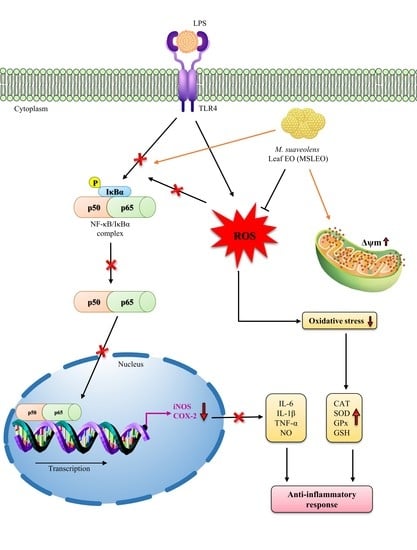
2. Results and Discussion

2.1. Chemical Composition of M. suaveolens Leaf Essential Oil (MSLEO)

MSLEO was pale-yellow in color and mint-like in smell, with a yield of 0.23% on a dry weight basis. The average oil yield was found to be higher compared to those determined for the same species by Mallavarapu et al. [45], Peerzada [46], and Bezerra et al. [40] but lower than those recorded by Malele et al. [47], Lohani et al. [48], and Xu et al. [39]. GC-MS analysis of the essential oil revealed that the oil is composed of 48 compounds, which account for 90.55% of the total essential oil (EO) (Table 1, Figure S1). The major constituents of MSLEO were β-caryophyllene (16.17%), phyllocladene (11.85%), abietatriene (11.46%), spathulenol (7.89%), 8,13-abietadien-18-ol (6.65%), and abietadiene (3.79%). The major chemical classes of compounds in the EO were diterpene hydrocarbons (28.82%), sesquiterpene hydrocarbons (25.74%), sesquiterpene alcohols (13.76%), and oxygenated diterpenes (13.34%). β-Caryophyllene has been detected to be the predominant component of essential oils of M. suaveolens by several researchers in the past [39,40,47,49]. This natural bicyclic sesquiterpene, present in many edible plants and essential oils, is known to be an effective anti-inflammatory agent [50,51,52,53]. By inhibiting the production of inflammatory mediators and regulating cell proliferation, β-caryophyllene exerts anti-inflammatory effects [54,55]. This led to the belief that β-caryophyllene or plants rich in β-caryophyllene have anti-inflammatory actions and may serve as new sources of drugs for treating viral infection, improving immunity, and reducing inflammation [56].
Among other major constituents of MSLEO, spathulenol is also known to exhibit anti-inflammatory effects, according to several researchers [58,59]. The essential oil of Psidium guineense, with spathulenol as the major constituent, exhibited strong anti-inflammatory activity against formalin-induced paw edema and carrageenan-induced mechanical hyperalgesia and paw edema in male Swiss mice [60]. Abietadiene and abietatriene, a group of aromatic abietanes, detected as dominant constituents of MSLEO, are also known to be anti-inflammatory in nature [61,62]. In addition, several minor constituents, like eucalyptol, β-selinene, caryophyllene oxide, and phytol, present in MSLEO have been reported to have anti-inflammatory activities [63,64,65]. The anti-inflammatory activity of MSLEO could be due to the predominance of β-caryophyllene, spathulenol, etc., or because of the synergistic effect of one or more major and minor phytoconstituents of the oil.
As can be seen from Table 1, MSLEO is mainly composed of terpenes and terpenoids, which are known to inhibit the NF-κB signaling pathway through IκB phosphorylation, DNA binding, p65 translocation, etc. [66,67,68,69]. These phytochemicals inhibit phosphorylation and IκB protein degradation, and subsequently, the active IκB-free NF-κB is translocated from the cytoplasm to the nucleus, leading to the transcription of proinflammatory cytokines [70]. β-Caryophyllene, a dominant constituent of MSLEO, is reported to act as an anti-inflammatory agent through the suppression of the main mediators of inflammation [52,55]. The expression of NF-ƙB is known to be downregulated by β-caryophyllene, thereby diminishing inflammatory and apoptotic responses [71,72,73,74]. Other major diterpenoid compounds, like phyllocladane, phytol, and abietanes, that were detected in MSLEO are reported to inhibit NF-κB activity by reducing the levels of cytokines and oxidative stress [63,75,76]. The anti-inflammatory effect of spathulenol has also been established through a mouse model of carrageenan-induced paw edema and pleurisy. Similarly, another minor constituent, sabinene, also showed anti-inflammatory activity by substantially regulating the production of NO and proinflammatory cytokines [77,78].

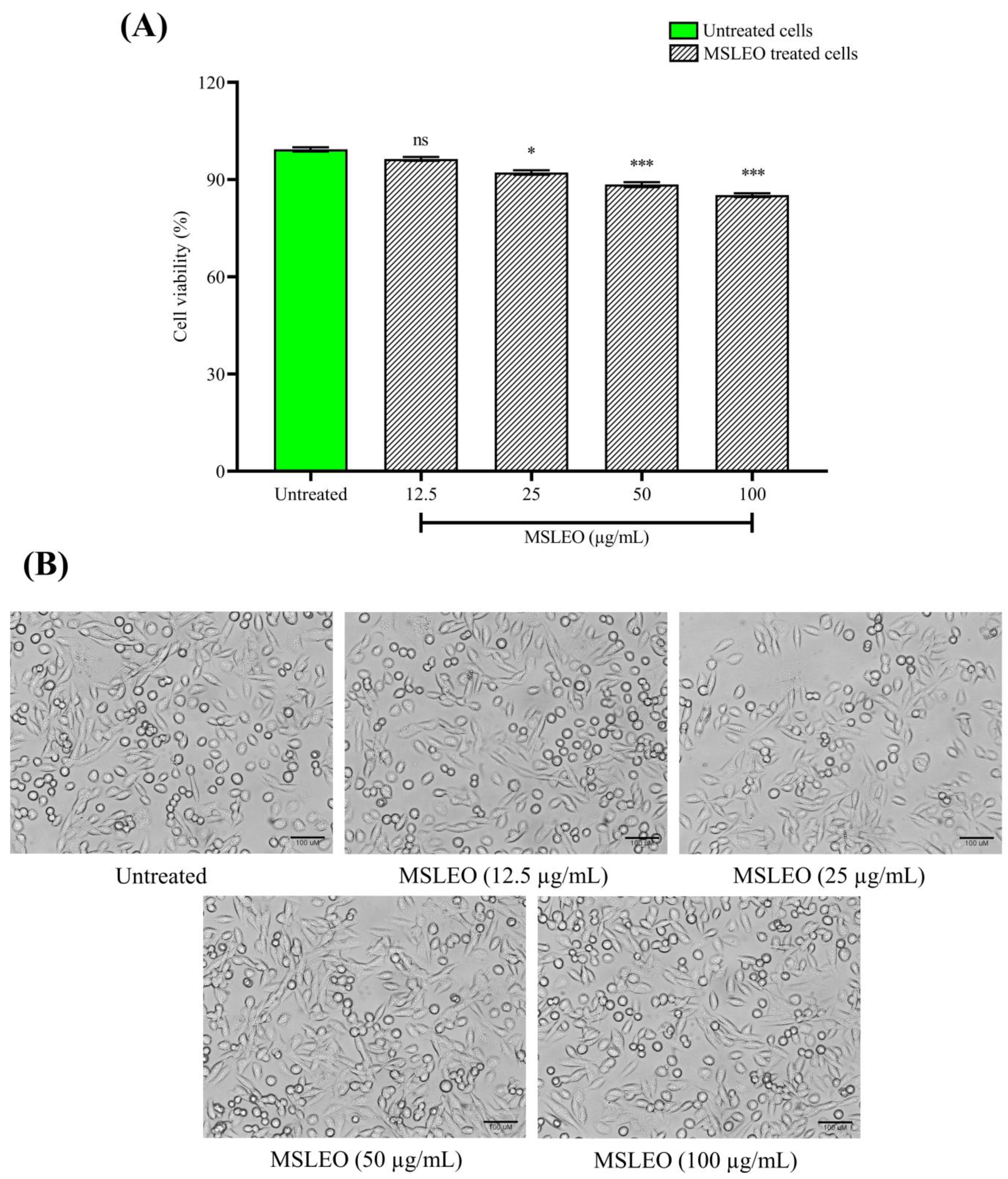
2.2. MSLEO Had No Impact on the Viability and Morphology of RAW 264.7 Cells

The MTT (3-[4,5-dimethylthiazol-2-yl]-2,5 diphenyl tetrazolium bromide) assay was performed to test the cytotoxicity of MSLEO. RAW 264.7 cells were treated with different doses of MSLEO between 12.5 µg/mL and 100 µg/mL (i.e., 12.5, 25, 50, and 100 µg/mL) to determine the level of toxicity and fix the non-toxic range of concentration to undertake anti-inflammatory assays. The half-maximal inhibitory concentration (IC50) of MSLEO was determined to be 324.17 µg/mL, as shown in Figure 1A. The results of the assay showed that MSLEO, up to a dose of 100 µg/mL, did not affect cell viability, which remained greater than 85%. Further, microscopic observation revealed that MSLEO, up to a 100 µg/mL concentration, did not show much cell spreading or pseudopodia formation, indicating that most of the cells were viable (Figure 1B). The results established that MSLEO did not adversely impact the viability and morphology of RAW 264.7 cells, and this could be due to the anti-inflammatory properties of MSLEO.
Based on these data, MSLEO at a lower concentration range (12.5 µg/mL) and a higher concentration range (100 µg/mL) were considered for the subsequent studies relating to inflammation and associated responses. Jena et al. [19] also observed no toxicity up to a 100 µg/mL concentration of the essential oil of Neocinnamomum caudatum. Similarly, treatment with 50 μg/mL Neolitsea sericea essential oil also maintained cell viability at 85% [79]. Using the MTT assay, Raka et al. [2] found that the viability of cells was not affected at all and remained greater than 90% with treatment with the Pingyin rose (Rosa rugosa) essential oil up to 100 µg/mL. In general, it is essential to know the optimal concentration of an essential oil before it is used as an anti-inflammatory agent [80].

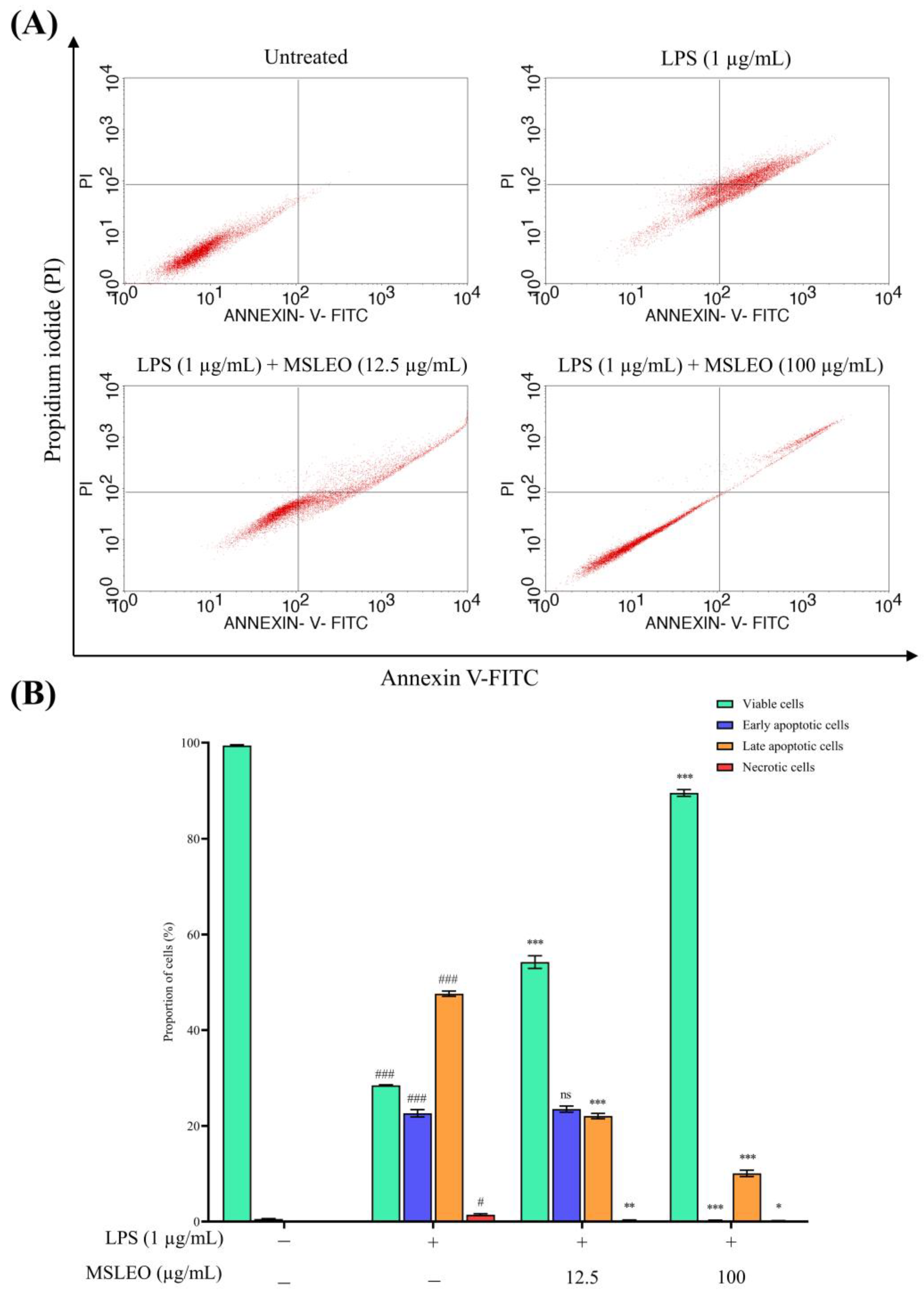
2.3. MSLEO Treatment Attenuates LPS-Induced Increase in Apoptosis

Apoptosis is the genetically regulated death of cells to ensure homeostasis and can be activated by two main pathways, i.e., death receptors and mitochondrial pathways. The essential oils of a number of plant species are reported to induce apoptosis marked by cytomorphological alterations like cell shrinkage, chromatin condensation, fragmentation of DNA accompanied by mitochondrial dysfunction, protein cleavage, etc. [81]. An abnormality in apoptosis may lead to several types of illnesses, including autoimmune diseases, neurodegenerative disorders, bacterial and viral infections, cardiac problems, and cancer [82]. An enhanced level of ROS generation, activation of protein kinase B, phosphorylated MAPKs, mitochondrial stress, and caspase activation are the main mechanisms of action by which plant essential oils induce programmed cell death [81].
In the present experiment, the externalization of phosphatidyl serine (PS) and cell membrane integrity was assessed by Annexin V–fluorescein isothiocyanate (FITC) and propidium iodide (PI) staining using a flow cytometer to study the effects of MSLEO on LPS-induced macrophage cells. When macrophage cells were activated with 1 µg/mL LPS for 24 h, the percentage of late (Annexin V+ve/PI+ve) apoptotic cells was significantly enhanced from 0.43% to 70.05%, and cell viability (Annexin V−ve/PI−ve) was drastically reduced from 99.57% to 28.61%, as depicted in Figure 2. Further, when these LPS-stimulated cells were co-cultured with a concentration of 12.5 µg/mL MSLEO, the proportion of apoptotic cells decreased from 71.39% to 45.09%, and the population of viable cells was nearly doubled from 28.61% to 54.73% in comparison to those treated with LPS only.
However, the treatment of murine macrophage cells with a dose of 100 µg/mL MSLEO drastically brought down the percentage of apoptotic cells from 70.05% to 10.14% and elevated the number of viable cells by more than 3 times (i.e., from 28.61% to 89.45%). Additionally, the proportion of necrotic cells decreased from 1.34% to 0.18% when LPS-induced macrophage cells were incubated with different concentrations of MSLEO. As reported earlier, the LPS model induces cell death by apoptosis through the autocrine secretion of TNF-α at the early stage and by the production of NO at the late apoptotic stage [83,84]. The data obtained from the Annexin V-FITC and PI double-staining assay suggested that MSLEO can attenuate the LPS-induced increase in apoptosis in a dose-dependent manner, consistent with the cytoprotective and anti-inflammatory activities of MSLEO.

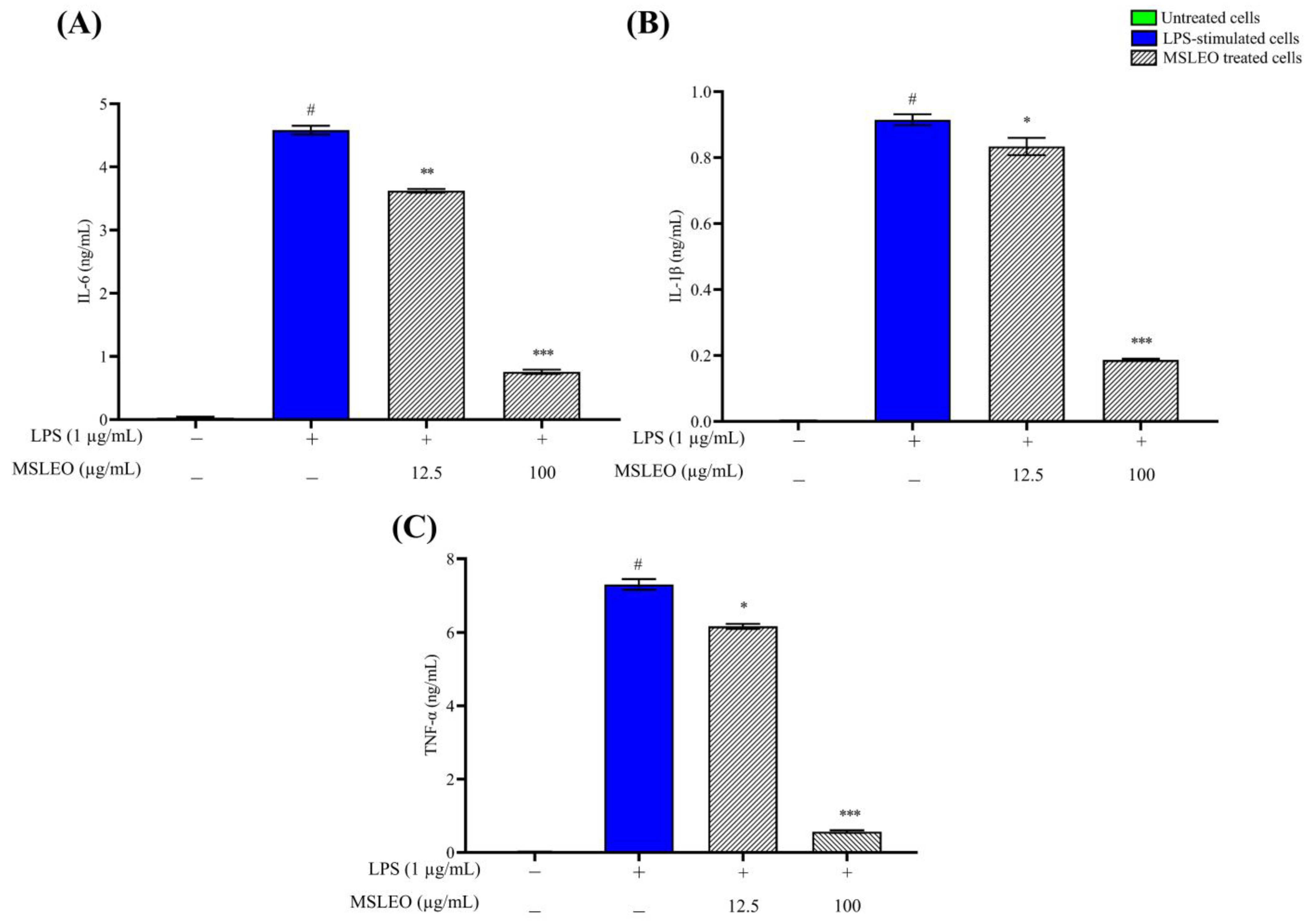
2.4. MSLEO Inhibits the LPS-Induced Increase in Expression of Proinflammatory Cytokines

One of the pathways through which plant essential oils primarily inhibit or attenuate inflammation is by downregulating the expression of the mRNA or protein of three key proinflammatory cytokines: IL-1β, IL-6, and TNF-α [85]. These cytokines regulate the immune response to inflammation through a complex network of interactions. They are stimulated in response to the invasion of the host by pathogenic microorganisms and are responsible for the recruitment and amplification of immune cells [86].
Being an endotoxin, LPS can activate macrophages and stimulate the release of several proinflammatory cytokines, which are involved in the upregulation of inflammatory reactions [87]. In the present investigation, the levels of expression of the above proinflammatory cytokines were first assessed in LPS-stimulated cells. Thereafter, the effect of MSLEO on the production of cytokines was measured by an ELISA (enzyme-linked immunosorbent assay) kit using a microplate reader. As depicted in Figure 3, after 24 h of exposure to LPS (1 µg/mL), significant increases in the expression levels of IL-6, IL-1β, and TNF-α were noticed as compared to the untreated group. Interestingly, incubation of these pre-activated macrophage cells treated with MSLEO at 12.5 and 100 µg/mL for 24 h was able to effectively reduce the levels of expression of these main proinflammatory cytokines in a dose-dependent manner. For instance, the administration of MSLEO at a 100 µg/mL concentration could reduce the levels of IL-6, IL-1β, and TNF-α by 6.1-fold, 4.8-fold, and 13.5-fold, respectively, compared to the LPS-treated groups.
The results of the present study are in agreement with the findings of earlier studies, where essential oils of Cinnamomum and Neocinnamomum species were found to suppress the production of the above proinflammatory cytokines generated by LPS depending on the concentration used [19,88,89]. The essential oils extracted from many other plant species, such as Citrus species, Artemisia fukudo, Chimonanthus praecox, Myrciaria tenella, Eucalyptus camaldulensis, Verbesina macrophylla, Lavandula angustifolia, Melaleuca alternifolia, and Cymbopogon martinii, have been reported to suppress the transcription of IL-6, IL-1β, and TNF-α by limiting their mRNA expression [12].
β-Caryophyllene, a major constituent of MSLEO, is known to exert strong anti-inflammatory effects by inhibiting the synthesis of proinflammatory mediators [90]. It is reported to inhibit the transcription of the above mediators in central nervous system immune effector cells (C6) treated at a dose of 10 mg/kg [50,91]. Further, the above data suggest that β-caryophyllene protects neurons from ischemia-induced cell death and acts as an anti-inflammatory agent. In addition, spathulenol and caryophyllene oxide in the essential oils have an important role in the process of the inhibition of these inflammatory mediators [92]. The anti-inflammatory activity of MSLEO through the suppression of the expression of different proinflammatory cytokines may be attributed to the dominance of β-caryophyllene, spathulenol, and other constituents and the synergistic effect of some major and minor constituents.

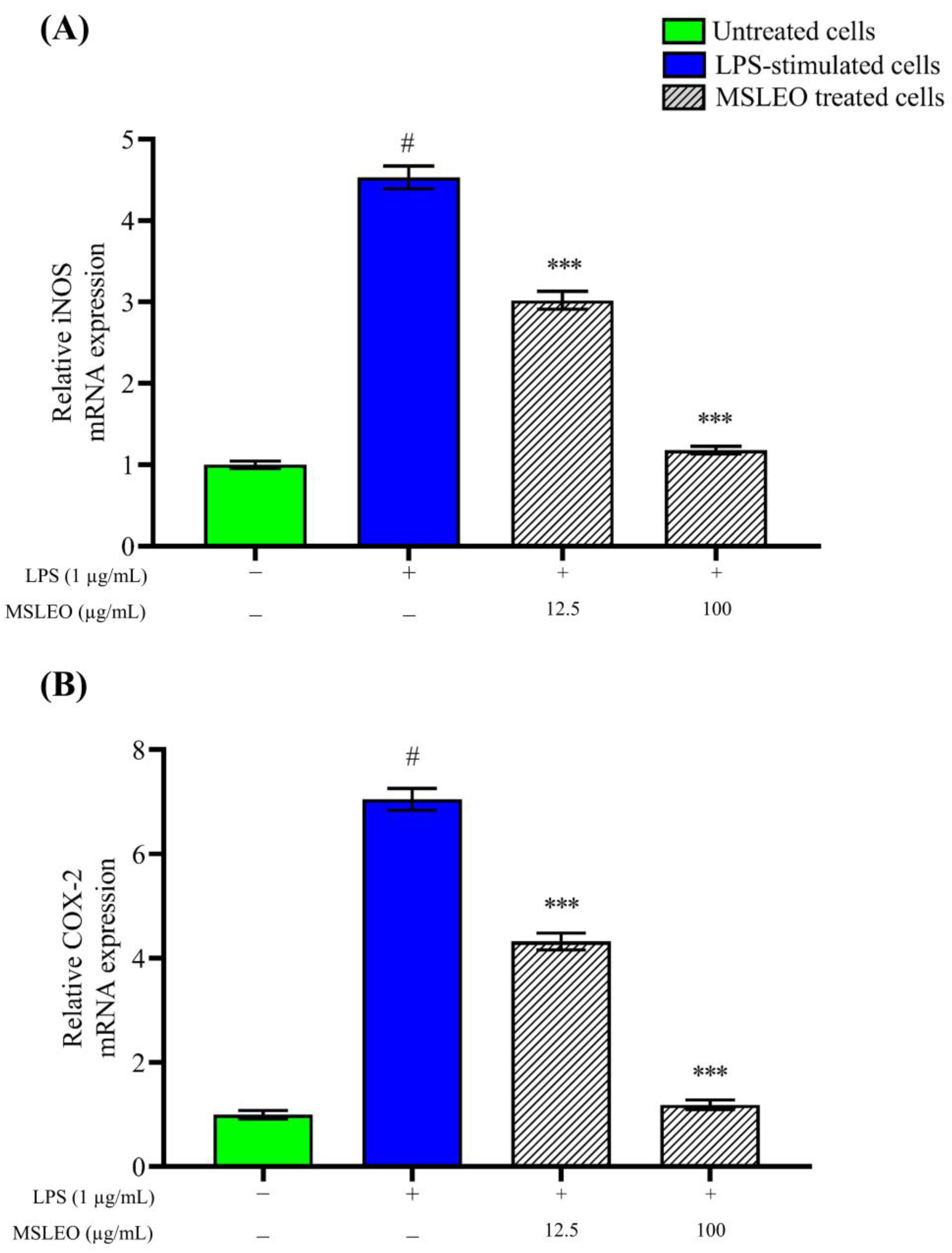
2.5. MSLEO Downregulates LPS-Induced Increase in iNOS and COX-2 mRNA Expression

In order to evaluate the anti-inflammatory potential of MSLEO, the levels of iNOS and COX-2 mRNA expression were estimated by real-time quantitative PCR (RT-qPCR). RAW 264.7 cells were treated with different concentrations of MSLEO (12.5 and 25 µg/mL) and with LPS and incubated for 24 h. The mRNA expression levels of iNOS and COX-2 were determined after incubation. As shown in Figure 4, the expression levels of iNOS and COX-2 increased many-fold upon incubation with 1 µg/mL LPS. When these pre-LPS-stimulated macrophages were treated with the minimum and maximum non-toxic doses of MSLEO (i.e., 12.5 and 100 µg/mL, respectively), the over-expression of iNOS and COX-2 mRNA was downregulated in a dose-dependent manner (Figure 4A,B). Interestingly, with a higher dose of MSLEO, a significant reduction in the expression of iNOS and COX-2 mRNA was observed as compared to the LPS-treated group, thereby restoring homeostasis.
iNOS, the most common isoform of NO, is produced when cells are stimulated with inflammatory cytokines and LPS [93,94]. Being a potent activator of iNOS, LPS interacts with Toll-like receptor 4 (TLR4) to activate macrophages [95]. Further, the role of the COX-2 enzyme is crucial in the process of the synthesis of PGE2, which is essentially an inflammatory mediator. As per Wei et al. [86], the production of PGE2 and NO is a closely linked process. In the mitigation of inflammatory conditions, the reduction in the levels of iNOS and COX-2 expression is considered crucial. One of the pathways by which plant essential oils primarily attenuate inflammatory responses is by inhibiting inducible nitric oxide synthase [96]. In addition, essential oils interfere with the arachidonic acid metabolic pathway and inhibit COX and LOX (Lipoxygenase) activity with a consequent reduction in PGE2, thus attenuating the deteriorating inflammatory condition [12]. Since NF-κB, in the activated state, facilitates the increased production of inflammatory cytokines, chemokines, and mediator enzymes [97], the regulation of the NF-κB signaling pathway has been widely used as a therapeutic route in the treatment of inflammatory conditions.
Kim et al. [98] found that the essential oil of Mentha arvensis exhibits anti-inflammatory activity by inhibiting proinflammatory cytokines and mediators through the suppression of COX-2 and iNOS expression. Similarly, the expression of the above two enzymes was found to be inhibited by essential oils of Chamaecyparis obtusa, Perilla frutescens, and Origanum vulgare [99,100]. The results of RT-qPCR and the concentration-dependent reduction in iNOS and COX-2 mRNA expression after treatment with MSLEO suggest that the oil has potent anti-inflammatory effects that can inhibit certain elements of the NF-κB pathway and ultimately minimize inflammatory responses.

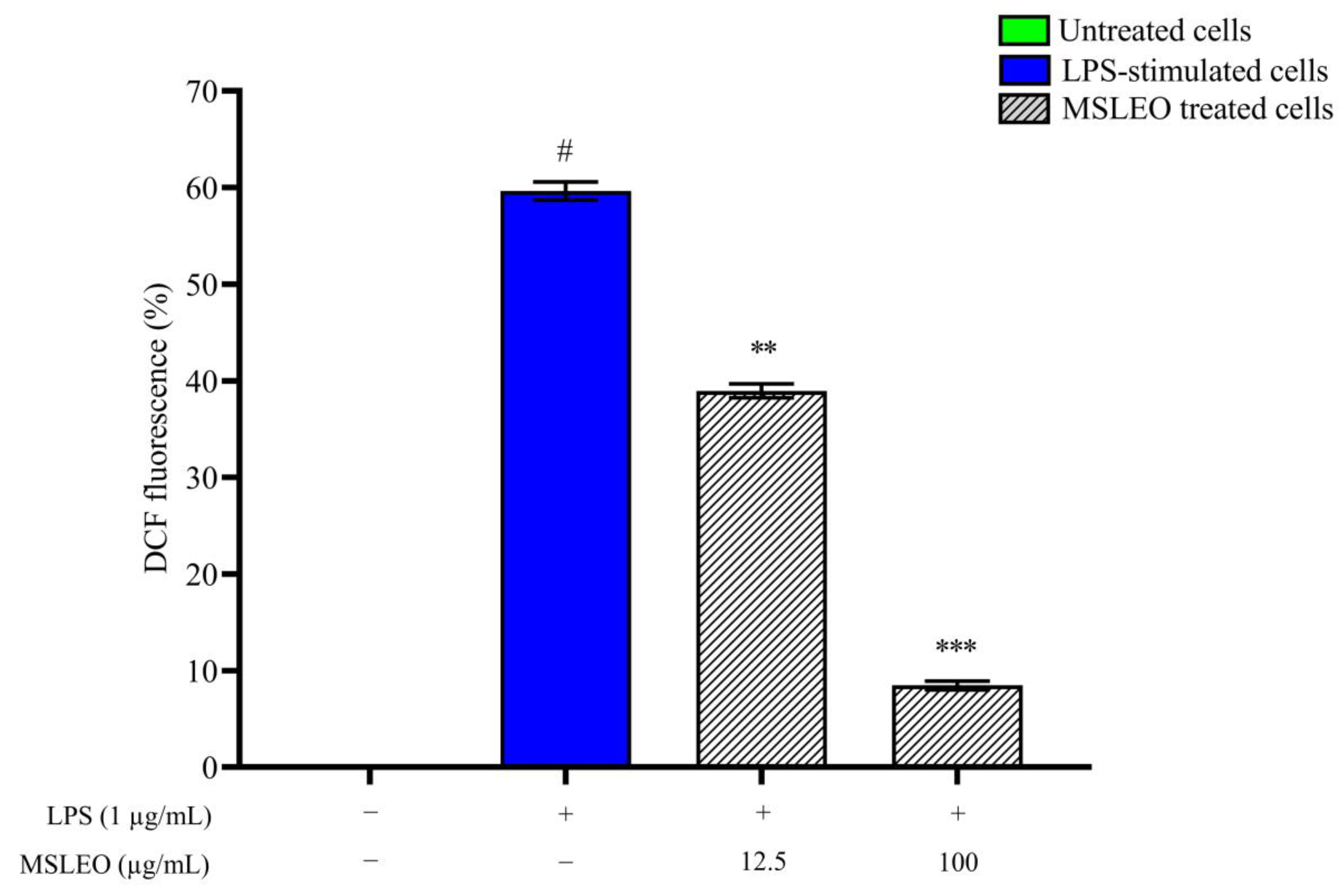
2.6. MSLEO Treatment Decreased LPS-Induced Increase in ROS Levels

Oxidative stress is reported to be associated with the pathogenesis of several metabolic and chronic disorders, including inflammatory conditions like cancer and cardiovascular disorders [101,102]. There is an increasing amount of evidence that ROS play a crucial role in the occurrence, progression, and resolution of the inflammatory response [103]. In addition, ROS act as signaling molecules for the regulation of cell survival, cell death, differentiation, cell signaling, and inflammation-related factor production [104]. However, prolonged or over-production of ROS by polymorphonuclear neutrophils (PMNs) at the inflammation site is responsible for endothelial injury, causing chronic inflammation underlying many neurodegenerative, cardiovascular, and metabolic diseases [105]. The majority of ROS are produced by aerobic cells during the mitochondrial respiratory chain and endogenous metabolic reactions [106,107], which are required to activate a variety of signaling pathways in both plants and animals. However, during LPS stimulation, RAW 264.7 cells produce excessive amounts of ROS/free radicals [108], and the endogenous antioxidants are inadequate to handle this condition, resulting in oxidative stress.
In the present study, the H2DCF-DA (2′,7′-dichlorodihydrofluorescein diacetate) cellular ROS assay kit was utilized to evaluate the effect of MSLEO on the intracellular ROS generation causing oxidative stress. Intracellular ROS oxidized non-fluorescent H2DCF-DA into fluorescent DCF (dichlorodihydrofluorescein), and the resulting fluorescence intensity was analyzed by a flow cytometer and correlated with the quantification of ROS produced during inflammation. As outlined in Figure 5, after 24 h of exposure to LPS (1 μg/mL), the percentage of RAW 264.7 cells emitting DCF fluorescence increased from 0.08% to 59.68%, indicating an escalated intracellular ROS level in LPS-treated cells in comparison to untreated cells. Meanwhile, intracellular ROS production in LPS-treated macrophages was substantially reduced upon the administration of 12.5 and 100 µg/mL MSLEO for 24 h. MSLEO at 12.5 µg/mL was able to suppress the intracellular ROS level by 1.5-fold, but at a higher concentration (100 µg/mL), the extent of ROS suppression was 7.1-fold in comparison with groups treated with LPS alone. The findings of this study are in agreement with the results obtained by earlier researchers, who found that essential oils of plant origin effectively neutralize intracellular ROS [109,110]. The reduction in NF-κB activity and the enhancement of mitochondrial membrane potential are indications of a reduction in the levels of ROS after treatment with MSLEO.
Inflammation and oxidative stress are pathophysiological events that are intricately related to each other [111,112]. In many chronic diseases, the co-existence of inflammation and oxidative stress has been reported. While cells liberate several reactive species at the site of inflammation, resulting in oxidative stress, many ROS/RNS can influence intracellular signaling cascades leading to the enhanced expression of proinflammatory genes [113,114]. The removal of pathogenic organisms from the body is the main purpose of inflammation, and for this, ROS production by phagocytes is important. While normal levels of ROS are important to reach homeostasis, normal levels of ROS are also equally important for killing pathogens. The uncontrolled production of ROS by different antioxidant mechanisms may lead to tissue damage. The antioxidant mechanism of the cell has a pivotal role to play in maintaining this intricate balance [112].

2.7. MSLEO Enhances Endogenous Antioxidant Enzyme Activities

Endogenous antioxidant enzymes, like glutathione peroxidase (GPx), catalase (CAT), and superoxide dismutase (SOD), counteract the pro-oxidant effects of ROS and thereby prevent damage to cells and tissues [103,115]. These antioxidant enzymes sequentially remove redundant ROS and prevent oxidative bursts. SOD initially catalyzes harmful superoxide radicals into molecular oxygen (O2) and hydrogen peroxide (H2O2). Subsequently, CAT or GPx converts H2O2 into the innocuous end-product, water [116].
In the present work, the effects of various concentrations of MSLEO on endogenous antioxidant enzyme (CAT, SOD, GPx, and GSH) activities were measured via ELISA using the LPS-induced RAW 264.7 cell model. The exposure of RAW 264.7 macrophage cells to 1 µg/mL LPS for 24 h resulted in a significant reduction in the antioxidant enzymes CAT, SOD, GPx, and GSH as compared to the untreated group (Figure 6). This reduction may be due to the innate immune response of macrophages to LPS, resulting in excessive production of ROS and RNS as a defense mechanism [117]. The long-term occurrence of excessive ROS and RNS or their inefficient scavenging impair the antioxidant enzymes’ ability to defend against damage from oxidative and nitrative stress [100], which in turn are linked to protein modifications, DNA damage, apoptosis, etc. [118,119].
However, when LPS-stimulated cells were co-cultured with varying dosages (12.5 and 100 µg/mL) of MSLEO, the levels of expression of CAT, SOD, GPx, and GSH were significantly elevated in a dose-independent manner. The antioxidant capacity of MSLEO could be due to the predominance of terpenes in the essential oil, which are considered natural antioxidants [120,121].
Jena et al. [19] reported that the essential oil of Neocinnamomum caudatum could ameliorate oxidative stress by upregulating the activities of CAT, SOD, GPx, and GSH. Similarly, the treatment of porcine small intestinal epithelial (IPEC-J2) cells with different doses of Oregano (Origanum vulgare) essential oil resulted in a substantial increase in SOD and CAT mRNA expression levels, as well as intracellular concentrations of SOD and GPx [122]. Working on bleomycin-induced pulmonary fibrosis in a murine model, Tavares et al. [123] demonstrated that Cymbopogon winterianus essential oil can significantly reduce inflammation in Bronchoalveolar Lavage Fluid (BALF), reduce malondialdehyde (MDA) levels, and increase SOD activity. The findings of this present experiment also revealed that MSLEO has the ability to reinstate the expression levels of the endogenous antioxidant enzymes by significantly increasing the production levels of CAT, SOD, GPx, and GSH compared to the LPS-stimulated group, which is essential for preventing many inflammation-related disorders [124].

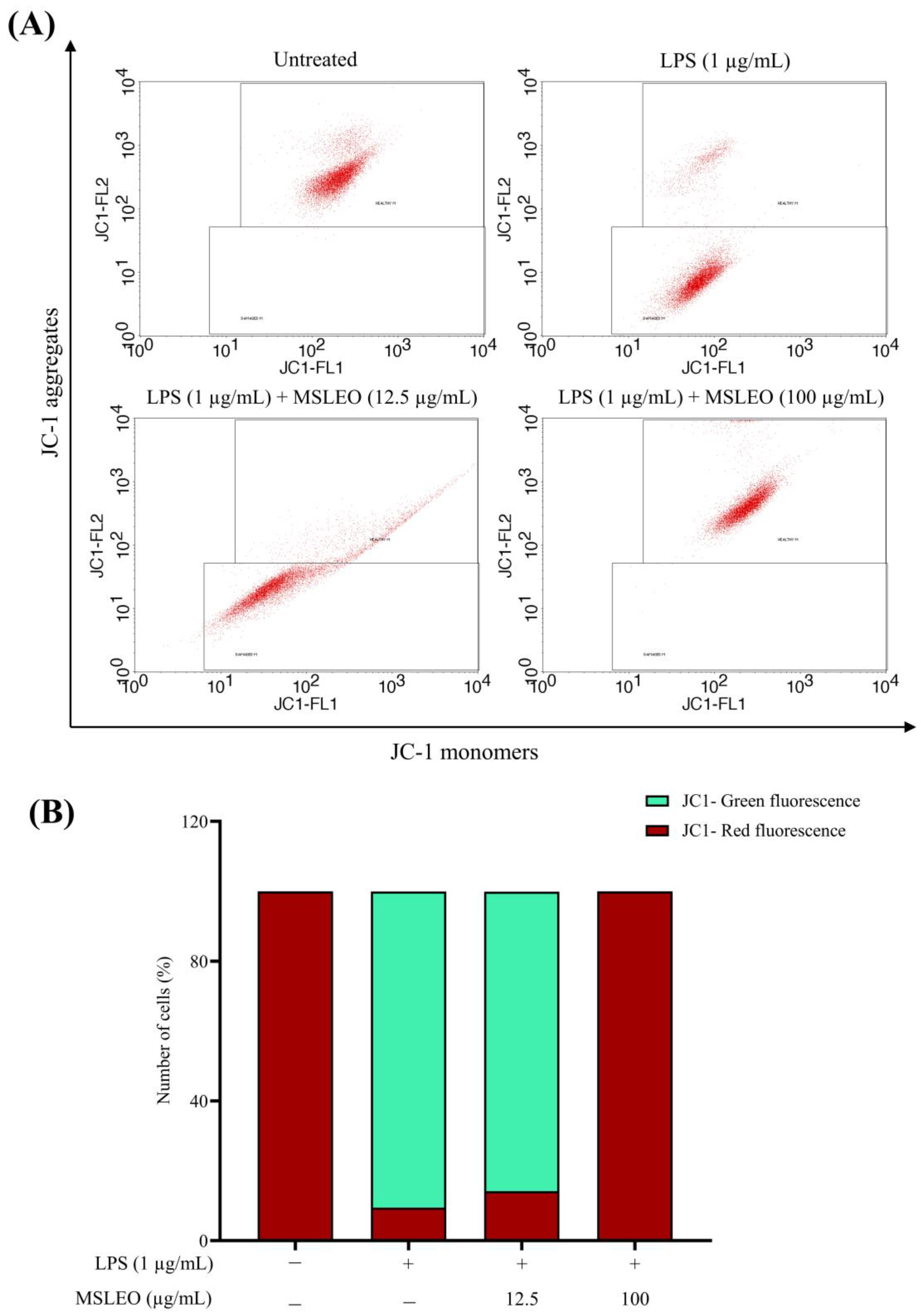
2.8. MSLEO Prevents Mitochondrial Membrane Potential (Δψm) from LPS-Stimulated Depolarization

The mitochondrial membrane potential (ΔΨm) is the most important component in the energy storage process during oxidative phosphorylation and the maintenance of mitochondrial homeostasis through mitochondrial removal by autophagy [125]. Therefore, cell health can be monitored from the mitochondrial membrane potential. The expression of key tricarboxylic acid (TCA)-cycle-related enzymes and mitochondrial membrane potential decreases when LPS is activated, whereas the levels of microtubule-associated protein LC3b and ROS increase [126]. The effect of MSLEO on mitochondrial damage and mitochondrial membrane potential (ΔΨm) resulting from LPS-induced inflammation were assessed using the JC-1 (5,5,6,6’-tetrachloro-1,1’,3,3’ tetraethylbenzimi-dazoylcarbocyanine iodide) Mitochondrial Membrane Potential (MMP) Assay Kit. JC-1 emitted red fluorescence when the membrane potential was normal, but in the event of disruption, it emitted green fluorescence.
When macrophage cells were subjected to LPS (1 µg/mL) for 24 h, a substantial decrease in gated cell populations emitting red fluorescence (9.46%) was observed compared to untreated cells (99.93%), indicating a lower membrane potential (Figure 7). This is because the aggregated form of JC-1 leaked out from the mitochondria into the cytoplasm as J-monomers, resulting in a decline in red fluorescence and an increase in green fluorescence, implying mitochondrial membrane dysfunction and depolarized MMP. Incubating these LPS-only-treated cells with MSLEO (12.5 and 100 µg/mL) for 24 h increased the level of red fluorescence. MSLEO, at 100 µg/mL, was also able to significantly increase the fraction of cells emitting red fluorescence up to 99.93%, pointing to higher mitochondrial membrane potential. This red spectral shift is due to an increased concentration of J-aggregates in mitochondria, suggesting the anti-inflammatory properties of M. suaveolens essential oil, which could regain the loss of mitochondrial membrane potential (Δψm) caused by LPS stimulation, depending on the concentration used. Neocinnamomum caudatum essential oil at concentrations of 12.5 and 25 µg/mL could significantly restore the mitochondrial membrane integrity [19]. de Carvalho et al. [127] found that the essential oil of Eugenia uniflora promoted mitochondrial membrane dysfunction in Drosophila melanogaster by inhibiting oxidative phosphorylation. Similarly, essential oils isolated from several species of Cistus are reported to ameliorate ultraviolet-B-induced mitochondrial damage and cellular senescence in human keratinocytes [128]. Essential oils containing β-caryophyllene as a major constituent in plant species, such as Pterodon emarginatus, Aloysia citrodora, Syzygium aromaticum, Eplingiella fruticosa, Ocimum basilicum, and Salvia rosmarinus, have the potential to counter oxidative stress and mitochondrial dysfunction and have possible links to neuroprotection [129].

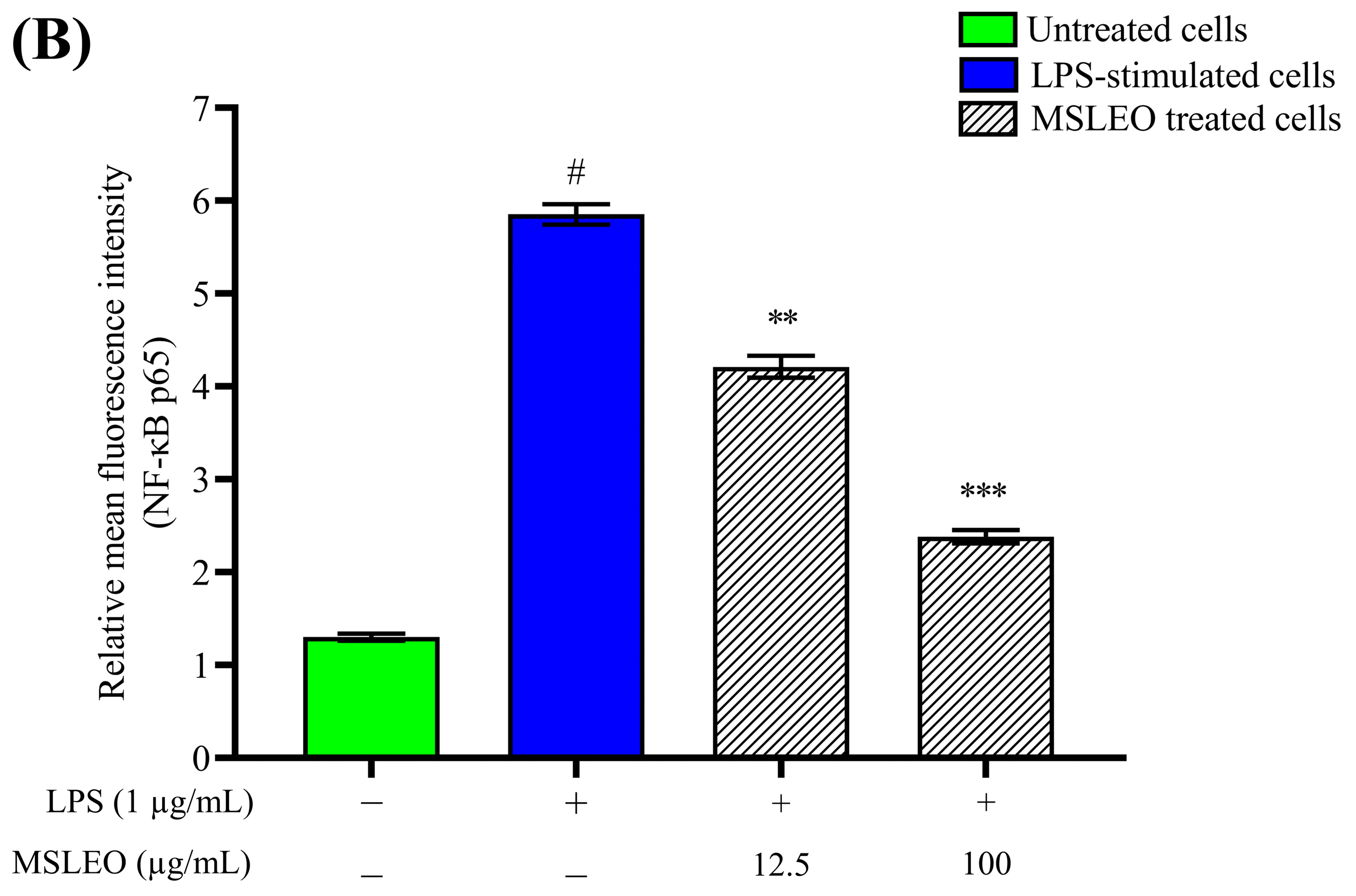
2.9. MSLEO Restricts NF-κB Nuclear Translocation in LPS-Stimulated RAW 264.7 Cells

NF-κB, a heterodimer complex consisting of p65 and p50 subunits, regulates multiple innate immune functions, in addition to contributing to inflammation, cancer, and nervous system function [130]. Once activated, NF-κB regulates the expression of several related genes and facilitates the over-production of proinflammatory mediators, thereby promoting cell growth, proliferation, apoptosis, and disease progression [131,132,133,134,135]. In view of this, understanding the mechanism of the regulation of the NF-κB pathway is essential to study the pathogenesis and treatment of several human diseases.
In the current work, murine macrophage cells were exposed to MSLEO for 24 h, followed by LPS treatment. The inhibitory effect of MSLEO on the NF-κB signaling pathway was evaluated by measuring the NF-κB-p65 fluorescence intensity using phycoerythrin (PE) fluorochrome. In order to observe the translocation of NF-κB p65 from the matrix of the cytoplasm to the nucleus, confocal microscopy was used. The cells were then immuno-stained with the p65 antibody and incubated for 12 h. The nucleus was stained with a Hoechst 33342 fluorescent solution and an FITC-conjugated secondary antibody. As illustrated in Figure 8, LPS (1 μg/mL) stimulation for 24 h triggered the translocation of NF-κB dimers to the nucleus, and further phosphorylation of p65 resulted in a substantial increase in the relative mean fluorescence intensity of NF-κB-p65 expression, increasing by 4.6-fold in comparison with the untreated cells. However, MSLEO treatment with 12.5 μg/mL and 100 μg/mL doses could nullify these changes by restricting the translocation of NF-κB dimers, thereby lowering the relative mean fluorescence intensity of NF-κB-p65 by 28.57% (1.4-fold) and 60% (2.5-fold), respectively. The results confirmed the anti-inflammatory potential of MSLEO in RAW 264.7 cells.
The NF-κB protein, being a central inflammatory mediator, has a major role in the pathogenesis of a good number of inflammatory disorders [136]. The essential oils of Eucalyptus species and Neocinnamomum caudatum are reported to attenuate LPS-stimulated inflammation in macrophages by inhibiting the activation of the NF-κB signaling pathway [19,137]. In another study by Mohamed et al. [78], it was shown that Achillea millefolium essential oil is responsible for the downregulation of the expression of TNF-α and IL-6 through the inhibition of NF-κB expression and the upregulation of peroxisome proliferator-activated receptor gamma (PPAR-γ). The mechanism of the anti-inflammatory effects of essential oils in several plant species, such as Blumea balsamifera, Thymus vulgaris, Chamaecyparis obtusa, Citrus aurantium var. amara, Zanthoxylum bungeanum, Abies holophylla, Perilla frutescens, Mentha pulegium, Eucalyptus spp., Mentha arvensis, Glechoma hederaceai, and Zingiber striolatum, has been found to be through the attenuation of the NF-κB signaling pathway [12].
The findings of the present study demonstrate that MSLEO exerts significant anti-inflammatory action by preventing the activation of the NF-κB pathway, which, in turn, inhibits the expression of proinflammatory cytokines, such as IL-1β, IL-6, and TNF-α; other inflammatory mediators, like iNOS and COX-2; and the regulation of the transcription of many genes, thereby regulating cell growth, differentiation, and apoptosis.

3. Materials and Methods

3.1. Collection of Plant Samples and Essential Oil Isolation

Fresh leaf samples of Mesosphaerum suaveolens were collected from plants in the flowering stage (Figure S2) from Balugaon, Chilika (85°15′54.2808″ E, 19°48′47.7144″ N; Altitude 17 m) of Khordha district, Odisha, India, in October 2021. The plant was identified by Prof. P. C. Panda, Taxonomist, and a voucher specimen (2417/CBT/Dt 13.10.2021) was housed in the herbaria of the Centre for Biotechnology, Siksha ‘O’ Anusandhan (Deemed to be University), Bhubaneswar, Odisha, India. The leaves were cut into small pieces, dried in the shade for 10 days, and made into fine powder. Then, 300 g of leaf powder was hydro-distilled in a Clevenger apparatus for 6 h. The isolated essential oil (MSLEO) was dried in anhydrous sodium sulfate (Na2SO4) and stored at 4 °C for subsequent analyses.

3.2. Culture and Maintenance of Cells

RAW 264.7 murine macrophage cells were procured from the National Centre for Cell Science (NCCS) Cell Repository, Pune, Maharashtra, India. The cells were cultured in Dulbecco’s Modified Eagle Medium (DMEM), high-glucose medium supplemented with 10% FBS, 1% antibiotic–antimycotic solution, and 1% L-glutamine (200 mM) and incubated in a humidified CO2 incubator with an environment of 18–20% O2, 5% CO2, and 37 °C. The cells were passaged every 2 days and allowed to proliferate for 24 h before the treatments.

3.3. GC-MS and GC-FID Analysis of MSLEO

The phytochemical composition of MSLEO was determined using a Clarus 580 Gas Chromatograph (Perkin-Elmer, Shelton, Connecticut, USA) coupled with an SQ8S MS detector. The analysis was conducted on an Elite-5 MS capillary column with an electron ionization source set at 70 eV. Helium was used as a carrier gas by maintaining the flow rate at 1 mL/min. A total of 0.1 µL of the undiluted essential oil was injected for GC-MS analysis. Initially, the oven temperature was set at 60 °C, which was gradually increased to 220 °C at a rate of 3 °C/min and held for 7 min. The injector temperature was set at 250 °C, and the transfer interface temperature was kept at 150 °C. The chemical compounds were identified by comparing the mass spectra of the detected compounds with the in-build NIST Mass Spectral library and by matching the retention indices (RIs) obtained by running straight-chain n-alkane series (C8–C20) under identical conditions with the published bibliographic literature [57].
Furthermore, to reinforce the reliability of the phytochemical identification of MSLEO, analysis was performed using the GC equipped with a Flame Ionization Detector (FID) while maintaining identical column conditions and programming parameters to those in the GC-MS analysis. To determine the response factors (RFs) of individual compounds, quantitative analysis was conducted with nonane, which served as the internal standard. The area percentages obtained from the GC-FID analysis were corrected using the response factors, which were established using representative reference standards for each group of chemicals under similar experimental conditions. The representative standard compounds used in the present study were as follows: eucalyptol (ether), 1-octen-3-ol (fatty alcohol), terpinen-4-ol (monoterpene alcohol), sabinene (monoterpene hydrocarbon), fenchone (monoterpene ketone), β-caryophyllene (sesquiterpene hydrocarbon), spathulenol (sesquiterpene alcohol), caryophyllene oxide (sesquiterpene oxides), hexahydrofarnesyl acetone (sesquiterpene ketone), abietatriene (diterpene hydrocarbons), phytol (diterpene alcohols), and dehydroabietol (oxygenated diterpenes). The response factor (RF) was calculated by using the formula:
RF = Canalyte/[(Aanalyte/Aistd)] × Cistd.
where Aanalyte and Canalyte are the GC peak area and the corresponding concentration of the standard compound representing a chemical group, and Aistd and Cistd are the GC peak area and concentration of the internal standard.

3.4. Cytotoxicity Assay

The MTT assay is a well-established quantitative colorimetric assay based on the conversion of MTT [3-(4,5-dimethylthiazol-2-yl)-2,5 diphenyl tetrazolium bromide] to an insoluble formazan crystal by succinate dehydrogenase to determine the toxicity level of a test compound in cultured cells [138]. In the present study, RAW 264.7 cells at a density of 2 × 10⁵ cells/mL were seeded in a 96-well plate, and the cells were allowed to proliferate and adhere properly for 24 h prior to treatment with the test compounds. The macrophage cells were then exposed to various doses of the essential oil (diluted with DMSO), MSLEO (12.5, 25, 50, and 100 µg/mL), and incubated at 37 °C for an additional 24 h in a 5% CO2 environment. After 24 h of incubation, the utilized medium was removed, and MTT reagent (0.5 mg/mL) was added to each well and further incubated for another 3 h. The medium was again taken out, 100 µL of DMSO was added to each well, and the plate was gently agitated until the complete dissolution of formazan crystals. Subsequently, the absorbance of the purple-colored formazan was measured with an ELISA plate reader at 570 nm. The cytotoxicity level of MSLEO was determined by comparing the absorbance of the MSLEO-treated cells with the absorbance of the untreated RAW 264.7 cells. The morphological alterations in RAW 264.7 cells after treatment with MSLEO were visualized under an inverted microscope (scale bar = 100 µm). The proportion of viable cells was determined by using the following mathematical expression:
C e l l   v i a b i l i t y % = A b s o r b a n c e   o f   t h e   t r e a t e d   c e l l s A b s o r b a n c e   o f   t h e   u n t r e a t e d   c e l l s × 100

3.5. Annexin V-FITC/PI Apoptosis Assay

Annexin V-FITC/PI staining is one of the most extensively used tests to evaluate apoptosis and necrosis in cells [139,140]. In the current experiment, the percentage of apoptotic/necrotic RAW 264.7 cells were determined following the manual instructions provided with the Annexin V-FITC apoptosis kit (BD Biosciences). The cells were plated at a density of 2.5 × 10⁵ cells/mL in a 24-well plate and cultured for 24 h at 37 °C in a CO2 incubator. Then, the spent medium was aspirated out, and the cells were stimulated with LPS (1 µg/mL) for 2 h, except for the untreated cells. Subsequently, the stimulated cells were exposed to the minimum (12.5 µg/mL) and maximum (100 µg/mL) doses of MSLEO and incubated for another 24 h under similar conditions to those described earlier. After that, the cells were washed twice with 1X phosphate-buffered saline (PBS) solution, harvested, and resuspended in polystyrene tubes containing 400 μL of binding buffer supplemented with 5 μL of Annexin V-FITC and 5 μL of propidium iodide (PI). Fluorescence-activated cell sorting (FACs) tubes were kept in the dark at room temperature for 15 min, and then the cells were analyzed with a flow cytometer at an excitation wavelength of 488 nm and emission wavelengths of 530 nm and 610 nm for Annexin V-FITC and PI fluorescence, respectively. The anti-apoptotic potential of MSEO was assessed based on the proportion of viable (Annexin V−ve/PI−ve), early apoptotic (Annexin V+ve/PI−ve), and late apoptotic/necrotic (Annexin V+ve/PI+ve) cells screened using BD CellQuest Pro Ver. 6.0 software (Becton, Dickinson, Heidelberg, Germany).

3.6. Proinflammatory Cytokine (IL-6, IL-1β, and TNF-α) Detection by ELISA

The levels of the expression of proinflammatory cytokines were evaluated using commercially available mouse immunoassay ELISA kits and referring to the manufacturer’s instructions (Abcam Co. Cambridge, UK). Initially, the cells were pretreated with LPS (1 μg/mL) for 2 h (except for the untreated group), and then these cells were subjected to lower and higher concentrations of MSLEO (12.5 and 100 µg/mL) for 24 h at 37 °C in the incubator. After incubation for 24 h, the culture medium was collected and centrifuged at 2000 rpm for 10 min at 4 °C. Afterward, supernatants were collected, and the absorbance was recorded at 450 nm to measure the levels of IL-6, IL-1β, and TNF-α. The results were expressed in ng/mL.

3.7. RNA Extraction and Real-Time Quantitative PCR (RT-qPCR)

RT-qPCR analysis was conducted to estimate the levels of the expression of iNOS and COX-2 mRNA in LPS-stimulated cells following the protocol of Syam et al. [141]. As described above, the cells were treated with two doses of MSLEO, i.e., 12.5 µg/mL and 100 µg/mL, and incubated for 24 h at 37 °C and 5% CO2. After incubation, total RNA was isolated from the cells using the Qiagen RNeasy kit (Hilden, Germany) following the instructions provided by the manufacturer. The isolated RNA was treated with DNAse to avoid any genomic contamination. The purity of RNA was checked spectrophotometrically by comparing the absorbances taken at 260 nm and 280 nm, and the total RNA was quantified with the help of QiaExpert (Hilden, Germany). Later, the IScript cDNA synthesis kit (Bio-Rad) was used for the synthesis of cDNA from the previously isolated RNA through reverse transcription. Then, PCR amplification of the target genes, i.e., COX-2, iNOS, and GADPH (housekeeping gene), was performed by employing the cDNA as a template and gene-specific forward and reverse primers (Table 2). The RT-qPCR analysis was carried out with a Qiagen Rotor-Gene Q 5plex HRM RT-qPCR using SYBR green fluorescent dye with a thermal cycle of initial denaturation at 95 °C for 5 min, followed by 40 cycles of denaturation at 95 °C for 10 s, annealing at 60 °C for 20 s, and extension at 72 °C for 20 s. The 2−ΔΔCT approach was used to measure and normalize the target genes’ expression levels, i.e., COX-2 and iNOS, relative to the internal housekeeping gene (GADPH) in the same sample.

3.8. Intracellular ROS Assay

D2CFH-DA (2,7-dichlorofluorescein diacetate) is a ROS-sensitive fluorescence indicator widely used for measuring intracellular ROS levels [142]. In viable cells, esterase cleaves DCFH-DA into DCFH, which is then oxidized by ROS to produce the highly fluorescent chemical DCF (2′,7′-dichlorofluorescein). Briefly, the cells were seeded at a density of 2.5 × 10⁵ cells/mL in a 24-well plate, pre-stimulated with LPS (1 μg/mL), and exposed to two concentrations of MSLEO (12.5 and 100 µg/mL) in 5% CO2 at 37 °C for 24 h. Subsequently, the used medium was taken out, and the cells were washed with 1X PBS and incubated for another 30 min with DCFH-DA (10 μM) away from light. DCFH-DA was then removed, and the cells were rinsed twice with cold PBS, trypsinized, and centrifuged. The collected pellets were then dissolved in PBS, and fluorescence intensity was measured using a flow cytometer (BD FACScan, BD Bioscience, San Jose, CA, USA).

3.9. Measurement of Endogenous Antioxidant Enzyme Activities by ELISA

Like in the endogenous antioxidant enzyme expression study, pre-stimulated cells grown to a density of 2 × 10⁵ cells/mL in a 24-well plate were subjected to two different doses (i.e., 12.5 µg/mL and 100 µg/mL) of MSLEO for 24 h at 37 °C and 5% CO2. The incubated cells were then washed with PBS, and the cellular lysates were collected using RIPA buffer and kept at −80 °C for further analysis. Following the manufacturer’s protocol, the activities of the endogenous antioxidant enzymes CAT, SOD, GPx, and GSH were measured spectrophotometrically using the cellular lysates. The activities of the endogenous antioxidant enzymes (CAT, SOD, GPx, and GSH) were expressed as mU/mL, U/mg, ng/mL, and µM protein, respectively.

3.10. Mitochondrial Membrane Potential (MMP) Assay

The mitochondrial membrane potential (△ψm) was evaluated using JC-1 dye as per the protocol described by Yuan et al. [143]. The cells (2.5 × 10⁵ cells/mL) were seeded in a 24-well plate overnight after being treated with 1 μg/mL LPS for 2 h. They were then treated with 12.5 µg/mL and 100 µg/mL MSLEO and incubated at 37 °C and 5% CO2 for 24 h. The cells were subsequently washed with 1X PBS, trypsinized, and centrifuged, and following that, 0.5 mL of JC-1 working solution was added to the collected pellets. After that procedure, the cells were once again rinsed with PBS, resuspended in 1 mL of fresh 1X PBS, and then examined using a flow cytometer (Becton Dickinson FACScan, BD Bioscience, USA). The mitochondrial membrane potential was estimated by comparing the proportion of gated cells emitting red fluorescence (JC-1 aggregates) and green fluorescence (JC-1 monomers) measured at 540/570 nm and 485/535 nm, respectively.

3.11. NF-κB Nuclear Translocation Assay by Confocal Microscopy

The cells were plated at a density of 2.5 × 10⁵ cells/mL in a 24-well plate and stimulated with LPS (1 μg/mL) for 2 h (except for the untreated ones). The pre-stimulated cells were then treated with 12.5 µg/mL and 100 µg/mL MSLEO and incubated for an additional time of 24 h at 37 °C in a 5% CO2 environment. The culture medium was subsequently taken out from each well, and cells were rinsed twice with 1X PBS; the cells grown in the wells were fixed using 0.5 mL of BD Cytofix/Cytoperm solution (BD Biosciences, 554714) at room temperature for 10 min. After washing the cells with 1X PBS (supplemented with 0.5% bovine and 0.1% sodium azide), the cells were incubated for 30 min with mouse anti-NF-κB p65 antibody (10 µL) conjugated with phycoerythrin fluorochrome (PE). For nuclear staining, cells were counterstained with 100 µL of DAPI solution (1 µg/ mL) and incubated for 10 min in the dark. After the final wash, the cells were visualized under a Zeiss LSM 880 (Carl Zeiss Microscopy GmbH, Jena, Germany) confocal microscope, and the expression level of NF-κB p65 was measured using Image J software version 1.54F.

3.12. Statistical Analysis

All the findings from the experiments are reflected as mean ± SD (standard deviation). GraphPad Prism version 8.0 software (Boston, MA, USA) was used to evaluate the statistical significance by ANOVA test, followed by Tukey’s multiple range test.

4. Conclusions

The present research unraveled the anti-inflammatory potential of Mesosphaerum suaveolens leaf essential oil (MSLEO) and the molecular mechanism of its action for the first time. The study revealed that MSLEO could inhibit LPS-induced inflammation and oxidative stress in RAW 264.7 macrophages through a reduction in the levels of the proinflammatory cytokines like IL-6, IL-1β, and TNF-α, the expression of inflammatory mediators such as iNOS and COX-2, and intracellular ROS production. MSLEO was also able to maintain the mitochondrial membrane integrity and inhibited the translocation of NF-κB from the cytosol to the nucleus, thereby downregulating the expression of proinflammatory genes, cytokines, and mediators. The findings of the current research indicate that MSLEO could serve as a promising therapeutic agent against inflammation-related responses. However, further in-depth laboratory-based research involving in vitro assays and in vivo models may be necessary to establish the therapeutic efficacy of the essential oil before initiating preclinical trials on its anti-inflammatory effects, leading to drug development.

Supplementary Materials

The following supporting information can be downloaded at: https://www.mdpi.com/article/10.3390/molecules28155817/s1, Figure S1: GC-MS chromatogram of the Mesosphaerum suaveolens leaf essential oil (MSLEO). Figure S2: Image of Mesosphaerum suaveolens plant.

Author Contributions

Conceptualization: P.C.P., S.N. and A.R.; methodology: O.M., S.J., P.K.D. and A.S.; formal analysis: O.M., A.R. and S.S.P.; writing—review and editing: O.M., P.C.P. and A.R.; supervision: S.N. and P.C.P. All authors have read and agreed to the published version of the manuscript.

Funding

This study was funded by the Department of Science and Technology, Government of India, under the INSPIRE Fellowship program awarded to O.M. (No. DST/INSPIRE Fellowship/2019/IF190495).

Institutional Review Board Statement

Not applicable.

Informed Consent Statement

Not applicable.

Data Availability Statement

The data presented in this work are available in the article.

Acknowledgments

The authors are grateful to M. R. Nayak and S. C. Si, Siksha ‘O’ Anusandhan (Deemed to be University) for providing the necessary facilities and encouragement.

Conflicts of Interest

The authors declare no conflict of interest.

Sample Availability

Samples of the compounds are not available from the authors.

References

  1. Chou, S.T.; Lai, C.C.; Lai, C.P.; Chao, W.W. Chemical composition, antioxidant, anti-melanogenic and anti-inflammatory activities of Glechoma hederacea (Lamiaceae) essential oil. Ind. Crop. Prod. 2018, 122, 675–685. [Google Scholar] [CrossRef]
  2. Raka, R.N.; Zhiqian, D.; Yue, Y.; Luchang, Q.; Suyeon, P.; Junsong, X.; Hua, W. Pingyin rose essential oil alleviates LPS-induced inflammation in RAW 264.7 cells via the NF-κB pathway: An integrated in vitro and network pharmacology analysis. BMC Complement. Med. Ther. 2022, 22, 272. [Google Scholar] [CrossRef]
  3. Chen, Z.; Bozec, A.; Ramming, A.; Schett, G. Anti-inflammatory and immune-regulatory cytokines in rheumatoid arthritis. Nat. Rev. Rheumatol. 2019, 15, 9–17. [Google Scholar] [CrossRef] [PubMed]
  4. Sorci-Thomas, M.G.; Thomas, M.J. Microdomains, inflammation, and atherosclerosis. Circ. Res. 2016, 118, 679–691. [Google Scholar] [CrossRef] [PubMed]
  5. Singh, N.; Baby, D.; Rajguru, J.P.; Patil, P.B.; Thakkannavar, S.S.; Pujari, V.B. Inflammation and cancer. Ann. Afr. Med. 2019, 18, 121. [Google Scholar] [CrossRef]
  6. Polednik, K.M.; Koch, A.C.; Felzien, L.K. Effects of essential oil from Thymus vulgaris on viability and inflammation in zebrafish embryos. Zebrafish 2018, 15, 361–371. [Google Scholar] [CrossRef]
  7. Hotamisligil, G.S. Inflammation and metabolic disorders. Nature 2006, 444, 860–867. [Google Scholar] [CrossRef]
  8. Abdulkhaleq, L.A.; Assi, M.A.; Abdullah, R.; Zamri-Saad, M.; Taufiq-Yap, Y.H.; Hezmee, M.N.M. The crucial roles of inflammatory mediators in inflammation: A review. Vet. World 2018, 11, 627. [Google Scholar] [CrossRef] [Green Version]
  9. Billack, B. Macrophage activation: Role of toll-like receptors, nitric oxide, and nuclear factor kappa B. Am. J. Pharm. Educ. 2006, 70, 102. [Google Scholar] [CrossRef] [Green Version]
  10. Bhattacharyya, A.; Chattopadhyay, R.; Mitra, S.; Crowe, S.E. Oxidative stress: An essential factor in the pathogenesis of gastrointestinal mucosal diseases. Physiol. Rev. 2014, 94, 329–354. [Google Scholar] [CrossRef] [Green Version]
  11. Aviello, G.; Knaus, U.G. ROS in gastrointestinal inflammation: Rescue or sabotage? Br. J. Pharmacol. 2017, 174, 1704–1718. [Google Scholar] [CrossRef] [Green Version]
  12. Zhao, Q.; Zhu, L.; Wang, S.; Gao, Y.; Jin, F. Molecular mechanism of the anti-inflammatory effects of plant essential oils: A systematic review. J. Ethnopharmacol. 2023, 307, 115829. [Google Scholar] [CrossRef]
  13. Huang, Z.; Xie, L.; Xu, Y.; Zhao, K.; Li, X.; Zhong, J.; Lu, Y.; Xu, X.; Goodin, S.; Zhang, K.; et al. Essential oils from Zingiber striolatum Diels attenuate inflammatory response and oxidative stress through regulation of MAPK and NF-κB signaling pathways. Antioxidants 2021, 10, 2019. [Google Scholar] [CrossRef] [PubMed]
  14. Zarghi, A.; Arfaei, S. Selective COX-2 inhibitors: A review of their structure-activity relationships. Iran. J. Pharm. Res. 2011, 10, 655. [Google Scholar] [CrossRef]
  15. Bindu, S.; Mazumder, S.; Bandyopadhyay, U. Non-steroidal anti-inflammatory drugs (NSAIDs) and organ damage: A current perspective. Biochem. Pharmacol. 2020, 180, 114147. [Google Scholar] [CrossRef] [PubMed]
  16. Cunningham, K.; Candelario, D.M.; Angelo, L.B. Nonsteroidal anti-inflammatory drugs: Updates on dosage formulations and adverse effects. Orthop. Nurs. 2020, 39, 408–413. [Google Scholar] [CrossRef] [PubMed]
  17. Reynolds, J.F.; Noakes, T.D.; Schwellnus, M.P.; Windt, A.; Bowerbank, P. Non-steroidal anti-inflammatory drugs fail to enhance healing of acute hamstring injuries treated with physiotherapy. S. Afr. Med. J. 1995, 85, 517–522. [Google Scholar] [PubMed]
  18. Maroon, J.C.; Bost, J.W.; Maroon, A. Natural anti-inflammatory agents for pain relief. Surg. Neurol. Int. 2010, 1, 80. [Google Scholar] [CrossRef] [Green Version]
  19. Jena, S.; Ray, A.; Mohanta, O.; Das, P.K.; Sahoo, A.; Nayak, S.; Panda, P.C. Neocinnamomum caudatum Essential oil ameliorates lipopolysaccharide-induced inflammation and oxidative stress in RAW 264.7 cells by inhibiting NF-κB activation and ROS production. Molecules 2022, 27, 8193. [Google Scholar] [CrossRef]
  20. Miguel, M.G. Antioxidant and anti-inflammatory activities of essential oils: A short review. Molecules 2010, 15, 9252–9287. [Google Scholar] [CrossRef] [Green Version]
  21. Zuo, X.; Gu, Y.; Wang, C.; Zhang, J.; Zhang, J.; Wang, G.; Wang, F. A systematic review of the anti-inflammatory and immunomodulatory properties of 16 essential oils of herbs. Evid. Based Complement. Altern. Med. 2020, 2020, 8878927. [Google Scholar] [CrossRef]
  22. Bhargava, V.V.; Patel, S.C.; Desai, K.S. Importance of terpenoids and essential oils in chemotaxonomic approach. Int. J. Herb. Med. 2013, 1, 14–21. [Google Scholar]
  23. Nóbrega de Almeida, R.; Agra, M.D.F.; Negromonte Souto Maior, F.; De Sousa, D.P. Essential oils and their constituents: Anticonvulsant activity. Molecules 2011, 16, 2726–2742. [Google Scholar] [CrossRef]
  24. POWO. Plants of the World Online. Facilitated by the Royal Botanic Gardens, Kew. Available online: http://www.plantsoftheworldonline.org (accessed on 7 June 2023).
  25. Padalia, H.; Srivastava, V.; Kushwaha, S.P.S. Modeling potential invasion range of alien invasive species, Hyptis suaveolens (L.) Poit. in India: Comparison of MaxEnt and GARP. Ecol. Inform. 2014, 22, 36–43. [Google Scholar] [CrossRef]
  26. Afreen, T.; Srivastava, P.; Singh, H.; Singh, J.S. Effect of invasion by Hyptis suaveolens on plant diversity and selected soil properties of a constructed tropical grassland. J. Plant Ecol. 2018, 11, 751–760. [Google Scholar] [CrossRef] [Green Version]
  27. Li, R.; Tang, G.; Liu, X.; Li, J.; Wang, D.; Ji, S. An ethnopharmacological review of Hyptis suaveolens (L.) Poit. Trop. J. Pharm. Res. 2020, 19, 1541–1550. [Google Scholar] [CrossRef]
  28. Mishra, P.; Sohrab, S.; Mishra, S.K. A review on the phytochemical and pharmacological properties of Hyptis suaveolens (L.) Poit. Future J. Pharm. Sci. 2021, 7, 65. [Google Scholar] [CrossRef]
  29. Sastri, B.N. The Wealth of India—Raw Materials; Council of Scientific and Industrial Research (CSIR): New Delhi, India, 1959; Volume V (H–K). [Google Scholar]
  30. Adda, C.; Atachi, P.; Hell, K.; Tamò, M. Potential use of the bushmint, Hyptis suaveolens, for the control of infestation by the pink stalk borer, Sesamia calamistis on maize in southern Benin, West Africa. J. Insect Sci. 2011, 11, 33. [Google Scholar] [CrossRef] [PubMed]
  31. Seyoum, A.; Pålsson, K.; Kung’a, S.; Kabiru, E.W.; Lwande, W.; Killeen, G.F.; Hassanali, A.; Knots, B.G.J. Traditional use of mosquito-repellent plants in western Kenya and their evaluation in semi-field experimental huts against Anopheles gambiae: Ethnobotanical studies and application by thermal expulsion and direct burning. Trans. R. Soc. Trop. Med. Hyg. 2002, 96, 225–231. [Google Scholar] [CrossRef] [PubMed]
  32. Aguirre, C.; Torres, I.; Mendoza-Hernández, G.; Garcia-Gasca, T.; Blanco-Labra, A. Analysis of protein fractions and some minerals present in Chan (Hyptis suaveolens L.) seeds. J. Food Sci. 2012, 77, 15–19. [Google Scholar] [CrossRef]
  33. Hsu, F.C.; Tsai, S.F.; Lee, S.S. Chemical investigation of Hyptis suaveolens seed, a potential antihyperuricemic nutraceutical, with assistance of HPLC-SPE-NMR. J. Food Drug Anal. 2019, 27, 897–905. [Google Scholar] [CrossRef] [PubMed]
  34. McNeil, M.; Facey, P.; Porter, R. Essential oils from the Hyptis genus—A review (1909–2009). Nat. Prod. Commun. 2011, 6, 1775–1796. [Google Scholar] [PubMed] [Green Version]
  35. Bezerra, J.W.A.; Rodrigues, F.C.; Lima Bezerra, J.J.; Vieira Pinheiro, A.A.; Almeida de Menezes, S.; Tavares, A.B.; Costa, A.R.; Augusta de Sousa Fernandes, P.; Bezerra da Silva, V.; Martins da Costa, J.G.; et al. Traditional uses, phytochemistry, and bioactivities of Mesosphaerum suaveolens (L.) Kuntze. Evid.-Based Complement. Altern. Med. 2022, 2022, 3829180. [Google Scholar] [CrossRef]
  36. Barbosa, L.C.A.; Martins, F.T.; Teixeira, R.R.; Polo, M.; Montanari, R.M. Chemical variability and biological activities of volatile oils from Hyptis suaveolens (L.) Poit. Agric. Conspec. Sci. 2013, 78, 1–10. [Google Scholar]
  37. Luz, T.R.S.A.; Leite, J.A.C.; de Mesquita, L.S.S.; Bezerra, S.A.; Silveira, D.P.B.; de Mesquita, J.W.C.; Gomes, R.E.C.; Vilanova, C.M.; de Sousa Ribeiro, M.N.; do Amaral, F.M.M.; et al. Seasonal variation in the chemical composition and biological activity of the essential oil of Mesosphaerum suaveolens (L.) Kuntze. Ind. Crop. Prod. 2020, 153, 112600. [Google Scholar] [CrossRef]
  38. Nantitanon, W.; Chowwanapoonpohn, S.; Okonogi, S. Antioxidant and antimicrobial activities of Hyptis suaveolens essential oil. Sci. Pharm. 2007, 75, 35–54. [Google Scholar] [CrossRef] [Green Version]
  39. Xu, D.H.; Huang, Y.S.; Jiang, D.Q.; Yuan, K. The essential oils chemical compositions and antimicrobial, antioxidant activities and toxicity of three Hyptis species. Pharm. Biol. 2013, 51, 1125–1130. [Google Scholar] [CrossRef] [Green Version]
  40. Bezerra, J.W.A.; Costa, A.R.; Da Silva, M.A.P.; Rocha, M.I.; Boligon, A.A.; Da Rocha, J.B.T.; Barros, L.M.; Kamdem, J.P. Chemical composition and toxicological evaluation of Hyptis suaveolens (L.) Poiteau (LAMIACEAE) in Drosophila melanogaster and Artemia salina. S. Afr. J. Bot. 2017, 113, 437–442. [Google Scholar] [CrossRef]
  41. Bayala, B.; Zouré, A.A.; Zohoncon, T.M.; Tinguerie, B.L.; Baron, S.; Bakri, Y.; Simpore, J.; Lobaccaro, J.M.A. Effects of extracts and molecules derived from medicinal plants of West Africa in the prevention and treatment of gynecological cancers: A review. Am. J. Cancer Res. 2020, 10, 2730. [Google Scholar] [PubMed]
  42. Grassi, P.; Reyes, T.S.U.; Sosa, S.; Tubaro, A.; Hofer, O.; Zitterl-Eglseer, K. Anti-inflammatory activity of two diterpenes of Hyptis suaveolens from El Salvador. Z. Naturforsch. C 2006, 61, 165–170. [Google Scholar] [CrossRef]
  43. Shenoy, R.; Shirwaikar, A. Anti-inflammatory and free radical scavenging studies of Hyptis suaveolens (Labiatae). Indian Drugs 2002, 39, 574–577. [Google Scholar]
  44. Machado, F.D.F.; de Oliveira Formiga, R.; de Morais Lima, G.R.; de Jesus, N.Z.T.; Júnior, E.B.A.; Marinho, A.F.; Tavares, J.F.; Santos, F.A.; Viana, A.F.S.C.; Araújo, A.A.; et al. Hyptis suaveolens (L.) Poit protects colon from TNBS-induced inflammation via immunomodulatory, antioxidant and anti-proliferative mechanisms. J. Ethnopharmacol. 2021, 265, 113153. [Google Scholar] [CrossRef] [PubMed]
  45. Mallavarapu, G.R.; Ramesh, S.; Kaul, P.N.; Bhattacharya, A.K.; Rajeswara Rao, B.R. The essential oil of Hyptis suaveolens (L.) Poit. J. Essent. Oil Res. 1993, 5, 321–323. [Google Scholar] [CrossRef]
  46. Peerzada, N. Chemical composition of the essential oil of Hyptis suaveolens. Molecules 1997, 2, 165–168. [Google Scholar] [CrossRef] [Green Version]
  47. Malele, R.S.; Mutayabarwa, C.K.; Mwangi, J.W.; Thoithi, G.N.; Lopez, A.G.; Lucini, E.I.; Zygadlo, J.A. Essential oil of Hyptis suaveolens (L.) Poit. from Tanzania: Composition and antifungal activity. J. Essent. Oil Res. 2003, 15, 438–440. [Google Scholar] [CrossRef]
  48. Lohani, H.; Andola, H.C.; Chauhan, N. Variations in essential oil composition and biological activity of Hyptis suaveolens Poit: A high value aromatic plant of the Himalaya. Med. Plants Int. J. Phytomed. 2011, 3, 311–314. [Google Scholar] [CrossRef]
  49. Martins, F.T.; Santos, M.H.D.; Polo, M.; Barbosa, L.C.D.A. Chemical variation in the essential oil of Hyptis suaveolens (L.) Poit., under cultivation condition. Quim. Nova 2006, 29, 1203–1209. [Google Scholar] [CrossRef] [Green Version]
  50. Chang, H.J.; Kim, J.M.; Lee, J.C.; Kim, W.K.; Chun, H.S. Protective effect of β-caryophyllene, a natural bicyclic sesquiterpene, against cerebral ischemic injury. J. Med. Food 2013, 16, 471–480. [Google Scholar] [CrossRef]
  51. Dahham, S.S.; Tabana, Y.M.; Ahamed, M.B.K.; Majid, A.M.A. In vivo anti-inflammatory activity of β-caryophyllene, evaluated by molecular imaging. Mol. Med. Chem. 2015, 1, 1–6. [Google Scholar] [CrossRef] [Green Version]
  52. Scandiffio, R.; Geddo, F.; Cottone, E.; Querio, G.; Antoniotti, S.; Gallo, M.P.; Maffei, M.E.; Bovolin, P. Protective effects of (E)-β-caryophyllene (BCP) in chronic inflammation. Nutrients 2020, 12, 3273. [Google Scholar] [CrossRef]
  53. Bell, S.J.; Gomez-Pinilla, F.; Ling, P.R. Beta-caryophyllene, an anti-inflammatory natural compound, improves cognition. J. Food Nutr. Sci. 2021, 3, 189–199. [Google Scholar]
  54. Brito, L.F.; Oliveira, H.B.M.; das Neves Selis, N.; e Souza, C.L.S.; Júnior, M.N.S.; de Souza, E.P.; Silva, L.S.C.D.; de Souza Nascimento, F.; Amorim, A.T.; Campos, G.B.; et al. Anti-inflammatory activity of β-caryophyllene combined with docosahexaenoic acid in a model of sepsis induced by Staphylococcus aureus in mice. J. Sci. Food Agric. 2019, 99, 5870–5880. [Google Scholar] [CrossRef] [PubMed]
  55. Francomano, F.; Caruso, A.; Barbarossa, A.; Fazio, A.; La Torre, C.; Ceramella, J.; Mallamaci, R.; Saturnino, C.; Iacopetta, D.; Sinicropi, M.S. β-Caryophyllene: A sesquiterpene with countless biological properties. Appl. Sci. 2019, 9, 5420. [Google Scholar] [CrossRef] [Green Version]
  56. Jha, N.K.; Sharma, C.; Hashiesh, H.M.; Arunachalam, S.; Meeran, M.F.; Javed, H.; Patil, C.R.; Goyal, S.N.; Ojha, S. β-Caryophyllene, a natural dietary CB2 receptor selective cannabinoid can be a candidate to target the trinity of infection, immunity, and inflammation in Covid-19. Front. Pharmacol. 2021, 12, 590201. [Google Scholar] [CrossRef]
  57. Adams, R.P. Identification of Essential Oil Components by Gas Chromatography/Mass Spectrometry; Allured Publishing: Carol Stream, IL, USA, 2007. [Google Scholar]
  58. Apel, M.A.; Lima, M.E.; Sobral, M.; Young, M.C.M.; Cordeiro, I.; Schapoval, E.E.; Henriques, A.T.; Moreno, P.R.H. Anti-inflammatory activity of essential oil from leaves of Myrciaria tenella and Calycorectes sellowianus. Pharm. Biol. 2010, 48, 433–438. [Google Scholar] [CrossRef]
  59. Nascimento, K.F.; Moreira, F.M.F.; Santos, J.A.; Kassuya, C.A.L.; Croda, J.H.R.; Cardoso, C.A.L.; do Carmo Vieira, M.; Ruiz, A.L.T.G.; Foglio, M.A.; de Carvalho, J.E.; et al. Antioxidant, anti-inflammatory, antiproliferative and antimycobacterial activities of the essential oil of Psidium guineense Sw. and spathulenol. J. Ethnopharmacol. 2018, 210, 351–358. [Google Scholar] [CrossRef]
  60. Santos, D.E.; Radai, J.A.S.; do Nascimento, K.F.; Formagio, A.S.N.; de Matos Balsalobre, N.; Ziff, E.B.; Castelon Konkiewitz, E.; Kassuya, C.A.L. Contribution of spathulenol to the anti-nociceptive effects of Psidium guineense. Nutr. Neurosci. 2022, 25, 812–822. [Google Scholar] [CrossRef]
  61. Fernandez, M.A.; Tornos, M.P.; Garcia, M.D.; De las Heras, B.; Villar, A.M.; Saenz, M.T. Anti-inflammatory activity of abietic acid, a diterpene isolated from Pimenta racemosa var. grissea. J. Pharm. Pharmacol. 2001, 53, 867–872. [Google Scholar] [CrossRef]
  62. González, M.A. Aromatic abietane diterpenoids: Their biological activity and synthesis. Nat. Prod. Rep. 2014, 32, 684–704. [Google Scholar] [CrossRef]
  63. Silva, R.O.; Sousa, F.B.M.; Damasceno, S.R.; Carvalho, N.S.; Silva, V.G.; Oliveira, F.R.M.; Sousa, D.P.; Aragão, K.S.; Barbosa, A.L.; Freitas, R.M.; et al. Phytol, a diterpene alcohol, inhibits the inflammatory response by reducing cytokine production and oxidative stress. Fundam. Clin. Pharmacol. 2014, 28, 455–464. [Google Scholar] [CrossRef] [Green Version]
  64. Martins, H.B.; Selis, N.D.N.; Nascimento, F.S.; Carvalho, S.P.D.; Gusmão, L.D.O.; Nascimento, J.D.S.; Brito, A.K.P.; Souza, S.I.D.; Oliveira, M.V.D.; Timenetsky, J.; et al. Anti-inflammatory activity of the essential oil citral in experimental infection with Staphylococcus aureus in a model air pouch. Evid.-Based Complement. Altern. Med. 2017, 2017, 2505610. [Google Scholar] [CrossRef] [PubMed] [Green Version]
  65. Sitarek, P.; Rijo, P.; Garcia, C.; Skała, E.; Kalemba, D.; Białas, A.J.; Szemraj, J.; Pytel, D.; Toma, M.; Wysokińska, H.; et al. Antibacterial, anti-inflammatory, antioxidant, and antiproliferative properties of essential oils from hairy and normal roots of Leonurus sibiricus L. and their chemical composition. Oxid. Med. Cell Longev. 2017, 2017, 7384061. [Google Scholar] [CrossRef] [PubMed] [Green Version]
  66. Salminen, A.; Lehtonen, M.; Suuronen, T.; Kaarniranta, K.; Huuskonen, J. Terpenoids: Natural inhibitors of NF-κB signaling with anti-inflammatory and anticancer potential. Cell. Mol. Life Sci. 2008, 65, 2979–2999. [Google Scholar] [CrossRef] [PubMed]
  67. De Cássia da Silveira e Sá, R.; Andrade, L.N.; de Sousa, D.P. A review on anti-inflammatory activity of monoterpenes. Molecules 2013, 18, 1227–1254. [Google Scholar] [CrossRef] [Green Version]
  68. De Santana Souza, M.T.; Almeida, J.R.G.D.S.; de Souza Araujo, A.A.; Duarte, M.C.; Gelain, D.P.; Moreira, J.C.F.; Dos Santos, M.R.V.; Quintans-Júnior, L.J. Structure-activity relationship of terpenes with anti-inflammatory profile—A systematic review. Basic Clin. Pharmacol. Toxicol. 2014, 115, 244–256. [Google Scholar] [CrossRef]
  69. Jain, H.; Dhingra, N.; Narsinghani, T.; Sharma, R. Insights into the mechanism of natural terpenoids as NF-κB inhibitors: An overview on their anticancer potential. Exp. Oncol. 2016, 38, 158–168. [Google Scholar] [CrossRef] [PubMed]
  70. Seo, E.J.; Fischer, N.; Efferth, T. Phytochemicals as inhibitors of NF-kB for treatment of Alzheimer’s disease. Pharmacol. Res. 2018, 129, 262–273. [Google Scholar] [CrossRef]
  71. Chen, B.; Li, H.; Ou, G.; Ren, L.; Yang, X.; Zeng, M. Curcumin attenuates MSU crystal-induced inflammation by inhibiting the degradation of IkappaB alpha and blocking mitochondrial damage. Arthritis Res. Ther. 2019, 21, 193. [Google Scholar] [CrossRef] [Green Version]
  72. Li, W.Y.; Yang, F.; Chen, J.H.; Ren, G.F. β-caryophyllene ameliorates MSU-induced gouty arthritis and inflammation through inhibiting NLRP3 and NF-κB signal pathway: In silico and in vivo. Front. Pharmacol. 2021, 12, 651305. [Google Scholar] [CrossRef]
  73. Liu, M.; Niu, W.; Oub, L. β-Caryophyllene ameliorates the Mycoplasmal pneumonia through the inhibition of NF-κB signal transduction in mice. Saudi J. Biol. Sci. 2021, 28, 4240–4246. [Google Scholar] [CrossRef]
  74. Younis, N.S. β-Caryophyllene ameliorates cyclophosphamide induced cardiac injury: The association of TLR4/NFκB and Nrf2/HO1/NQO1 pathways. J. Cardiovasc. Dev. Dis. 2022, 9, 133. [Google Scholar] [CrossRef] [PubMed]
  75. Chiu, L.C.; Wang, J.Y.; Lin, C.H.; Hsu, C.H.; Lin, L.C.; Fu, S.L. Diterpenoid compounds isolated from Chloranthus oldhamii Solms exert anti-inflammatory effects by inhibiting the ikk/nf-κb pathway. Molecules 2021, 26, 6540. [Google Scholar] [CrossRef] [PubMed]
  76. Claro-Cala, C.M.; Grao-Cruces, E.; Toscano, R.; Millan-Linares, M.C.; Montserrat-de la Paz, S.; Martin, M.E. Acyclic diterpene phytol from hemp seed oil (Cannabis sativa L.) exerts anti-inflammatory activity on primary human monocytes-macrophages. Foods 2022, 11, 2366. [Google Scholar] [CrossRef] [PubMed]
  77. Valente, J.; Zuzarte, M.; Gonçalves, M.J.; Lopes, M.C.; Cavaleiro, C.; Salgueiro, L.; Cruz, M.T. Antifungal, antioxidant and anti-inflammatory activities of Oenanthe crocata L. essential oil. Food Chem. Toxicol. 2013, 62, 349–354. [Google Scholar] [CrossRef] [PubMed]
  78. Mohamed, M.E.; Elsayed, S.A.; Madkor, H.R.; Eldien, H.M.S.; Mohafez, O.M. Yarrow oil ameliorates ulcerative colitis in mice model via regulating the NF-κB and PPAR-γ pathways. Intest. Res. 2021, 19, 194. [Google Scholar] [CrossRef]
  79. Yoon, W.J.; Moon, J.Y.; Kang, J.Y.; Kim, G.O.; Lee, N.H.; Hyun, C.G. Neolitsea sericea essential oil attenuates LPS-induced inflammation in RAW 264.7 macrophages by suppressing NF-κB and MAPK activation. Nat. Prod. Commun. 2010, 5, 1934578X1000500835. [Google Scholar] [CrossRef] [PubMed]
  80. Fraternale, D.; Teodori, L.; Rudov, A.; Prattichizzo, F.; Olivieri, F.; Guidarelli, A.; Albertini, M.C. The in vitro activity of Angelica archangelica L. essential oil on inflammation. J. Med. Food 2018, 21, 1238–1243. [Google Scholar] [CrossRef]
  81. Sharifi-Rad, J.; Sureda, A.; Tenore, G.C.; Daglia, M.; Sharifi-Rad, M.; Valussi, M.; Tundis, R.; Sharifi-Rad, M.; Loizzo, M.R.; Ademiluyi, A.O.; et al. Biological activities of essential oils: From plant chemoecology to traditional healing systems. Molecules 2017, 22, 70. [Google Scholar] [CrossRef]
  82. Xu, X.; Lai, Y.; Hua, Z.C. Apoptosis and apoptotic body: Disease message and therapeutic target potentials. Biosci. Rep. 2019, 39, BSR20180992. [Google Scholar] [CrossRef] [Green Version]
  83. Xaus, J.; Comalada, M.; Valledor, A.F.; Lloberas, J.; López-Soriano, F.; Argilés, J.M.; Bogdan, C.; Celada, A. LPS induces apoptosis in macrophages mostly through the autocrine production of TNF-α. Blood 2000, 95, 3823–3831. [Google Scholar] [CrossRef]
  84. George, L.; Ramasamy, T.; Sirajudeen, K.N.S.; Manickam, V. LPS-induced apoptosis is partially mediated by hydrogen sulphide in RAW 264.7 murine macrophages. Immunol. Investig. 2019, 48, 451–465. [Google Scholar] [CrossRef] [PubMed]
  85. Turner, M.D.; Nedjai, B.; Hurst, T.; Pennington, D.J. Cytokines and chemokines: At the crossroads of cell signalling and inflammatory disease. Biochim. Biophys. Acta Mol. Cell Res. 2014, 1843, 2563–2582. [Google Scholar] [CrossRef] [PubMed] [Green Version]
  86. Wei, J.; Zhang, X.; Bi, Y.; Miao, R.; Zhang, Z.; Su, H. Anti-inflammatory effects of cumin essential oil by blocking JNK, ERK, and NF-κB signaling pathways in LPS-stimulated RAW 264.7 cells. Evid. Based Complement. Alternat. Med. 2015, 2015, 474509. [Google Scholar] [CrossRef]
  87. Raetz, C.R.; Whitfield, C. Lipopolysaccharide endotoxins. Annu. Rev. Biochem. 2002, 71, 635–700. [Google Scholar] [CrossRef] [PubMed] [Green Version]
  88. Hao, X.; Sun, W.; Ke, C.; Wang, F.; Xue, Y.; Luo, Z.; Wang, X.; Zhang, J.; Zhang, Y. Anti-inflammatory activities of leaf oil from Cinnamomum subavenium in vitro and in vivo. Biomed. Res. Int. 2019, 2019, 1823149. [Google Scholar] [CrossRef] [Green Version]
  89. Budiastuti, B.; Sukardiman, S.; Primaharinastiti, R.; Nurcholida, R. Anti-inflammatory activity of Cinnamon bark oil (Cinnamomum Burmannii (Nees & T. Nees) Blume) from blume from Lombok, Timur, Indonesia. Pharmacogn. J. 2021, 13, 1005–1013. [Google Scholar] [CrossRef]
  90. Youssef, D.A.; El-Fayoumi, H.M.; Mahmoud, M.F. Beta-caryophyllene protects against diet-induced dyslipidemia and vascular inflammation in rats: Involvement of CB2 and PPAR-γ receptors. Chem. Biol. Interact. 2019, 297, 16–24. [Google Scholar] [CrossRef]
  91. Machado, K.D.C.; Islam, M.T.; Ali, E.S.; Rouf, R.; Uddin, S.J.; Dev, S.; Shilpi, J.A.; Shill, M.C.; Reza, H.M.; Das, A.K.; et al. A systematic review on the neuroprotective perspectives of beta-caryophyllene. Phytother. Res. 2018, 32, 2376–2388. [Google Scholar] [CrossRef]
  92. Chao, L.K.; Hua, K.F.; Hsu, H.Y.; Cheng, S.S.; Liu, J.Y.; Chang, S.T. Study on the anti-inflammatory activity of essential oil from leaves of Cinnamomum osmophloeum. J. Agric. Food Chem. 2005, 53, 7274–7278. [Google Scholar] [CrossRef]
  93. Bogdan, C. Nitric oxide and the immune response. Nat. Immunol. 2001, 2, 907–916. [Google Scholar] [CrossRef]
  94. Webb, J.L.; Harvey, M.W.; Holden, D.W.; Evans, T.J. Macrophage nitric oxide synthase associates with cortical actin but is not recruited to phagosomes. Infect. Immun. 2001, 69, 6391–6400. [Google Scholar] [CrossRef] [PubMed] [Green Version]
  95. Wan, P.; Xie, M.; Chen, G.; Dai, Z.; Hu, B.; Zeng, X.; Sun, Y. Anti-inflammatory effects of dicaffeoylquinic acids from Ilex kudingcha on lipopolysaccharide-treated RAW264.7 macrophages and potential mechanisms. Food Chem. Toxicol. 2019, 126, 332–342. [Google Scholar] [CrossRef] [PubMed]
  96. Nakai, K.; Kadiiska, M.B.; Jiang, J.J.; Stadler, K.; Mason, R.P. Free radical production requires both inducible nitric oxide synthase and xanthine oxidase in LPS-treated skin. Proc. Natl. Acad. Sci. USA 2006, 103, 4616–4621. [Google Scholar] [CrossRef] [PubMed]
  97. Liou, H.C. Regulation of the immune system by NF-κB and IκB. BMB Rep. 2002, 35, 537–546. [Google Scholar] [CrossRef] [Green Version]
  98. Kim, S.Y.; Han, S.D.; Kim, M.; Mony, T.J.; Lee, E.S.; Kim, K.M.; Choi, S.H.; Hong, S.H.; Choi, J.W.; Park, S.J. Mentha arvensis essential oil exerts anti-inflammatory in LPS-stimulated inflammatory responses via inhibition of ERK/NF-κB signaling pathway and anti-atopic dermatitis-like effects in 2, 4-dinitrochlorobezene-induced BALB/c mice. Antioxidants 2021, 10, 1941. [Google Scholar] [CrossRef]
  99. Raha, S.; Kim, S.M.; Lee, H.J.; Lee, S.J.; Heo, J.D.; Venkatarame Gowda Saralamma, V.; Ha, S.E.; Kim, E.H.; Mun, S.P.; Kim, G.S. Essential oil from Korean Chamaecyparis obtusa leaf ameliorates respiratory activity in Sprague-Dawley rats and exhibits protection from NF-κB-induced inflammation in WI38 fibroblast cells. Int. J. Mol. Med. 2019, 43, 393–403. [Google Scholar] [CrossRef]
  100. Xu, Y.; Shi, Y.; Huang, J.; Gu, H.; Li, C.; Zhang, L.; Liu, G.; Zhou, W.; Du, Z. The essential oil derived from Perilla frutescens (L.) Britt. attenuates imiquimod–induced psoriasis-like skin lesions in BALB/c mice. Molecules 2022, 27, 2996. [Google Scholar] [CrossRef]
  101. Rodrigues, R.D.O.; Yaochite, J.N.U.; Braga, M.A.; Sousa, A.R.D.; Sasahara, G.L.; Fonseca, S.G.D.C.; Araújo, T.D.D.V.; Santiago, G.M.P.; Sousa, L.M.D.; Carvalho, J.L.D.; et al. Antioxidant and anti-inflammatory activities of Bauhinia ungulata L. (Fabaceae) on LPS-stimulated RAW 264.7 cells. Pharmacogn. J. 2019, 11, 37–42. [Google Scholar] [CrossRef] [Green Version]
  102. Tian, Y.; Zhou, S.; Takeda, R.; Okazaki, K.; Sekita, M.; Sakamoto, K. Anti-inflammatory activities of amber extract in lipopolysaccharide-induced RAW 264.7 macrophages. Biomed. Pharmacother. 2021, 141, 111854. [Google Scholar] [CrossRef]
  103. Nogueira, V.; Hay, N. Molecular pathways: Reactive oxygen species homeostasis in cancer cells and implications for cancer therapy. Clin. Cancer Res. 2013, 19, 4309–4314. [Google Scholar] [CrossRef] [Green Version]
  104. Thannickal, V.J.; Fanburg, B.L. Reactive oxygen species in cell signaling. Am. J. Physiol. Lung Cell. Mol. Physiol. 2000, 279, L1005–L1028. [Google Scholar] [CrossRef] [PubMed] [Green Version]
  105. Chelombitko, M.A. Role of reactive oxygen species in inflammation: A minireview. Mosc. Univ. Biol. Sci. Bull. 2018, 73, 199–202. [Google Scholar] [CrossRef] [Green Version]
  106. Poyton, R.O.; Ball, K.A.; Castello, P.R. Mitochondrial generation of free radicals and hypoxic signaling. Trends Endocrinol. Metab. 2009, 20, 332–340. [Google Scholar] [CrossRef] [PubMed]
  107. Reuter, S.; Gupta, S.C.; Chaturvedi, M.M.; Aggarwal, B.B. Oxidative stress, inflammation, and cancer: How are they linked? Free Radic. Biol. Med. 2010, 49, 1603–1616. [Google Scholar] [CrossRef] [Green Version]
  108. Bognar, E.; Sarszegi, Z.; Szabo, A.; Debreceni, B.; Kalman, N.; Tucsek, Z.; Sumegi, B.; Gallyas, F., Jr. Antioxidant and anti-inflammatory effects in RAW264.7 macrophages of malvidin, a major red wine polyphenol. PLoS ONE 2013, 8, e65355. [Google Scholar] [CrossRef]
  109. Hsouna, A.B.; Dhibi, S.; Dhifi, W.; Saad, R.B.; Brini, F.; Hfaidh, N.; Mnif, W. Essential oil from halophyte Lobularia maritima: Protective effects against CCl 4-induced hepatic oxidative damage in rats and inhibition of the production of proinflammatory gene expression by lipopolysaccharide-stimulated RAW 264.7 macrophages. RSC Adv. 2019, 9, 36758–36770. [Google Scholar] [CrossRef]
  110. Avola, R.; Granata, G.; Geraci, C.; Napoli, E.; Graziano, A.C.E.; Cardile, V. Oregano (Origanum vulgare L.) essential oil provides anti-inflammatory activity and facilitates wound healing in a human keratinocytes cell model. Food Chem. Toxicol. 2020, 144, 111586. [Google Scholar] [CrossRef]
  111. Castellani, P.; Balza, E.; Rubartelli, A. Inflammation, DAMPs, tumor development, and progression: A vicious circle orchestrated by redox signaling. Antioxid. Redox Signal. 2014, 20, 1086–1097. [Google Scholar] [CrossRef]
  112. Mittal, M.; Siddiqui, M.R.; Tran, K.; Reddy, S.P.; Malik, A.B. Reactive oxygen species in inflammation and tissue injury. Antioxid. Redox Signal. 2014, 20, 1126–1167. [Google Scholar] [CrossRef] [Green Version]
  113. Arrate, M.P.; Rodriguez, J.M.; Tran, T.M.; Brock, T.A.; Cunningham, S.A. Cloning of human junctional adhesion molecule 3 (JAM3) and its identification as the JAM2 counter-receptor. J. Biol. Chem. 2001, 276, 45826–45832. [Google Scholar] [CrossRef] [Green Version]
  114. Aronis, A.; Melendez, J.A.; Golan, O.; Shilo, S.; Dicter, N.; Tirosh, O. Potentiation of Fas-mediated apoptosis by attenuated production of mitochondria-derived reactive oxygen species. Cell Death Differ. 2003, 10, 335–344. [Google Scholar] [CrossRef] [PubMed] [Green Version]
  115. Qin, S.; Xu, Y.; Nie, Z.; Liu, H.; Gao, W.; Li, C.; Zhao, P. Metabolomic and antioxidant enzyme activity changes in response to cadmium stress under boron application of wheat (Triticum aestivum). Environ. Sci. Pollut. Res. 2022, 29, 34701–34713. [Google Scholar] [CrossRef] [PubMed]
  116. Nandi, A.; Yan, L.J.; Jana, C.K.; Das, N. Role of catalase in oxidative stress- and age-associated degenerative diseases. Oxidative Med. Cell. Longev. 2019, 2019, 9613090. [Google Scholar] [CrossRef] [Green Version]
  117. Paiva, C.N.; Bozza, M.T. Are reactive oxygen species always detrimental to pathogens? Antioxid. Redox. Signal. 2014, 20, 1000–1037. [Google Scholar] [CrossRef] [Green Version]
  118. Valko, M.; Leibfritz, D.; Moncol, J.; Cronin, M.T.; Mazur, M.; Telser, J. Free radicals and antioxidants in normal physiological functions and human disease. Int. J. Biochem. Cell Biol. 2007, 39, 44–84. [Google Scholar] [CrossRef] [PubMed]
  119. Nita, M.; Grzybowski, A. The role of the reactive oxygen species and oxidative stress in the pathomechanism of the age-related ocular diseases and other pathologies of the anterior and posterior eye segments in adults. Oxid. Med. Cell. Longev. 2016, 2016, 3164734. [Google Scholar] [CrossRef] [Green Version]
  120. Maestri, D.M.; Nepote, V.; Lamarque, A.L.; Zygadlo, J.A. Natural products as antioxidants. In Phytochemistry: Advances in Research; Imperato, F., Ed.; Research Signopost: Kerala, India, 2006; pp. 105–135. [Google Scholar]
  121. Miguel, M.G. Antioxidant activity of medicinal and aromatic plants. A review. Flavour Fragr. J. 2010, 25, 291–312. [Google Scholar] [CrossRef]
  122. Zou, Y.; Wang, J.; Peng, J.; Wei, H. Oregano essential oil induces SOD1 and GSH expression through Nrf2 activation and alleviates hydrogen peroxide-induced oxidative damage in IPEC-J2 cells. Oxid. Med. Cell. Longev. 2016, 2016, 13. [Google Scholar] [CrossRef] [Green Version]
  123. Tavares, L.A.; Rezende, A.A.; Santos, J.L.; Estevam, C.S.; Silva, A.M.; Schneider, J.K.; Cunha, J.L.; Droppa-Almeida, D.; Correia-Neto, I.J.; Cardoso, J.C.; et al. Cymbopogon winterianus essential oil attenuates bleomycin-induced pulmonary fibrosis in a murine model. Pharmaceutics 2021, 13, 679. [Google Scholar] [CrossRef]
  124. Kamatou, G.P.; Viljoen, A.M. A review of the application and pharmacological properties of α-bisabolol and α-bisabolol-rich oils. J. Am. Oil Chem. Soc. 2010, 87, 1–7. [Google Scholar] [CrossRef]
  125. Zorova, L.D.; Popkov, V.A.; Plotnikov, E.Y.; Silachev, D.N.; Pevzner, I.B.; Jankauskas, S.S.; Babenko, V.A.; Zorov, S.D.; Balakireva, A.V.; Juhaszova, M.; et al. Mitochondrial membrane potential. Anal. Biochem. 2018, 552, 50–59. [Google Scholar] [CrossRef] [PubMed]
  126. Ji, D.; Yin, J.Y.; Li, D.F.; Zhu, C.T.; Ye, J.P.; Pan, Y.Q. Effects of inflammatory and anti-inflammatory environments on the macrophage mitochondrial function. Sci. Rep. 2020, 10, 20324. [Google Scholar] [CrossRef] [PubMed]
  127. de Carvalho, N.R.; Rodrigues, N.R.; Macedo, G.E.; Bristot, I.J.; Boligon, A.A.; de Campos, M.M.; Cunha, F.A.; Coutinho, H.D.; Klamt, F.; Merritt, T.J.; et al. Eugenia uniflora leaf essential oil promotes mitochondrial dysfunction in Drosophila melanogaster through the inhibition of oxidative phosphorylation. Toxicol. Res. 2017, 6, 526–534. [Google Scholar] [CrossRef] [Green Version]
  128. Ledrhem, M.; Nakamura, M.; Obitsu, M.; Hirae, K.; Kameyama, J.; Bouamama, H.; Gadhi, C.; Katakura, Y. Essential oils derived from Cistus species activate mitochondria by inducing SIRT1 expression in human keratinocytes, leading to senescence inhibition. Molecules 2022, 27, 2053. [Google Scholar] [CrossRef]
  129. Ullah, H.; Di Minno, A.; Santarcangelo, C.; Khan, H.; Daglia, M. Improvement of oxidative stress and mitochondrial dysfunction by β-caryophyllene: A focus on the nervous system. Antioxidants 2021, 10, 546. [Google Scholar] [CrossRef] [PubMed]
  130. Zhang, L.; Ye, M.; Shi, Y.; Zhu, H.; Chi, L.; Pan, C.; Xu, Y.; Zheng, X.; Xiang, H.; Li, C. Phytochemical components and biological activities of essential oils from three selected medicinal plants. Ind. Crop. Prod. 2021, 160, 113127. [Google Scholar] [CrossRef]
  131. Bottex-Gauthier, C.; Pollet, S.; Favier, A.; Vidal, D.R. The Rel/NF-kappa-B transcription factors: Complex role in cell regulation. Pathol. Biol. 2002, 50, 204–211. [Google Scholar] [CrossRef]
  132. Liu, T.; Zhang, L.; Joo, D.; Sun, S.C. NF-κB signaling in inflammation. Signal Transduct. Target. Ther. 2017, 2, 17023. [Google Scholar] [CrossRef] [Green Version]
  133. Wan, F.; Lenardo, M.J. The nuclear signaling of NF-κB: Current knowledge, new insights, and future perspectives. Cell Res. 2010, 20, 24–33. [Google Scholar] [CrossRef] [Green Version]
  134. Nam, N.H. Naturally occurring NF-κB inhibitors. Mini Rev. Med. Chem. 2006, 6, 945–951. [Google Scholar] [CrossRef]
  135. Shih, R.H.; Wang, C.Y.; Yang, C.M. NF-kappaB signaling pathways in neurological inflammation: A mini review. Front. Mol. Neurosci. 2015, 8, 77. [Google Scholar] [CrossRef] [PubMed] [Green Version]
  136. Labbozzetta, M.; Notarbartolo, M.; Poma, P. Can NF-κB be considered a valid drug target in neoplastic diseases? Our point of view. Int. J. Mol. Sci. 2020, 21, 3070. [Google Scholar] [CrossRef] [PubMed]
  137. Ho, C.L.; Li, L.H.; Weng, Y.C.; Hua, K.F.; Ju, T.C. Eucalyptus essential oils inhibit the lipopolysaccharide-induced inflammatory response in RAW264.7 macrophages through reducing MAPK and NF-κB pathways. BMC Complement. Med. Ther. 2020, 20, 200. [Google Scholar] [CrossRef] [PubMed]
  138. Van Meerloo, J.; Kaspers, G.J.; Cloos, J. Cell sensitivity assays: The MTT assay. Methods Mol. Biol. 2011, 731, 237–245. [Google Scholar] [CrossRef]
  139. Reutelingsperger, C.P.M.; Van Heerde, W.L. Annexin V, the regulator of phosphatidylserine-catalyzed inflammation and coagulation during apoptosis. Cell. Mol. Life Sci. 1997, 53, 527–532. [Google Scholar] [CrossRef]
  140. Komoriya, A.; Packard, B.Z.; Brown, M.J.; Wu, M.L.; Henkart, P.A. Assessment of caspase activities in intact apoptotic thymocytes using cell-permeable fluorogenic caspase substrates. J. Exp. Med. 2000, 191, 1819–1828. [Google Scholar] [CrossRef]
  141. Syam, S.; Bustamam, A.; Abdullah, R.; Sukari, M.A.; Hashim, N.M.; Mohan, S.; Looi, C.Y.; Wong, W.F.; Yahayu, M.A.; Abdelwahab, S.I. β Mangostin suppress LPS-induced inflammatory response in RAW 264.7 macrophages in vitro and carrageenan-induced peritonitis in vivo. J. Ethnopharmacol. 2014, 153, 435–445. [Google Scholar] [CrossRef]
  142. Baek, S.H.; Park, T.; Kang, M.G.; Park, D. Anti-inflammatory activity and ROS regulation effect of sinapaldehyde in LPS-stimulated RAW 264.7 macrophages. Molecules 2020, 25, 4089. [Google Scholar] [CrossRef]
  143. Yuan, Z.H.; Liang, Z.E.; Wu, J.; Yi, J.E.; Chen, X.J.; Sun, Z.L. A potential mechanism for the anti-apoptotic property of koumine involving mitochondrial pathway in LPS-mediated RAW 264.7 macrophages. Molecules 2016, 21, 1317. [Google Scholar] [CrossRef] [Green Version]
Figure 1. Cytotoxicity and morphological alteration in RAW 264.7 cells after treatment with different concentrations of MSLEO. (A) Cell viability as measured by MTT assay by treating cells with different concentrations of MSLEO (12.5–100 µg/mL) and incubating them for 24 h. (B) Morphological alterations in cells visualized under an inverted microscope after treatment with various concentrations of MSLEO (scale bar = 100 µm). Each value indicates the mean ± SD of 3 independent experiments. Statistical significance was measured by one-way ANOVA followed by the Tukey test. ns > 0.5, * p < 0.05, *** p < 0.001 between the untreated and MSLEO-treated cells.
Molecules 28 05817 g001
Figure 2. Effect of MSLEO on LPS-induced apoptosis. RAW 264.7 cells were pretreated with LPS (1 µg/mL), followed by different doses of MSLEO (12.5 and 100 µg/mL) for 24 h, stained with FITC-conjugated Annexin V and PI, and analyzed by flow cytometry. (A) Flow cytometry dot plots showing apoptosis of RAW 264.7 cells in different treatment groups (B). The proportion of viable cells, necrotic cells, and apoptotic cells (including early and late apoptotic cells) in different treatment groups determined by analyzing 10,000 gated cells using the flow cytometric method. Each value indicates the mean ± SD of three independent experiments. Statistical significance was determined by one-way ANOVA followed by the Tukey test. # p < 0.05, ### p < 0.001 between the untreated and LPS-treated (1 µg/mL) cells; ns > 0.5, * p < 0.05, ** p < 0.01, *** p < 0.001 between the LPS (1 µg/mL) and MSLEO-treated cells.
Molecules 28 05817 g002
Figure 3. Effect of MSLEO on production of proinflammatory cytokines. The cells were pretreated with LPS (1 µg/mL) and then incubated with different dosages (12.5 and 100 µg/mL) of MSLEO for 24 h. The expression patterns of (A) IL-6, (B) IL-1β, and (C) TNF-α were determined using ELISA. Each value indicates the mean ± SD of three independent experiments. Statistical significance was calculated by one-way ANOVA followed by the Tukey test. # p < 0.001 between the untreated and LPS-treated (1 µg/mL) cells; * p < 0.05, ** p < 0.01, *** p < 0.001 between the LPS (1 µg/mL) and MSLEO-treated cells.
Molecules 28 05817 g003
Figure 4. Effect of MSLEO on iNOS and COX-2 expression. RAW 264.7 cells were stimulated with LPS (1 µg/mL) for 2 h, and then they were treated with MSLEO at 12.5 µg/mL and 100 µg/mL concentrations for 24 h. The relative mRNA expression of (A) iNOS and (B) COX-2 determined by RT-qPCR analysis. Each value indicates the mean ± SD of three independent experiments. Statistical significance was calculated by one-way ANOVA followed by the Tukey test. # p < 0.001 between the untreated cells and LPS-treated (1 µg/mL) cells; *** p < 0.001 between the LPS (1 µg/mL) and MSLEO-treated cells.
Molecules 28 05817 g004
Figure 5. Effect of MSLEO on intracellular ROS levels in LPS-stimulated RAW 264.7 cells. The pre-LPS-stimulated (1 µg/mL) murine macrophages were incubated with 12.5 µg/mL and 100 µg/mL MSLEO for 24 h, followed by treatment with the DCFH-DA (10 µM) fluorescent probe for 30 min and analysis by flow cytometry. Representative bar graphs show the mean DCFH-DA fluorescence intensity of different treatment groups. Each value indicates the mean ± SD of three independent experiments. Statistical significance was calculated by one-way ANOVA followed by the Tukey test. # p < 0.001 between the untreated and LPS-treated (1 µg/mL) cells; ** p < 0.01, and *** p < 0.001 between the standard LPS (1 µg/mL) and MSLEO-treated cells.
Molecules 28 05817 g005
Figure 6. Effect of various concentrations (12.5–100 µg/mL) of MSLEO on the activity of endogenous antioxidant enzymes: (A) catalase (CAT); (B) superoxide dismutase (SOD); (C) glutathione peroxidase (GPx); and (D) glutathione (GSH). Each value indicates the mean ± SD of three independent experiments. Statistical significance was calculated by one-way ANOVA followed by the Tukey test. # p < 0.001 between the untreated and LPS-treated (1 µg/mL) cells; * p < 0.05, ** p < 0.01, and *** p < 0.001 between the LPS (1 µg/mL) and MSLEO-treated LPS-induced cells.
Molecules 28 05817 g006
Figure 7. Effect of MSLEO on mitochondrial membrane potential (△ψm). RAW 264.7 cells activated by LPS (1 µg/mL) were treated with 12.5 µg/mL and 100 µg/mL MSLEO for 24 h. Following incubation, cells were stained with JC-1 dye, and fluorescence intensity was measured using a flow cytometer. (A) Representative flow cytometry dot plots of JC-1 staining in different treatment groups. (B) Quantification of mitochondrial membrane potential by comparing the percentage of gated cells emitting red fluorescence and green fluorescence in different treatment groups. Each value indicates the mean ± SD of three independent experiments. Statistical significance was determined by one-way ANOVA followed by the Tukey test.
Molecules 28 05817 g007
Figure 8. Effect of MSLEO on NF-κB-p65 translocation. (A) Pre-LPS-stimulated cells were incubated with different concentrations (i.e., 12.5 µg/mL and 100 µg/mL) of MSLEO for 24 h. After incubation, cells were stained with PE mouse anti-NF-κB p65 antibody conjugated with phycoerythrin fluorochrome (PE), and nuclei were counterstained with DAPI and incubated for 10 min. Cells were visualized under a confocal microscope (scale bar = 100 µM) to detect the localization of NF-κB-p65 by immunofluorescence analysis. (B) Bar graph representing the relative mean fluorescence intensity of NF-κB-p65 in untreated, LPS-alone-treated, and LPS + MSLEO-treated RAW 264.7 cells. Each value indicates the mean ± SD of three independent experiments. Statistical significance was calculated by one-way ANOVA followed by the Tukey test. # p < 0.001 between the untreated and LPS-treated (1 µg/mL) cells; ** p < 0.01 and *** p < 0.001 between the LPS (1 µg/mL) and MSLEO-treated LPS-induced cells.
Molecules 28 05817 g008aMolecules 28 05817 g008b
Table 1. Chemical composition of Mesosphaerum suaveolens leaf essential oil (MSLEO).
S. No.Compound aRi bRi cPeak Area %RF d
1Sabinene9669750.21 ± 0.011.0
21-Octen-3-ol9739790.34 ± 0.011.5
3Eucalyptol102810311.70 ± 0.041.3
4Fenchone108310860.14 ± 0.011.3
5Terpinen-4-ol117511770.76 ± 0.021.3
6α-Cubebene133713510.17 ± 0.011.0
7α-Copaene136713760.87 ± 0.031.0
8β-Elemene138013900.41 ± 0.021.0
9α-Gurjunene139614090.16 ± 0.011.0
10β-Caryophyllene1418141916.17 ± 0.621.0
11trans-Bergamotene142614341.82 ± 0.071.0
12α-Humulene144614541.40 ± 0.051.0
13allo-Aromadendrene145014600.28 ± 0.011.0
14β-Chamigrene146214770.15 ± 0.011.0
15γ-Muurolene147014790.51 ± 0.021.0
16β-Selinene147914901.25 ± 0.031.0
17Viridiflorene148114960.17 ± 0.011.0
18α-Selinene148714981.86 ± 0.041.0
19δ-Cadinene150815230.54 ± 0.021.0
20Isocaryophyllene oxide153915270.20 ± 0.011.5
21Spathulenol157515787.89 ± 0.281.3
22Caryophyllene oxide157715832.04 ± 0.081.5
23Viridiflorol158415920.23 ± 0.011.3
24Humulene epoxide II159816080.46 ± 0.011.5
2510-epi-γ-Eudesmol160616230.38 ± 0.021.3
26β-Atlantol162316080.46 ± 0.021.3
27Caryophylla-4(12),8(13)-dien-5β-ol162716400.24 ± 0.011.3
28Selin-11-en-4-α-ol164916592.10 ± 0.071.3
2914-Hydroxy-9-epi-(E)-caryophyllene166216691.02 ± 0.031.3
30(Z)-α-trans-Bergamotol168116901.46 ± 0.051.3
31Hexahydrofarnesyl acetone182918360.42 ± 0.021.3
32Rimuene189418960.52 ± 0.021.4
33(3Z)-Cembrene A193519660.18 ± 0.011.4
34Manoyl oxide197619771.90 ± 0.061.4
35Phyllocladene2004201711.85 ± 0.431.4
36Abieta-8,12-diene201520220.16 ± 0.011.4
37Abietatriene2054205611.46 ± 0.521.4
38Kaur-16-ene205920610.46 ± 0.031.4
39Abietadiene208520873.79 ± 0.181.4
40Phytol211421141.70 ± 0.081.3
41Abieta-8(14),13(15)-diene214021540.41 ± 0.021.4
42Isopimara-7,15-dien-3-one222522270.61 ± 0.021.4
43Dehydroabietal225622750.19 ± 0.011.4
44Isopimarol230323050.34 ± 0.031.3
458,13-Abietadien-18-ol231823246.65 ± 0.321.4
464-Epiabietol234923440.43 ± 0.011.4
47Dehydroabietol235823683.58 ± 0.161.4
48Abietol239224010.57 ± 0.021.3
Ether1.70 ± 0.04
Fatty alcohol0.34 ± 0.01
Monoterpene alcohol0.76 ± 0.02
Monoterpene hydrocarbon0.21 ± 0.01
Monoterpene ketone0.14 ± 0.01
Sesquiterpene hydrocarbons25.74 ± 0.54
Sesquiterpene alcohols13.76 ± 0.29
Sesquiterpene oxides2.70 ± 0.06
Oxygenated sesquiterpene0.42 ± 0.02
Diterpene hydrocarbons28.82 ± 0.87
Diterpene alcohols2.62 ± 0.08
Oxygenated diterpenes13.34 ± 0.38
Total identified90.55 ± 1.89
Data are represented as mean ± SD (n = 3). a Compounds are listed in order of elution on Elite-5 MS column. b RI experimentally calculated using straight-chain n-alkane series (C8–C20). c RI obtained from literature [57]. d FID response factor (RF).
Table 2. Sequences of primer sets used in RT-qPCR.
Target Gene Primer Sequence
iNOSForward5′-CTTCAACACCAAGGTTGTCTGCA-3′
Reverse5′-ATGTCATGAGCAAAGGCGCAGAA-3′
COX-2Forward5′-CACTACATCCTGACCCACTT-3′
Reverse5′-ATGCTCCTGCTTGAGTATGT-3′
GAPDHForward5′-GCAAAGTGGAGATTGTTGCCATC-3′
Reverse5′-CATATTTCTCGTGGTTCACACCC-3′
Disclaimer/Publisher’s Note: The statements, opinions and data contained in all publications are solely those of the individual author(s) and contributor(s) and not of MDPI and/or the editor(s). MDPI and/or the editor(s) disclaim responsibility for any injury to people or property resulting from any ideas, methods, instructions or products referred to in the content.

