
Chemical Composition and In Vitro Antioxidant Activity and Anti-Acetylcholinesterase Activity of Essential Oils from Tadehagi triquetrum (L.) Ohashi


Abstract
:1. Introduction
2. Results
2.1. Identification of Chemical Compositions of Essential Oils
2.2. Antioxidant Activity of Essential Oils
2.2.1. ABTS Assay
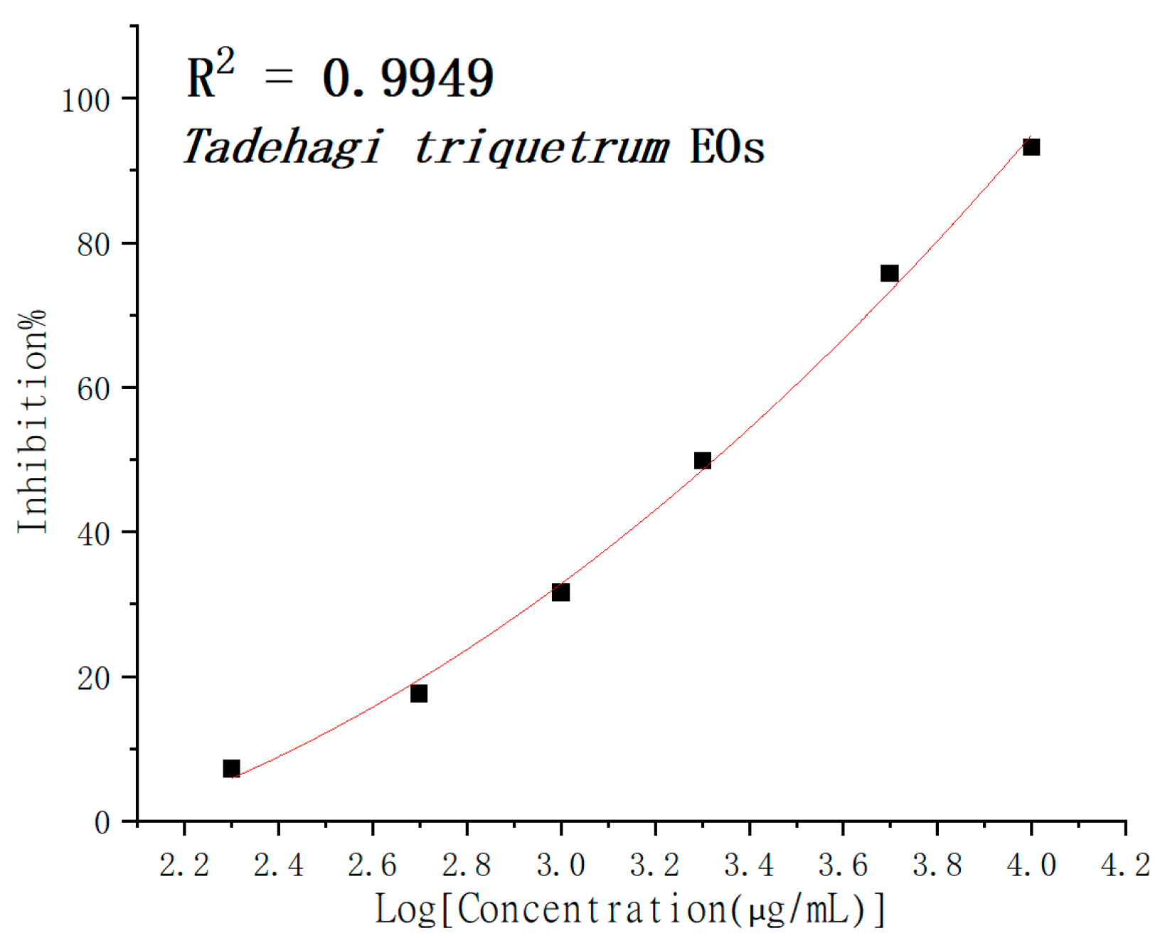
2.2.2. DPPH Assay
2.2.3. FRAP Assay
2.3. Antiacetylcholinesterase Activity
3. Discussion
4. Materials and Methods
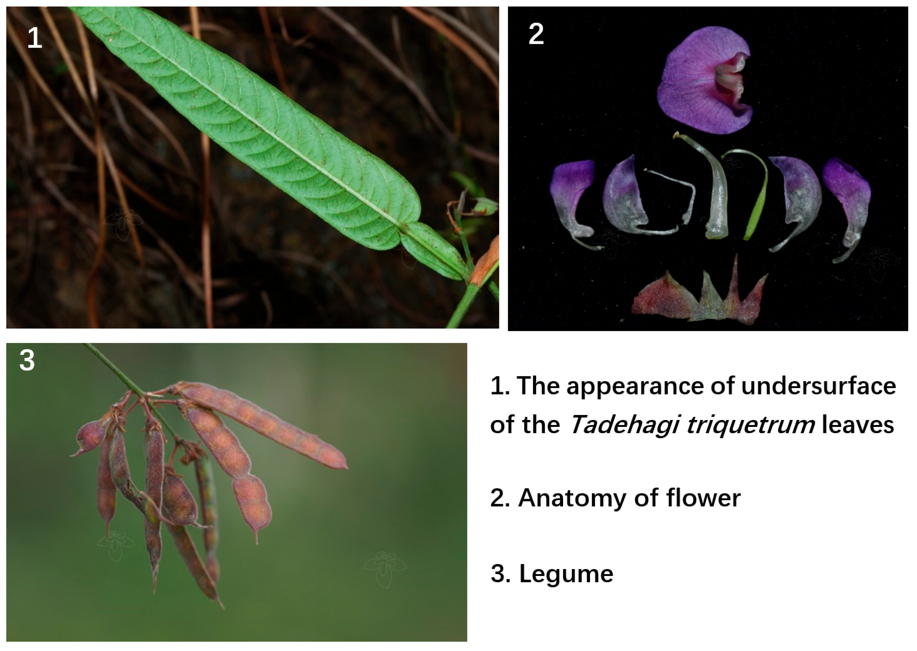
4.1. Plant Material
4.2. Extraction of Eseential Oils
4.3. Gas Chromatography–Mass Spectrometry (GC–MS) Analysis
4.4. Antioxidant Activity Test
4.4.1. ABTS Assay
4.4.2. DPPH Assay
4.4.3. FRAP Assay
4.5. Evaluation of Antiacetylcholinesterase Activity
5. Conclusions
Author Contributions
Funding
Institutional Review Board Statement
Informed Consent Statement
Data Availability Statement
Acknowledgments
Conflicts of Interest
Sample Availability
References
- Teofilovic, B.; Grujic-Letic, N.; Golocorbin-Kon, S.; Stojanovic, S.; Vastag, G.; Gadzuric, S. Experimental and chemometric study of antioxidant capacity of basil (Ocimum basilicum) extracts. Ind. Crops Prod. 2017, 100, 176–182. [Google Scholar] [CrossRef]
- Amorati, R.; Valgimigli, L. Methods to Measure the Antioxidant Activity of Phytochemicals and Plant Extracts. J. Agric. Food Chem. 2018, 66, 3324–3329. [Google Scholar] [CrossRef]
- De Oliveira, V.S.; Ferreira, F.S.; Cople, M.C.R.; Labre, T.D.; Augusta, I.M.; Gamallo, O.D.; Saldanha, T. Use of Natural Antioxidants in the Inhibition of Cholesterol Oxidation: A Review. Compr. Rev. Food Sci. Food Saf. 2018, 17, 1465–1483. [Google Scholar] [CrossRef] [Green Version]
- Olszowy, M. What is responsible for antioxidant properties of polyphenolic compounds from plants. Plant Physiol. Biochem. 2019, 144, 135–143. [Google Scholar] [CrossRef]
- Fierascu, R.C.; Ortan, A.; Fierascu, I.C.; Fierascu, I. In vitro and in vivo evaluation of antioxidant properties of wild-growing plants. A short review. Curr. Opin. Food Sci. 2018, 24, 1–8. [Google Scholar] [CrossRef]
- Kamath, B.R.; Sabeena, K. In vitro Study on Antioxidant Activity of Methanolic Leaf Extract of Piper Betle Linn. J. Evol. Med. Dent. Sci. 2018, 7, 2865–2869. [Google Scholar]
- Mitic, M.; Lazarevic-Pasti, T. Does the application of acetylcholinesterase inhibitors in the treatment of Alzheimer’s disease lead to depression. Expert Opin. Drug Metab. Toxicol. 2021, 17, 841–856. [Google Scholar] [CrossRef]
- Yang, Y.; Liu, M.; Li, J.; Zhang, Y.Q.; Lu, L. Screening of Some Medicinal Plants for Acetylcholinesterase Inhibition and Antioxidant Activity. Chin. J. Exp. Tradit. Med. Formulae 2013, 19, 213–218. [Google Scholar]
- Stuchbury, G.; Munch, G. Alzheimer’s associated inflammation, potential drug targets and future therapies. J. Neural Transm. 2005, 112, 429–453. [Google Scholar] [CrossRef]
- Vina, J.; Lloret, A.; Orti, R.; Alonso, D. Molecular bases of the treatment of Alzheimer’s disease with antioxidants: Prevention of oxidative stress. Mol. Aspects Med. 2004, 25, 117–123. [Google Scholar] [CrossRef]
- Aye, M.M.; Aung, H.T.; Sein, M.M.; Armijos, C. A Review on the Phytochemistry, Medicinal Properties and Pharmacological Activities of 15 Selected Myanmar Medicinal Plants. Molecules 2019, 24, 293. [Google Scholar] [CrossRef] [Green Version]
- Ding, H.; Shi, L.Y.; Chen, Y.; Lu, X.; Feng, B.M.; Wang, Y.Q.; Yu, D.Y. Analysis of Anti-allergic Asthma Constituents in Tadehagi triquetrum Leaves. Chin. J. Exp. Tradit. Med. Formulae 2017, 23, 30–35. [Google Scholar]
- Tang, A.C.; Chen, X.Y.; Lu, Q.Y.; Zheng, N.; Wei, Y.F.; Wu, X.Y. Antihepatotoxic Effect of Tadehaginoside, Extracted from Tadehagi triquetrum (L.), Against CCl4-Lesioned Rats Through Activating the Nrf2 Signaling Pathway and Attenuating the Inflammatory Response. Inflammation 2014, 37, 1006–1014. [Google Scholar] [CrossRef]
- Zhang, X.; Chen, C.; Li, Y.; Chen, D.; Dong, L.; Na, W.; Wu, C.; Zhang, J.; Li, Y. Tadehaginosides, A-J, phenylpropanoid glucosides from Tadehagi triquetrum, enhance glucose uptake via the upregulation of PPARγ and GLUT-4 in C2C12 myotubes. J. Nat. Prod. 2016, 79, 1249–1258. [Google Scholar] [CrossRef]
- Saeio, K.; Chaiyana, W.; Okonogi, S. Antityrosinase and antioxidant activities of essential oils of edible Thai plants. Drug Discov. Ther. 2011, 5, 144–149. [Google Scholar] [CrossRef] [Green Version]
- Dawidowicz, A.L.; Olszowy, M. Antioxidant properties of BHT estimated by ABTS assay in systems differing in pH or metal ion or water concentration. Eur. Food Res. Technol. 2011, 232, 837–842. [Google Scholar] [CrossRef] [Green Version]
- Parejo, L.; Codina, C.; Petrakis, C.; Kefalas, P. Evaluation of scavenging activity assessed by Co(II)/EDTA-induced luminol chemiluminescence and DPPH center dot (2,2-diphenyl-1-picrylhydrazyl) free radical assay. J. Pharmacol. Toxicol. Methods 2000, 44, 507–512. [Google Scholar] [CrossRef]
- Benzie, I.F.F.; Strain, J.J. The ferric reducing ability of plasma (FRAP) as a measure of “antioxidant power”: The FRAP assay. Anal. Biochem. 1996, 239, 70–76. [Google Scholar] [CrossRef] [Green Version]
- Kus, C.; Tas, M.; Kucukaydin, S.; Tel-Cayan, G.; Duru, M.E. Chemical analysis and in vitro antioxidant and anticholinesterase activities of essential oils and extracts from different parts of Erica manipuliflora. J. Res. Pharm. 2019, 23, 1098–1105. [Google Scholar]
- Harada, H.; Yamashita, U.; Kurihara, H.; Fukushi, E.; Kawabata, J.; Kamei, Y. Antitumor activity of palmitic acid found as a selective cytotoxic substance in a marine red alga. Anticancer Res. 2002, 22, 2587–2590. [Google Scholar]
- Boubaker, J.; Ben Toumia, I.; Sassi, A.; Bzouich-Mokded, I.; Mazgar, S.G.; Sioud, F.; Bedoui, A.; Skhiri, S.S.; Ghedira, K.; Chekir-Ghedira, L. Antitumoral Potency by Immunomodulation of Chloroform Extract from Leaves of Nitraria retusa, Tunisian Medicinal Plant, via its Major Compounds β-sitosterol and Palmitic Acid in BALB/c Mice Bearing Induced Tumor. Nutr. Cancer 2018, 70, 650–662. [Google Scholar] [CrossRef]
- Vazquez-Jimenez, J.G.; Chavez-Reyes, J.; Romero-Garcia, T.; Zarain-Herzberg, A.; Valdes-Flores, J.; Galindo-Rosales, J.M.; Rueda, A.; Guerrero-Hernandez, A.; Olivares-Reyes, J.A. Palmitic acid but not palmitoleic acid induces insulin resistance in a human endothelial cell line by decreasing SERCA pump expression. Cell. Signal. 2016, 28, 53–59. [Google Scholar] [CrossRef]
- Nadathur, S.R.; Carney, J.R.; Gould, S.J.; Bakalinsky, A.T. Palmitic acid is the major fatty acid responsible for significant anti-N-methyl-N′-nitro-N-nitrosoguanidine (MNNG) activity in yogurt. Mutat. Res./Environ. Mutagen. Relat. Subj. 1996, 359, 179–189. [Google Scholar] [CrossRef]
- Zhao, M.; Zhang, X.H.; Gao, L.; Song, Y.F.; Xu, C.; Yu, C.X.; Shao, S.S.; Zhao, J.J. Palmitic Acid Downregulates Thyroglobulin (Tg), Sodium Iodide Symporter (NIS), and Thyroperoxidase (TPO) in Human Primary Thyrocytes: A Potential Mechanism by Which Lipotoxicity Affects Thyroid? Int. J. Endocrinol. 2018, 2018, 4215848. [Google Scholar] [CrossRef]
- Yu, Y.; Liu, T.H.; Liu, L.X.; Chen, Y.; Tang, J.; Peng, W.H.; Tan, H. Application of the mushroom volatile 1-octen-3-ol to suppress a morel disease caused by Paecilomyces penicillatus. Appl. Microbiol. Biotechnol. 2022, 106, 4787–4799. [Google Scholar] [CrossRef]
- Lee, K.G.; Shibamoto, T. Antioxidant Properties of Aroma Compounds Isolated from Soybeans and Mung Beans. Agric. Food Chem. 2000, 48, 4290–4293. [Google Scholar] [CrossRef]
- Haidar, R.; Roudet, J.; Bonnard, O.; Dufour, M.C.; Corio-Costet, M.F.; Fert, M.; Gautier, T.; Deschamps, A.; Fermaud, M. Screening and modes of action of antagonistic bacteria to control the fungal pathogen Phaeomoniella chlamydospora involved in grapevine trunk diseases. Microbiol. Res. 2016, 192, 172–184. [Google Scholar] [CrossRef]
- Xiong, C.; Li, Q.; Li, S.H.; Chen, C.; Chen, Z.Q.; Huang, W.L. In vitro Antimicrobial Activities and Mechanism of 1-Octen-3-ol against Food-related Bacteria and Pathogenic Fungi. J. Oleo Sci. 2017, 66, 1041–1049. [Google Scholar] [CrossRef] [Green Version]
- Almeida, A.P.; Muzitano, M.F.; Costa, S.S. 1-octen-3-O-alpha-L-arabinopyranosyl-(1 -> 6)-beta-glucopyranoside, a minor substance from the leaves of Kalanchoe pinnata (Crassulaceae). Rev. Bras. Farmacogn. Braz. J. Pharmacogn. 2006, 16, 485–489. [Google Scholar] [CrossRef] [Green Version]
- Liu, X.Y.; Chen, X.B.; Chen, G.Y. Research Progress in Bioactivity and Synthesis of β-caryophyllene and Its Derivatives. Chem. Ind. For. Prod. 2012, 32, 104–110. [Google Scholar]
- Alsaud, N.; Shahbaz, K.; Farid, M. Evaluation of deep eutectic solvents in the extraction of beta-caryophyllene from New Zealand Manuka leaves (Leptospermum scoparium). Chem. Eng. Res. Des. 2021, 166, 97–108. [Google Scholar] [CrossRef]
- Benovit, S.C.; Silva, L.L.; Salbego, J.; Loro, V.L.; Mallmann, C.A.; Baldisserotto, B.; Flores, E.M.M.; Heinzmann, B.M. Anesthetic activity and bio-guided fractionation of the essential oil of Aloysia gratissima (Gillies & Hook.) Tronc. in silver catfish Rhamdia quelen. An. Acad. Bras. Cienc. 2015, 87, 1675–1689. [Google Scholar]
- Parodi, T.V.; Gressler, L.T.; Silva, L.D.; Becker, A.G.; Schmidt, D.; Caron, B.O.; Heinzmann, B.M.; Baldisserotto, B. Chemical composition of the essential oil of Aloysia triphylla under seasonal influence and its anaesthetic activity in fish. Aquac. Res. 2020, 51, 2515–2524. [Google Scholar] [CrossRef]
- Legault, J.; Pichette, A. Potentiating effect of beta-caryophyllene on anticancer activity of alpha-humulene, isocaryophyllene and paclitaxel. J. Pharm. Pharmacol. 2007, 59, 1643–1647. [Google Scholar] [CrossRef]
- Arul, S.; Rajagopalan, H.; Ravi, J.; Dayalan, H. Beta-Caryophyllenc Suppresses Ovarian Cancer Proliferation by Inducing Cell Cycle Arrest and Apoptosis. Anti Cancer Agents Med. Chem. 2020, 20, 1530–1537. [Google Scholar] [CrossRef]
- Kiran, I.; Durceylan, Z.; Kirimer, N.; Baser, K.H.C.; Noma, Y.; Demirci, F. Biotransformation of alpha-Cedrol and Caryophyllene Oxide by the Fungus Neurospora crassa. Nat. Prod. Commun. 2010, 5, 515–518. [Google Scholar]
- Jassal, K.; Kaushal, S.; Rashmi; Rani, R. Antifungal potential of guava (Psidium guajava) leaves essential oil, major compounds: Beta-caryophyllene and caryophyllene oxide. Arch. Phytopathol. Plant Prot. 2021, 54, 2034–2050. [Google Scholar] [CrossRef]
- Ramachandhiran, D.; Sankaranarayanan, C.; Murali, R.; Babukumar, S.; Vinothkumar, V. β-Caryophyllene promotes oxidative stress and apoptosis in KB cells through activation of mitochondrial-mediated pathway–An in-vitro and in-silico study. Arch. Physiol. Biochem. 2022, 128, 148–162. [Google Scholar] [CrossRef]
- Nie, C.N.; Gao, Y.; Du, X.; Bian, J.L.; Li, H.; Zhang, X.; Wang, C.M.; Li, S.Y. Characterization of the effect of cis-3-hexen-1-ol on green tea aroma. Sci Rep. 2020, 10, 15506. [Google Scholar] [CrossRef]
- Zhang, T.; Fang, K.; Ni, H.; Li, T.; Li, L.J.; Li, Q.B.; Chen, F. Aroma enhancement of instant green tea infusion using β-glucosidase and β-xylosidase. Food Chem. 2020, 315, 126287. [Google Scholar] [CrossRef]
- Li, W.Z.; Yang, L.; Shen, X.W.; Yuan, Y.H.; Yuan, G.H.; Luo, M.H.; Guo, X.R. Prescription screening and field evaluation of broad spectrum attractants of scarab beetles from Ricinus communis. Chin. J. Eco Agric. 2013, 21, 480–486. [Google Scholar] [CrossRef]
- He, B.Q.; Zhang, Y.Y.; Zhuang, Y.B.; Wei, A.H.; Li, R.D.; Zhang, S.Y. α-Glucosidase inhibitory activity of the extracts from Tadehagi triquetrum. Nat. Prod. Res. Dev. 2020, 32, 2026–2030. [Google Scholar]
- De Boer, H.J.; Vongsombath, C.; Kafer, J. A Fly in the Ointment: Evaluation of Traditional Use of Plants to Repel and Kill Blowfly Larvae in Fermented Fish. PLoS ONE 2011, 6, e29521. [Google Scholar] [CrossRef]
- Chang, L.W.; Chu, C.C.; Chu, H.L.; Wu, H.C.; Duh, P.D. Comparison of the protective effects of seven selected herbs against oxidative stress. J. Coastal Life Med. 2016, 3, 564–569. [Google Scholar]
- Liu, W.W.; Ren, H.; Cao, X.L.; Xu, C.M.; Wang, Q.E. Progress in Evaluation Techniques for Antioxidant Activity of Natural Products in vitro. Food Sci. 2010, 31, 415–419. [Google Scholar]
- Gülçin, I.; Berashvili, D.; Gepdiremen, A. Antiradical and antioxidant activity of total anthocyanins from Perilla pankinensis decne. J. Ethnopharmacol. 2005, 101, 287–293. [Google Scholar] [CrossRef]
- Zhang, X.; Zhao, X.H. Antioxidant Activities of Some Polyphenols Evaluated by Different Chemical Methods and Correlation Analysis. Food Sci. 2008, 29, 85–89. [Google Scholar]
- Lou, Y.H.; Zha, X.M.; Zhang, L.L.; Zhang, Y.; Xu, Y.G.; Zhang, L.Y. Advances in Acetylcholesterase Inhibitors Derived from Natural Products. Prog. Pharm. Sci. 2012, 36, 385–393. [Google Scholar]
- Antolovich, M.; Prenzler, P.D.; Patsalides, E.; McDonald, S.; Robards, K. Methods for testing antioxidant activity. Analyst 2002, 127, 183–198. [Google Scholar] [CrossRef]
- Schlesier, K.; Harwat, M.; Bohm, V.; Bitsch, R. Assessment of Antioxidant Activity by Using Different In Vitro Methods. Free Radic. Res. 2002, 36, 177–187. [Google Scholar] [CrossRef]
- Ellman, G.L.; Courtney, K.D.; Andres, V.; Featherstone, R.M. A new and rapid colorimetric determination of acetylcholinesterase activity. Biochem. Pharmacol. 1961, 7, 88–95. [Google Scholar] [CrossRef]


| No. | RT | Compound | RI a | RI b | Area (%) | Identification Method |
|---|---|---|---|---|---|---|
| 1 | 4.602 | (E)-2-Hexenal | 854 | 854 | 0.83% | RRI, MS |
| 2 | 4.711 | 3-Hexen-1-ol | 858 | 856 | 4.60% | RRI, MS |
| 3 | 5.038 | 1-Hexanol | 871 | 868 | 3.53% | RRI, MS |
| 4 | 6.124 | Sorbaldehyde | 912 | 911 | 0.37% | RRI, MS |
| 5 | 7.995 | 3,5,5-Trimethyl-hex-2-ene | 978 | 985 | 0.23% | RRI, MS |
| 6 | 8.361 | 1-Octen-3-ol | 989 | 980 | 14.22% | RRI, MS |
| 7 | 8.432 | 2,2-Dimethylhexanal | 991 | 993 | 0.74% | RRI, MS |
| 8 | 9.528 | Eucalyptol | 1031 | 1032 | 1.71% | RRI, MS |
| 9 | 10.728 | (Z)-5-Undecene | 1073 | 1079 | 0.28% | RRI, MS |
| 10 | 11.530 | Linalool | 1101 | 1099 | 3.24% | RRI, MS |
| 11 | 11.601 | Nonanal | 1104 | 1104 | 0.68% | RRI, MS |
| 12 | 12.463 | Pinocarveol | 1140 | 1139 | 0.47% | RRI, MS |
| 13 | 13.187 | endo-Borneol | 1167 | 1171 | 0.67% | RRI, MS |
| 14 | 13.833 | α-Terpineol | 1191 | 1189 | 1.61% | RRI, MS |
| 15 | 14.536 | β-Cyclocitral | 1220 | 1220 | 0.45% | RRI, MS |
| 16 | 14.754 | Nerol | 1230 | 1228 | 0.30% | RRI, MS |
| 17 | 15.392 | Geraniol | 1257 | 1255 | 0.60% | RRI, MS |
| 18 | 16.086 | Bornyl acetate | 1285 | 1285 | 0.23% | RRI, MS |
| 19 | 17.537 | α-Terpinyl acetate | 1350 | 1350 | 1.17% | RRI, MS |
| 20 | 18.099 | Copaene | 1375 | 1376 | 0.53% | RRI, MS |
| 21 | 18.328 | Damascenone | 1385 | 1386 | 1.36% | RRI, MS |
| 22 | 18.437 | β-Elemene | 1390 | 1391 | 1.31% | RRI, MS |
| 23 | 18.786 | β-Longipinene | 1406 | 1403 | 0.29% | RRI, MS |
| 24 | 19.124 | Caryophyllene | 1423 | 1419 | 7.27% | RRI, MS |
| 25 | 19.779 | Humulene | 1455 | 1454 | 1.24% | RRI, MS |
| 26 | 19.828 | (E)-β-Famesene | 1458 | 1457 | 0.77% | RRI, MS |
| 27 | 19.915 | Alloaromadendren | 1462 | 1461 | 0.28% | RRI, MS |
| 28 | 19.986 | Precocene I | 1465 | 1466 | 0.76% | RRI, MS |
| 29 | 20.352 | Germacrene D | 1482 | 1481 | 0.26% | RRI, MS |
| 30 | 20.401 | β-Selinene | 1485 | 1486 | 0.37% | RRI, MS |
| 31 | 20.450 | β-Ionone | 1487 | 1491 | 0.60% | RRI, MS |
| 32 | 20.875 | α-Farnesene | 1509 | 1508 | 0.39% | RRI, MS |
| 33 | 21.012 | γ-Cadinene | 1516 | 1513 | 0.30% | RRI, MS |
| 34 | 21.197 | δ-Cadinene | 1526 | 1524 | 0.88% | RRI, MS |
| 35 | 22.332 | Spathulenol | 1583 | 1577 | 0.37% | RRI, MS |
| 36 | 22.432 | Caryophyllene oxide | 1588 | 1581 | 2.85% | RRI, MS |
| 37 | 22.603 | Himbaccol | 1596 | 1591 | 0.29% | RRI, MS |
| 38 | 22.905 | β-Oplopenone | 1613 | 1606 | 0.82% | RRI, MS |
| 39 | 23.439 | Oxacyclotetradeca-4,11-diyne | 1642 | 1639 | 0.55% | RRI, MS |
| 40 | 23.756 | α-Cadinol | 1659 | 1653 | 0.26% | RRI, MS |
| 41 | 23.838 | Precocene II | 1663 | 1658 | 0.29% | RRI, MS |
| 42 | 24.121 | Aromadendrene oxide-(2) | 1678 | 1678 | 0.27% | RRI, MS |
| 43 | 24.776 | Pentadecanal | 1714 | 1715 | 0.60% | RRI, MS |
| 44 | 25.692 | Tetradecanoic acid | 1766 | 1768 | 0.33% | RRI, MS |
| 45 | 27.045 | Perhydrofarnesyl acetone | 1844 | 1844 | 0.61% | RRI, MS |
| 46 | 27.411 | Pentadecanoic acid | 1866 | 1867 | 0.51% | RRI, MS |
| 47 | 28.289 | Farnesyl acetone | 1919 | 1918 | 0.39% | RRI, MS |
| 48 | 28.737 | Isophytol | 1947 | 1948 | 0.28% | RRI, MS |
| 49 | 29.528 | Hexadecanoic acid | - | 1964 | 22.70% | MS |
| 50 | 30.662 | Heptadecanoic acid | 2069 | 2071 | 0.27% | RRI, MS |
| 51 | 31.399 | Phytol | 2117 | 2114 | 4.22% | RRI, MS |
| 52 | 31.890 | (Z)-18-Octadec-9-enolide | 2183 | 2154 | 6.10% | RRI, MS |
| 53 | 31.961 | Mandenol | - | 2159 | 3.08% | MS |
| 54 | 32.206 | Octadecanoic acid | 2172 | 2172 | 1.28% | RRI, MS |
| 55 | 32.424 | Isopropyl oleate | 2187 | 2192 | 0.24% | RRI, MS |
| 56 | 34.879 | Octadecanamide | 2365 | 2374 | 0.34% | RRI, MS |
| 57 | 40.668 | Squalene | 2831 | 2827 | 0.33% | RRI, MS |
| 58 | 43.739 | Hentriacontane | - | 3100 | 0.27% | MS |
| Activity Test | EOs of T. triquetrum (L.) Ohashi | BHT | Trolox |
|---|---|---|---|
| ABTS IC50 (mg/mL) | 2.12 ± 0.05 | 0.015 ± 0.0003 | 0.009 ± 0.0005 |
| DPPH IC50 (mg/mL) | 4.73 ± 0.91 | 0.043 ± 0.0012 | 0.012 ± 0.0008 |
| FRAP Antioxidant Capacity (mM/g) | 117.42 ± 8.10 | ||
| AChE IC50 (mg/mL) | >0.25 |
Disclaimer/Publisher’s Note: The statements, opinions and data contained in all publications are solely those of the individual author(s) and contributor(s) and not of MDPI and/or the editor(s). MDPI and/or the editor(s) disclaim responsibility for any injury to people or property resulting from any ideas, methods, instructions or products referred to in the content. |
© 2023 by the authors. Licensee MDPI, Basel, Switzerland. This article is an open access article distributed under the terms and conditions of the Creative Commons Attribution (CC BY) license (https://creativecommons.org/licenses/by/4.0/).
Share and Cite
Song, W.; Xu, Z.; Gao, P.; Liu, X. Chemical Composition and In Vitro Antioxidant Activity and Anti-Acetylcholinesterase Activity of Essential Oils from Tadehagi triquetrum (L.) Ohashi. Molecules 2023, 28, 2734. https://doi.org/10.3390/molecules28062734
Song W, Xu Z, Gao P, Liu X. Chemical Composition and In Vitro Antioxidant Activity and Anti-Acetylcholinesterase Activity of Essential Oils from Tadehagi triquetrum (L.) Ohashi. Molecules. 2023; 28(6):2734. https://doi.org/10.3390/molecules28062734
Chicago/Turabian StyleSong, Wenzhi, Ziyue Xu, Peizhong Gao, and Xu Liu. 2023. "Chemical Composition and In Vitro Antioxidant Activity and Anti-Acetylcholinesterase Activity of Essential Oils from Tadehagi triquetrum (L.) Ohashi" Molecules 28, no. 6: 2734. https://doi.org/10.3390/molecules28062734