
Design, Synthesis, Antifungal Activity, and 3D-QSAR Study of Novel Quinoxaline-2-Oxyacetate Hydrazide
Abstract
:
1. Introduction

2. Results and Discussion
3. Materials and Methods
4. Conclusions
Supplementary Materials
Author Contributions
Funding
Institutional Review Board Statement
Informed Consent Statement
Data Availability Statement
Conflicts of Interest
References
- Li, L.; Zhu, X.-M.; Zhang, Y.-R.; Cai, Y.-Y.; Wang, J.-Y.; Liu, M.-Y.; Wang, J.-Y.; Bao, J.-D.; Lin, F.-C. Research on the Molecular Interaction Mechanism between Plants and Pathogenic Fungi. Int. J. Mol. Sci. 2022, 23, 4658. [Google Scholar] [CrossRef] [PubMed]
- Wu, Y.-L.; Han, L.-J.; Wu, X.-M.; Jiang, W.; Liao, H.; Xu, Z.; Pan, C.-P. Trends and perspectives on general Pesticide analytical chemistry. Adv. Agrochem. 2022, 1, 113–124. [Google Scholar] [CrossRef]
- Strange, R.N.; Scott, P.R. Plant Disease: A Threat to Global Food Security. Annu. Rev. Phytopathol. 2005, 43, 83–116. [Google Scholar] [CrossRef] [PubMed]
- Wang, X.; Fu, X.; Chen, M.; Wang, A.; Yan, J.; Mei, Y.; Wang, M.; Yang, C. Novel 1,3,5-thiadiazine-2-thione derivatives containing a hydrazide moiety: Design, synthesis and bioactive evaluation against phytopathogenic fungi in vitro and in vivo. Chin. Chem. Lett. 2019, 30, 1419–1422. [Google Scholar] [CrossRef]
- Swain, T. Secondary Compounds as Protective Agents. Annu. Rev. Plant Biol. 1977, 28, 479–501. [Google Scholar] [CrossRef]
- Zhang, P.; Duan, C.-B.; Jin, B.; Ali, A.S.; Han, X.; Zhang, H.; Zhang, M.-Z.; Zhang, W.-H.; Gu, Y.-C. Recent advances in the natural products-based lead discovery for new agrochemicals. Adv. Agrochem. 2023, 2, 324–339. [Google Scholar] [CrossRef]
- Arif, T.; Bhosale, J.D.; Kumar, N.; Mandal, T.K.; Bendre, R.S.; Lavekar, G.S.; Dabur, R. Natural products—Antifungal agents derived from plants. J. Asian Nat. Prod. Res. 2009, 11, 621–638. [Google Scholar] [CrossRef] [PubMed]
- Roberts, D.P.; Mattoo, A.K. Sustainable Agriculture—Enhancing Environmental Benefits, Food Nutritional Quality and Building Crop Resilience to Abiotic and Biotic Stresses. Agriculture 2018, 8, 8. [Google Scholar] [CrossRef]
- Dhameliya, T.M.; Chudasma, S.J.; Patel, T.M.; Dave, B.P. A review on synthetic account of 1,2,4-oxadiazoles as anti-infective agents. Mol. Divers. 2022, 26, 2967–2980. [Google Scholar] [CrossRef]
- Xu, H.; Fan, L.-L. Synthesis and antifungal activities of novel 5,6-dihydro-indolo[1,2-a]quinoxaline derivatives. Eur. J. Med. Chem. 2011, 46, 1919–1925. [Google Scholar] [CrossRef]
- Jiang, Y.; Zhu, C.-H.; Xia, Z.-H.; Zhao, H.-Q. Quinoxalinone-1,2,3-triazole derivatives as potential antifungal agents for plant anthrax disease: Design, synthesis, antifungal activity and SAR study. Adv. Agrochem. 2023. [Google Scholar] [CrossRef]
- El-Zahabi, H.S.A. Synthesis, Characterization, and Biological Evaluation of Some Novel Quinoxaline Derivatives as Antiviral Agents. Arch. Pharm. 2017, 350, 1700028–1700040. [Google Scholar] [CrossRef] [PubMed]
- Zhao, Y.; Cheng, G.; Hao, H.; Pan, Y.; Liu, Z.; Dai, M.; Yuan, Z. In vitro antimicrobial activities of animal-used quinoxaline 1,4-di-N-oxides against mycobacteria, mycoplasma and fungi. BMC Vet. Res. 2016, 12, 186–198. [Google Scholar] [CrossRef]
- Moreno, E.; Ancizu, S.; Pérez-Silanes, S.; Torres, E.; Aldana, I.; Monge, A. Synthesis and antimycobacterial activity of new quinoxaline-2-carboxamide 1,4-di-N-oxide derivatives. Eur. J. Med. Chem. 2010, 45, 4418–4426. [Google Scholar] [CrossRef]
- Shekhar, A.C.; Rao, P.S.; Narsaiah, B.; Allanki, A.D.; Sijwali, P.S. Emergence of pyrido quinoxalines as new family of antimalarial agents. Eur. J. Med. Chem. 2014, 77, 280–287. [Google Scholar] [CrossRef]
- Wilhelmsson, L.M.; Kingi, N.; Bergman, J. Interactions of Antiviral Indolo[2,3-b]quinoxaline Derivatives with DNA. J. Med. Chem. 2008, 51, 7744–7750. [Google Scholar] [CrossRef]
- De Clercq, E. Toward Improved Anti-HIV Chemotherapy: Therapeutic Strategies for Intervention with HIV Infections. J. Med. Chem. 1995, 38, 2491–2517. [Google Scholar] [CrossRef] [PubMed]
- Borah, B.; Chowhan, L.R. Recent advances in the transition-metal-free synthesis of quinoxalines. RSC Adv. 2021, 11, 37325–37353. [Google Scholar] [CrossRef]
- Montana, M.; Mathias, F.; Terme, T.; Vanelle, P. Antitumoral activity of quinoxaline derivatives: A systematic review. Eur. J. Med. Chem. 2019, 163, 136–147. [Google Scholar] [CrossRef]
- Pereira, J.A.; Pessoa, A.M.; Cordeiro, M.N.; Fernandes, R.; Prudencio, C.; Noronha, J.P.; Vieira, M. Quinoxaline, its derivatives and applications: A State of the Art review. Eur. J. Med. Chem. 2015, 97, 664–672. [Google Scholar] [CrossRef]
- Tariq, S.; Alam, O.; Synthesis, M.A. Anti-inflammatory, p38α MAP kinase inhibitory activities and molecular docking studies of quinoxaline derivatives containing triazole moiety. Bioorg. Chem. 2018, 76, 343–358. [Google Scholar] [CrossRef] [PubMed]
- Hu, Y.; Wang, K.; MacMillan, J.B. Hunanamycin A, an Antibiotic from a Marine-Derived Bacillus hunanensis. Org. Lett. 2013, 15, 390–393. [Google Scholar] [CrossRef] [PubMed]
- Xia, R.; Guo, T.; He, J.; Chen, M.; Su, S.; Jiang, S.; Tang, X.; Chen, Y.; Xue, W. Antimicrobial evaluation and action mechanism of chalcone derivatives containing quinoxaline moiety. Monatsh. Chem. 2019, 150, 1325–1334. [Google Scholar] [CrossRef]
- Chang, J.; Liu, Y.; Zhang, T.; Chen, Z.; Fang, H.; Hua, X. A Comprehensive Investigation of Hydrazide and Its Derived Structures in the Agricultural Fungicidal Field. J. Agric. Food Chem. 2023, 71, 8297–8316. [Google Scholar] [CrossRef] [PubMed]
- Niu, J.-B.; Quan, C.-H.; Liu, Y.; Yu, G.-X.; Yang, J.-J.; Li, Y.-R.; Zhang, Y.-B.; Qi, Y.-Q.; Song, J.; Jin, C.-Y.; et al. Discovery of N-aryl sulphonamide-quinazoline derivatives as anti-gastric cancer agents in vitro and in vivo via activating the Hippo signalling pathway. J. Enzym. Inhib. Med. Chem. 2021, 36, 1715–1731. [Google Scholar] [CrossRef]
- Sharma, P.C.; Sharma, D.; Sharma, A.; Saini, N.; Goyal, R.; Ola, M.; Chawla, R.; Thakur, V.K. Hydrazone comprising compounds as promising anti-infective agents: Chemistry and structure-property relationship. Mater. Today Chem. 2020, 18, 100349–100369. [Google Scholar] [CrossRef]
- Stierli, D.; Eberle, M.; Lamberth, C.; Jacob, O.; Balmer, D.; Gulder, T. Quarternary α-cyanobenzylsulfonamides: A new subclass of CAA fungicides with excellent anti-Oomycetes activity. Bioorg. Med. Chem. 2021, 30, 115965. [Google Scholar] [CrossRef] [PubMed]
- Wang, Z.-W.; Zhao, L.-X.; Ma, P.; Ye, T.; Fu, Y.; Ye, F. Fragments recombination, design, synthesis, safener activity and CoMFA model of novel substituted dichloroacetylphenyl sulfonamide derivatives. Pest Manag. Sci. 2021, 77, 1724–1738. [Google Scholar] [CrossRef] [PubMed]
- Chen, Z.; Fang, H.; Chang, J.; Zhang, T.; Cui, Y.; Zhang, L.; Sui, J.; Ma, Q.; Su, P.; Wang, J.; et al. Natural Alkaloid Waltherione F-Derived Hydrazide Compounds Evaluated in an Agricultural Fungicidal Field. J. Agric. Food Chem. 2023, 71, 12333–12345. [Google Scholar] [CrossRef]
- Yao, X.; Zhang, R.; Lv, B.; Wang, W.-W.; Liu, Z.; Hu, Z.; Design, D.L. Synthesis and biological evaluation of thiazole and imidazo[1,2-a]pyridine derivatives containing a hydrazone substructure as potential agrochemicals. Adv. Agrochem. 2023, 2, 154–162. [Google Scholar] [CrossRef]
- Chen, Y.-J.; Ma, K.-Y.; Du, S.-S.; Zhang, Z.-J.; Wu, T.-L.; Sun, Y.; Liu, Y.-Q.; Yin, X.-D.; Zhou, R.; Yan, Y.-F.; et al. Antifungal Exploration of Quinoline Derivatives against Phytopathogenic Fungi Inspired by Quinine Alkaloids. J. Agric. Food Chem. 2021, 69, 12156–12170. [Google Scholar] [CrossRef] [PubMed]
- Kumar, A.; Dhameliya, T.M.; Sharma, K.; Patel, K.A.; Hirani, R.V.; Bhatt, A.J. Sustainable approaches towards the synthesis of quinoxalines: An update. J. Mol. Struct. 2022, 1259, 132732–132747. [Google Scholar] [CrossRef]
- Mao, X.; Wu, Z.; Bi, C.; Wang, J.; Zhao, F.; Gao, J.; Hou, Y.; Zhou, M. Molecular and Biochemical Characterization of Pydiflumetofen-Resistant Mutants of Didymella bryoniae. J. Agric. Food Chem. 2020, 68, 9120–9130. [Google Scholar] [CrossRef] [PubMed]
- Yang, Y.D.; He, Y.H.; Ma, K.Y.; Li, H.; Zhang, Z.J.; Sun, Y.; Wang, Y.L.; Hu, G.F.; Wang, R.X.; Liu, Y.Q. Design and Discovery of Novel Antifungal Quinoline Derivatives with Acylhydrazide as a Promising Pharmacophore. J. Agric. Food Chem. 2021, 69, 8347–8357. [Google Scholar] [CrossRef] [PubMed]
- Wang, X.; Dai, Z.C.; Chen, Y.F.; Cao, L.L.; Yan, W.; Li, S.K.; Wang, J.X.; Zhang, Z.G. Synthesis of 1,2,3-triazole hydrazide derivatives exhibiting anti-phytopathogenic activity. Eur. J. Med. Chem. 2017, 126, 171–182. [Google Scholar] [CrossRef] [PubMed]
- Wang, X.; Wang, A.; Qiu, L.; Chen, M.; Lu, A.; Li, G.; Yang, C.; Xue, W. Expedient Discovery for Novel Antifungal Leads Targeting Succinate Dehydrogenase: Pyrazole-4-formylhydrazide Derivatives Bearing a Diphenyl Ether Fragment. J. Agric. Food Chem. 2020, 68, 14426–14437. [Google Scholar] [CrossRef]
- Yang, C.; Sun, S.; Li, W.; Mao, Y.; Wang, Q.; Duan, Y.; Csuk, R.; Li, S. Bioactivity-Guided Subtraction of MIQOX for Easily Available Isoquinoline Hydrazides as Novel Antifungal Candidates. J. Agric. Food Chem. 2023, 71, 11341–11349. [Google Scholar] [CrossRef]
- Zhi, X.Y.; Zhang, Y.; Li, Y.F.; Liu, Y.; Niu, W.P.; Li, Y.; Zhang, C.R.; Cao, H.; Hao, X.J.; Yang, C. Discovery of Natural Sesquiterpene Lactone 1-O-Acetylbritannilactone Analogues Bearing Oxadiazole, Triazole, or Imidazole Scaffolds for the Development of New Fungicidal Candidates. J. Agric. Food Chem. 2023, 71, 11680–11691. [Google Scholar] [CrossRef]


| Compd. | B. cinrea | A. solani | G. zeae | R. solani | C. orbiculare | A. alternata |
|---|---|---|---|---|---|---|
| 1 | 86.1 ± 1.4 | 89.5 ± 1.2 | 95.8 ± 0.8 | 91.9 ± 0.9 | 100 | 96.4 ± 0.5 |
| 2 | 75.9 ± 0.5 | 48.1 ± 1.3 | 93.1 ± 0.6 | 94.4 ± 0.8 | 97.7 ± 0.5 | 100 |
| 3 | 88.8 ± 1.6 | 79.3 ± 0.7 | 59.7 ± 2.2 | 97.2 ± 0.7 | 100 | 91.2 ± 0.7 |
| 4 | 74.4 ± 1.6 | 66.4 ± 2.4 | 65.0 ± 1.2 | 77.4 ± 1.3 | 87.3 ± 1.1 | 85.0 ± 1.5 |
| 5 | 39.6 ± 3.0 | 40.7 ± 0.7 | 85.3 ± 0.6 | 96.4 ± 0.7 | 81.9 ± 0.6 | 60.9 ± 0.5 |
| 6 | 97.0 ± 0.6 | 80.6 ± 0 | 26.3 ± 0.7 | 70.2 ± 0.8 | 100 | 95.3 ± 2 |
| 7 | 80.8 ± 1.7 | 85.0 ± 0.7 | 99.7 ± 0.6 | 100 | 51.3 ± 1.0 | 100 |
| 8 | 10.2 ± 0.5 | 28.8 ± 1.0 | <10 | 39.5 ± 0.8 | <10 | <10 |
| 9 | 85.6 ± 1.4 | 73.7 ± 1.2 | 29.2 ± 1.3 | 97.2 ± 0.7 | 56.2 ± 1.1 | 46.9 ± 0.5 |
| 10 | 21.9 ± 1.1 | 47.9 ± 0.7 | 31.9 ± 2.0 | 81.9 ± 2.1 | 50 ± 0.6 | 41.1 ± 0.7 |
| 11 | 14.7 ± 1.6 | 58.6 ± 1.0 | 12.8 ± 1.2 | 81.0 ± 0.7 | 83.5 ± 0.5 | 60.4 ± 0.7 |
| 12 | 52.2 ± 1.0 | 56.5 ± 1.9 | 25.6 ± 1.2 | 73.9 ± 2.2 | 50.1 ± 0.7 | 72.0 ± 0.6 |
| 13 | 56.7 ± 1.0 | 42.1 ± 0.7 | 25.8 ± 1.9 | 75.0 ± 0.8 | 35.6 ± 0.6 | 73.6 ± 0.8 |
| 14 | 28.0 ± 1.2 | 60.7 ± 0.7 | 23.9 ± 0.8 | 79.6 ± 1.2 | 34.4 ± 2.6 | 77.1 ± 0.7 |
| 15 | 80.6 ± 1.7 | 6.4 ± 0.7 | 92.2 ± 1.2 | 98.0 ± 0.7 | 91.7 ± 0.6 | 63.2 ± 0.7 |
| 16 | 70.6 ± 1.1 | 62.1 ± 0.7 | 93.6 ± 0.6 | 94.4 ± 0.8 | 95.6 ± 0.6 | 97.9 ± 0.7 |
| 17 | 74.6 ± 1.7 | 66.4 ± 0.7 | 84.7 ± 1.1 | 96.4 ± 0.7 | 60.1 ± 2.9 | 48.1 ± 0.7 |
| 18 | 81.6 ± 1.4 | 65.1 ± 0.8 | 91.4 ± 0.6 | 98.0 ± 1.3 | 94.8 ± 0.7 | 87.8 ± 2.4 |
| 19 | 20.4 ± 0.7 | 33.6 ± 0.7 | 42.2 ± 2.3 | 73.4 ± 0.8 | 18.8 ± 0.7 | 23.1 ± 0.7 |
| 20 | 93.8 ± 0.7 | 94.9 ± 1.4 | 48.1 ± 4.3 | 98.4 ± 0.7 | 100 | 63.8 ± 3.4 |
| 21 | 55.5 ± 2.0 | 35.0 ± 0.7 | 45.0 ± 1.0 | 73.4 ± 0.8 | 47.3 ± 0.5 | 33.1 ± 0.5 |
| 22 | 60.7 ± 2.4 | 76.4 ± 0.7 | 30.8 ± 0.8 | 97.2 ± 0.7 | 34.6 ± 0.6 | 39.6 ± 0.7 |
| 23 | 75.4 ± 0.7 | 69.1 ± 0.7 | 80.8 ± 1.6 | 97.6 ± 0.8 | 100 | 32.4 ± 0.7 |
| 24 | 93.7 ± 0.6 | 84.9 ± 1.2 | 25.8 ± 1.3 | 92.7 ± 0.7 | 67.2 ± 3.4 | 74.3 ± 0.6 |
| 25 | 41.5 ± 1.1 | 19.3 ± 0.7 | 15.8 ± 2.5 | 100 | 49.4 ± 0.6 | 50.7 ± 0.7 |
| 26 | 76.6 ± 1.1 | 62.1 ± 0.7 | 98.9 ± 0.8 | 100 | 100 | 91.9 ± 0.5 |
| 27 | 69.9 ± 2.6 | 46.4 ± 0.7 | 89.7 ± 2.2 | 95.2 ± 0.9 | 90.6 ± 0.6 | 56.3 ± 0.7 |
| 28 | 44.5 ± 2.0 | 39.3 ± 0.7 | 42.5 ± 1.6 | 78.0 ± 2.2 | 79.4 ± 0.6 | 45.8 ± 0.8 |
| 29 | <10 | 20.0 ± 1.0 | <10 | 100 | 14 ± 0.5 | <10 |
| 30 | 61.7 ± 0.7 | 20.7 ± 0.7 | 33.1 ± 0.6 | 100 | 39.4 ± 0.6 | 54.9 ± 0.7 |
| 31 | <10 | 23.7 ± 4.4 | 27.5 ± 4.4 | 96.8 ± 1.1 | 18.2 ± 1.1 | 22.2 ± 2.3 |
| 32 | 46.6 ± 1.1 | 54.6 ± 1.1 | 78.9 ± 2.6 | 83.9 ± 0.9 | 70.6 ± 1.4 | 60.2 ± 2.6 |
| 33 | 55.2 ± 3.2 | 53.3 ± 1.4 | 68.9 ± 1.9 | 92.5 ± 6.3 | 70.4 ± 1.3 | 48.4 ± 1.8 |
| 34 | 50.5 ± 1.5 | 70.7 ± 0.6 | 85.5 ± 1.1 | 100 | 65.9 ± 1.6 | 45.1 ± 1.4 |
| 35 | 14.1 ± 2.0 | 41.1 ± 0.8 | 24.8 ± 1.1 | 100 | 39.6 ± 1.7 | 29.3 ± 1.6 |
| 36 | 57.8 ± 3.3 | 86.1 ± 1.2 | 91.2 ± 0.5 | 100 | 100 | 92.0 ± 1.9 |
| pyrimethanil | 75.1 ± 1.0 | 43.1 ± 0.7 | 32.6 ± 1.8 | 89.4 ± 0.7 | 15.2 ± 1.1 | 27.2 ± 1.5 |
| Pathogen | Compd. | EC50 | Pathogen | Compd. | EC50 |
|---|---|---|---|---|---|
| B. Cinerea | 6 | 3.31 ± 0.18 | R. solani | 1 | 0.20 ± 0.07 |
| 20 | 4.36 ± 0.10 | 2 | 0.19 ± 0.04 | ||
| 26 | 4.90 ± 0.05 | 3 | 0.58 ± 0.03 | ||
| pyrimethanil | 3.39 ± 0.22 | 4 | 0.17 ± 0.11 | ||
| A. solani | 20 | 4.42 ± 0.09 | 5 | 0.26 ± 0.07 | |
| carbendazim | 5.46 ± 0.14 | 6 | 0.58 ± 0.16 | ||
| G. zeae | 1 | 0.94 ± 0.03 | 7 | 0.65 ± 0.08 | |
| 2 | 1.22 ± 0.06 | 9 | 0.84 ± 0.20 | ||
| 7 | 1.21 ± 0.02 | 10 | 2.22 ± 0.06 | ||
| 15 | 0.87 ± 0.03 | 11 | 1.41 ± 0.11 | ||
| 16 | 1.17 ± 0.08 | 12 | 1.05 ± 0.04 | ||
| 18 | 1.77 ± 0.04 | 13 | 0.20 ± 0.01 | ||
| 28 | 1.27 ± 0.06 | 14 | 0.39 ± 0.08 | ||
| 38 | 1.54 ± 0.09 | 15 | 0.16 ± 0.04 | ||
| pyrimethanil | 2.20 ± 0.11 | 16 | 0.36 ± 0.12 | ||
| C. orbiculare | 1 | 1.84 ± 0.07 | 17 | 0.32 ± 0.23 | |
| 2 | 1.32 ± 0.06 | 18 | 0.52 ± 0.03 | ||
| 3 | 3.35 ± 0.22 | 19 | 0.33 ± 0.11 | ||
| 6 | 8.39 ± 0.15 | 20 | 0.54 ± 0.14 | ||
| 15 | 1.01 ± 0.11 | 21 | 0.15 ± 0.09 | ||
| 16 | 1.35 ± 0.07 | 22 | 0.47 ± 0.04 | ||
| 18 | 3.84 ± 0.10 | 23 | 0.54 ± 0.14 | ||
| 20 | 3.86 ± 0.13 | 24 | 0.66 ± 0.11 | ||
| 23 | 1.36 ± 0.14 | 25 | 0.68 ± 0.05 | ||
| 26 | 1.61 ± 0.12 | 26 | 0.55 ± 0.05 | ||
| 27 | 1.03 ± 0.11 | 27 | 0.18 ± 0.04 | ||
| 36 | 2.23 ± 0.08 | 28 | 0.15 ± 0.08 | ||
| carbendazim | 2.32 ± 0.11 | 29 | 1.21 ± 0.11 | ||
| A. alternata | 1 | 1.54 ± 0.12 | 30 | 1.05 ± 0.09 | |
| 2 | 10.75 ± 0.05 | 31 | 0.80 ± 0.11 | ||
| 3 | 7.35 ± 0.21 | 32 | 0.23 ± 0.05 | ||
| 6 | 12.09 ± 0.05 | 33 | 0.38 ± 0.07 | ||
| 7 | 1.99 ± 0.08 | 34 | 0.36 ± 0.05 | ||
| 16 | 4.82 ± 0.25 | 35 | 0.55 ± 0.23 | ||
| 28 | 2.85 ± 0.06 | 36 | 0.50 ± 0.15 | ||
| pyrimethanil | 2.07 ± 0.15 | pyrimethanil | 0.21 ± 0.10 |
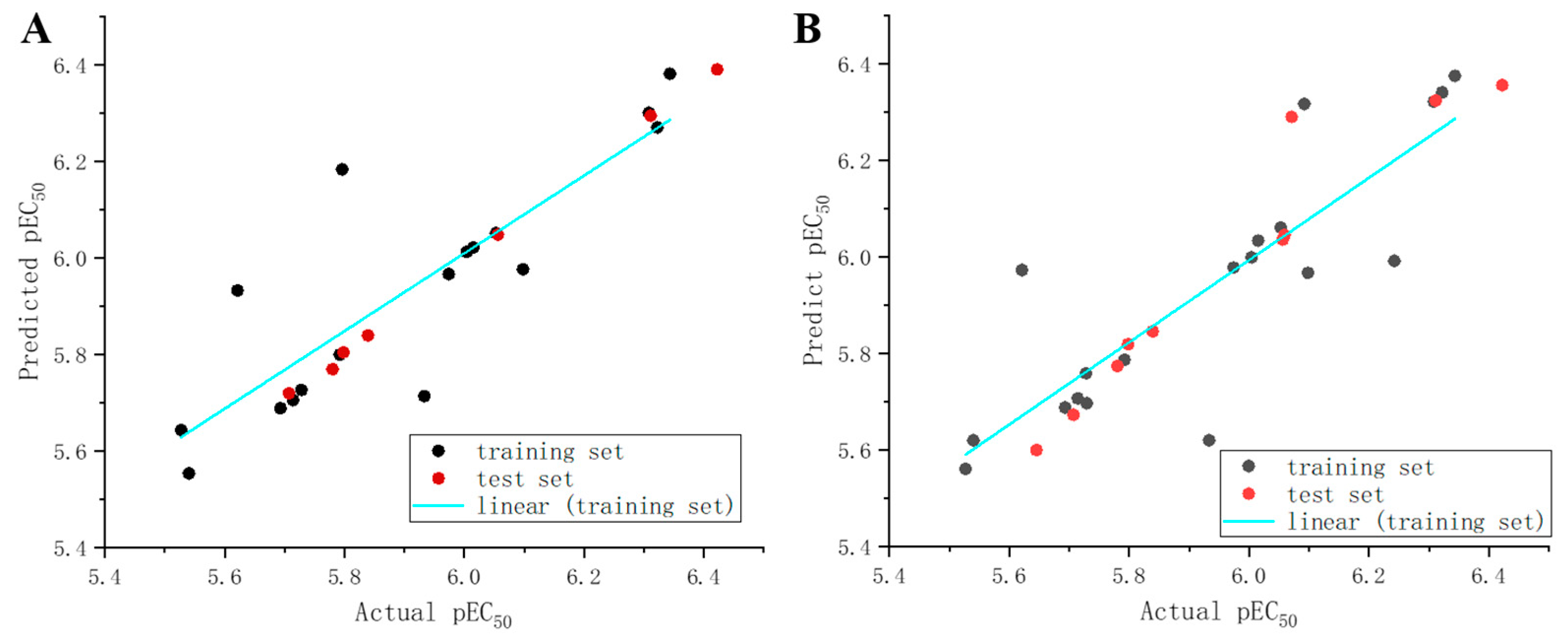
| Statistical Parameter | CoMFA | CoMSIA | Validation Criteria |
|---|---|---|---|
| q2 a | 0.843 | 0.845 | >0.5 |
| r2 b | 0.997 | 0.985 | >0.8 |
| s c | 0.025 | 0.038 | |
| F d | 144.141 | 125.981 | |
| ONC e | 14 | 7 |
| Compd. | EC50 | pEC50 | CoMFA | CoMSIA | ||
|---|---|---|---|---|---|---|
| Pred pEC50 b | Residual | Pred pEC50 b | Residual | |||
| 1 * | 0.20 ± 0.07 | 6.311 | 6.295 | 0.016 | 6.324 | −0.013 |
| 2 | 0.19 ± 0.04 | 6.343 | 6.382 | −0.039 | 6.375 | −0.032 |
| 3 | 0.58 ± 0.03 | 5.728 | 5.727 | 0.001 | 5.759 | −0.031 |
| 4 | 0.17 ± 0.11 | 6.322 | 6.271 | 0.051 | 6.341 | −0.019 |
| 5 * | 0.26 ± 0.07 | 6.071 | 6.376 | −0.305 | 6.29 | −0.219 |
| 6 | 0.58 ± 0.16 | 5.729 | 6.193 | −0.464 | 5.697 | 0.032 |
| 9 * | 2.22 ± 0.06 | 5.645 | 6.209 | −0.564 | 5.6 | 0.045 |
| 12 | 1.05 ± 0.04 | 5.621 | 5.933 | −0.312 | 5.973 | −0.352 |
| 13 | 0.20 ± 0.01 | 6.015 | 6.022 | −0.007 | 6.034 | −0.019 |
| 14 * | 0.39 ± 0.08 | 6.056 | 6.049 | 0.007 | 6.036 | 0.02 |
| 15 | 0.12 ± 0.04 | 6.098 | 5.977 | 0.121 | 5.967 | 0.131 |
| 16 | 0.36 ± 0.12 | 5.974 | 5.967 | 0.007 | 5.978 | −0.004 |
| 17 | 0.32 ± 0.23 | 6.004 | 6.013 | −0.009 | 5.999 | 0.005 |
| 18 | 0.52 ± 0.03 | 5.792 | 5.800 | −0.008 | 5.787 | 0.005 |
| 19 | 0.33 ± 0.11 | 6.053 | 6.052 | 0.001 | 6.061 | −0.008 |
| 20 * | 0.54 ± 0.14 | 5.78 | 5.77 | 0.01 | 5.774 | 0.006 |
| 22 | 0.47 ± 0.04 | 5.933 | 5.714 | 0.219 | 5.62 | 0.313 |
| 23 * | 0.54 ± 0.14 | 5.798 | 5.805 | −0.007 | 5.819 | −0.021 |
| 24 | 0.66 ± 0.11 | 5.714 | 5.706 | 0.008 | 5.707 | 0.007 |
| 25 * | 0.68 ± 0.05 | 5.707 | 5.72 | −0.013 | 5.673 | 0.034 |
| 26 | 0.55 ± 0.05 | 5.796 | 6.184 | −0.388 | 6.253 | −0.457 |
| 27 | 0.18 ± 0.04 | 6.308 | 6.301 | 0.007 | 6.322 | −0.014 |
| 28 * | 0.15 ± 0.08 | 6.422 | 6.391 | 0.031 | 6.356 | 0.066 |
| 29 | 1.21 ± 0.11 | 5.527 | 5.644 | −0.117 | 5.561 | −0.034 |
| 30 | 1.05 ± 0.09 | 5.540 | 5.554 | −0.014 | 5.62 | −0.08 |
| 31 | 0.80 ± 0.11 | 5.693 | 5.689 | 0.004 | 5.688 | 0.005 |
| 32 | 0.23 ± 0.05 | 6.242 | 6.048 | 0.194 | 5.992 | 0.25 |
| 33 * | 0.38 ± 0.07 | 6.059 | 6.33 | −0.271 | 6.045 | 0.014 |
| 34 | 0.36 ± 0.05 | 6.092 | 6.33 | −0.238 | 6.317 | −0.225 |
| 35 * | 0.55 ± 0.23 | 5.839 | 5.84 | −0.001 | 5.846 | −0.007 |
Disclaimer/Publisher’s Note: The statements, opinions and data contained in all publications are solely those of the individual author(s) and contributor(s) and not of MDPI and/or the editor(s). MDPI and/or the editor(s) disclaim responsibility for any injury to people or property resulting from any ideas, methods, instructions or products referred to in the content. |
© 2024 by the authors. Licensee MDPI, Basel, Switzerland. This article is an open access article distributed under the terms and conditions of the Creative Commons Attribution (CC BY) license (https://creativecommons.org/licenses/by/4.0/).
Share and Cite
Teng, P.; Li, Y.; Fang, R.; Zhu, Y.; Dai, P.; Zhang, W. Design, Synthesis, Antifungal Activity, and 3D-QSAR Study of Novel Quinoxaline-2-Oxyacetate Hydrazide. Molecules 2024, 29, 2501. https://doi.org/10.3390/molecules29112501
Teng P, Li Y, Fang R, Zhu Y, Dai P, Zhang W. Design, Synthesis, Antifungal Activity, and 3D-QSAR Study of Novel Quinoxaline-2-Oxyacetate Hydrazide. Molecules. 2024; 29(11):2501. https://doi.org/10.3390/molecules29112501
Chicago/Turabian StyleTeng, Peng, Yufei Li, Ruoyu Fang, Yuchuan Zhu, Peng Dai, and Weihua Zhang. 2024. "Design, Synthesis, Antifungal Activity, and 3D-QSAR Study of Novel Quinoxaline-2-Oxyacetate Hydrazide" Molecules 29, no. 11: 2501. https://doi.org/10.3390/molecules29112501