Article

New Hydroxylactones and Chloro-Hydroxylactones Obtained by Biotransformation of Bicyclic Halolactones and Their Antibacterial Activity

by
Małgorzata Grabarczyk
1,*,
Anna Duda-Madej
2,*,
Fedor Romanenko
1,
Gabriela Maciejewska
3,
Wanda Mączka
1,
Agata Białońska
4 and
Katarzyna Wińska
1
1
Department of Food Chemistry and Biocatalysis, Wrocław University of Environmental and Life Sciences, Norwida 25, 50-375 Wrocław, Poland
2
Department of Microbiology, Faculty of Medicine, Wroclaw Medical University, Chałubińskiego 4, 50-368 Wrocław, Poland
3
Faculty of Chemistry, Wrocław University of Technology, Wybrzeże Wyspiańskiego 27, 50-370 Wrocław, Poland
4
Faculty of Chemistry, University of Wrocław, F. Joliot-Curie 14, 50-383 Wrocław, Poland
*
Authors to whom correspondence should be addressed.
Molecules 2024, 29(12), 2820; https://doi.org/10.3390/molecules29122820
Submission received: 25 April 2024 / Revised: 10 June 2024 / Accepted: 11 June 2024 / Published: 13 June 2024

Abstract

:
The aim of this study was to obtain new halolactones with a gem-dimethyl group in the cyclohexane ring (at the C-3 or C-5 carbon) and a methyl group in the lactone ring and then subject them to biotransformations using filamentous fungi. Halolactones in the form of mixtures of two diasteroisomers were subjected to screening biotransformations, which showed that only compounds with a gem-dimethyl group located at the C-5 carbon were transformed. Strains from the genus Fusarium carried out hydrolytic dehalogenation, while strains from the genus Absidia carried out hydroxylation of the C-7 carbon. Both substrates and biotransformation products were then tested for antimicrobial activity against multidrug-resistant strains of both bacteria and yeast-like fungi. The highest antifungal activity against C. dubliniensis and C. albicans strains was obtained for compound 5b, while antimicrobial activity against S. aureus MRSA was obtained for compound 4a.

Graphical Abstract

1. Introduction

Lactones are a large group of naturally occurring compounds. They are characterized by a variety of biological activities [1,2,3]. Of particular interest are lactones containing halogen atoms in their structure. Such compounds are usually obtained by chemical synthesis, although sometimes they can also be isolated from living organisms. Synthetic halolactones show antiproliferative activity [4], antimicrobial activity, cytotoxicity [5] and the ability to inhibit photosynthesis [6,7]. On the other hand, bromolactones of natural origin, isolated from the tunicates of Pseudodistoma antinboja, showed antimicrobial activity [8].
Hydroxylactones are another group of lactones characterized by biological activity, and they are often isolated from natural sources. Studies have shown that they exhibit antimicrobial [9,10,11,12,13], fungistatic [14] and cytotoxic [15] activity. Hydroxylactones have also been found to affect the pancreas [16] or liver [17].
Hydroxylactones can also be obtained by biotransformation of halolactones. Such reactions can proceed by hydrolytic dehalogenation or hydroxylation of an inactivated carbon atom leaving a halogen atom in the molecule. An example of the first reaction is the transformation of bicyclic lactones with a halogenoethylcyclohexane group catalyzed by Absidia glauca strain AM177 [18]. Another example is the transformation of bicyclic halolactones with one methyl group in the cyclohexane ring carried out by strains Fusarium culmorum AM10 and Cunninghamella japonica AM472 [19]. An analogous course of reactions was observed for bicyclic halolactones with two methyl groups in the cyclohexane ring when strains of the genus Fusarium were used for biotransformation: F. culmorum AM10, F. avenaceum AM11, F. oxysporum AM13, F. scirpi AM199, F. solani AM203 and Syncephalastrum racemosum AM105 [20].
An example of hydroxylation reactions with a halogen atom left in the molecule can be seen in biotransformations of halolactones obtained from β-citral, in which the halogen atom was located in the side chain, while the cyclohexane ring was hydroxylated [21]. Such reactions are also possible for bicyclic lactones with the halogen atom located at the secondary carbon, in the cyclohexane ring. Halo-hydroxylactones were obtained by biotransforming lactones with three methyl groups in the cyclohexane ring and a methyl group in the lactone ring. In this case, the biocatalyst was Pleurotus ostreatus strain PB7’96 [22]. Analogs of the above compounds containing one methyl group each in both rings (cyclohexane and lactone rings) biotransformed using A. cylindrospora strain AM336 yielded halo-hydroxylactones [23].
Continuing our research, we obtained further halolactones with the gem-dimethylcyclohexane system by chemical synthesis, and from them—by biotransformation—hydroxylactones. Knowing that both halo- and hydroxylactones can exhibit antimicrobial activity, we subjected the compounds we obtained both by chemical synthesis and by biotransformation to such tests.

2. Results

2.1. Obtaining of Substrates and Analysis of Their Structures

In order to obtain substrates for biotransformation in organic synthesis, two known allylic alcohols 1a and 1b were used as substrates [24]. These alcohols were previously obtained from commercially available 4,4-dimethylcyclohes-1,3-dione. Both alcohols were subjected to Claisen rearrangement reaction with orthopropionate modification. This reaction yielded ester 2a and ester 2b, both as a mixture of diastereoisomers A and B in the proportions 72%:28% (compound 2a) and 20%:80% (compound 2b), respectively (Figures S1 and S2). The formation of each ester as two diastereoisomers was due to the presence of a methyl group in the side chain. In the next step, the esters were subjected to base hydrolysis to obtain two acids, 3a and 3b. These acids were also mixtures of diastereoisomers A and B in the proportions 74%:26% (compound 3a) and 22%:78% (compound 3b) (Figures S3 and S4). In the final step, both acids 3a and 3b were lactonized to obtain the corresponding halolactones, which were also mixtures of the two diastereoisomers. From acid 3a, chlorolactone 4a (72% A and 28% B) (Figure S5), bromolactone 5a (76% A and 24% B) (Figure S6) and iodolactone 6a (96% A and 4% B) (Figure S7) were obtained. Acid 3b gave rise to chlorolactone 4b (22% A and 78% B) (Figure S8), bromolactone 5b (14% A and 86% B) (Figure S9) and iodolactone 6b (23% A and 77% B) (Figure S10). A diagram of the synthesis of the above halolactones is shown in Figure 1.
In order to accurately analyze the structures of the obtained halolactones, they were separated into separate diastereoisomers. Analysis of the 1H NMR spectra of chlorolactone 4a, bromolactone 5a and iodolactone 6a (Figures S31–S62) revealed that the cyclohexane ring assumes a slightly deformed chair conformation in all cases. In the case of the A isomer, the signal from the H-1 proton has a multiplet or triplet shape with a coupling constant J = 6.0 Hz, while the signal from the H-2 proton has a narrow multiplet shape. This indicates the axial position of proton H-1 and the equatorial position of proton H-2. In the case of isomer B, the signals coming from protons H-1 and H-2 are broad multiplets, indicating their axial orientation. A comparison of data on the structures of the lactones obtained here with the known structure of their previously obtained analog [20] made it possible to determine that the lactone ring assumes a cis orientation in relation to the cyclohexane ring and a trans orientation with respect to one of the methyl groups located at the C-5 carbon. Performing NOESY analyses, in turn, made it possible to determine the spatial orientation of the H-7 proton and the CH3-11 methyl group. The spectra of isomer A show couplings occurring between atoms H-1 and H-2, H-1 and H-6, and H-1 and H-7. This means that the H-7 proton is in a cis position relative to protons H-1, H-2 and H-6, while the CH3-11 group lies across the plane of the lactone ring. An analogous analysis performed for isomer B showed that disagreements occur between protons H-1 and H-6 and H-2 and H-7. This indicates that in this case, proton H-7 is in the trans position relative to proton H-1 and proton H-6 and cis relative to proton H-2, while the CH3-11 group lies in the plane of the lactone ring. The spatial structures of both stereoisomers of halolactones 4a, 5a and 6a are shown in Figure 2.
In turn, when analyzing the 1H NMR spectra of chlorolactone 4b, bromolactone 5b and iodolactone 6b (Figures S63–S95), it can be seen that the cyclohexane ring assumes a chair conformation. In the case of isomer A, the signal from the H-1 proton is a doublet with coupling constants of 3.6 and 3.6 Hz for compounds 4b and 5b, and 4.0 and 3.2 Hz for compound 6b, while signals coming from proton H-2 are narrow multiplets. This means that both the H-1 proton and H-2 proton adopt an equatorial orientation. The signals coming from the H-1 proton of isomer B are doublets with coupling constants of 9. 6 and 6.8 Hz for compound 4b, 10.0 and 7.6 Hz for compound 5b, and 10.4 and 7.2 Hz for compound 6b. The signals coming from proton H-2 are doublets (isomer B) with large coupling constants of 9.6, 10.0 and 10.4 Hz for compounds 4b, 5b and 6b, respectively. This indicates their diaxial orientation. Performing NOESY analyses allowed us to confirm that two different diastereoisomers were formed in this case. In the case of isomer A, couplings are visible between protons H-1 and H-2, H-1 and H-6, and H-1 and H-7. This means that proton H-7 is in the trans position relative to protons H-1 and H-6, and cis relative to proton H-2, so the CH3-11 group lies across the plane of the lactone ring. In the case of isomer B, interactions are seen for protons H-1 and H-6, and H-2 and H-7. This indicates the cis orientation of proton H-7 relative to proton H-2, and trans relative to protons H-1 and H-6, and thus the positioning of the CH3-11 group lies in the plane of the lactone ring. The structures of halolactones 4b, 5b and 6b are shown in Figure 3.
For one of the isomers, specifically isomer B of iodolactone 6b, crystallographic studies were performed. In this way, its structure was unambiguously determined, and it was found that there are two molecules in the independent part of the elementary cell (Figure 4). The absolute configurations of these two molecules were determined as 1S, 2S, 6R, 7R or 1R, 2R, 6S, 7S. Knowing the configurations of the chiral centers of one of the compounds, it was therefore possible to determine the configurations of the other halolactones obtained during the synthesis. The B isomers of chlorolactone 4b and bromolactone 5b have a configuration identical to that of iodolactone 6b, described above. The A isomer of compounds 4b6b has the configuration 1S, 2R, 6R, 7S or 1R, 2S, 6R, 7R. For compounds 4a6a, isomer A has the configuration 1S, 2R, 6R, 7S or 1R, 2S, 6S, 7R, while isomer B has the configuration 1S, 2S, 6R, 7R or 1R, 2R, 6S, 7S.

2.2. Screening Biotransformation of Halolactones 4a,b6a,b

The following strains of filamentous fungi from the collection of the Department of Food Chemistry and Biotechnology were used in the first stage of the study, i.e., screening biotransformation: Fusarium culmorum AM10, F. avenaceum AM12, F. equiseti AM16, F. semitectum AM20, F. oxysporum AM21, F. solani AM203, Absidia coerulea AM93, A. glauca AM177, A. cylindrospora AM336. The results of biotransformations of lactones 4a6a (as mixtures of diastereoisomers A and B) carried out by strains of the genus Fusarium and strains of the genus Absidia are shown in Table 1 and Table 2, respectively. Biotransformation conducted on compounds 4b6b (also as mixtures of diastereoisomers A and B) did not yield any products; in all cases, after 7 days of transformation, only substrates were present in the reaction mixture. Therefore, further studies were conducted only on compounds 4a6a.
In an analysis of the data shown in Table 1, it can be concluded that the course of biotransformation is mainly affected by the type of substituent. The presence of a substituent, which was a chlorine atom, resulted in the absence of any reaction. It was definitely different when a bromine or iodine atom was present as a substituent in the lactone molecule. Bromolactone 5a and iodolactone 6a were transformed by all six strains, yielding as products hydroxylactones 7a and 8a (or in three cases only 7a with respect to iodolactone).
The use of strains of the genus Absidia (Table 2) as a biocatalyst gave different results. In this case, the course of the reaction was mainly determined by the type of biocatalyst used. The A. coerulea AM93 strain was not capable of transforming any of the substrates. In the case of the A. glauca AM177 strain, the formation of both hydroxylactones 7a and 8a in small amounts and the products of C-7 carbon hydroxylation, i.e., halo-hydroxylactones 9a, 10a and 11a, was observed. The most efficient biotransformation was carried out by the the A. cylindrospora AM336 strain, which converted substrates exclusively to halo-hydroxylactones 9a, 10a and 11a with good yield.
In parallel with conducting screening biotransformations, two control trials were performed. The first consisted of adding substrate (halolactones 4a6b) to a sterile medium without the addition of fungus. The second consisted of incubating the filamentous fungi used for biotransformation in a sterile medium without the addition of substrate. Both experiments were conducted for 7 days, with samples taken as in the case of the screening procedure. It was found that the reaction mixture always contained only substrate, which means that hydrolytic dehalogenation does not occur without the presence of microorganisms. It was also found that the compounds observed as biotransformation products were not secondary metabolites produced by the tested strains.

2.3. Preparative Biotransformation of Halolactones 4a6a: Analysis of the Structures of the Obtained Derivatives

Conducting screening biotransformations made it possible to select strains that were used for the next step, i.e., preparative biotransformation which allows determining the structures of the resulting products. Considering the efficiency of the reaction, the best results were obtained for the F. avenaceum AM12 and A. cylindrospora AM336 strains. The first one was capable of carrying out hydrolytic dehalogenation, while the second one was able to carry out hydroxylation reactions. The results of experiments conducted using the F. avenaceum AM12 strain are shown in Table 3.
The use of the F. avenaceum AM12 strain made it possible to obtain hydroxylactone 7a from both substrates and, in the case of iodolactone 6a, additionally hydroxylactone 8a (Figure 5).
By analyzing the 1H NMR spectra of both hydroxylactones (Figures S96–S105), it can be concluded that compound 7a was formed from the A isomer of the substrate, while compound 8a was obtained from the B isomer. The slightly deformed chair conformation of the cyclohexane ring was preserved in all cases. In the molecule of iodolactone 6a isomer A, the iodine atom occupied the axial position, while the H-2 proton occupied an equatorial position. Thus, the hydrolytic dehalogenation reaction mechanism, which is analogous to that of SN2, resulted in the approach of the hydroxyl group from the equatorial side. Thus, the introduction of a hydroxyl group into the equatorial position of the C-2 carbon did not change the conformation of the cyclohexane ring, but only slightly deformed it. The structures of the compounds in question are shown in Figure 6.
In the case of the B isomer of iodolactone 6a, the iodine atom was in an equatorial position, bringing the hydroxyl group into the axial position. The signal from the H-2 proton present on the NMR spectrum is a broad multiplet located in the range of 3.79–3.84 ppm, indicating its axial orientation. This means that in this case there was a conformational change in the cyclohexane ring, resulting in the hydroxyl group being in the equatorial position. This is also confirmed by changes in the position of the signals coming from the H-6 and H-1 protons. The broad multiplet coming from the H-6 proton lying in the range of 1.96–2.00 ppm has shifted towards the lower field, towards 2.44 ppm, while the narrow multiplet coming from the H-1 proton (4.78–4.81 ppm) has shifted towards the higher field, becoming a broad multiplet (4.35–4.40 ppm). The structures of the compounds in question are shown in Figure 7.
The second strain used for preparative biotransformation was the A. cylindrospora AM336 strain. The results of this stage of the study are shown in Table 4.
The use of A. cylindrospora strain AM336 as a biocatalyst allowed the preparation of halo-hydroxylactones 9a11a. In all three cases, the halogen atom present in the lactone molecule remained in its position, while the hydroxyl group was introduced into the tertiary carbon C-7 (Figure 8).
By analyzing the 1H NMR spectra of halo-hydroxylactones 9a11a (Figures S106–S120), it can be concluded that these compounds were formed from the A isomer of the corresponding halolactones. In all cases, the cyclohexane ring assumes a chair conformation. The insertion site of the hydroxyl group is evidenced by the absence of a signal coming from the H-7 proton, as well as a shift of the signal coming from the CH3-11 methyl group toward the lower field. There was a definite change in the signals coming from protons H-2 and H-1, with the former shifting toward a higher field and the latter toward a lower field. In addition, the shape of the signal coming from the H-2 proton indicates a change in its orientation from equatorial to axial. These changes indicate a change in the conformation of the cyclohexane ring and the lactone ring connected to it. As a result of this change, the methyl group of CH3-11 was located in the plane of the lactone ring, while the newly introduced hydroxyl group was located across the plane of the lactone ring. The structures of the obtained compounds are shown in Figure 9.
All substrates used for biotransformation were racemic mixtures. Therefore, it was necessary to verify whether the products obtained by biotransformation were characterized by enantiomeric excess. Accordingly, the compounds were analyzed using a chiral column, and their optical rotation was also verified. The results are presented in Table 5.
Analyzing the data included in Table 5, it can be seen that when the hydroxyl group takes the place of the halogen atom (hydrolytic dehalogenation), the resulting products are characterized by an average enantiomeric excess (48.9–60.4%). On the other hand, when a hydroxyl group is inserted into a tertiary carbon atom (hydroxylation), the resulting products can be described as being racemic mixtures, as their enantiomeric excesses were in the range of 1.1–6.1% (Figures S121–S126).

2.4. Biological Tests

Both halolactones with a gem-dimethyl group in the cyclohexane ring and a methyl group in the lactone ring (tested as mixtures of diastereoisomers A and B) and their hydroxyl derivatives showed varying activity against hospital-acquired multidrug-resistant isolates. The resistance profile of strains tested during experiments is presented in Table 6.
The activity of tested lactones ranged from 64 to >512 µg/mL.
All tested compounds were inactive or very weakly active (MIC50 ≥ 512 µg/mL) against Gram-negative strains, i.e., E. coli ESBL, P. aeruginosa ESBL and A. baumannii.
Against Gram-positive strains showing MR resistance, chlorolactone 4a, bromolactone 5a and bromo-hydroxylactone 10a showed the most promise, with an MIC50 against S. aureus of 128 µg/mL for the first compound and 256 µg/mL for the other two. In addition, hydroxylactone 7a (MIC50 = 256 µg/mL) was active against S. epidermidis. Moreover, this compound also inhibited the growth of E. faecalis VRE, reaching MIC50 = 256 µg/mL, while the other tested compounds remained inactive against this bacterial strain. Furthermore, all tested lactones showed no activity and/or at very low levels against S. haemolyticus (MIC50 ≥ 512 µg/mL).
It is interesting to note that compound 4a showed a twice lower MIC50 against S. aureus MRSA than against the reference strain S. aureus ATCC25923, with MIC50 values of 128 µg/mL and 256 µg/mL, respectively. Chlorolactone 4a is therefore noteworthy, as it may prove to be an alternative in the fight against infections with the etiology of resistant strains.
The highest activity among the tested lactones was obtained against yeast-like fungal strains. Bromolactone 5b showed the most interesting MIC50 of 64 and 128 µg/mL for C. dubliniensis and C. albicans, respectively. In addition, iodolactone 6a also proved active against C. albicans, MIC50 = 256 µg/mL. The remaining compounds showed no (MIC50 > 512 µg/mL) or slightly reduced (MIC50 = 512 µg/mL) antifungal activity.
In an analysis of MIC90 values, compounds 4a and 5a were the most active, showing MICs of 128 µg/mL and 256 µg/mL, respectively, against S. aureus MRSA. In addition, compound 5b reduced the activity of both C. albicans and C. dubliniensis, showing an MIC50 of 256 µg/mL.
The tested lactones did not show lethal activity against the bacterial and fungal strains used in the study. Only compound 4a showed an MBC of 256 µg/mL against S. aureus MRSA.
The MIC50, MIC90 and MBC/MFC values are presented in Table 7.
Separated A and B isomers of these compounds, which showed activity against the tested strains, were taken for further antimicrobial activity studies. The A isomer of chlorolactone 4a proved to be more active compared to the B isomer and the starting compound against the S. aureus MR strain. The MIC50 value was 64, >512 and 128 µg/mL for 4a-A, 4a-B and 4a, respectively. Similar relationships were observed for bromolactone 5a (starting MIC50 = 256 µg/mL). Isomer A showed higher activity than isomer B, MIC50 = 128 µg/mL vs. >512 µg/mL, respectively. Thus, for both compounds, the A isomers proved to be an order of magnitude more active than the starting compounds, 64 vs. 128 µg/mL for 4a-A vs. 4a and 128 vs. 256 µg/mL for 5a-A vs. 5a.
The same compounds also showed activity against S. aureus strain ATCC25923; MIC50 = 256 µg/mL for compounds 4a and 5a. After separation, the A isomers were also more active than the B isomers. For compound 4a, the A isomer was found to be as much as twice as active as the starting compound (MIC50 = 64 µg/mL). In contrast, for compound 5a, isomer A was an order of magnitude more active (MIC50 = 128 µg/mL) than 5a. In both cases, the B isomers were inactive (MIC50 > 512 µg/mL).
The A isomer of iodolactone 6a (only this isomer could be isolated as a pure compound) and the A and B isomers of bromolactone 5b were taken for further studies of antifungal activity. Compound 6a showing activity against C. albicans 31 strain (MIC50 = 256 µg/mL) proved less active for isomer A (MIC50 > 512 µg/mL). On the other hand, compound 5b, which showed an MIC50 of 128 µg/mL against this strain, had higher activity for isomer B than for isomer A after separation into isomers (MIC50 = 256 µg/mL vs. MIC50 = 64 µg/mL, isomer A vs. isomer B, respectively). In contrast, the compound’s activity against C. dubliniensis 1745, which was initially 64 µg/mL, was not confirmed for any of the individual isomers. Isomer A proved to be twice as active as 5b, while isomer B showed no activity against this strain (MIC50 > 512 µg/mL).
The results obtained are shown in Table 8.

3. Discussion

Biotransformations carried out on halolactones make it possible to obtain hydroxyl derivatives of these compounds. These derivatives can be formed by hydrolytic dehalogenation or hydroxylation. Hydrolytic dehalogenation reactions are carried out by hydrolytic dehalogenases, enzymes responsible for replacing the halogen atom with a hydroxyl group derived from water [25,26]. The mechanism of this reaction involves the formation of a covalent bond between the substrate and the aspartate residue located in the enzyme’s active center, with the elimination of the halogen. The resulting oxoester is then hydrolyzed under the influence of water, releasing the product. In turn, hydroxylation reactions are catalyzed by cytochrome P450 monooxygenases (CYP). CYP enzymes, which come in many different variants, are capable of regioselective hydroxylation of the neutral C-H bond, especially in molecules of terpenoid compounds [27,28,29,30]. Hydroxylation reactions can also be catalyzed by unspecific peroxygenases (UPOs), enzymes often found in fungi [31].
The strains of the genus Fusarium used in the studies presented here were capable of hydrolytic dehalogenation to varying degrees. In the case of halolactones 4a6a, i.e., compounds with a gem-dimethyl grouping at the C-5 carbon, the formation of two hydroxylactones 7a and 8a (from isomers A and B, respectively) was observed. An analysis of the structures of the substrates as well as the obtained products proved that in the case of isomer A, the halogen atom occupied the axial position, so the hydroxyl group could be introduced into the equatorial position. The conformation of the cyclohexane ring did not change. In the case of isomer B, the halogen atom was in the equatorial position, and the hydroxyl group in the product also occupied the equatorial position. This means that during the biotransformation of isomer B, the conformation of the cyclohexane ring changed. Such a course of the hydrolytic dehalogenation reaction has been observed by us before [19,32].
The second type of reaction occurring during biotransformation was hydroxylation, during which strains of the genus Absidia converted bromolactone 5a and iodolactone 6a into the corresponding halo-hydroxylactones. In this case, only the formation of products was observed in which the hydroxyl group was inserted into the tertiary carbon of C-7. Moreover, it can be concluded that only the A isomer, in which the methyl group of CH3-11 lay in the plane of the lactone ring, underwent this reaction. It is worth noting that in this case, the conformation of the cyclohexane ring changed. Similar reactions were previously observed when the fungi Pleurotus ostreatus or Absidia cylindrospora AM336 were used as a biocatalyst [22,23].
When analyzing the course of biotransformation, attention should also be paid to the structure of the substrate, i.e., the type of substituent, which is a halogen atom, and the location of the methyl groups in the cyclohexane ring. Fungi of the genus Fusarium capable of conducting hydrolytic dehalogenation did not transform chlorolactone 4a, while bromolactone 5a and iodolactone 6a were transformed into hydroxylactones 7a and 8a. This is consistent with our previous experiments, during which it was observed that chlorolactones were the least transformed compounds, while bromo- and iodolactones were more transformed [19,20,33]. These differences can be linked to the magnitudes of the ionic radii of chlorine, bromine and iodine. The enzyme’s active center adjusts better to larger ionic radii (bromine, iodine) than smaller ones (chlorine) [25]. Fungi of the genus Absidia were capable of transforming all three substrates 4a6a into the corresponding halo-hydroxylactones 9a11a. In the case of hydroxylation of the C-7 carbon located in the lactone ring, the type of halogen is not important.
The second element that has a significant impact on the course of biotransformation is the structure of the substrate, specifically the location of the methyl groups in the cyclohexane ring. Already at the screening stage, it was noted that only compounds with a gem-dimethyl group located at the C-5 carbon undergo biotransformation. Their analogs with two methyl groups located at the C-3 carbon did not undergo any transformation. Analogous behavior was previously observed for compounds having one methyl group at the C-5 carbon (they biotransformed) or at the C-3 carbon (they did not undergo any transformation) [33].
In an analysis of the results of the biological tests, it can be seen that the structure of the compound affects its antimicrobial activity. The presence of a gem-dimethyl moiety at the C-5 carbon promoted the inhibition of the growth of Gram-positive bacteria, while at the C-3 carbon—yeast-like fungi. The presence of a chlorine or bromine atom in the lactone molecule (compounds 4a, 5a) resulted in inhibition of the growth of S. aureus bacteria, both the reference strain and the strain showing MR-type resistance. An analogous effect was observed for compound 10a, in which a bromine atom and a hydroxyl group were present. The introduction of a hydroxyl group in place of a halogen atom (compound 7a) resulted in growth inhibition of the bacteria E. faecalis and S. epidermidis. In contrast, bromolactone 5b showed the highest activity against the yeast-like fungi C. albicans and C. dubliniensis.
Further tests carried out on the separated A and B isomers showed that the A isomers of the tested compounds have higher activity against Gram-positive bacterial strains (Staphylococcus spp.). However, the starting compounds are more active against strains of the genus Candida. Separation into individual fractions did not affect the antifungal activity.

4. Materials and Methods

4.1. General Methods

The progress of the reaction, the course of biotransformation and the purity of the obtained products were determined by using thin-layer chromatography (TLC) and gas chromatography (GC). TLC plates (TLC Silica gel 60 F254, Merck, Darmstadt, Germany) and eluent in the form of a mixture of hexane/acetone in different volume ratios were used for thin-layer chromatography. The presence of the tested compounds was checked using a cerium inducer (1% Ce(SO4)2, 2% H3[P(Mo3O10)4] in 10% H2SO4). The reaction mixtures were purified using column chromatography with silica gel (Kieselgel 60, Merck, Darmstadt, Germany, mesh 230–400) and eluent in the form of a mixture of hexane/acetone at different volume ratios. GC analyses were carried out on an Agilent Technologies 6890N instrument gas chromatograph (Varian, Agilent Technologies, Santa Clara, CA, USA) using an SGE BP5 GC column (cross-linked methyl silicone gum 30 m × 0.25 mm × 0.25 µm). The injector and detector (FID) temperatures were 150 °C and 300 °C, respectively. The temperature program used to monitor the course of the synthesis (for compounds 2a, 3a, 2b, 3b) was as follows: initial column temperature 100 °C (hold for 1 min), ramp 100–220 °C at 25 °C/min, ramp 220–300 °C at 40 °C/min. For halolactones 4a6a, the program was as follows: injector 200 °C, detector (FID) 300 °C, 120 °C hold 5 min, ramp 5 °C/min to 200 °C, ramp 40 °C/min to 300 °C. On the other hand, for halolactones 4b6b, the following program was used: injector 250 °C, detector (FID) 300 °C, 160 °C hold 1 min, ramp 15 °C/min to 230 °C, ramp 30 °C/min to 300 °C. When analyzing the biotransformation progress, a different program variant was used: injector 150 °C and detector (FID) 300 °C, initial column temperature 130 °C (hold for 1 min), ramp 130–260 °C at 25 °C/min, ramp 260–300 °C at 40 °C/min. A CP-cyclodextrin-B-325 chiral column (30 m × 0.25 mm × 0.25 µm) (Supelco, Bellefonte, PA, USA) was used to determine the enantiomeric excesses of biotransformation products. For all compounds, the temperature of the injector and detector (FID) was 200 °C. The temperature program for hydroxylactone 7a was as follows: 140 °C (hold 10 min), 140–190 °C (rate 1 °C/min), and 190–200 °C (rate 10 °C/min). For hydroxylactone 8a it was: 140 °C (hold 20 min), 140–180 °C (rate 1 °C/min), and 180–200 °C (rate 20 °C/min). In turn, for halo-hydroxylactones, the following conditions were used: 140 °C (hold 1 min), 140–200 °C (rate 1 °C/min). All compounds were subjected to HR-MS analysis using a Waters LCT Premier XE instrument (ESI ionization, Waters, Milford, MA, USA). A JEOL DeltaTM 400 MHz spectrometer (JEOL USA, Inc., Peabody, MA, USA) and a Bruker AvanceTM 600 MHz spectrometer (Bruker, Rheinstetten, Germany) were used to obtain NMR spectra, using CDCl3 as a solvent. The optical rotation of biotransformation products was determined using a Jasco P-2000 polarimeter (Jasco, Easton, PA, USA); measurements were performed for chloroform solutions, at concentrations expressed in g/100 mL.

4.2. Organic Synthesis

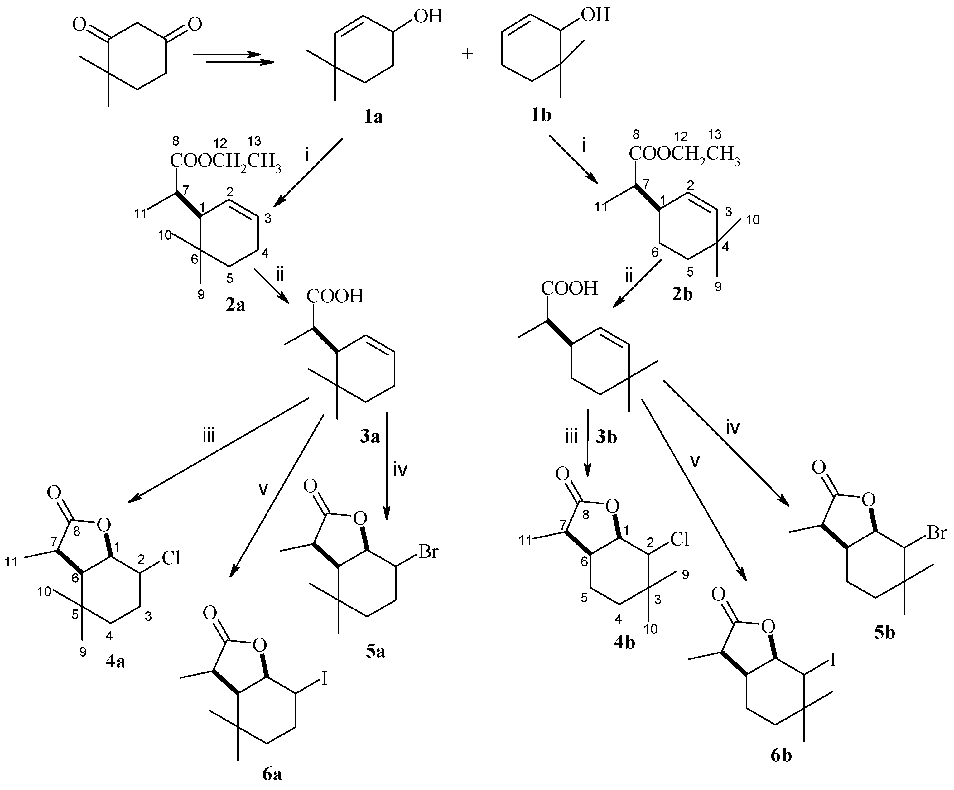
(1R, 7S or 1S, 7R)-Ethyl ester of (4,4,7-trimethylcyclohex-2-en-1-yl)acetic acid (isomer A) and (1R, 7R or 1S, 7S)-Ethyl Ester of (4,4,7-trimethylcyclohex-2-en-1-yl)acetic acid (isomer B) (2a), The known allylic alcohol 1a [24] was subjected to Claisen rearrangement with orthopropionate modification. Briefly, a mixture of alcohol (3.5 g), triethyl orthopropionate (20 cm3) and a catalytic amount of propionic acid was heated at 138 °C for 30 h. The progress of the reaction was monitored by TLC and GC. The obtained ester was purified by column chromatography using a 19:1 mixture of hexane and acetone as eluent. Ester 2a (3.9 g, 68%) was obtained as a pair of diastereoisomers A and B in the ratio 72%:28%. The spectral data of the obtained compound 2a are as follows: 1H NMR (600 MHz, CDCl3): 0.86 (s, 3H, CH3-9B), 0.87 (s, 3H, CH3-9A), 0.94 (s, 3H, CH3-10B), 0.96 (s, 3H, CH3-10A), 1.10–1.14 (M, 2H, CH2-5B), 1.09 (d, J = 7.2 Hz, 3H, CH3-11B), 1.20–1.26 (m, 9H, CH3-13A, CH3-13B, CH3-11A), 1.33–1.42 (m, 2H, CH2-4B), 1.58 (m, 2H, CH2-5A), 1.96–2.00 (m, 2H, CH2-4A), 2.27–2.37 (m, 2H, H-1A and H-1B), 2.58 (qd, J = 6.8 and 4.8 Hz, 1H, H-7B), 2.71 (qd, J = 7.2 and 3.6 Hz, 1H, H-7A), 4.06 (q, J = 7.2 Hz,2H, CH2-12A), 4.11 (q, J = 6.8 Hz,2H, CH2-12B), 5.49–5.54 (m, 2H, H-3A and H-3B), 5.69–5.74 (m, 2H, H-2A and H-3B), 13C NMR (151 MHz, CDCl3): 9.26 (C-11B), 14.19 (C-11A), 14.61 (C-13B), 18.71 (C-13A), 22.78 (C-4A), 23.07 (C-9A), 24.74 (C-9B), 25.56 (C-4B), 28.79 (C-10B), 29.04 (C-10A), 29.18 (C-6B), 29.62 (C-6A), 34.82 (C-5B), 36.03 (C-5A), 39.86 (C-7B), 39.95 (C-7A), 45.90 (C-1B), 49.85 (C-1A), 60.02 (C-12A), 60.36 (C-12B), 126.34 (C-2A), 126.77 (C-2B), 139.02127.03 (C-3A), 127.55 (C-3B), 176.20 (C-8A), 177.55 (C-8B), ESIHRMS: calculated for C13H22O2, 211.1698 (M + H)+, found 211.1700.
(1R, 7S or 1S, 7R)-(4,4,7-trimethylcyclohex-2-en-1-yl)acetic acid (isomer A) and (1R, 7R or 1S, 7S)-(4,4,7-trimethylcyclohex-2-en-1-yl)acetic acid (isomer B) (3a), Ester 2a (3.9 g) was given alkaline hydrolysis according to a known procedure [32]. Briefly, the ester and 5% potassium hydroxide solution in ethanol were heated for 6 h under reflux conditions. After the reaction was completed, the ethanol was evaporated, the residue was dissolved in water, acidified with 1 mol hydrochloric acid, and then extracted with diethyl ether. As a result, 1.5 g (44%) of acid 3a was obtained, as a pair of isomers A and B in the ratio 74%:26%. The spectral data of compound 3a are as follows: 1H NMR (400 MHz, CDCl3): 0.89 (s, 3H, CH3-9A), 0.92 (s, 3H, CH3-9B), 0.96 (s, 3H, CH3-10A), 0.98 (s, 3H, CH3-10B), 1.11 (d, J = 7.2 Hz, 3H, CH3-11A), 1.27 (d, J = 7.6 Hz, 3H, CH3-11B), 1.29–1.34 (m, 1H, CH2-5B), 1.98–2.00 (m, 2H, CH2-4A), 2.04–2.06 (m, 1H, H-1B), 2.38–2.40 (m, 1H, H-1A), 2.64–2.70 (qd, J = 7.2 and 4.4 Hz, 1H, H-7a), 2.73–2.76 (qd, J = 7.2 and 4.0 Hz, 1H, H-7B), 5.49–5.50 (m, 1H, H-3B), 5.51–5.53 (m, 1H, H-3A), 5.69–5.70 (m, 1H, H-2B), 5.73–5.77 (m, 1H, H-2A), 13C NMR (151 MHz, CDCl3): 14.10 (C-11A), 17.82 (C-11B), 22.75 (C-9A), 23.32 (C-4A), 24.70 (C-9B), 29.12 (C-10A), 29.60 (C-10B), 32.31 (C-6A), 32.60 (C-6B), 34.93 (C-5A), 35.79 (C-5B), 39.45 (C-7A), 40.11 (C-7B), 45.80 (C-1B), 45.80 (C-4B), 49.50 (C-1A), 126.30 (C-2A), 126.72 (C-2B), 127.03 (C-3B), 127.90 (C-3B), 182.36 (C-8B), 183.870 (C-8A), ESIHRMS: calculated for C11H18O2, m/z 183.1385 (M + H)+, found 183.1378.
(1S, 2R, 6R, 7S or 1R, 2S, 6S, 7R)-2-Chloro-3,3,7-trimethyl-9-oxabicyclo[4.3.0]nonan-8-one (isomer A) and (1S, 2S, 6R, 7R or 1R, 2R, 6S, 7S)-2-Chloro-3,3,7-trimethyl-9-oxabicyclo[4.3.0]nonan-8-one (isomer B) (4a), Chlorolactone was prepared according to a known procedure [32]. Briefly, acid 3a (0.5 g) and N-chlorosuccinimide (0.9 g) dissolved in THF (30 mL) were stirred for 24 h at room temperature. The reaction mixture was diluted with water, and the product was extracted with diethyl ether. After purification of the reaction product by column chromatography, using a mixture of hexane/acetone 3:1 as eluent, 0.35 g (59%) of chlorolactone 4a was obtained as a pair of isomers A and B, occurring in the ratio 72%:28%. The spectral data of compound 4a are as follows: Isomer A: 1H NMR (600 MHz, CDCl3): 1.09 (s, 3H, CH3-9), 1.11 (s, 3H, CH3-10), 1.41–1.44 (m, 1H, one of CH2-4), 1.46 (d, J = 7.2 Hz, 3H, CH3-11), 1.59–1.62 (m, 1H, one of CH2-4), 1.88-1.94 (m, 1H, one of CH2-3), 2.15–2.20 (m, 1H, CH2-3), 2.47 (dd, J = 7.2 and 6.0 Hz, 1H, H-6), 2.76 (q, J = 7.8 Hz, 1H, H-7), 4.23–4.28 (m, H-2), 4.49–4.52 (m, 1H, H-1), 13C NMR (151 MHz, CDCl3): 14.07 (C-11), 24.67 (C-9), 27.83 (C-3), 30.80 (C-5), 32.38 (C-10), 34.22 (C-4), 39.64 (C-7), 47.58 (C-6), 58.11 (C-2), 82.26 (C-1), 178.93 (C-8). Isomer B: 1H NMR (400 MHz, CDCl3): 1.04 (s, 3H, CH3-10), 1.08 (s, 3H, CH3-9), 1.33 (d, J = 6.8 Hz, 3H, CH3-11), 1.42–1.53 (m, 1H, CH2-4), 1.84–1.92 (m, 1H, one of CH2-3), 2.01–2.04 (m, 1H, H-6), 2.06–2.11 (m, 1H, one of CH2-3), 2.36–2.44 (m, 1H, H-7), 3.62–3.69 (m, 1H, H-2), 4.97 (dd, J = 9.2 and 7.2 Hz, 1H, H-1), 13C NMR (100 MHz, CDCl3): 17.29 (C-11), 28.15 (C-9), 29.481 (C-10), 29.85 (C-3), 32.70(C-5), 34.29 (C-4), 35.90 (C-7), 54.62 (C-6), 60.75 (C-2), 81.62 (C-1), 178.85 (C-8). ESIHRMS: calculated for C11H17ClO2, m/z 217.0995 and 219.0969 (M + H)+, found 217.0999 and 219.0970, calculated for C11H17ClO2Na, m/z 239.0815 and 241.0788 (M + H)+, found 239.0823 and 241.0790.
(1S, 2R, 6R, 7S or 1R, 2S, 6S, 7R)-2-Bromo-3,3,7-trimethyl-9-oxabicyclo[4.3.0]nonan-8-one (isomer A) and (1S, 2S, 6R, 7R or 1R, 2R, 6S, 7S)-2-Bromo-3,3,7-trimethyl-9-oxabicyclo[4.3.0]nonan-8-one (isomer B) (5a), Bromolactone was obtained according to a known procedure [32]. Briefly, acid 3a (0.5 g) and N-bromosuccinimide (0.7 g) dissolved in THF (30 mL) were stirred for 24 h at room temperature. Water was added to the reaction mixture and the product was extracted with diethyl ether. After purification of the reaction product by column chromatography, using a mixture of hexane/acetone 3:1 as eluent, 0.54g (75%) of bromolactone 5a was obtained as a pair of isomers A and B, occurring in the ratio 76%:24%. The spectral data of compound 5a are as follows: Isomer A: 1H NMR (600 MHz, CDCl3): 1.09 (s, 3H, CH3-9), 1.12 (s, 3H, CH3-10), 1.43–1.46 (m, 1H, one of CH2-4), 1.47 (d, J = 7.2 Hz, 3H, CH3-11), 1.62 (m, 1H, one of CH2-4), 2.00–2.06 (m, 3H, one of CH2-3), 2.21–2.27 (m 1H, one of CH2-3), 2.49 (dd, J = 7.2 and 6.6 Hz, 1H, H-6), 2.77 (q, J = 7.2 Hz, 1H, H-7), 4.36 (m, H-2), 4.61–4.65 (t, J = 6.0 Hz, 1H, H-1), 13C NMR (151 MHz, CDCl3): 17.18 (C-11), 28.08 (C-3), 29.47 (C-5), 30.81 (C-9), 32.45 (C-10), 35.69 (C-4), 39.82 (C-7), 49.70 (C-6), 50.12 (C-2), 82.47 (C-1), 178.92 (C-8). Isomer B: 1H NMR (600 MHz, CDCl3): 1.06 (s, 3H, CH3-10), 1.12 (s, 3H, CH3-9), 1.37 (d, J = 6.6 Hz, 3H, CH3-11), 1.42–1.44 (m, 1H, one of CH2-4), 1.49–1.54 (m, 1H, one of CH2-4), 2.02–2.10 (m, 2H, one of CH2-3, H-6), 2.16–2.19 (m, 1H, one of CH2-3), 2.45–2.51 (m, 1H, H-7), 3.74–3.79 (m, 1H, H-2), 4.66 (dd, J = 9.6 and 7.2 Hz, 1H, H-1), 13C NMR (151 MHz, CDCl3): 17.14 (C-11), 28.08 (C-9), 29.047 (C-10), 30.72 (C-3), 32.64 (C-5), 35.22 (C-4), 35.62 (C-7), 51.72 (C-2), 54.84 (C-6), 81.70 (C-1), 178.65 (C-8). ESIHRMS: calculated for C11H17BrO2, m/z 261.0490 and 263.0473 (M + H)+, found 261.0475 and 263.0453, calculated for C11H17BrO2Na, m/z 283.0310 and 285.0290 (M + H)+, found 283.0304 and 285.0284.
(1S, 2R, 6R, 7S or 1R, 2S, 6S, 7R)-2-Iodo-3,3,7-trimethyl-9-oxabicyclo[4.3.0]nonan-8-one (isomer A) and (1S, 2S, 6R, 7R or 1R, 2R, 6S, 7S)-2-Iodo-3,3,7-trimethyl-9-oxabicyclo[4.3.0]nonan-8-one (isomer B) (6a), Iodolactone was prepared according to a known procedure [32]. Briefly, acid 3a (0.5 g) was dissolved in 25 mL diethyl ether and stirred for 1 h with 25 mL of 0.5 mol sodium bicarbonate solution. Then, 1 g of iodine and 2 g of potassium iodide in 20 mL of water were added to the solution, and the whole mixture was stirred for 24 h. After the reaction was completed, the product was extracted with diethyl ether. After purification of the compound by column chromatography, using a mixture of hexane/acetone 3:1 as eluent, 0.48g (57%) of iodolactone 6a was obtained as a pair of isomers A and B, occurring in the ratio 96%:4%. The spectral data of compound 6a are as follows: Isomer A: 1H NMR (400 MHz, CDCl3): 1.04 (s, 3H, CH3-9), 1.09 (s, 3H, CH3-10), 1.41–1.48 (m, 2H CH2-4), 1.45 (d, J = 7.2 Hz, 3H, CH3-11), 2.03–2.17 (m, 2H, CH2-4), 2.09–2.14 (m, 2H, CH2-3), 2.43 (dd, J = 7.2 and 6.0 Hz, 1H, H-6), 2.71 (q, J = 7.8 Hz, 1H, H-7), 4.42 (m, H-2), 4.70 (m, 1H, H-1), 13C NMR (100 MHz, CDCl3): 14.42 (C-11), 25.18 (C-9), 28.58 (C-2), 30.87(C-3), 32.66 (C-10), 37.34 (C-4), 40.09 (C-7), 47.73 (C-6), 54.77 (C-5), 84.06 (C-1), 179.06 (C-8). Isomer B: 1H NMR (400 MHz, CDCl3): 1.04 (s, 3H, CH3-9), 1.09 (s, 3H, CH3-10), 1.41–1.49 (m, 2H, CH2-4), 1.45 (d, J = 7.6 Hz, 3H, CH3-11), 1.92–1.97 (m, 2H, one of CH2-3), 2.11–2.15 (m, 1H, CH2-6), 2.20–2.22 (m, 1H, one of CH2-3), 2.48–2.55 (qm, 1H, H-7), 3.74–3.81 (m, H-2), 4.69–4.72 (m, H-1), 13C NMR (100 MHz, CDCl3): 17.10 (C-11), 25.18 (C-9), 28.12 (C-2), 29.74 (C-10), 30.87 (C-5), 33.02 (C-3), 37.34 (C-4), 40.01 (C-7), 47.69 (C-6), 82.91 (C-1), 179.06 (C-8). ESIHRMS: calculated for C11H17IO2, m/z 309.0352 (M + H)+, found 309.0352, calculated for C11H17IO2Na, m/z 331.0171 (M + H)+, found 331.0161.
(1R, 7S or 1R, 7R)-Ethyl ester of (4,4,7-trimethylcyclohex-2-en-1-yl)acetic acid (isomer A) and (1R, 7R or 1S, 7S)-Ethyl ester of (4,4,7-trimethylcyclohex-2-en-1-yl)acetic acid (isomer B) (2b), The known allylic alcohol 1b [24] (1.5 g) was subjected to Claisen rearrangement with orthopropionate modification analogously as described for ester 1a. Ester 2b (1.6 g) was obtained as a mixture of two diastereoisomers A and B in the ratio 20%:80% in 65% yield. The spectral data of compound 2b are as follows: 1H NMR (400 MHz, CDCl3): 0.93 (s, 3H, CH3-9), 0.94 (s, 3H, CH3-10), 1.09 (d, J = 6.8 Hz, 3H, CH3-11A), 1.11 (d, J = 6.8 Hz, 3H, CH3-11B), 1.24 (t, J = 7.6 Hz, 3H, CH3-12), 1.36–1.42 (m, 2H, one of CH2-5, one of CH2-6), 1.47–1.52 (m, 1H, one of CH2-6), 1.54–1.53 (m, 1H, one of CH2-5), 2.29–2.37 (m, 2H, H-1 and H-7), 4.12 (q, 2H, CH2-12), 5.28–5.31 (dm, J = 10.0 Hz, 1H, H-3), 5.43–5.45 (dm, J = 8.4 Hz, 1H, H-2),13C NMR (100 MHz, CDCl3): 13.63 (C-11A), 13.92 (C-11B), 14.39 (C-13A and C-13B), 22.39 (C-5A), 24.07 (C-5B), 28.77 (C-9A), 29.00 (C-9B), 30.52 (C-10B), 30.71 (C-10A), 31.66 (C-4A), 31.74 (C-4B), 36.27 (C-6B), 36.39 (C-6A), 38.48 (C-1A), 38.51 (C-1B), 44.06 (C-7B), 44.21 (C-7A), 60.22 (C-12A and C-12B), 125.60 (C-3A), 126.80 (C-3B), 139.02 (C-2A), 139.31 (C-2B), 176.07 (C-8B), 176.16 (C-8A), ESIHRMS: calculated for C13H22O2, m/z 211.1698 (M + H)+, found 211.1700.
(1R, 7S or 1R, 7R)-(4,4,7-trimethylcyclohex-2-en-1-yl)acetic acid (isomer A) and (1R, 7R or 1S, 7S)-(4,4,7-trimethylcyclohex-2-en-1-yl)acetic acid (isomer B) (3b), Ester 2b (1.6g) was given an alkaline hydrolysis, analogously as described for acid 2a. Acid 3b (0.7g) was obtained as a mixture of two diastereoisomers A and B in the ratio 22%:78% in 50% yield. The spectral data of compound 3b are as follows: 1H NMR (400 MHz, CDCl3): 0.90 (s, 3H, CH3-10B), 0.94 (s, 3H, CH3-9A), 0.96 (s, 3H, CH3-10A,), 0.96(s, 3H, CH3-9B), 1.12 (d, J = 6.8 Hz, 3H, CH3-11A), 1.16 (d, J = 6.8 Hz, 3H, CH3-11B), 1.38–1.43 (m, 4H, one of CH2-5A, one of CH2-6A, one of CH2-5B, one of CH2-6B), 1.49–1.53 (m, 2H, one of CH2-6A, one of CH2-6B), 1.63–1.68 (m, 2H, one of CH2-5A, one of CH2-5B), 2.37–2.44 (m, 4H, H-1A, H-1B, H-7A and H-7B), 5.33–5.35 (dm, J = 10.4 Hz, 2H, H-3A, H-3B), 5.46–5.49 (dm, J = 10.0 Hz, 2H, H-2A, H-2B), 13C NMR (100 MHz, CDCl3): 13.19 (C-11A), 13.62 (C-11B), 22.05 (C-5A), 24.19 (C-5B), 28.77 (C-9A), 29.00 (C-9B), 30.49 (C-10B), 30.69 (C-10A), 31.65 (C-4A), 31.75 (C-4B), 43.90 (C-6B), 43.00 (C-6A), 38.19 (C-1A), 38.19 (C-1B), 43.90 (C-7B), 44.00 (C-7A), 125.12 (C-3A), 126.63 (C-3B), 139.36 (C-2A), 139.71 (C-2B), 182.31 (C-8B), 182.39 (C-8A), ESIHRMS: calculated for C11H18O2, m/z 183.1385 (M + H)+, found 183.1376.
(1S, 2R, 6R, 7S or 1R, 2S, 6R, 7R)-2-Chloro-3,3,7-trimethyl-9-oxabicyclo[4.3.0]nonan-8-one (isomer A) and (1S, 2S, 6R, 7R or 1R, 2R, 6S, 7S)-2-Chloro-3,3,7-trimethyl-9-oxabicyclo[4.3.0]nonan-8-one (isomer B) (4b), Acid 3b (0.2g) was subjected to chlorolactonization in the same manner as given for compound 3a. The product 4b was obtained as a mixture of two diastereoisomers A and B in the ratio 22%:78% with a yield of 0.19 g (80%) with the following spectral data: Isomer A: 1H NMR (400 MHz, CDCl3): 1.07 (s, 3H, CH3-9), 1.07 (s, 3H, CH3-10), 1.18 (d, J = 7.2 Hz, 3H, CH3-11), 1.24–1.30 (m, 2H, CH2-4), 1.56–1.70 (m, 2H, CH2-5), 2.60–2.67 (m, 1H, H-6), 2.75 (q, J = 7.2 Hz, 1H, H-7), 4.10 (m, 1H, H-2), 4.57 (dd, J = 3.6 and 3.6 Hz, 1H, H-1), 13C NMR (100 MHz, CDCl3): 9.56 (C-11), 18.95 (C-4), 25.64 (C-9), 30.17 (C-10), 30.57 (C-5), 34.10 (C-3), 35.49 (C-6), 40.97 (C-7), 65.22 (C-2), 80.83 (C-1), 178.31 (C-8). Isomer B: 1H NMR (400 MHz, CDCl3): 1.00 (s, 3H, CH3-9), 1.07 (s, 3H, CH3-10), 1.20 (d, J = 6.4 Hz, 3H, CH3-11), 1.46 (ddd, J = 14.4, 4.4 and 4.4 Hz, 1H, one of CH2-4), 1.55 (ddd, J = 14.4, 4.8. and 2.4 Hz, 1H, one of CH2-4), 1.64–1.69 (m, 1H, one of CH2-5), 1.85–1.94 (m, 1H, one of CH2-5), 2.36–2.48 (m, 2H, H-6, H-7), 3.56 (d, J = 9.6 Hz, 1H, H-2), 4.48 (dd, J = 9.6 and 6.8 Hz, 1H, H-1), 13C NMR (100 MHz, CDCl3): 13.48 (C-11), 18.54 (C-9), 19.89 (C-5), 29.60 (C-10), 34.00 (C-4), 35.74 (C-6), 36.97 (C-3), 43.53 (C-7), 70.83 (C-2), 81.59 (C-1), 178.30 (C-8). ESIHRMS: calculated for C11H17ClO2, m/z 217.0995 and 219.0969 (M + H)+, found 217.0999 and 219.0970, calculated for C11H17ClO2Na, m/z 239.0815 and 241.0788 (M + H)+, found 239.0823 and 241.079.
(1S, 2R, 6R, 7S or 1R, 2S, 6R, 7R)-2-Bromo-3,3,7-trimethyl-9-oxabicyclo[4.3.0]nonan-8-one (isomer A) and (1S, 2S, 6R, 7R or 1R, 2R, 6S, 7S)-2-Bromo-3,3,7-trimethyl-9-oxabicyclo[4.3.0]nonan-8-one (isomer B) (5b), Acid 3b (0.2 g) underwent bromolactonization analogously as described for compound 3a. The resulting product 5b was formed as a mixture of two diastereoisomers A and B in the ratio 14%:86% with a yield of 0.26 g (90%) with the following spectral data: Isomer A: 1H NMR (600 MHz, CDCl3): 1.14 (s, 3H, CH3-9), 1.18 (s, 3H, CH3-10), 1.22 (d, J = 7.2 Hz, 3H, CH3-11), 1.29–1.34 (m, 2H, one of CH2-4, one of CH2-5), 1.61–1.66 (m, 1H, CH2-4), 1.71–1.76 (m, 1H, CH2-5), 2.74–2.76 (m, 1H, H-6), 2.80 (q, J = 7.2 Hz, 1H, H-7), 4.32 (m, 1H, H-2), 4.78 (dd, J = 3.6 and 3.6 Hz, 1H, H-1), 13C NMR (151 MHz, CDCl3): 9.51 (C-11), 19.03 (C-4), 25.69 (C-10), 31.07 (C-5), 32.48 (C-9), 33.96 (C-3), 36.45 (C-6), 41.17 (C-7), 60.33 (C-2), 81.21 (C-1), 178.31 (C-8). Isomer B: 1H NMR (600 MHz, CDCl3): 1.08 (s, 3H, CH3-9), 1.12 (s, 3H, CH3-10), 1.24 (d, J = 6.8 Hz, 3H, CH3-11), 1.52 (ddd, J = 14.0, 4.8 and 4.4 Hz, 1H, one of CH2-4), 1.63–1.67 (m, 1H, one of CH2-4), 1.70–1.74 (m, 1H, one of CH2-5), 1.92–1.99 (m, 1H, one of CH2-5), 2.36–2.40 (m, 1H, H-6), 2.50 (q, J = 6.8 Hz, 1H, H-7), 3.74 (d, J = 10.0 Hz, 1H, H-2), 4.67 (dd, J = 10.0 and 7.6 Hz, 1H, H-1), 13C NMR (151 MHz, CDCl3): 13.31 (C-11), 19.69 (C-9), 19.92 (C-5), 30.98 (C-10), 33.60 (C-4), 35.41 (C-7), 36.78 (C-3), 43.69 (C-6), 65.34 (C-2), 81.77 (C-1), 178.05 (C-8). ESIHRMS: calculated for C11H17BrO2, m/z 261.0490 and 263.0471 (M + H)+, found 261.0485 and 263.0465, calculated for C11H17BrO2Na, m/z 283.0310 and 285.0290 (M + H)+, found 283.0305 and 285.0285.
(1S, 2R, 6R, 7S or 1R, 2S, 6R, 7R)-2-Iodo-3,3,7-trimethyl-9-oxabicyclo[4.3.0]nonan-8-one (isomer A) and (1S, 2S, 6R, 7R or 1R, 2R, 6S, 7S)-2-Iodo-3,3,7-trimethyl-9-oxabicyclo[4.3.0]nonan-8-one (isomer B) (6b), Acid 3b (0.3 g) was submitted to iodolactonization in the same way as compound 3a. The product 6b was formed as a mixture of two diastereoisomers A and B in the ratio 23%:77% with a yield of 0.24 g (48%). The spectral data are as follows: Isomer A: 1H NMR (400 MHz, CDCl3): 1.13 (s, 3H, CH3-9), 1.17 (d, J = 6.8 Hz, 3H, CH3-11), 1.22 (s, 3H, CH3-10), 1.24–1.29 (m, 1H, one ofCH2-4), 1.60–1.69 (m, 2H, one of CH2-4 and CH2-5), 2.76 (q, J = 6.8 Hz, 1H, H-7), 2.80–2.85 (m, 1H, H-6), 4.53–4.54 (m, 1H, H-2), 4.94 (dd, J = 4.0 and 3.2 Hz, 1H, H-1), 13C NMR (100 MHz, CDCl3): 9.62 (C-11), 19.35 (C-5), 22.73 (C-3), 24.85 (C-10), 32.07 (C-4), 35.57 (C-6), 36.99 (C-9), 41.69 (C-7), 45.00 (C-2), 83.48 (C-1), 178.67 (C-8). Isomer B: 1H NMR (400 MHz, CDCl3): 1.05 (s, 3H, CH3-9), 1.06 (s, 3H, CH3-10), 1.19 (d, J = 6.8 Hz, 3H, CH3-11), 1.48–1.57 (m, 1H, one of CH2-4), 1.64–1.73 (m, 2H, one of CH2-4 and one of CH2-5), 1.88–1.98 (m, 1H, one of CH2-5), 2.19–2.24 (m, 1H, H-6), 2.52 (q, J = 6.8 Hz, 1H, H-7), 3.84 (d, J = 10.4 Hz, 1H, H-2), 4.77 (dd, J = 10.4 and 7.2 Hz, 1H, H-1), 13C NMR (100 MHz, CDCl3): 13.24 (C-11), 20.21 (C-5), 21.92 (C-10), 31.81 (C-4), 33.84 (C-9), 34.99 (C-7), 36.62 (C-3), 43.43 (C-6), 48.49 (C-2), 83.00 (C-1), 177.92 (C-8). ESIHRMS: calculated for C11H17IO2, m/z 309.0352 (M + H)+, found 309.0352.

4.3. X-ray Diffraction Data

Single-crystal X-ray diffraction data were collected at 100 K on Rigaku XtaLAB Synergy R DW system (HyPix-Arc 150, Rigaku, Wrocław, Poland) κ-geometry diffractometers using Cu Kα radiation. Data reduction and analysis were carried out with the CrysAlis Pro program [33]. The structures were solved by direct methods and refined with the full-matrix least-squares technique using the SIR2019 version 19.02 [34] and Shelxl-2018/3 [35] programs. Hydrogen atoms were placed at calculated positions, and before the last cycle of refinement, all H atoms were fixed and were allowed to ride on their parent atoms. Anisotropic displacement parameters were refined for all non-hydrogen atoms.
Crystal data for B isomer of 2-iodo-3,3,7-trimethyl-9-oxabicyclo[4.3.0]nonan-8-one: C11H17IO2, M = 308.14, monoclinic, P21/c, a = 7.384(2) Å, b = 21.772(4) Å, c = 14.745(4) Å, β = 90.93(2)°, V = 2370.2(10) Å3, Z = 8, Dc = 1.727 Mg m−3, T = 100(2) K, R = 0.0360, wR = 0.0957 (3978 reflections with I > 2σ(I)) for 253 variables, CCDC 2359086.

4.4. Biotransformation

4.4.1. Microorganisms

Biotransformation reactions were carried out using strains of filamentous fungi from the collection of the Department of Food Chemistry and Biocatalysis of Wrocław University of Life Sciences. There were the strains of the genus Fusarium: F. culmorum AM10, F. avenaceum AM12, F. equiseti AM16, F. semitectum AM20, F. oxysporum AM21, F. solani AM203, and strains from the genus Absidia: A. coerulea AM93, A. glauca AM177, A. cylindrospora AM336. These strains were cultured on Sabouraud Agar (0.5 g of aminobac, 0.5 g of peptone, 4 g of glucose and 1.5 g of agar dissolved in 100 mL of water) at 28 °C and stored at 4 °C after growth.

4.4.2. Screening Biotransformation

Biotransformation was carried out in 300 mL Erlenmeyer flasks containing 100 mL of Sabouraud medium, consisting of 3 g of glucose and 1 g of peptone dissolved in 100 mL of water. After inoculation of the medium with the given microorganism, the mycelium was allowed to grow for three days. After this time, 10 mg of halolactone 4a,b6a,b dissolved in 1 mL of acetone was added to each flask. Shaken culture was continued for another seven days, and after three, five and seven days, about 30 mL of medium was taken with the mycelium. This mixture was extracted with dichloromethane (15 mL), dried with magnesium sulfate and analyzed by GC using a SGE BP5 column.

4.4.3. Preparative Biotransformation

To 10 Erlenmeyer flasks, each containing 100 mL of Sabouraud medium with overgrown mycelium, 10 mg each (for a total of 100 mg) of bromo-5a or iodolactone 6a dissolved in 10 mL of acetone was added. Shaken culture was carried out for 7 days, after which the combined contents of ten flasks were extracted with dichloromethane (3 × 30 mL). The combined organic fractions were dried with anhydrous magnesium sulfate. After evaporation of the solvent in vacuo, the products were purified by column chromatography using a mixture of hexane/acetone 6:1 as eluent. The spectral data of the compounds obtained are shown below.
(1S, 2S, 6R, 7S or 1R, 2R, 6S, 7R)-2-Hydroxy-3,3,7-trimethyl-9-oxabicyclo[4.3.0]nonan-8-one (7a), 1H NMR (400 MHz, CDCl3): 1.01 (s, 6H, CH3-9 and CH3-10), 1.19–1.27 (m, 1H, one of CH2-4), 1.39–1.41 (m, 1H, one of CH2-4), 1.43 (d, J = 7.2 Hz, 3H, CH3-11), 1.61–1.71 (m, 1H, one of CH2-3), 1.76–1.83 (m, 1H, one of CH2-3), 2.15 (s, 1H, OH), 2.18 (dd, J = 6.8 and 4.4 Hz, 1H, H-6), 2.82 (q, J = 7.2 Hz, 1H, H-7), 3.74 (ddd, J = 10.4, 5.2 and 5.2, 1H, H-2), 4.42 (t, J = 4.4 Hz, 1H, H-1), 13C NMR (151 MHz, CDCl3): 11.87 (C-11), 22.78 (C-9), 26.11 (C-3), 31.53 (C-10), 31.92 (C-5), 38.81 (C-4), 42.20 (C-7), 47.07 (C-6), 69.30 (C-2), 80.52 (C-1), 179.27 (C-8), ESIHRMS: calculated for C11H18O3, m/z 199.1334 (M + H)+, found 199.1330, calculated for C11H18O3Na, m/z 221.1154 (M + H)+, found 221.1134.
(1S, 2R 6R, 7R or 1R, 2S, 6S, 7S)-2-Hydroxy-3,3,7-trimethyl-9-oxabicyclo[4.3.0]nonan-8-one (8a), 1H NMR (400 MHz, CDCl3): 1.06 (s, 3H, CH3-9), 1.09 (s, 3H, CH3-10), 1.24–1.26 (m, 1H, one of CH2-4), 1.40 (d, J = 7.2 Hz, 3H, CH3-11), 1.45–1.53 (m, 1H, one of CH2-4), 1.56–1.65 (m, 1H, one of CH2-3), 1.86–1.93 (m, 1H, one of CH2-3), 1.99 (s, 1H, OH), 2.44 (dd, J = 8.4 and 7.2 Hz, 1H, H-6), 2.69 (q, J = 8.0 Hz, 1H, H-7), 3.85–3.91 (m, 1H, H-2), 4.35–4.40 (m, 1H, H-1), 13C NMR (151 MHz, CDCl3): 15.19 (C-11), 23.09 (C-5), 26.35 (C-3), 30.38 (C-10), 32.86 (C-4), 34.22 (C-9), 38.35 (C-7), 48.33 (C-6), 71.55 (C-2), 84.28 (C-1), 180.24 (C-8), ESIHRMS: calculated for C11H18O3, m/z 199.1334 (M + H)+, found 199.1330.
(1S, 2R, 6R, 7R or 1R, 2S, 6S, 7S)-2-Chloro-7-hydroxy-3,3,7-trimethyl-9-oxabicyclo[4.3.0]nonan-8-one (9a), 1H NMR (400 MHz, CDCl3): 1.10 (s, 3H, CH3-9), 1.22 (s, 3H, CH3-10), 1.40–1.44 (m, 1H, one of CH2-4), 1.59 (d, J = 7.2 Hz, 3H, CH3-11), 1.66 (td, J = 14.0 and 3.6 Hz, 1H, one of CH2-4), 1.92 (qd, J = 14.0 and 4.0 Hz, 1H, one of CH2-3), 2.12–2.17 (m, 1H, one of CH2-3), 2.54 (d, J = 8.4 Hz, 1H, H-6), 2.76 (s, 1H, OH), 3.92 (ddd, J = 12.8, 10.0, 5.2 Hz, 1H, H-2), 4.61 (dd, J = 10.0 and 8.4 Hz, 1H, H-1), 13C NMR (151 MHz, CDCl3): 24.08 (C-11), 26.56 (C-10), 30.08 (C-3), 30.31 (C-9), 32.84 (C-5), 35.06 (C-4), 55.09 (C-6), 59.71 (C-2), 73.88 (C-7), 81.16 (C-1), 179.60 (C-8), ESIHRMS: calculated for C11H17ClO3, m/z 233.0945 and 235.0918 (M + H)+, found 233.0933 and 235.0893.
(1S, 2R, 6R, 7R or 1R, 2S, 6S, 7S)-2-Bromo-7-hydroxy-3,3,7-trimethyl-9-oxabicyclo[4.3.0]nonan-8-one (10a), 1H NMR (400 MHz, CDCl3): 1.10 (s, 3H, CH3-9), 1.21 (s, 3H, CH3-10), 1.24–1.29 (m, 1H, one of CH2-4), 1.60 (d, J = 7.2 Hz, 3H, CH3-11), 1.66 (td, J = 14.0 and 3.6 Hz, 1H, one of CH2-4), 2.09 (qd, J = 13.2 and 4.0 Hz, 1H, one of CH2-3), 2.22–2.25 (m, 1H, one of CH2-3), 2.50 (dd, J = 8,4 and 1.2 Hz, 1H, H-6), 2.72 (s, 1H, OH), 4.00 (ddd, J = 12.8, 10.0, 5.6 Hz, 1H, H-2), 4.74 (dd, J = 10.0 and 8.4 Hz, 1H, H-1), 13C NMR (151 MHz, CDCl3): 24.35 (C-11), 26.67 (C-10), 30.07 (C-9), 31.23 (C-3), 32.84 (C-5), 35.03 (C-4), 50.59 (C-2), 55.39 (C-6), 73.86 (C-7), 81.30 (C-1), 179.44 (C-8), ESIHRMS: calculated for C11H17BrO3, m/z 277.0439 and 279.0420 (M + H)+, found 277.0425 and 279.0387.
(1S, 2R, 6R, 7R or 1R, 2S, 6S, 7S)-2-Iodo-7-hydroxy-3,3,7-trimethyl-9-oxabicyclo[4.3.0]nonan-8-one (11a), 1H NMR (400 MHz, CDCl3): 1.10 (s, 3H, CH3-9), 1.20 (s, 3H, CH3-10), 1.24 (m, 1H, one of CH2-4), 1.64 (d, J = 7.2 Hz, 3H, CH3-11), 1.65 (td, J = 14.0 and 4.0 Hz, 1H, one of CH2-4), 2.25 (qd, J = 13.6 and 4.0 Hz, 2H, CH2-3), 2.38 (d, J = 8.4 Hz, 1H, H-6), 2.58 (s, 1H, OH), 3.95 (ddd, J = 12.8, 10.8, 5.6 Hz, 1H, H-2), 4.61 (dd, J = 10.4 and 8.0 Hz, 1H, H-1), 13C NMR (151 MHz, CDCl3): 24.85 (C-11), 26.93 (C-10), 26.98 (C-2), 30.04 (C-9), 32.83 (C-5), 33.50 (C-3), 37.30 (C-4), 55.24 (C-6), 73.88 (C-7), 82.42 (C-1), 179.12 (C-8), ESIHRMS: calculated for C11H17IO3, m/z 325.0301 (M + H)+, found 325.0309.

4.5. Biological Tests

4.5.1. Microbial Strains

Multidrug-resistant (MDR) clinical bacterial strains, namely methicillin-resistant (MR) Staplylococcus aureus, S. epidermidis and S. haemolyticus; vancomycin-resistant Enterococcus faecalis (VRE); beta-lactamase-producing (ESBL) Eschericha coli and Pseudomonas aeruginosa; and Acinetobacter baumannii, as well as two strains of yeast-like fungi—Candida albicans and C. dubliniensis—were used to test antimicrobial activity. In addition, a reference strain of S. aureus ATCC25923 was used in the study. All strains needed for the determinations can be found in the Museum of Microorganisms of the Department of Microbiology of the Medical University of Wrocław.
The bacterial strains were stored at −80 °C. They were revived in overnight culture in Tryptic Soy Broth (TSB) media (OXOID, Hampshire, UK) or Sabouraud Broth (SB) using shaking (MaxQTM 6000 incubator shaker, Thermo Scientific, Waltham, MA, USA) at 125 rpm and 37 °C. For each experiment, a fresh 18–20 h culture was prepared on Tryptic Soy Agar (TSA, OXOID, UK) or Sabouraud Agar (SA), which was then transferred to 3 mL of fresh Mueller Hinton broth (MHB, OXOID, Hampshire, UK) or SB for bacteria and fungi, respectively. The optical density of each culture was set at 108 CFU/mL and then diluted to a concentration representing the primary inoculum, 106 CFU/mL.

4.5.2. Determination of Antimicrobial Activity

Determination of Inhibitory Concentration

MIC (minimum inhibitory concentration) values for the tested compounds were determined using the broth microdilution method, according to the standards of the European Committee for Antimicrobial Susceptibility Testing, (EUCAST) [36,37]. Stock solutions (20 mg/mL) of the tested lactones were prepared in DMSO (Chempur, Piekary Śląskie, Poland). Doubly concentrated compounds were diluted at concentrations ranging from 2 µg/mL to 1024 µg/mL in 96-well titration plates.
Culture in MHB or SB (105 CFU/mL) was added to an appropriate liquid medium with twice-concentrated compounds. The following controls were set for the assays: blank (MHB/SB medium); negative/background compound (MHB/SB medium with the halolactones under study); growth (bacterial culture in MHB/SB) and solvent (bacterial/fungal culture in MHB/SB with the appropriate concentration of DMSO). Each experiment was performed as 3 independent experiments, each in 3 replicates.
The contents of the titration plate were stirred for 20 min (2000 rpm) and incubated for 18 h at 37 °C. The optical density (OD600nm) was then read (ASYS UVM340 reader, BIOCHROM Ltd., Cambridge, UK). The MIC value was calculated from the optical density counted for the test compound (taking into account the blank and background of the compound) relative to the growth control. On this basis, MIC50 and MIC90 were determined, i.e., the concentration of the modified lactone that contributed to killing the test strain at 50% and 90%, respectively.

Determination of the Killing Concentration

In order to determine the MBC (minimum bactericidal concentration) and MFC (minimum fungicidal concentration) values of the tested compounds, 10 µL of the reaction mixture was seeded from each well of the titer plate onto TSA or SA and incubated for 18–20 h at 37 °C.

5. Conclusions

As a result of several stages of organic synthesis, two groups of racemic halolactones with two methyl groups in the cyclohexane ring (at the C-5 carbon or C-3 carbon) and a methyl group in the lactone ring (C-7 carbon) were obtained. Halolactones with substituents at the C-5 carbon underwent biotransformation to hydroxylactones or halo-hydroxylactones. Their analogs with substituents at the C-3 carbon did not undergo any transformation under the influence of filamentous fungi. The biological activity tests showed that halolactones containing a chlorine or bromine atom with a gem-dimethyl moiety at the C-5 carbon in their structure are capable of inhibiting the growth of bacteria, while those containing bromolactone with two methyl groups at the C-3 carbon are capable of inhibiting yeast-like fungi. Among the biotransformation products, bromo-hydroxylactone with a gem-dimethyl moiety at the C-5 carbon was the only one to show antimicrobial activity.

Supplementary Materials

The following supporting information can be downloaded at https://www.mdpi.com/article/10.3390/molecules29122820/s1: Figures S1–S10: Chromatograms of substrates, Figures S11–S30: 1H NMR, COSY, HMQC, 13C NMR, HRMS spectrum of esters and acids, Figures S31–S95: 1H NMR, COSY, HMQC, 13C NMR, NOESY, HRMS spectrum of halolactones, Figures S96–S105: 1H NMR, COSY, HMQC, 13C NMR, HRMS spectrum of hydroxylactones, Figures S106–S120: 1H NMR, COSY, HMQC, 13C NMR, HRMS spectrum of halo-hydroxylactones, Figures S121–S126: Chiral chromatograms of hydroxylactones and halo-hydroxylactones.

Author Contributions

Conceptualization, M.G.; investigation, M.G., A.D.-M., F.R., W.M. and K.W.; analysis, M.G., G.M. and A.B.; writing—original draft preparation, M.G.; writing—review and editing, M.G., A.D.-M., W.M. and K.W.; supervision, K.W. All authors have read and agreed to the published version of the manuscript.

Funding

This research received no external funding.

Institutional Review Board Statement

Not applicable.

Informed Consent Statement

Not applicable.

Data Availability Statement

Data are contained within the article and Supplementary Materials.

Conflicts of Interest

The authors declare no conflicts of interest.

References

  1. Sartori, S.K.; Diaz, M.A.N.; Diaz-Muñoz, G. Lactones: Classification, Synthesis, Biological Activities, and Industrial Applications. Tetrahedron 2021, 84, 132001. [Google Scholar] [CrossRef]
  2. Mazur, M.; Masłowiec, D. Antimicrobial Activity of Lactones. Antibiotics 2022, 11, 1327. [Google Scholar] [CrossRef] [PubMed]
  3. Abood, S.; Eichelbaum, S.; Mustafi, S.; Veisaga, M.L.; López, L.A.; Barbieri, M. Biomedical Properties and Origins of Sesquiterpene Lactones, with a Focus on Dehydroleucodine. Nat. Prod. Commun. 2017, 12, 995. [Google Scholar] [CrossRef]
  4. Mazur, M.; Włoch, A.; Bahri, F.; Pruchnik, H.; Pawlak, A.; Obmińska-Mrukowicz, B.; Maciejewska, G.; Gładkowski, W. Chemoenzymatic Synthesis of Enantiomeric, Bicyclic δ-Halo-γ-lactones with a Cyclohexane Ring, Their Biological Activity and Interaction with Biological Membranes. Biomolecules 2020, 10, 95. [Google Scholar] [CrossRef] [PubMed]
  5. Kamizela, A.; Gawdzik, B.; Urbaniak, M.; Lechowicz, Ł.; Białońska, A.; Kutniewska, S.E.; Gonciarz, W.; Chmiela, M. New γ-Halo-δ-lactones and δ-Hydroxy-γ-lactones with Strong Cytotoxic Activity. Molecules 2019, 24, 1875. [Google Scholar] [CrossRef] [PubMed]
  6. Varejão, J.O.; Barbosa, L.C.; Ramos, G.Á.; Varejão, E.V.; King-Díaz, B.; Lotina-Hennsen, B. New rubrolide analogues as inhibitors of photosynthesis light reactions. J. Photochem. Photobiol. B. 2015, 145, 11–18. [Google Scholar] [CrossRef] [PubMed]
  7. Barbosa, L.C.; Maltha, C.R.; Lage, M.R.; Barcelos, R.C.; Donà, A.; Carneiro, J.W.; Forlani, G. Synthesis of rubrolide analogues as new inhibitors of the photosynthetic electron transport chain. J. Agric. Food Chem. 2012, 60, 10555–10563. [Google Scholar] [CrossRef]
  8. Wang, W.; Kim, H.; Nam, S.J.; Rho, B.J.; Kang, H. Antibacterial butenolides from the Korean tunicate Pseudodistoma antinboja. J. Nat. Prod. 2012, 75, 2049–2054. [Google Scholar] [CrossRef] [PubMed]
  9. Mohamed, T.A.; Hegazy, M.E.F.; Abd El Aty, A.A.; Ghabbour, H.A.; Alsaid, M.S.; Shahat, A.A.; Paré, P.W. Antimicrobial sesquiterpene lactones from Artemisia sieberi. J. Asian Nat. Prod. Res. 2017, 19, 1093–1101. [Google Scholar] [CrossRef]
  10. Bach, S.M.; Fortuna, M.A.; Attarian, R.; de Trimarco, J.T.; Catalán, C.A.; Av-Gay, Y.; Bach, H. Antibacterial and cytotoxic activities of the sesquiterpene lactones cnicin and onopordopicrin. Nat. Prod. Commun. 2011, 6, 163–166. [Google Scholar] [CrossRef]
  11. Wang, A.; Yin, R.; Zhou, Z.; Gu, G.; Dai, J.; Lai, D.; Zhou, L. Eremophilane-Type Sesquiterpenoids From the Endophytic Fungus Rhizopycnis vagum and Their Antibacterial, Cytotoxic, and Phytotoxic Activities. Front Chem. 2020, 26, 596889. [Google Scholar] [CrossRef] [PubMed]
  12. Béni, Z.; Dékány, M.; Kovács, B.; Csupor-Löffler, B.; Zomborszki, Z.P.; Kerekes, E.; Szekeres, A.; Urbán, E.; Hohmann, J.; Ványolós, A. Bioactivity-Guided Isolation of Antimicrobial and Antioxidant Metabolites from the Mushroom Tapinella atrotomentosa. Molecules 2018, 23, 1082. [Google Scholar] [CrossRef] [PubMed]
  13. Bai, M.; Huang, G.L.; Mei, R.Q.; Wang, B.; Luo, Y.P.; Nong, X.H.; Chen, G.Y.; Zheng, C.J. Bioactive Lactones from the Mangrove-Derived Fungus Penicillium sp. TGM112. Mar. Drugs 2019, 17, 433. [Google Scholar] [CrossRef] [PubMed]
  14. Tarman, K.; Palm, G.J.; Porzel, A.; Merzweiler, K.; Arnold, N.; Wessjohann, L.A.; Unterseher, M.; Lindequist, U. Helicascolide C, a new lactone from an Indonesian marine algicolous strain of Daldinia eschscholzii (Xylariaceae, Ascomycota). Phytochem. Lett. 2012, 5, 83–86. [Google Scholar] [CrossRef]
  15. Huang, X.; Awano, Y.; Maeda, E.; Asada, Y.; Takemoto, H.; Watanabe, T.; Kojima-Yuasa, A.; Kobayashi, Y. Cytotoxic activity of two natural sesquiterpene lactones, isobutyroylplenolin and arnicolide D, on human colon cancer cell line HT-29. Nat. Prod. Res. 2014, 28, 914–916. [Google Scholar] [CrossRef] [PubMed]
  16. Wang, Y.N.; Xia, G.Y.; Wang, L.Y.; Ge, G.B.; Zhang, H.W.; Zhang, J.F.; Wu, Y.Z.; Lin, S. Purpurolide A, 5/5/5 Spirocyclic Sesquiterpene Lactone in Nature from the Endophytic Fungus Penicillium purpurogenum. Org. Lett. 2018, 20, 7341–7344. [Google Scholar] [CrossRef] [PubMed]
  17. Gangadhar, K.N.; Rodrigues, M.J.; Pereira, H.; Gaspar, H.; Malcata, F.X.; Barreira, L.; Varela, J. Anti-Hepatocellular Carcinoma (HepG2) Activities of Monoterpene Hydroxy Lactones Isolated from the Marine Microalga Tisochrysis Lutea. Mar. Drugs 2020, 18, 567. [Google Scholar] [CrossRef] [PubMed]
  18. Mazur, M.; Zych, K.M.; Obmińska-Mrukowicz, B.; Pawlak, A. Microbial Transformations of Halolactones and Evaluation of Their Antiproliferative Activity. Int. J. Mol. Sci. 2023, 24, 7587. [Google Scholar] [CrossRef] [PubMed]
  19. Wińska, K.; Grabarczyk, M.; Mączka, W.; Żarowska, B.; Maciejewska, G.; Dancewicz, K.; Gabryś, B.; Anioł, M. Biotransformation of lactones with methylcyclohexane ring and their biological activity. Appl. Sci. 2017, 7, 12. [Google Scholar] [CrossRef]
  20. Grabarczyk, M.; Wińska, K.; Mączka, W.; Żarowska, B.; Białońska, A.; Anioł, M. Hydroxylactones with the gem-dimethylcyclohexane system—synthesis and antimicrobial activity. Arab. J. Chem. 2019, 12, 2280–2288. [Google Scholar] [CrossRef]
  21. Mazur, M.; Gładkowski, W.; Srček, V.G.; Radošević, K.; Maciejewska, G.; Wawrzeńczyk, C. Regio- and enantioselective microbial hydroxylation and evaluation of cytotoxic activity of β-cyclocitral-derived halolactones. PLoS ONE 2017, 12, e0183429. [Google Scholar] [CrossRef]
  22. Grabarczyk, M.; Mączka, W.; Wińska, K.; Żarowska, B.; Maciejewska, G.; Gębarowska, E.; Pietr, S.J. Antimicrobial chloro-hydroxylactones derived from the biotransformations of bicyclic halolactones by cultures of Pleurotus ostreatus. Bioorg. Chem. 2020, 104, 104250. [Google Scholar] [CrossRef] [PubMed]
  23. Grabarczyk, M.; Mączka, W.; Maciejewska, G.; Wińska, K. New Bromo- and Iodo-Hydroxylactones with Two Methyl Groups Obtained by Biotransformation of Bicyclic Halolactones. Catalysts 2021, 11, 73. [Google Scholar] [CrossRef]
  24. Wińska, K.; Grudniewska, A.; Chojnacka, A.; Białońska, A.; Wawrzeńczyk, C. Enzymatic resolution of racemic secondary cyclic allylic alcohols. Tetrahedron Asymmetry 2010, 21, 670–678. [Google Scholar] [CrossRef]
  25. Agarwal, V.; Miles, Z.D.; Winter, J.M.; Eustáquio, A.S.; El Gamal, A.A.; Moore, B.S. Enzymatic Halogenation and Dehalogenation Reactions: Pervasive and Mechanistically Diverse. Chem. Rev. 2017, 117, 5619–5674. [Google Scholar] [CrossRef] [PubMed]
  26. Zakary, S.; Oyewusi, H.A.; Huyop, F. Dehalogenases for pollutant degradation: A mini review. J. Trop. Life Sci. 2021, 11, 7–24. [Google Scholar] [CrossRef]
  27. Permana, D.; Kitaoka, T.; Ichinose, H. Conversion and synthesis of chemicals catalyzed by fungal cytochrome P450 monooxygenases: A review. Biotechnol. Bioeng. 2023, 120, 1725–1745. [Google Scholar] [CrossRef] [PubMed]
  28. Aminudin, N.I.; Ridzuan, M.; Susanti, D.; Azira, Z.; Abidin, Z. Biotransformation of sesquiterpenoids: A recent insight. J. Asian Nat. Prod. Res. 2022, 24, 103–145. [Google Scholar] [CrossRef]
  29. Renata, H. Synthetic utility of oxygenases in site-selective terpenoid functionalization. J. Ind. Microbiol. Biotechnol. 2021, 48, kuab002. [Google Scholar] [CrossRef]
  30. Hussain, R.; Ahmed, M.; Khan, T.A.; Akhter, Y. Fungal P 450 monooxygenases-the diversity in catalysis and their promising roles in biocontrol activity. Appl. Microbiol. Biotechnol. 2020, 104, 989–999. [Google Scholar] [CrossRef]
  31. Hofrichter, M.; Kellner, H.; Pecyna, M.J.; Ullrich, R. Fungal unspecific peroxygenases: Heme-thiolate proteins that combine peroxidase and cytochrome P450 properties. In Monooxygenase, Peroxidase and Peroxygenase Properties and Mechanisms of Cytochrome P450; Springer: Cham, Switzerland, 2015; pp. 341–368. [Google Scholar]
  32. Grabarczyk, M.; Wińska, K.; Mączka, W.; Żołnierczyk, A.K.; Żarowska, B.; Anioł, M. Lactones with methylcyclohexane system obtained by chemical and microbiological methods and their antimicrobial activity. Molecules 2015, 20, 3335–3353. [Google Scholar] [CrossRef] [PubMed]
  33. CrysAlis PRO. CrysAlis PRO, version 1.171.41.80a; Rigaku Oxford Diffraction: Cedar Park, TX, USA, 2020. [Google Scholar]
  34. Burla, M.C.; Caliandro, R.; Carrozzini, B.; Cascarano, G.L.; Cuocci, C.; Giacovazzo, C.; Mallamo, M.; Mazzone, A.; Polidori, G. Crystal structure determination and refinement via SIR2014. J. Appl. Cryst. 2015, 48, 306–309. [Google Scholar] [CrossRef]
  35. Sheldrick, G.M. Crystal Structure Refinement with SHELXL. Acta Crystallogr. C Struct. Chem. 2015, 71, 3–8. [Google Scholar] [CrossRef] [PubMed]
  36. The European Committee on Antimicrobial Susceptibility Testing Clinical Breakpoints—Bacteria (v 10.0). 2020. Available online: https://www.eucast.org/fileadmin/src/media/PDFs/EUCAST_files/Breakpoint_tables/v_10.0_Breakpoint_Tables.pdf (accessed on 25 September 2023).
  37. EUCAST. Antifungal Susceptibility Testing (AFST). Available online: https://www.eucast.org/ast_of_fungi (accessed on 25 September 2023).
Figure 1. Synthesis of (±)-halolactones 4a6a and 4b6b, i: CH3CH2C(OCH2CH3)3, C2H5COOH, 155 °C; ii: KOH, EtOH; iii: NCS, THF; iv: NBS, THF; v: NaHCO3, H2O/(C2H5)2O, I2, KI.
Molecules 29 02820 g001
Figure 2. The spatial structures of diastereoisomers A and B of halolactones 4a6a.
Molecules 29 02820 g002
Figure 3. The spatial structures of diastereoisomers A and B of halolactones 4b6b.
Molecules 29 02820 g003
Figure 4. The structures of isomer B of halolactone 6b.
Molecules 29 02820 g004
Figure 5. Products obtained during biotransformation of lactones 4a6a by F. avenaceum AM12 strain.
Molecules 29 02820 g005
Figure 6. The formation of hydroxylactone 7a.
Molecules 29 02820 g006
Figure 7. The formation of hydroxylactone 8a.
Molecules 29 02820 g007
Figure 8. Products obtained by biotransformation of lactones 4a6a by A. cylindrospora AM336 strain.
Molecules 29 02820 g008
Figure 9. Spatial structures of halo-hydroxylactones 9a11a obtained by biotransformation of lactones 5a6a by A. cylindrospora AM336 strain.
Molecules 29 02820 g009
Table 1. Results of screening biotransformation of lactones 4a6a (mixtures of diastereoisomers A and B) carried out for 7 days by strains of the genus Fusarium (in % according to GC).
StrainChlorolactone 4aProductBromolactone 5aHydroxylactone 7aHydroxylactone 8aIodolactone 6aHydroxylactone 7aHydroxylactone 8a
F. culmorum AM10100-69.626.34.181.212.66.2
F. avenaceum AM12100-42.250.96.950.432.916.7
F. equiseti AM16100-69.224.26.475.624.4-
F. semitectum AM20100-87.49.43.291.38.7-
F. oxysporum AM21100-78.015.36.790.010.0-
F. solani AM203100-79.217.43.477.415.07.6
Table 2. Results of screening biotransformation of lactones 4a6a (mixtures of diastereoisomers A and B) carried out for 7 days by strains of the genus Absidia (in % according to GC).
StrainChlorolactone 4aChloro-Hydroxylactone 9aBromolactone 5aHydroxylactone 7aHydroxylactone 8aBromo-Hydroxylactone 10aIodolactone 6aHydroxylactone 7aHydroxylactone 8aIodo-Hydroxylactone 11a
A. coerulea AM93100-100---100---
A. glauca AM17780.219.858.35.810.525.450.813.413.022.8
A. cylindrospora AM33659.041.036.4--63.630.7--69.3
Table 3. Results of the preparative-scale biotransformation of lactones 5a and 6a (mixtures of diastereoisomers A and B) by F. avenaceum AM12 after 7 days of incubation (in % according to GC).
SubstrateUnreacted Substrate (%)Lactone 7a (%)Isolated Yield (g/%)Lactone 8a (%)Isolated Yield (g/%)
Bromolactone 5a50.444.60.02/31.15.0-
Iodolactone 6a42.339.10.017/26.418.60.0066/10.3
Table 4. Results of the preparative-scale biotransformation of lactones 4a, 5a and 6a (mixtures of diastereoisomers A and B) by A. cylindrospora AM336 after 7 days of incubation (in % according to GC).
SubstrateUnreacted Substrate (%)ProductIsolated Yield (g/%)
Lactone 9a (%)Lactone 10a (%)Lactone 11a (%)
Chlorolactone 4a51.148.9--0.0326/30.4
Bromolactone 5a41.4-58.6-0.026/24.5
Iodolactone 6a36.3--63.70.03/28.5
Table 5. The values of enantiospecificity and optical purity of lactones 7a11a.
SubstrateProductee (%) a 20 D
Bromolactone 5aHydroxylactone 7a48.9−23.12 (C = 0.95, CHCl3)
Iodolactone 6aHydroxylactone 7a56.3−23.88 (C = 0.80, CHCl3)
Iodolactone 6aHydroxylactone 8a60.4+12.86 (C = 0.31, CHCl3)
Chlorolactone 4aChloro-hydroxylactone 9a1.6−1.12 (C = 1.55, CHCl3)
Bromolactone 5aBromo-hydroxylactone 10a1.1−1.62 (C = 1.25, CHCl3)
Iodolactone 6aIodo-hydroxylactone 11a6.1−2.57 (C = 0.78, CHCl3)
Table 6. Resistance profile of the strains used in the study.
StrainResistance ProfileSensitivity Profile
Staphylococcus aureus S16FOX; ERY; CLI; CIPVAN; GM; SXT; NET; TEC; LZD
Enterococcus faecalis 37VREVAN; GMAMP; IMP; TEC
Staphylococcus epidermidis S22FOX; ERY; CLI; SXTCIP; VAN; GM; NET; TEC; TET
Staphylococcus haemolyticus 9GM; ERY; MYVAN
Escherichia coli 1471CIP; GM; TOB; CTX; CAZ; CXM; AMC; AZM; FEP; TZP; MEM; AMK; SAMSXT; IMP; ETP; DOR
Pseudomonas aeruginosa 5894pIMP; MEM; DOR; CIP; GM; TOB; AMK; NET; FEP; TZP; CAZ; AZMCST
Acinetobacter baumannii 2800CIP; GM; SXT; TOB; CAZ; FEP; TZP; MEM; LVX; PIP; AMK; IMPCST
Candida albicans 31ITC (I)5FC; AMB; KTC; FLC; MIC
Candida dubliniensis 17455FC AMB, MIC; KTC; ITC; FLC
AMB (amphotericin B); AMC (amoxicillin/clavulanic acid); AMK (amikacin); AMP (ampicillin); AZM (aztreonam); CAZ (ceftazidime); CIP (ciprofloxacin); CLI (clindamycin); CST (colistin); CTX (cefotaxim); CXM (cefuroxime); DOR (doripenem); ETM (ertapenem); ERY (erythromycin); FEP (cefepime); FLC (fluconazole); FOX (cefoxitin); GM (gentamicin); IMP (imipenem); ITC (itraconazole); KET (ketoconazole); LVX (levofloxacin); LZD (linezolid); MEM (meropenem); MIC (miconazole); MY (lincomycin); NET (netilmicin); PIP (piperacillin); SAM (ampicillin/sulbactam); SXT (trimethoprim-sulfamethoxazole); TEC (teicoplanin); TET (tetracycline); TOB (tobramycin); VAN (vancomycin); VRC (voriconazole); TZP (piperacillin/tazobactam); 5FC (fluorocytozine); I—intermediate.
Table 7. Antibacterial and antifungal activity of tested strains of clinical origin.
The Values of MIC50/MIC90 (MBC/MFC) [µg/mL]
LactoneS. aureus MRSAE. faecalis VRES. epidermidis MRCNSS. aureus ATCC25923S. haemolyticus MRCNSC. albicansC. dubliniensisE. coli ESBLP. aeruginosa ESBLA. baumannii
4a128/128 (256)>512/>512 (>512)>512/>512 (>512)256/256 (>512)512/>512 (>512)>512/>512 (>512)>512/>512 (>512)>512/>512 (>512)>512/>512 (>512)>512/>512 (>512)
5a256/256 (>512)512/>512 (>512)>512/>512 (>512)256/512 (512)512/512 (512)512/512 (512)512/512 (512)>512/>512 (>512)>512/>512 (>512)512/>512 (>512)
6a512/512 (>512)512/>512 (>512)512/>512 (>512)512/>512 (>512)512/>512 (>512)256/>512 (>512)512/>512 (>512)>512/>512 (>512)>512/>512 (>512)>512/>512 (>512)
7a512/512 (>512)256/512 (>512)256/512 (>512)>512/>512 (>512)>512/>512 (>512)>512/>512 (>512)>512/>512 (>512)>512/>512 (>512)>512/>512 (>512)>512/>512 (>512)
8a>512/>512 (>512)>512/>512 (>512)>512/>512 (>512)>512/>512 (>512)>512/>512 (>512)>512/>512 (>512)>512/>512 (>512)>512/>512 (>512)>512/>512 (>512)>512/>512 (>512)
9a>512/>512 (>512)>512/>512 (>512)512/>512 (>512)>512/>512 (>512)>512/>512 (>512)>512/>512 (>512)>512/>512 (>512)>512/>512 (>512)>512/>512 (>512)>512/>512 (>512)
10a256/>512 (>512)>512/>512 (>512)512/>512 (>512)>512/>512 (>512)>512/>512 (>512)>512/>512 (>512)>512/>512 (>512)>512/>512 (>512)>512/>512 (>512)>512/>512 (>512)
11a512/>512 (>512)512/>512 (>512))>512/>512 (>512)>512/>512 (>512)>512/>512 (>512)>512/>512 (>512)>512/>512 (>512)>512/>512 (>512)>512/>512 (>512)>512/>512 (>512)
4b>512/>512 (>512)>512/>512 (>512)>512/>512 (>512)>512/>512 (>512)>512/>512 (>512)>512/>512 (>512)>512/>512 (>512)>512/>512 (>512)>512/>512 (>512)>512/>512 (>512)
5b512/>512 (>512)512/512 (>512)512/>512 (>512)>512/>512 (>512)512/>512 (>512)128/256 (512)64/256 (512)>512/>512 (>512)>512/>512 (>512)512/>512 (>512)
6b512/>512 (>512)512/>512 (>512)512/>512 (>512)>512/>512 (>512)512/>512 (>512)>512/>512 (>512)512/>512 (>512)>512/>512 (>512)>512/>512 (>512)>512/>512 (>512)
Table 8. Antibacterial and antifungal activity of selected strains of clinical origin.
Values MIC50 [µg/mL]
LactoneS. aureus MRSAS. aureus ATCC 25923C. albicans 31C. dubliniensis 1745
4a-A6464NTNT
4a-B>512>512NTNT
5a-A128128NTNT
5a-B>512>512NTNT
6a-ANTNT>512NT
5b-ANTNT256256
5b-BNTNT64>512
NT—not tested.
Disclaimer/Publisher’s Note: The statements, opinions and data contained in all publications are solely those of the individual author(s) and contributor(s) and not of MDPI and/or the editor(s). MDPI and/or the editor(s) disclaim responsibility for any injury to people or property resulting from any ideas, methods, instructions or products referred to in the content.

