
Very Strong Hydrogen Bond in Nitrophthalic Cocrystals

 , , , and
Abstract
:1. Introduction
2. Results
2.1. Crystal Structures of the Studied Cocrystals
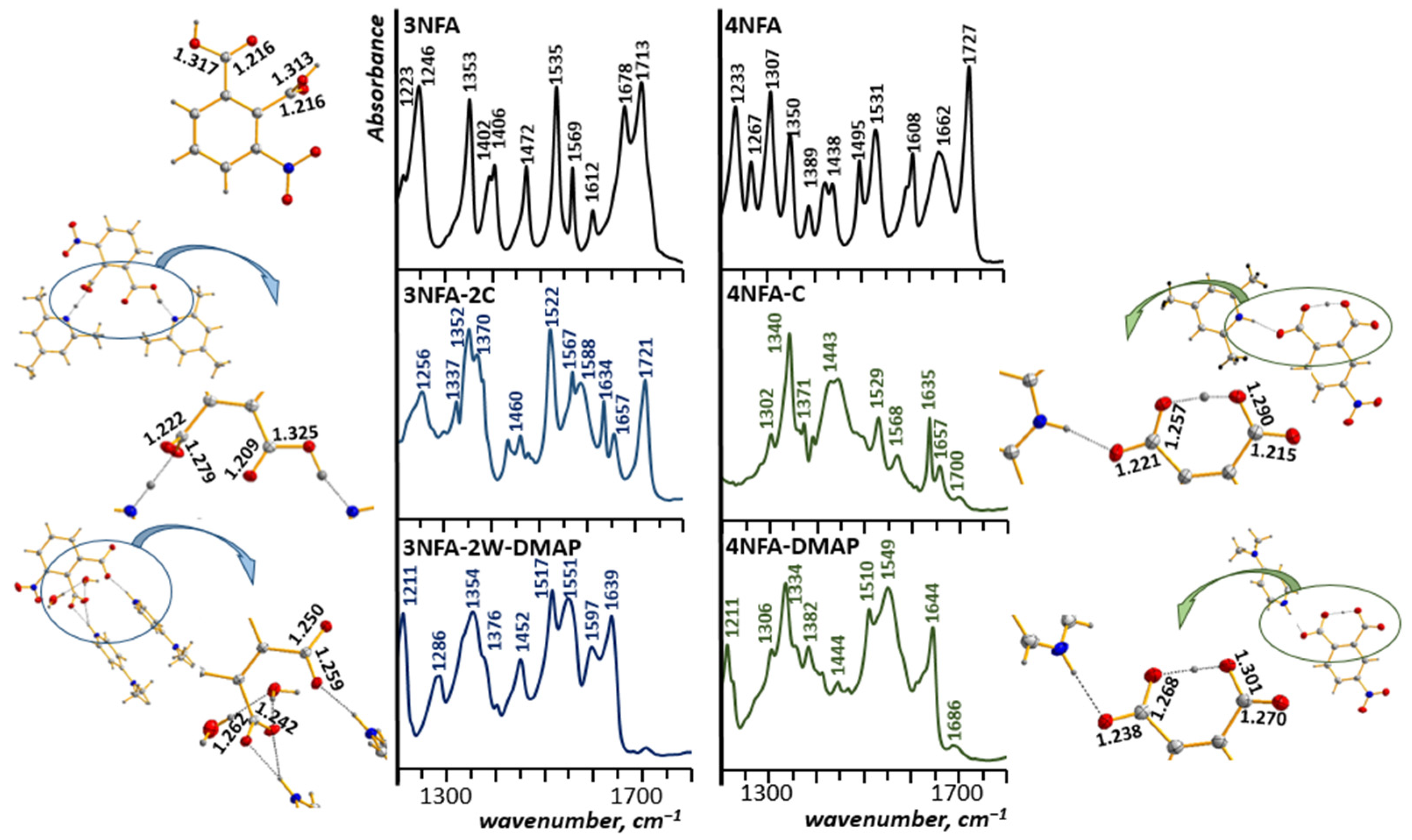
2.2. Infrared and Raman Spectra of the Studied Cocrystals
2.3. CP-MD Simulations of the Studied Complexes
3. Discussion
3.1. Structural Analysis of Hydrogen Bonds in Studied Cocrystals
3.2. Spectral Analysis of Hydrogen Bonds in Studied Cocrystals
3.2.1. Positions of the ν(C=O) and νas(CO2−) Bands vs. the Stoichiometry and Geometry of the Studied Cocrystals
3NFA-2C and 3NFA-2W-2DMAP Cocrystals vs. Their Spectra
4NFA-C and 4NFA-DMAP Cocrystals vs. Their Spectra
3.3. Potential Energy Curve Calculation for Proton Transfer in Hydrogen Bonds
3.4. CP-MD Simulations in Solid State Analysis of Hydrogen Bonds
4. Materials and Methods
4.1. Compounds and Solvent
4.2. Single Crystal X-ray Structure Determination of Complexes
4.3. Raman and Infrared Measurements
4.4. CP-MD in the Crystalline Phase and DFT Calculations
5. Conclusions
Author Contributions
Funding
Institutional Review Board Statement
Informed Consent Statement
Data Availability Statement
Acknowledgments
Conflicts of Interest
Appendix A

| Crystal Data | CCDC 22999111 (3NFA-2C) | CCDC 2301402 (3NFA-2W-2DMAP) | CCDC 2299110 (4NFA-C) | CCDC 2302801 (4NFA-DMAP) |
|---|---|---|---|---|
| Empirical formula | C24H27N3O6; C8H4NO6, C8H11N, C8H12N | C22H29N5O8; C8H3NO6, 2(C7H11N2), 2(H2O) | C16H16N2O6; C8H4NO6, C8H12N | C15H15N3O6; C8H4NO6, C7H11N2 |
| Formula weight | 453.48 | 491.50 | 332.31 | 333.30 |
| Temperature | 100(2) K | 100(2) K | 100(2) K | 100(2) K |
| Wavelength | 1.54184 Å | 0.71073 Å | 1.54184 Å | 0.71073 Å |
| Crystal system | Monoclinic | Triclinic | Orthorhombic | Triclinic |
| Space group | P 21/c (No.14) | P-1 (No.2) | Pnma (62) | P-1 (No.2) |
| Unit cell dimensions | a = 7.821(3) Å b = 41.778(3) Å c = 7.253(2) Å β = 109.47(3)° | a = 8.202(3) Å b = 11.125(3) Å c = 13.637(2) Å α = 70.93(4)° β = 85.62(3)° γ = 82.18(3)° | a = 15.8962(5) Å b = 6.6134(3) Å c = 14.6385(5) Å | a = 8.3181(3) Å b = 9.3553(3) Å c = 9.5025(4) Å α = 97.950(3)° β = 92.029(4)° γ = 93.273(3)° |
| Volume | 2234.4(11) Å3 | 1164.4(6) Å3 | 1538.92(10) Å3 | 730.47(5) Å3 |
| Z | 4 | 2 | 4 | 2 |
| Density (calculated) | 1.348 Mg/m3 | 1.402 Mg/m3 | 1.434 Mg/m3 | 1.515 Mg/m3 |
| Absorption coefficient | 0.809 mm−1 | 0.108 mm−1 | 0.941 mm−1 | 0.119 mm−1 |
| F (000) | 960 | 520 | 696 | 348 |
| Crystal size | 0.20 × 0.20 × 0.10 mm3 | 0.150 × 0.100 × 0.070 mm3 | 0.197 × 0.098 × 0.051 mm3 | 0.150 × 0.110 × 0.050 mm3 |
| Theta range for data collection | 2.115 to 73.241° | 1.581 to 28.938° | 4.105 to 73.021° | 2.166 to 28.924° |
| Reflections collected | 25595 | 19572 | 5533 | 9647 |
| Independent reflections | 4332 [R(int) = 0.0245] | 5571 [R(int) = 0.0361] | 1604 [R(int) = 0.0198] | 3419 [R(int) = 0.0334] |
| Completeness to theta | 67.684° to 98.8% | 1.581 to 28.938° | 67.684° to 99.8% | 2.166 to 28.924° |
| Refinement method | Full-matrix least-squares on F2 | Full-matrix least-squares on F2 | Full-matrix least-squares on F2 | Full-matrix least-squares on F2 |
| Data/restraints/parameters | 4332/0/308 | 5571/0/320 | 1604/0/145 | 3419/0/219 |
| Goodness-of-fit on F2 | 0.997 | 1.073 | 1.075 | 1.027 |
| Final R indices [I > 2sigma(I)] | R1 = 0.0643, wR2 = 0.1510 | R1 = 0.0457, wR2 = 0.1079 | R1 = 0.0449, wR2 = 0.1185 | R1 = 0.0487, wR2 = 0.1038 |
| R indices (all data) | R1 = 0.0665, wR2 = 0.1518 | R1 = 0.0695, wR2 = 0.1299 | R1 = 0.0520, wR2 = 0.1233 | R1 = 0.0709, wR2 = 0.1135 |
| Extinction coefficient | n/a | n/a | n/a | n/a |
| Largest diff. peak and hole | 0.317 and −0.343 e.Å−3 | 0.310 and −0.289 e.Å−3 | 0.236 and −0.238 e.Å−3 | 0.299 and −0.274 e.Å−3 |
References
- Pimentel, G.C.; McClellan, A.L. The Hydrogen Bond; Reinhold Pub. Corp.: New York, NY, USA, 1960. [Google Scholar]
- Jeffery, G.A.; Saenger, W. Hydrogen Bonding in Biological Structures; Springer: Berlin, Germany, 1991. [Google Scholar] [CrossRef]
- Maréchal, Y. The Hydrogen Bond and the Water Molecule: The Physics and Chemistry of Water, Aqueous and Bio Media; Elsevier: Amsterdam, The Netherlands, 2007. [Google Scholar] [CrossRef]
- Grabowski, S.J. Understanding Hydrogen Bonds: Theoretical and Experimental Views; RSC: Cambridge, UK, 2020. [Google Scholar]
- Wójcik, M.J.; Ozaki, Y. Spectroscopy and Computational of Hydrogen-Bonded Systems; Wiley-VCH GmbH: Weinheim, Germany, 2023. [Google Scholar] [CrossRef]
- Desiraju, G.R.; Steiner, T. The Weak Hydrogen Bond in Structural Chemistry and Biology; Oxford University Press: Oxford, UK, 1999. [Google Scholar]
- Schuster, P.; Zundel, G.; Sandorfy, C. The Hydrogen Bond; North-Holland: Amsterdam, The Netherlands, 1976. [Google Scholar]
- Hynes, J.T.; Klinman, J.P.; Limbach, H.-H.; Schowen, R.L. Hydrogen-Transfer Reactions; Wiley-VCH Verlag GmbH & Co. KGaA: Weinheim, Germany, 2007. [Google Scholar] [CrossRef]
- Lynden-Bell, R.M.; Morris, S.C.; Barrow, J.D.; Finney, J.L.; Harper, C.L., Jr. Water and Life. The Unique Properties of H2O; CRC Press: Boca Raton, FL, USA; Taylor & Francis Group: London, UK, 2010. [Google Scholar]
- Antonov, L. Tautomerism: Concepts and Applications in Science and Technology; Wiley-VCH Verlag GmbH & Co. KGaA: Weinheim, Germany, 2016. [Google Scholar]
- Gilli, G.; Gilli, P. The Nature of the Hydrogen Bond; Oxford University Press: Oxford, UK, 2009. [Google Scholar] [CrossRef]
- Scheiner, S. Hydrogen Bonding: A Theoretical Perspective; Oxford University Press: Oxford, UK, 1997. [Google Scholar] [CrossRef]
- Kohen, A.; Limbach, H.H. Isotope Effects in Chemistry and Biology; CRC Press: Boca Raton, FL, USA, 2006. [Google Scholar] [CrossRef]
- Pihko, P.M. Hydrogen Bonding in Organic Synthesis; Wiley-VCH Verlag GmbH & Co. KGaA: Weinheim, Germany, 2009. [Google Scholar] [CrossRef]
- Vladilo, G.; Hassanali, A. Hydrogen Bonds and Life in the Universe. Life 2018, 8, 1. [Google Scholar] [CrossRef] [PubMed]
- Speakman, J.C. Acid salts of carboxylic acids, crystals with some “very short” hydrogen bonds. In Progress in Theory, Struct. Bond; Herigonte, P.v., Smith, D.W., Mayer, U., Gutmann, V., Speakman, J.C., Harnung, S.E., Schäffer, C.E., Eds.; Springer: Berlin/Heidelberg, Germany, 1972; Volume 12, pp. 141–199. [Google Scholar] [CrossRef]
- Speakman, J.C. Some “very short” hydrogen bonds. Chem. Commun. (Lond.) 1967, 32b–33. [Google Scholar] [CrossRef]
- Hadži, D. Infrared spectra of strongly hydrogen-bonded systems. Pure Appl. Chem. 1965, 11, 435–453. [Google Scholar] [CrossRef]
- Macdonald, A.L.; Speakman, J.C.; Hadži, D. Crystal structures of the acid salts of some monobasic acids. Part XIV. Neutron-diffraction studies of potassium hydrogen bis(trifluoroacetate) and potassium deuterium bis(trifluoroacetate): Crystals with short and symmetrical hydrogen bonds. J. Chem. Soc. Perkin Trans. 1972, 2, 825–832. [Google Scholar] [CrossRef]
- Cleland, W.W.; Kreevoy, M.M. Low-barrier hydrogen-bonds and enzymatic catalysis. Science 1994, 264, 1887–1890. [Google Scholar] [CrossRef] [PubMed]
- Cleland, W.W.; Frey, P.A.; Gerlt, J.A. The low barrier hydrogen bond in enzymatic catalysis. J. Biol. Chem. 1998, 273, 25529–25532. [Google Scholar] [CrossRef] [PubMed]
- Hur, O.; Leja, C.; Dunn, M. Evidence of a low-barrier hydrogen bond in the tryptophan synthase catalytic mechanism. Biochemistry 1996, 35, 7378–7386. [Google Scholar] [CrossRef]
- Wu, Z.R.; Ebrahimian, S.; Zawrotny, M.E.; Thornburg, L.D.; Perez-Alvarado, G.C.; Brothers, P.; Pollack, R.M.; Summers, M.F. Solution Structure of 3-Oxo-Δ5-Steroid Isomerase. Science 1997, 276, 415. [Google Scholar] [CrossRef]
- Fersht, A.R.; Shi, J.-P.; Knill-Jones, J.; Lowe, D.M.; Wilkinson, A.J.; Blow, D.M.; Brick, P.; Carter, P.; Waye, M.M.Y.; Winter, G. Hydrogen bonding and biological specificity analysed by protein engineering. Nature 1985, 314, 235–238. [Google Scholar] [CrossRef]
- Yamaguchi, S.; Kamikubo, H.; Kurihara, K.; Kuroki, R.; Niimura, N.; Shimizu, N.; Yamazaki, Y.; Kataoka, M. Low-barrier hydrogen bond in photoactive yellow protein. Proc. Natl. Acad. Sci. USA 2009, 106, 440–444. [Google Scholar] [CrossRef]
- Chakalov, E.R.; Shekurov, R.P.; Miluykov, V.A.; Tolstoy, P.M. Evidence of extremely short hydrogen bond in the homoconjugated ferrocene-1,1′-diyl-bisphosphinic acid anion: Sign change of the H/D isotope effect on the 31P NMR chemical shift. Phys. Chem. Chem. Phys. 2023, 25, 29486–29495. [Google Scholar] [CrossRef] [PubMed]
- Tupikina, E.Y.; Sigalov, M.V.; Alkhuder, O.; Tolstoy, P.M. Charge Relay Without Proton Transfer: Coupling of Two Short Hydrogen Bonds via Imidazole in Models of Catalytic Triad of Serine Protease Active Site. Chem. Phys. Chem. 2023, 25, e202300970. [Google Scholar] [CrossRef] [PubMed]
- Sigalov, M.; Shainyan, B.; Krief, P.; Ushakov, I.; Chipanina, N.; Oznobikhina, L. Intramolecular interactions in dimedone and phenalen-1,3-dione adducts of 2(4)-pyridinecarboxaldehyde: Enol–enol and ring-chain tautomerism, strong hydrogen bonding, zwitterions. J. Mol. Struct. 2011, 1006, 234–246. [Google Scholar] [CrossRef]
- Wilson, C.C.; Thomas, L.H.; Morrison, C.A. A symmetric hydrogen bond revisited: Potassium hydrogen maleate by variable temperature, variable pressure neutron diffraction and plane-wave DFT methods. Chem. Phys. Lett. 2003, 381, 102–108. [Google Scholar] [CrossRef]
- Steiner, T.; Majerz, I.; Wilson, C.C. First O-H-N Hydrogen Bond with a Centered Proton Obtained by Thermally Induced Proton Migration. Angew. Chem. Int. Ed. 2001, 40, 2651. [Google Scholar] [CrossRef]
- Schiøtt, B.; Iversen, B.B.; Madsen, G.K.H.; Bruice, T.C. Characterization of the short strong hydrogen bond in benzoylacetone by ab initio calculations and accurate diffraction experiments. Implications for the electronic nature of low-barrier hydrogen bonds in enzymatic reactions. J. Am. Chem. Soc. 1998, 120, 12117–12124. [Google Scholar] [CrossRef]
- Wilson, C.C. Interesting proton behaviour in molecular structures. Variable temperature neutron diffraction and ab initio study of acetylsalicylic acid: Characterising librational motions and comparing protons in different hydrogen bonding potentials. New J. Chem. 2002, 26, 1733–1739. [Google Scholar] [CrossRef]
- Wozniak, K.; Mallinson, P.R.; Smith, G.T.; Wilson, C.C.; Grech, E. Role of C—H O hydrogen bonds in the ionic complexes of 1,8-bis(dimethylamino)naphthalene. J. Phys. Org. Chem. 2003, 16, 764–771. [Google Scholar] [CrossRef]
- Schiøtt, B.; Iversen, B.B.; Madsen, G.K.H.; Larsen, F.K.; Bruice, T.C. On the electronic nature of low-barrier hydrogen bonds in enzymatic reactions. Proc. Natl. Acad. Sci. USA 1998, 95, 12799–12802. [Google Scholar] [CrossRef]
- Vishweshwar, P.; Jagadeesh Babu, N.; Nangia, A.; Mason, S.A.; Puschmann, H.; Mondal, R.; Howard, J.A.K. Variable Temperature Neutron Diffraction Analysis of a Very Short O−H···O Hydrogen Bond in 2,3,5,6-Pyrazinetetracarboxylic Acid Dihydrate: Synthon-Assisted Short Oacid−H···Owater Hydrogen Bonds in a Multicenter Array. J. Phys. Chem. A 2004, 108, 9406–9416. [Google Scholar] [CrossRef]
- Parkin, A.; Wozniak, K.; Wilson, C.C. From Proton Disorder to Proton Migration: A Continuum in the Hydrogen Bond of a Proton Sponge in the Solid State. Cryst. Grow. Des. 2007, 7, 1393–1398. [Google Scholar] [CrossRef]
- Takusagawa, F.; Koetzle, T.F. Neutron diffraction study of quinolinic acid recrystallized from D2O: Evaluation of temperature and isotope effects in the structure. Acta Crystallogr. 1979, B35, 2126–2135. [Google Scholar] [CrossRef]
- D’Ascenzo, L.; Auffinger, P. A comprehensive classification and nomenclature of carboxyl–carboxyl(ate) supramolecular motifs and related catemers: Implications for biomolecular systems. Acta Crystallogr. 2015, B71, 164–175. [Google Scholar] [CrossRef]
- Aakeroy, C.B. Crystal Engineering: Strategies and Architectures. Acta Crystallogr. 1997, B53, 569–586. [Google Scholar] [CrossRef]
- Saunders, L.K.; Nowell, H.; Hatcher, L.E.; Shepherd, H.J.; Teat, S.J.; Allan, D.R.; Raithby, P.R.; Wilson, C.C. Exploring short strong hydrogen bonds engineered in organic acid molecular crystals for temperature dependent proton migration behaviour using single crystal synchrotron X-ray diffraction (SCSXRD). CrystEngComm 2019, 21, 5249–5260. [Google Scholar] [CrossRef]
- Lorente, P.; Shenderovich, I.G.; Buntkowsky, G.; Golubev, N.S.; Denisov, G.S.; Limbach, H.-H. 1H/15N NMR chemical shielding, dipolar 15N,2H coupling and hydrogen bond geometry correlations in a novel series of hydrogen bonded acid-base complexes of collidine with carboxylic acids. Magn. Reson. Chem. 2001, 39, S18–S29. [Google Scholar] [CrossRef]
- Tolstoy, P.M.; Schah-Mohammedi, P.; Smirnov, S.N.; Golubev, N.S.; Denisov, G.S.; Limbach, H.-H. Characterization of Fluxional Hydrogen Bonded Complexes of Acetic Acid and Acetate by NMR: Geometries, Isotope and Solvent Effects. J. Am. Chem. Soc. 2004, 126, 5621–5634. [Google Scholar] [CrossRef]
- Tolstoy, P.M.; Smirnov, S.N.; Shenderovich, I.G.; Golubev, N.S.; Denisov, G.S.; Limbach, H.-H. NMR Studies of Solid State-Solvent and H/D Isotope Effects on Hydrogen Bond Geometries of 1:1 Complexes of Collidine with Carboxylic Acids. J. Mol. Struct. 2004, 700, 19–27. [Google Scholar] [CrossRef]
- Andreeva, D.V.; Ip, B.; Gurinov, A.; Tolstoy, P.M.; Denisov, G.S.; Shenderovich, I.G.; Limbach, H.-H. Geometrical features of hydrogen bonded complexes involving sterically hindered pyridines. J. Phys. Chem. A 2006, 110, 10872–10879. [Google Scholar] [CrossRef]
- Tolstoy, P.M.; Guo, J.; Koeppe, B.; Golubev, N.S.; Denisov, G.S.; Smirnov, S.N.; Limbach, H.-H. Geometries and Tautomerism of OHN Hydrogen Bonds in Polar Solution probed by H/D Isotope Effects on 13C NMR Chemical Shifts. J. Phys. Chem. A 2010, 114, 10775–10782. [Google Scholar] [CrossRef]
- Pylaeva, S.; Allolio, C.; Koeppe, B.; Denisov, G.S.; Limbach, H.-H.; Sebastiani, D.; Tolstoy, P.M. Proton transfer in a short hydrogen bond caused by solvation shell fluctuations: An ab initio MD and NMR/UV study of an (OHO)-bonded system. Phys. Chem. Chem. Phys. 2015, 17, 4634–4644. [Google Scholar] [CrossRef]
- Koeppe, B.; Pylaeva, S.A.; Allolio, C.; Sebastiani, D.; Nibbering, E.T.J.; Denisov, G.S.; Limbach, H.-H.; Tolstoy, P.M. Polar solvent fluctuations drive proton transfer in hydrogen bonded complexes of carboxylic acid with pyridines: NMR, IR and ab initio MD study. Phys. Chem. Chem. Phys. 2017, 19, 1010–1028. [Google Scholar] [CrossRef] [PubMed]
- Frantsuzov, I.; Johnson, M.R.; Trommsdorff, H.P.; Horsewill, A.J. Proton Tunnelling in the Hydrogen Bonds of the Benzoic Acid Dimer: 18O Substitution and Isotope Effects of the Heavy Atom Framework. J. Phys. Chem. B 2014, 118, 7777–7784. [Google Scholar] [CrossRef]
- Huyskens, P.L.; Zeegers-Huyskens, T. Molecular Associations and Acid-Base Equilibriums. J. Chim. Phys. Phys.-Chim. Biol. 1964, 61, 81–86. [Google Scholar] [CrossRef]
- Gilli, P.; Pretto, L.; Bertolasi, V.; Gilli, G. Predicting hydrogen bond strengths from acid-base molecular properties. The pKa slide rule: Toward the solution of a long-lasting problem. Acc. Chem. Res. 2009, 42, 33–44. [Google Scholar] [CrossRef]
- Bhogala, B.R.; Basavoju, S.; Nangia, S. Tape and layer structures in cocrystals of some di- and tricarboxylic acids with 4,4′-bipyridines and isonicotinamide. From binary to ternary cocrystals. CrystEngComm 2005, 7, 551–562. [Google Scholar] [CrossRef]
- Cruz-Cabeza, A.J.; Lusi, M.; Wheatcroft, H.P.; Bond, A.D. The role of solvation in proton transfer reactions: Implications for predicting salt/co-crystal formation using the ΔpKa rule. Faraday Discuss. 2022, 235, 446–466. [Google Scholar] [CrossRef] [PubMed]
- Cruz-Cabeza, A.J. Acid–base crystalline complexes and the pKa rule. CrystEngComm 2012, 14, 6362–6365. [Google Scholar] [CrossRef]
- Jóźwiak, K.; Jezierska, A.; Panek, J.J.; Goremychkin, E.A.; Tolstoy, P.M.; Shenderovich, I.G.; Filarowski, A. Inter- vs. intra-molecular hydrogen bond patterns and proton dynamics in phthalic acid associates. Molecules 2020, 25, 4720. [Google Scholar] [CrossRef]
- Jóźwiak, K.; Jezierska, A.; Panek, J.J.; Kochel, A.; Filarowski, A. Inter- vs. Intra-Molecular Hydrogen Bond in Complexes of Nitrophthalic Acids with Pyridine. Int. J. Mol. Sci. 2023, 24, 5248. [Google Scholar] [CrossRef]
- Perrin, D.D. Dissociation Constants of Organic Bases in Aqueous Solutions; Butterworths: London, UK, 1972. [Google Scholar]
- Essery, J.M.; Schofield, K. 769. The influence of steric factors on the properties of 4-aminopyridine derivatives. J. Chem. Soc. 1961, 3939–3953. [Google Scholar] [CrossRef]
- McKinnon, J.J.; Spackman, M.A.; Mitchell, A.S. Novel tools for visualizing and exploring intermolecular interactions in molecular crystals. Acta Crystallogr. 2004, B60, 627–668. [Google Scholar] [CrossRef] [PubMed]
- Glidewell, C.; Low, J.N.; Skakle, J.M.S.; Wardell, J.L. 3-Nitrophthalic acid: C(4) and R22(8) motifs of O-H⋯O hydrogen bonds generate sheets which are linked by C-H⋯O hydrogen bonds. Acta Crystallogr. 2003, C59, o144–o146. [Google Scholar] [CrossRef]
- Smith, G.; Wermuth, U.D.; Young, D.J.; White, J.M. The 1:1 proton-transfer compounds of 4-(phenyldiazenyl)aniline (aniline yellow) with 3-nitrophthalic, 4-nitrophthalic and 5-nitroisophthalic acids. Acta Crystallogr. 2008, C64, o123–o127. [Google Scholar] [CrossRef] [PubMed]
- Smith, G.; Wermuth, U.D. Proton-transfer compounds of isonipecotamide with the aromatic dicarboxylic acids 4-nitrophthalic, 4,5-dichlorophthalic, 5-nitroisophthalic and terephthalic acid. Acta Crystallogr. 2011, C67, o259–o264. [Google Scholar] [CrossRef] [PubMed]
- Filatova, E.A.; Gulevskaya, A.V.; Pozharskii, A.F.; Ermolenko, E.A.; Ozeryanskii, V.A.; Misharev, A.D. Synthesis of 2-Aryl- and 2,7-Diaryl-1,8-bis(dimethylamino)naphthalenes. Overview of the “Buttressing effect” in 2,7-Disubstituted Proton Sponges. ChemistrySelect 2020, 5, 9932–9945. [Google Scholar] [CrossRef]
- Pozharskii, A.F.; Ryabtsova, O.V.; Ozeryanskii, V.A.; Degtyarev, A.V.; Kazheva, O.N.; Alexandrov, G.G.; Dyachenko, O.A. Organometallic Synthesis, Molecular Structure, and Coloration of 2,7-Disubstituted 1,8-Bis(dimethylamino)naphthalenes. How Significant Is the Influence of “Buttressing Effect” on Their Basicity? J. Org. Chem. 2003, 68, 10109–10122. [Google Scholar] [CrossRef] [PubMed]
- Ozeryanskii, V.A.; Marchenko, A.V.; Pozharskii, A.F.; Filarowski, A.; Spiridonova, D.V. Combination of “buttressing” and “clothespin” effects for reaching the shortest NHN hydrogen bond in proton sponge cations. J. Org. Chem. 2021, 86, 3637–3647. [Google Scholar] [CrossRef]
- Buemi, G.; Zuccarello, F. Importance of steric effect on the hydrogen bond strength of malondialdehyde and acetylacetone 3-substituted derivatives. An ab initio study. Electron. J. Theoret. Chem. 1997, 2, 302–314. [Google Scholar] [CrossRef]
- Kwocz, A.; Panek, J.J.; Jezierska, A.; Hetmańczyk, Ł.; Pawlukojć, A.; Kochel, A.; Lipkowski, P.; Filarowski, A. A molecular roundabout: Triple cycle-arranged hydrogen bonds in light of experiment and theory. New J. Chem. 2018, 42, 19467–19477. [Google Scholar] [CrossRef]
- Martyniak, A.; Panek, J.J.; Jezierska-Mazzarello, A.; Filarowski, A. Triple hydrogen bonding in a circular arrangement: Ab initio, DFT and first-principles MD studies of tris-hydroxyaryl enamines. J. Comp.-Aided Mol. Des. 2012, 9, 1045–1053. [Google Scholar] [CrossRef]
- Bolvig, S.; Wozniak, K.; Hansen, P.E. Steric compression effects of intramolecularly hydrogen bonded o-hydroxy acyl aromatics. An X-ray and 13C-NMR study. J. Mol. Struct. 2005, 749, 155–168. [Google Scholar] [CrossRef]
- Hansen, P.E.; Spanget-Larsen, J. NMR and IR Investigations of Strong Intramolecular Hydrogen Bonds. Molecules 2017, 22, 552. [Google Scholar] [CrossRef]
- Hansen, P.E.; Ibsen, S.N.; Kristensen, T.; Bolvig, S. Deuterium and 18O isotope effects on 13C chemical shifts of sterically hindered and/or intra-molecularly hydrogen-bonded o-hydroxy acyl aromatics. Magn. Res. Chem. 1994, 32, 399–408. [Google Scholar] [CrossRef]
- Filarowski, A.; Koll, A.; Kochel, A.; Kalenik, J.; Hansen, P.E. The intramolecular hydrogen bond in ortho-hydroxy acetophenones. J. Mol. Struct. 2004, 700, 67–72. [Google Scholar] [CrossRef]
- Majewska, P.; Pająk, J.; Rospenk, M.; Filarowski, A. Intra- versus intermolecular hydrogen bonding equilibrium in 2-hydroxy-N,N-diethylbenzamide. J. Phys. Org. Chem. 2009, 22, 130–137. [Google Scholar] [CrossRef]
- Novak, A. Hydrogen bonding in solids correlation of spectroscopic and crystallographic data. Struct. Bond. 1974, 18, 177–216. [Google Scholar]
- Marechal, Y.; Durig, J. Vibration Spectra and Structure; Elsevier: Amsterdam, The Netherland, 1997. [Google Scholar]
- Iogansen, A.V. Direct proportionality of the hydrogen bonding energy and the intensification of the stretching v(XH) vibration in infrared spectra. Spectrochim. Acta A 1999, 55, 1585–1612. [Google Scholar] [CrossRef]
- Rozenberg, M.S. IR spectra and hydrogen bond energies of crystalline acid salts of carboxylic acids. Spectrochim. Acta A 1996, 52, 1559–1563. [Google Scholar] [CrossRef]
- Howard, J.; Tomkinson, J.; Eckert, J.; Goldstone, J.A.; Taylor, A.D. Inelastic neutron scattering studies of some intramolecular hydrogen bonded complexes: A new correlation of γ(OHO) vs. R (OO). J. Chem. Phys. 1983, 78, 3150–3155. [Google Scholar] [CrossRef]
- Jóźwiak, K.; Jezierska, A.; Panek, J.J.; Łydżba-Kopczyńska, B.; Filarowski, A. Renewed spectroscopic and theoretical research of hydrogen bonding in ascorbic acid. Spectrochim. Acta A 2024, 320, 124585. [Google Scholar] [CrossRef]
- Dega-Szafran, Z.; Dulewicz, E. Infrared and 1H nuclear magnetic resonance studies of hydrogen bonds in some pyridine trifluoroacetates and their deuteriated analogues in dichloromethane. J. Chem. Soc. Perkin Trans. 2 1983, 3, 345–351. [Google Scholar] [CrossRef]
- Barczyński, P.; Dega-Szafran, Z.; Szafran, M. Spectroscopic differences between molecular (O–H⋯N) and ionic pair (O−⋯H–N+) hydrogen complexes. J. Chem. Soc. Perkin Trans. 1985, 2, 765–771. [Google Scholar] [CrossRef]
- Gołdyn, M.; Bartoszak-Adamska, E.; Skowronek, J.; Komasa, A.; Lewandowska, A.; Dega-Szafran, Z.; Cofta, G. Synthesis and structural characteristic of pyridine carboxylic acid adducts with squaric acid. CrystEngComm 2022, 24, 7821–7832. [Google Scholar] [CrossRef]
- Hunger, L.; Al Sheakh, L.; Fritsch, S.; Villinger, A.; Ludwig, R.; Harville, P.; Moss, O.; Lachowicz, A.; Johnson, M.A. Spectroscopic Evidence for Doubly Hydrogen-Bonded Cationic Dimers in the Solid, Liquid, and Gaseous Phases of Carboxyl-Functionalized Ionic Liquids. J. Phys. Chem. B 2024, 128, 5463–5471. [Google Scholar] [CrossRef]
- Hunger, L.; Al-Sheakh, L.; Zaitsau, D.H.; Verevkin, S.P.; Appelhagen, A.; Villinger, A.; Ludwig, R. Dissecting Noncovalent Interactions in Carboxyl-Functionalized Ionic Liquids Exhibiting Double and Single Hydrogens Bonds Between Ions of Like Charge. Chem. Eur. J. 2022, 28, e202200949. [Google Scholar] [CrossRef]
- Zięba, S.; Mizera, A.; Markiewicz, K.H.; Dubis, A.T.; Ławniczak, P.; Gzella, A.; Siergiejczyk, L.; Łapiński, A. Effect of Azole Counterions on Thermal and Transport Properties of the Hydrated Salts of Hemimelitic Acid. J. Phys. Chem. C 2023, 127, 24403–24410. [Google Scholar] [CrossRef]
- Flakus, H.T.; Hachuła, B. The source of similarity of the IR spectra of acetic acid in the liquid and solid-state phases. Vib. Spectrosc. 2011, 56, 170–176. [Google Scholar] [CrossRef]
- Flakus, H.T.; Hachuła, B.; Hołaj-Krzak, J.T.; Al-Agel, F.A.; Rekik, N. “Long-distance” H/D isotopic self-organization phenomena in scope of the infrared spectra of hydrogen-bonded terephthalic and phthalic acid crystals. Spectrochim. Acta A 2017, 173, 65–74. [Google Scholar] [CrossRef]
- Flakus, H.T.; Hachuła, B.; Hołaj-Krzak, J.T. Long-distance inter-hydrogen bond coupling effects in the polarized IR spectra of succinic acid crystals. Spectrochim. Acta A 2015, 142, 126–134. [Google Scholar] [CrossRef]
- Vener, M.V.; Kuhn, O.; Bowman, J.M. Vibrational spectrum of the formic acid in the OH stretch region. A model 3D study. Chem. Phys. Lett. 2001, 349, 562–570. [Google Scholar] [CrossRef]
- Fillaux, F.; Limage, M.H.; Romain, F. Quantum proton transfer and interconversion in the benzoic acid crystal: Vibrational spectra, mechanism and theory. Chem. Phys. 2002, 276, 181–210. [Google Scholar] [CrossRef]
- Bournay, J.; Marechal, Y. Anomalous isotope effect in the H bonds of acetic acid dimers. J. Chem. Phys. 1973, 59, 5077–5087. [Google Scholar] [CrossRef]
- Issaoui, N.; Rekik, N.; Oujia, B.; Wójcik, M.J. Theoretical Infrared Line Shapes of H-Bonds within the Strong Anharmonic Coupling Theory. Fermi Resonances Effects. Int. J. Quant. Chem. 2010, 110, 2583–2602. [Google Scholar] [CrossRef]
- Rodziewicz, P.; Doltsinis, N.L. Formic Acid Dimerization: Evidence for Species Diversity from First Principles Simulations. J. Phys. Chem. A 2009, 113, 6266–6274. [Google Scholar] [CrossRef] [PubMed]
- Zundel, G. Easily Polarizable Hydrogen Bonds—Their Interactions with the Environment—IR Continuum and Anomalous Large Conductivity. In The Hydrogen Bond: Recent Developments in Theory and Experiments; Schuster, P., Zundel, G., Sandorfy, C., Eds.; North-Holland: Amsterdam, The Netherland, 1976; Volume 2, pp. 683–766. [Google Scholar]
- Badger, R.M.; Bauer, S.H. Spectroscopic Studies of the Hydrogen Bond. II. The Shift of the O–H Vibrational Frequency in the Formation of the Hydrogen Bond. J. Chem. Phys. 1937, 5, 839–851. [Google Scholar] [CrossRef]
- Haurie, M.; Novak, A.J. Étude par spectroscopie infrarouge et Raman des complexes de l’acide acétique avec des accepteurs de proton. Chim. Phys. 1967, 64, 679–687. [Google Scholar] [CrossRef]
- Gusakova, G.V.; Denisov, G.S.; Smolyanskii, A.L. Spectroscopic investigation of the reaction of acetic and isobutyric acids with tertiary amines. J. Appl. Spectrosc. 1972, 17, 1321–1325. [Google Scholar] [CrossRef]
- Gusakova, G.V.; Denisov, G.S.; Smolyanskii, A.L. A spectroscopic study of the interaction of isobutyric acid with pyridine and dioxan. J. Appl. Spectrosc. 1971, 14, 628–632. [Google Scholar] [CrossRef]
- Perrin, C.L.; Nielson, J.B. Asymmetry of Hydrogen Bonds in Solutions of Monoanions of Dicarboxylic Acids. J. Am. Chem. Soc. 1997, 119, 12734–12741. [Google Scholar] [CrossRef]
- Vener, M.V.; Shenderovich, I.G.; Rykounov, A.A. A qualitative study of the effect of a counterion and polar environment on the structure and spectroscopic signatures of a hydrated hydroxyl anion. Theor. Chem. Acc. 2013, 132, 1361. [Google Scholar] [CrossRef]
- Perrin, C.L. Are Short, Low-Barrier Hydrogen Bonds Unusually Strong? Acc. Chem. Res. 2010, 43, 1550–1557. [Google Scholar] [CrossRef] [PubMed]
- Shenderovich, I.G. Actual Symmetry of Symmetric Molecular Adducts in the Gas Phase, Solution and in the Solid State. Symmetry 2021, 13, 756. [Google Scholar] [CrossRef]
- Cook, J.L.; Hunter, C.A.; Low, C.M.R.; Perez-Velasco, A.; Vinter, J.G. Solvent Effects on Hydrogen Bonding. Angew. Chem. Int. Ed. 2007, 46, 3706–3709. [Google Scholar] [CrossRef] [PubMed]
- Martyniak, A.; Majerz, I.; Filarowski, A. Peculiarities of quasi-aromatic hydrogen bonding. RSC Adv. 2012, 2, 8135–8144. [Google Scholar] [CrossRef]
- Yi, X.; Chen, W.; Xiao, Y.; Liu, F.; Yu, X.; Zheng, A. Spectroscopically Visualizing the Evolution of Hydrogen-Bonding Interactions. J. Am. Chem. Soc. 2023, 145, 27471–27479. [Google Scholar] [CrossRef]
- Majerz, I. Proton Transfer Influence on Geometry and Electron Density in Benzoic Acid-Pyridine Complexes. Hevl. Chim. Acta 2016, 99, 286–295. [Google Scholar] [CrossRef]
- Majerz, I.; Gutmann, M. Intermolecular OHN hydrogen bond with a proton moving in 3-methylpyridinium 2,6-dichloro-4-nitrophenolate. RSC Adv. 2015, 5, 95576–95584. [Google Scholar] [CrossRef]
- Majerz, I.; Gutmann, M. Mechanism of proton transfer in the strong OHN intermolecular hydrogen bond. RSC Adv. 2011, 1, 219–228. [Google Scholar] [CrossRef]
- Barnes, A.J.; Legon, A.C. Proton transfer in amine hydrogen halide complexes: Comparison of low temperature matrices with the gas phase. J. Mol. Struct. 1998, 448, 101–106. [Google Scholar] [CrossRef]
- Andrews, L.; Wang, X.; Mielke, Z. Infrared Spectrum of the H3N-HCl Complex in Solid Ne, Ne/Ar, Ar, and Kr. Matrix Effects on a Strong Hydrogen-Bonded Complex. J. Phys. Chem. A 2001, 105, 6054–6064. [Google Scholar] [CrossRef]
- Marx, D.; Tuckerman, M.E.; Hutter, J.; Parrinello, M. The nature of the hydrated excess proton in water. Nature 1999, 397, 601–604. [Google Scholar] [CrossRef]
- Tuckerman, M.; Marx, D.; Klein, M.L.; Parrinello, M. On the Quantum Nature of the Shared Proton in Hydrogen Bonds. Science 1997, 275, 817–820. [Google Scholar] [CrossRef] [PubMed]
- Dopieralski, P.D.; Latajka, Z.; Olovsson, I. Proton Transfer Dynamics in Crystalline Maleic Acid from Molecular Dynamics Calculations. J. Chem. Theory Comput. 2010, 6, 1455–1461. [Google Scholar] [CrossRef] [PubMed]
- Neumann, M.A.; Craciun, S.; Corval, A.; Johnson, M.R.; Horsewil, A.J.; Benderskii, V.A.; Trommsdorff, H.P. Proton Dynamics and the Tautomerization Potential in Benzoic Acid Crystals. Ber. Busenges. Phys. Chem. 1998, 102, 325–334. [Google Scholar] [CrossRef]
- Udagawa, T.; Tanaka, H.; Kuwahata, K.; Tachikawa, M. Location of the Shared Proton in Proton-Bound Dimer Compound of Hydrogen Sulfate and Formate: Path Integral Molecular Dynamics Study. J. Phys. Chem. A 2024, 128, 2103–2110. [Google Scholar] [CrossRef] [PubMed]
- Brela, M.; Stare, J.; Pirc, G.; Sollner-Dolenc, M.; Boczar, M.; Wójcik, M.J.; Mavri, J. Car−Parrinello Simulation of the Vibrational Spectrum of a Medium Strong Hydrogen Bond by Two-Dimensional Quantization of the Nuclear Motion: Application to 2-Hydroxy-5-nitrobenzamide. J. Phys. Chem. B 2012, 116, 4510–4518. [Google Scholar] [CrossRef] [PubMed]
- Stare, J.; Panek, J.; Eckert, J.; Grdadolnik, J.; Mavri, J.; Hadži, D. Proton Dynamics in the Strong Chelate Hydrogen Bond of Crystalline Picolinic Acid N-Oxide. A New Computational Approach and Infrared, Raman and INS Study. J. Phys. Chem. 2008, 112, 1576–1586. [Google Scholar] [CrossRef] [PubMed]
- Brela, M.Z.; Wójcik, M.J.; Boczar, M.; Witek, Ł.; Yasuda, M.; Ozaki, Y. Car–Parrinello Molecular Dynamics Simulations of Infrared Spectra of Crystalline Vitamin C with Analysis of Double Minimum Proton Potentials for Medium-Strong Hydrogen Bonds. J. Phys. Chem. B 2015, 119, 7922–7930. [Google Scholar] [CrossRef] [PubMed]
- Rikagu Oxford Diffraction. CrysAlisPro; Agilent Technologies Inc.: Yarnton, Oxfordshire, UK, 2018. [Google Scholar]
- Sheldrick, G.M. SHELXT—Integrated space-group and crystal structure determination. Acta Crystallogr. 2015, A71, 3–8. [Google Scholar] [CrossRef]
- Sheldrick, G.M. Crystal Structure Refinement with SHELXL. Acta Crystallogr. 2015, C71, 3–8. [Google Scholar] [CrossRef]
- Diamond—Crystal and Molecular Structure Visualization. Crystal Impact—Dr. H. Putz & Dr. K. Brandenburg GbR, Germany. Available online: https://www.crystalimpact.com/diamond (accessed on 20 January 2023).
- Frisch, M.J.; Trucks, G.W.; Schlegel, H.B.; Scuseria, G.E.; Robb, M.A.; Cheeseman, J.R.; Scalmani, G.; Barone, V.; Petersson, G.A.; Nakatsuji, H.; et al. Gaussian 16, Revision C.01; Gaussian, Inc.: Wallingford, CT, USA, 2016. [Google Scholar]
- Becke, A.D. Density-functional thermochemistry. III. The role of exact exchange. J. Chem. Phys. 1993, 98, 5648–5652. [Google Scholar] [CrossRef]
- Lee, C.; Yang, W.; Parr, R.G. Development of the Colle-Salvetti Correlation-Energy Formula into a Functional of the Electron Density. Phys. Rev. 1993, 37, B785–B789. [Google Scholar] [CrossRef] [PubMed]
- Frisch, M.J.; Pople, J.A.; Binkley, J.S. Self-consistent molecular orbital methods 25. Supplementary functions for Gaussian basis sets. J. Chem. Phys. 1984, 80, 3265–3269. [Google Scholar] [CrossRef]
- Grimme, S. Semiempirical GGA-type density functional constructed with a long-range dispersion correction. J. Comput. Chem. 2006, 27, 1787–1799. [Google Scholar] [CrossRef] [PubMed]
- Schaftenaar, G.; Noordik, J.H. Molden: A pre- and post-processing program for molecular and electronic structures. J. Comput. Aided Mol. Des. 2000, 14, 123–134. [Google Scholar] [CrossRef] [PubMed]
- CPMD 4.3, Copyright IBM Corp. (1990–2019) Copyright MPI für Festkoerperforschung Stuttgart (1997–2001). Available online: http://www.cpmd.org (accessed on 12 September 2022).
- Perdew, J.P.; Ernzerhof, M.; Burke, K. Rationale for mixing exact exchange with density functional approximations. J. Chem. Phys. 1996, 105, 9982–9985. [Google Scholar] [CrossRef]
- Troullier, N.; Martins, J.L. Efficient pseudopotentials for plane-wave calculations. Phys. Rev. B 1991, 43, 1993–2006. [Google Scholar] [CrossRef]
- Nosé, S. A unified formulation of the constant temperature molecular dynamics methods. J. Chem. Phys. 1984, 81, 511–519. [Google Scholar] [CrossRef]
- Hoover, W.G. Canonical dynamics: Equilibrium phase-space distributions. Phys. Rev. A 1985, 31, 1695–1697. [Google Scholar] [CrossRef]
- Humphrey, W.; Dalke, A.; Schulten, K. Visual Molecular Dynamics. J. Mol. Graph. 1996, 14, 33–38. [Google Scholar] [CrossRef]
- Mercury—Crystal Structure Visualisation. Available online: http://www.ccdc.cam.ac.uk/Solutions/CSDSystem/Pages/Mercury.aspx (accessed on 15 September 2022).
- Williams, T.; Kelley, C. Gnuplot 4.4: An Interactive Plotting Program. 2010. Available online: http://www.gnuplot.info/docs_4.4/gnuplot.pdf (accessed on 12 September 2022).


| Cocrystal | D-H⋯A | Type of HB | d(D-H) | d(AH) | d(DA) | Θ(DHA) |
|---|---|---|---|---|---|---|
| 3NFA-2C | O(5)-H(5)⋯N(11) | inter | 1.07 | 1.60 | 2.653(3) | 164 |
| N(22)-H(22)⋯O(3) | - | 0.88 | 1.67 | 2.543(4) | 174 | |
| 3NFA-2W-2DMAP | O(1W)-H(1W)⋯O(2) | inter | 0.85 | 1.97 | 2.801(2) | 165 |
| O(1W)-H(2W)⋯O(4) | - | 0.85 | 1.90 | 2.730(2) | 167 | |
| O(2W)-H(3W)⋯O(1W) | - | 0.85 | 1.96 | 2.809(2) | 177 | |
| N(22)-H(22)⋯O(3) | - | 0.88 | 1.78 | 2.655(2) | 173 | |
| N(22)-H(22)⋯O(4) | - | 0.88 | 2.54 | 3.151(2) | 127 | |
| N(32)-H(32)⋯O(1) | - | 0.88 | 1.83 | 2.678(2) | 161 | |
| 4NFA-C | O(1)-H(1)⋯O(4) | intra | 1.35 | 1.07 | 2.410(2) | 171 |
| N(11)-H(11)⋯O(2) | inter | 0.88 | 1.78 | 2.654(3) | 169 | |
| 4NFA-DMAP | O(4)-H(5)⋯O(5) | intra | 1.32 | 1.10 | 2.409(1) | 169 |
| N(3)-H(3)⋯O(3) | inter | 0.86 | 1.93 | 2.761(2) | 163 |
| Cocrystal | Numbering C-O/C=O | Bond Distance d(C-O/C=O) |
|---|---|---|
| 3NFA-2C | C(8)-O(5) | 1.325 |
| C(8)=O(6) | 1.209 | |
| C(7)-O(3) | 1.279 | |
| C(7)=O(4) | 1.222 | |
| 3NFA-2W-2DMAP | C(8)-O(3) | 1.262 |
| C(8)=O(4) | 1.242 | |
| C(7)-O(1) | 1.259 | |
| C(7)=O(2) | 1.250 | |
| 4NFA-C | C(8)-O(4) | 1.290 |
| C(8)=O(3) | 1.215 | |
| C(7)-O(1) | 1.257 | |
| C(7)=O(2) | 1.221 | |
| 4NFA-DMAP | C(8)-O(5) | 1.301 |
| C(8)=O(6) | 1.270 | |
| C(7)-O(4) | 1.268 | |
| C(7)=O(3) | 1.238 |
Disclaimer/Publisher’s Note: The statements, opinions and data contained in all publications are solely those of the individual author(s) and contributor(s) and not of MDPI and/or the editor(s). MDPI and/or the editor(s) disclaim responsibility for any injury to people or property resulting from any ideas, methods, instructions or products referred to in the content. |
© 2024 by the authors. Licensee MDPI, Basel, Switzerland. This article is an open access article distributed under the terms and conditions of the Creative Commons Attribution (CC BY) license (https://creativecommons.org/licenses/by/4.0/).
Share and Cite
Jóźwiak, K.; Jezierska, A.; Panek, J.J.; Kochel, A.; Łydżba-Kopczyńska, B.; Filarowski, A. Very Strong Hydrogen Bond in Nitrophthalic Cocrystals. Molecules 2024, 29, 3565. https://doi.org/10.3390/molecules29153565
Jóźwiak K, Jezierska A, Panek JJ, Kochel A, Łydżba-Kopczyńska B, Filarowski A. Very Strong Hydrogen Bond in Nitrophthalic Cocrystals. Molecules. 2024; 29(15):3565. https://doi.org/10.3390/molecules29153565
Chicago/Turabian StyleJóźwiak, Kinga, Aneta Jezierska, Jarosław J. Panek, Andrzej Kochel, Barbara Łydżba-Kopczyńska, and Aleksander Filarowski. 2024. "Very Strong Hydrogen Bond in Nitrophthalic Cocrystals" Molecules 29, no. 15: 3565. https://doi.org/10.3390/molecules29153565
APA StyleJóźwiak, K., Jezierska, A., Panek, J. J., Kochel, A., Łydżba-Kopczyńska, B., & Filarowski, A. (2024). Very Strong Hydrogen Bond in Nitrophthalic Cocrystals. Molecules, 29(15), 3565. https://doi.org/10.3390/molecules29153565