Article

Differences in the Renal Accumulation of Radiogallium-Labeled (Glu)14 Peptides Containing Different Optical Isomers of Glutamic Acid

1
Institute for Frontier Science Initiative, Kanazawa University, Kakuma-machi, Kanazawa 920-1192, Ishikawa, Japan
2
Graduate School of Medical Sciences, Kanazawa University, Kakuma-machi, Kanazawa 920-1192, Ishikawa, Japan
3
Department of Nuclear Medicine, Institute of Medical, Pharmaceutical and Health Sciences, Kanazawa University, Takara-machi 13-1, Kanazawa 920-8641, Ishikawa, Japan
*
Author to whom correspondence should be addressed.
Molecules 2024, 29(17), 3993; https://doi.org/10.3390/molecules29173993
Submission received: 25 July 2024 / Revised: 15 August 2024 / Accepted: 21 August 2024 / Published: 23 August 2024

Abstract

:
Acidic amino acid peptides have a high affinity for bone. Previously, we demonstrated that radiogallium complex-conjugated oligo-acidic amino acids possess promising properties as bone-seeking radiopharmaceuticals. Here, to elucidate the effect of stereoisomers of Glu in Glu-containing peptides [(Glu)14] on their accumulation in the kidney, the biodistributions of [67Ga]Ga-N,N′-bis-[2-hydroxy-5-(carboxyethyl)benzyl]ethylenediamine-N,N′-diacetic acid-conjugated (l-Glu)14 ([67Ga]Ga-HBED-CC-(l-Glu)14), [67Ga]Ga-HBED-CC-(d-Glu)14, [67Ga]Ga-HBED-CC-(dl-Glu)14, and [67Ga]Ga-HBED-CC-(d-Glu-l-Glu)7 were compared. Although the accumulation of these compounds in the bone was comparable, their kidney accumulation and retention were strikingly different, with [67Ga]Ga-HBED-CC-(d-Glu-l-Glu)7 exhibiting the lowest level of kidney accumulation among these compounds. Repeated d- and l-peptides may be a useful method for reducing renal accumulation in some cases.

1. Introduction

Bone scintigraphy using bone-seeking radiopharmaceuticals, such as [99mTc]Tc-MDP, has been a longstanding and effective method for detecting bone metastases because of its high sensitivity [1,2,3]. [99mTc]Tc-MDP is a multinuclear complex that consists of a bisphosphonate compound with a remarkable affinity for bone, which is coupled with 99mTc, a gamma ray-emitting radionuclide used for imaging. In the case of [99mTc]Tc-MDP, 99mTc seamlessly coordinates with the bisphosphonate, serving as a carrier that specifically targets bone metastases [4].
In pursuit of advancing bone-seeking radiopharmaceuticals via innovative drug design, our and other research groups and have successfully synthesized and assessed stable radiometal complex-conjugated bisphosphonate compounds [5,6,7,8,9,10,11,12,13,14,15,16,17]. They exhibited superior pharmacokinetics as bone-seeking radiopharmaceuticals. Concurrently, the potential of acidic amino acid peptides, specifically oligo-aspartic acids and oligo-glutamic acid molecules, which exhibit a notable affinity for bone, was explored [18,19]. Importantly, we demonstrated that radiogallium complex-conjugated oligo-aspartic acids and radiogallium complex-conjugated oligo-γ-carboxyglutamic acid molecules containing 1,4,7,10-tetraazacyclododecane-1,4,7,10-tetraacetic acid (DOTA) or N,N′-bis-[2-hydroxy-5-(carboxyethyl)benzyl]ethylenediamine-N,N′-diacetic acid (HBED-CC) as a chelator, to obtain a stable gallium complex, possess promising properties as bone-seeking radiopharmaceuticals [20,21,22]. Moreover, radiogallium-labeled hybrid types of peptides (between tumor-targeting and bone-seeking oligo-aspartic acids molecules) were also developed [23,24,25].
In our investigations, the bone accumulation patterns were comparable among [67Ga]Ga-DOTA-(l-Asp)14, [67Ga]Ga-DOTA-(d-Asp)14, [67Ga]Ga-DOTA-(l-Glu)14, and [67Ga]Ga-DOTA-(d-Glu)14, as they exhibited a similar high affinity for hydroxyapatite. Conversely, a distinct order emerged regarding their kidney accumulation: [67Ga]Ga-DOTA-(l-Asp)14 = [67Ga]Ga-DOTA-(d-Asp)14 < [67Ga]Ga-DOTA-(d-Glu)14 < [67Ga]Ga-DOTA-(l-Glu)14 [21]. In addition, a recent study reported by our group revealed that [67Ga]Ga-HBED-CC-(l-Glu)14 exhibited a higher kidney accumulation than [67Ga]Ga-HBED-CC-(dl-Gla)14 [22]. That is to say, Glu peptides are more highly accumulated in the kidney than other acidic acid peptides. There could be differences in renal accumulation depending on the optical activity of Glu. However, the mechanism underlying these differences remains unclear.
In this study, to elucidate the effect of stereoisomers of Glu in radiogallium complex-conjugated (Glu)n on their accumulation in the kidney and to find a strategy to reduce the renal accumulation in some peptide-based radiolabeled compounds, the biodistribution of [67Ga]Ga-HBED-CC-(l-Glu)14, [67Ga]Ga-HBED-CC-(d-Glu)14, [67Ga]Ga-HBED-CC-(dl-Glu)14 (which is composed of racemic Glu), and [67Ga]Ga-HBED-CC-(d-Glu-l-Glu)7 (which is composed of repeated d-Glu and l-Glu) was compared. The structure of them is shown in Figure 1. Although we were interested in 68Ga (t1/2 = 68 min) for PET imaging, 67Ga (t1/2 = 3.3 days) was used in this essential study as an alternative radionuclide because of its long half-life.

2. Results

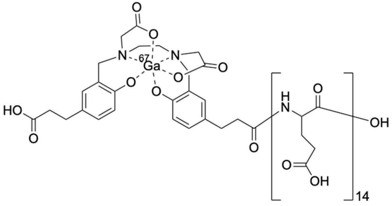
2.1. Synthesis of [67Ga]Ga-HBED-CC-(d-Glu)14, [67Ga]Ga-HBED-CC-(dl-Glu)14, and [67Ga]Ga-HBED-CC-(d-Glu-l-Glu)7

All [67Ga]Ga-HBED-CC-conjugated Glu peptides were synthesized with radiochemical yields greater than 95%; thus, they were used for further experiments without purification as having a radiochemical purity > 95%. Their radiochromatograms are shown in Figure S1.

2.2. Hydroxyapatite-Binding Assay

To determine the affinity of the compounds for hydroxyapatite, the hydroxyapatite-binding assay was performed. The hydroxyapatite-binding rates of [67Ga]Ga-HBED-CC-(l-Glu)14, [67Ga]Ga-HBED-CC-(d-Glu)14, [67Ga]Ga-HBED-CC-(dl-Glu)14, and [67Ga]Ga-HBED-CC-(d-Glu-l-Glu)7 are illustrated in Figure 2. The binding profiles of each tracer were similar.

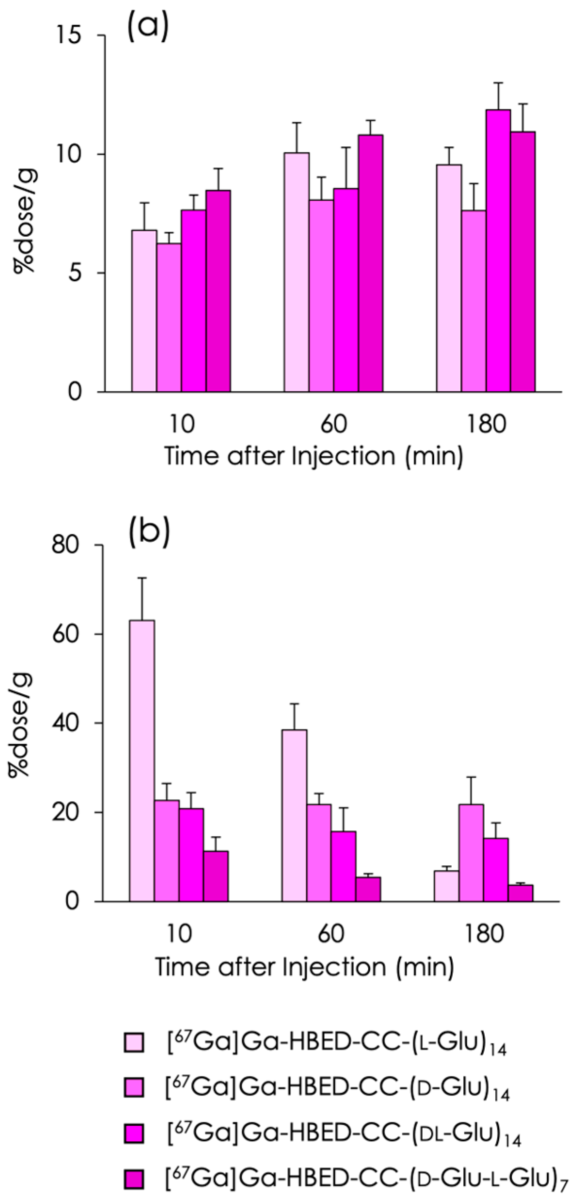
2.3. Biodistribution Experiments

The accumulations of [67Ga]Ga-HBED-CC-(l-Glu)14, [67Ga]Ga-HBED-CC-(d-Glu)14, [67Ga]Ga-HBED-CC-(dl-Glu)14, and [67Ga]Ga-HBED-CC-(d-Glu-l-Glu)7 in the bones and kidneys of normal mice are reported in Figure 3, and detailed data pertaining to their biodistribution are displayed in Tables S1–S3 and a previous paper [22]. All 67Ga-labeled peptides accumulated and were retained in the bone at high levels, and their accumulation rates were similar (Figure 3a). In turn, the renal accumulation of the tracers was largely dependent on the optical isomer of the constituent amino acid, Glu (Figure 3b). In all [67Ga]Ga-HBED-CC-(Glu)14 compounds, radioactivity in all tissues except the bones and kidneys was low (Tables S1–S3).

3. Discussion

Generally, peptides composed of d-type amino acids are more stable in vivo than peptides composed of l-type amino acids because peptidases recognize and metabolize the latter [26]. Therefore, studies have often been performed to improve the stability of peptides by replacing l-amino acids with d-amino acids [27]. In our previous study, repeated d-Asp, i.e., (d-Asp)n, was initially synthesized in an attempt to achieve a higher bone accumulation compared to (l-Asp)n as bone-targeting carriers. However, the accumulation of [67Ga]Ga-DOTA-(d-Asp)n and [67Ga]Ga-DOTA-(l-Asp)n in bone was hardly different because the two types of tracers showed extremely rapid clearance from the blood [21]. Similarly, in the present study, [67Ga]Ga-HBED-CC-(l-Glu)14, [67Ga]Ga-HBED-CC-(d-Glu)14, [67Ga]Ga-HBED-CC-(dl-Glu)14, and [67Ga]Ga-HBED-CC-(d-Glu-l-Glu)7 exhibited a similar bone accumulation (Figure 3) caused by rapid clearance and a similar affinity for hydroxyapatite as expected.
Several studies have investigated the differences in the renal accumulation of peptides composed of l-amino acids and d-amino acids. One study reported that the antimicrobial peptide composed of d-amino acid named danalexin resulted in prolonged retention in the rat kidney compared with the counterpart peptide, ranalexin, which is composed of l-amino acid [28]. That article indicates that the observed prolonged retention is attributable to the increased stability of the peptide and its increased resistance to peptidolysis. In turn, the initial renal uptake of d-amino acid peptides compared with l-amino acid peptides tends to be lower. This difference is primarily caused by the altered stereochemistry of d-amino acids, which affects their recognition and transport by renal uptake mechanisms, such as specific transporters and receptors [29]. In this study, [67Ga]Ga-HBED-CC-(l-Glu)14 exhibited a very high initial uptake into the kidney, with the accumulation gradually decreasing thereafter in a time-dependent manner. In contrast, [67Ga]Ga-HBED-CC-(d-Glu)14 showed a moderate uptake in the kidney and the accumulation was retained. These results are consistent with the above-mentioned descriptions provided in previous articles.
[67Ga]Ga-HBED-CC-(d-Glu-l-Glu)7 was synthesized as a peptide in which the d-Glu and l-Glu were alternately repeated. [67Ga]Ga-HBED-CC-(d-Glu-l-Glu)7 exhibited the lowest radioactivity in the kidney among all [67Ga]Ga-HBED-CC-(Glu)14 compounds, although the explanation for this observation remains unknown. It is known that poly-l-glutamic acid could be α-helical or have random coil peptide conformation depending on conditions [30]. The presence of d-type amino acids induces conformational preferences not followed by peptides consisting of naturally abundant l-type amino acids [31]. Therefore, we assume that repeating the d-Glu and l-Glu may have a very different conformation from oligo-l-glutamic acid, making it less susceptible to transporter recognition for kidney uptake. This is only speculation; further studies are needed to elucidate the mechanism.
Because [67Ga]Ga-HBED-CC-(d-Glu-l-Glu)7 showed an equivalent bone accumulation compared with the remaining [67Ga]Ga-HBED-CC conjugated (Glu)14 compounds, [67Ga]Ga-HBED-CC-(d-Glu-l-Glu)7 possessed the most ideal pharmacokinetics as a bone-seeking radiopharmaceutical. Although the compounds assessed in this study targeted hydroxyapatite in the bone and, therefore, no difference in target directivity should exist between d-Glu and l-Glu, the d- and l-peptides may not be easily interchangeable when targeting receptors and enzymes, etc. This is the reason for the limited number of targets that can be used. If target directivity is maintained, repeated d- and l-peptides may be a useful method for reducing the renal accumulation of peptides. However, the reduction of renal uptake using repeated d- and l-peptides has only been confirmed for Glu-peptides in this study. Therefore, future studies are needed to determine whether this strategy can be applied to other peptides.
A typical method of reducing the renal accumulation of radiopharmaceuticals includes coinfusion of lysine and arginine [32]. This method is used in clinical [177Lu]Lu-DOTATATE for radiation protection of kidneys [33]. Administration of the mixture of lysine and arginine is a relatively safe and effective method. However, the long time required to administer the mixture of lysine and arginine and its high dosage volume are problems. Another method to reduce renal radioactivity is introducing a cleavable linkage by enzymes on the renal brush border membrane [34,35]. This method is an excellent scientific approach; however, the drug design is complex. The method suggested in this study may overcome these problems. However, as mentioned, it is expected to be used in limited cases. It may be an option for reducing renal accumulation of radiopharmaceuticals.
A [67Ga]Ga-HBED-CC-(dl-Glu)14 peptide composed of racemic glutamic acid was also synthesized and evaluated. [67Ga]Ga-HBED-CC-(dl-Glu)14 exhibited a similar kidney uptake to [67Ga]Ga-HBED-CC-(d-Glu)14, and its radioactivity decreased gradually. As [67Ga]Ga-HBED-CC-(dl-Glu)14 is a mixture of various isomers, it was difficult to assess the relationship between its structure and renal radioactivity. It seems to be reasonable to assume that the radioactivity of [67Ga]Ga-HBED-CC-(dl-Glu)14 in the kidney was intermediate compared with the remaining [67Ga]Ga-HBED-CC-(Glu)14 compounds.
[67Ga]Ga-DOTA-(l-Glu)14 also showed a very high initial uptake in the kidney, and its accumulation was retained in a previous report [20]. The difference in retention observed between [67Ga]Ga-HBED-CC-(l-Glu)14 and [67Ga]Ga-DOTA-(l-Glu)14 may have been depended on the difference in the lipophilicity of their complexes. In turn, the renal accumulation and retention of [67Ga]Ga-DOTA-(l-Asp)14 and [67Ga]Ga-DOTA-(d-Asp)14 were not high and were similar. The mechanisms of uptake of oligo-aspartic acid and oligo-glutamic acid may be different.

4. Materials and Methods

4.1. Materials

Electrospray ionization mass spectra (ESI-MS) were obtained with a JEOL JMS-T100TD (JEOL Ltd., Tokyo, Japan). Purification of peptides was performed using an HPLC system (LC-20AD pump, SPD-20A UV detector at a wavelength of 220 nm, and CTO-20A column oven maintained at 40 °C; Shimadzu, Kyoto, Japan). Thin layer chromatography (TLC) analyses were performed with silica plates (Art 5553, Merck, Darmstadt, Germany). Fmoc-l-Glu(OtBu)-OH and Fmoc-d-Glu(OtBu)-OH were purchased from AmBeed (Arlington Heights, IL, USA). 2-Chlorotrityl chloride resin was purchased from Watanabe Chemical Industries, Ltd. (Hiroshima, Japan). N,N-Diisopropylethylamine (DIEA) was purchased from Nacalai Tesque (Kyoto, Japan). 1,3-Diisopropylcarbodiimide (DIPCDI) and 1-hydroxybenzotriazole hydrate (HOBt) were purchased from Kokusan Chemical Co., Ltd. (Tokyo, Japan). Other reagents were of reagent grade and used as received.

4.2. Synthesis of HBED-CC-(d-Glu)14, HBED-CC-(dl-Glu)14, and HBED-CC-(d-Glu-l-Glu)7

HBED-CC-(d-Glu)14, HBED-CC-(dl-Glu)14, and HBED-CC-(d-Glu-l-Glu)7 were synthesized according to a previous report [22]. Namely, the protected peptidyl resin was manually constructed using an Fmoc-based solid-phase methodology based on a previously described method [25]. After the construction of the peptide chain on the resin, the Fmoc protecting group was removed using 20% piperidine in DMF. Subsequently, a mixture containing 2.5 equivalents of HBED-CC-tris(tert-butyl) ester (which was synthesized according to the previous report [36]), DIPCDI, and HOBt in DMF was added and allowed to react at room temperature for 1.5 h. After the cleavage of the peptides from the resin and the deprotection using triisopropylsilane and trifluoroacetic acid (TFA) (5:95), the crude products were purified using reversed-phase (RP) HPLC on a Cosmosil 5C18-AR-II column (10 × 150 mm; Nacalai Tesque) at a flow rate of 4 mL/min with a gradient mobile phase of 20–60% methanol in water containing 0.1% TFA over 20 min, respectively. Chromatograms were obtained by monitoring the UV absorption at a wavelength of 220 nm. The fractions containing HBED-CC-(d-Glu)14, HBED-CC-(dl-Glu)14, and HBED-CC-(d-Glu-l-Glu)7 were determined by mass spectrometry and collected. The solvent was removed by lyophilization to provide HBED-CC conjugated Glu peptides as white powders.
  • MS (ESI+) analysis of HBED-CC-(d-Glu)14 calcd for C96H132N16O52 [M + 2H]2+: m/z = 1170.9 found 1170.8, yield: 3%.
  • MS (ESI+) analysis of HBED-CC-(dl-Glu)14 calcd for C96H132N16O52 [M + 2H]2+: m/z = 1170.9 found 1170.9, yield: 3%.
  • MS (ESI+) analysis of HBED-CC-(d-Glu-l-Glu)7 calcd for C96H132N16O52 [M + 2H]2+: m/z = 1170.9 found 1170.7, yield: 1%.

4.3. Preparation of [67Ga]Ga-HBED-CC-(d-Glu)14, [67Ga]Ga-HBED-CC-(dl-Glu)14, and [67Ga]Ga-HBED-CC-(d-Glu-l-Glu)7

[67Ga]Ga-citrate was purchased from Nihon Medi-Physics Co., Ltd. (Tokyo, Japan), and converted [67Ga]GaCl3 by using Sep-Pak® Silica Plus Light Cartridge (Waters Co., Ltd., Milford, MA, USA) according to a previous report [37,38]. Approximately 50 μg of HBED-CC-(d-Glu)14, HBED-CC-(dl-Glu)14, or HBED-CC-(d-Glu-l-Glu)7 was dissolved in 80 μL of 1 M N-2-hydroxyethylpiperazine-N-2-ethanesulfonic acid (HEPES) solution (pH 5.0), and 20 μL of [67Ga]GaCl3 solution in 0.1 M HCl was added and allowed to react at 80 °C for 10 min. The radiochemical purities of 67Ga-HBED-CC conjugated Glu peptides were determined via TLC analyses using silica plates with acetonitrile and H2O (1:1) as a developing solvent. RP-HPLC was performed with a Cosmosil 5C18-II column (4.6 × 150 mm) at a flow rate of 1 mL/min with a gradient mobile phase of 20% ethanol in water containing 10 mM tetrabutylammoniumhydroxide (TBAH) to 55% ethanol in water containing 10 mM TBAH for 20 min.

4.4. Hydroxyapatite-Binding Assays

Hydroxyapatite-binding assays were performed according to procedures described previously [7]. Briefly, hydroxyapatite beads (Bio-Gel; Bio-Rad, Hercules, CA, USA) were suspended in Tris/HCl-buffered saline (50 mM, pH 7.4) at 1 mg/mL, 2.5 mg/mL, 10 mg/mL, and 25 mg/mL. For the solutions of [67Ga]Ga-HBED-CC-(d-Glu)14, [67Ga]Ga-HBED-CC-(dl-Glu)14, and [67Ga]Ga-HBED-CC-(d-Glu-l-Glu)7, the ligand concentrations were adjusted to 19.5 µM by adding the corresponding HBED-CC conjugated Glu peptides. A solution of each [67Ga]Ga-HBED-CC-(d-Glu)14, [67Ga]Ga-HBED-CC-(dl-Glu)14, and [67Ga]Ga-HBED-CC-(d-Glu-l-Glu)7 (200 µL each) was added to 200 μL of the hydroxyapatite suspension, and the samples were gently shaken for 1 h at room temperature. After centrifugation at 10,000× g for 5 min, the radioactivity of the supernatants was measured using an auto well gamma counter (ARC-7010B, ALOKA Co., Ltd., Tokyo, Japan). Control experiments were performed using the same procedure without hydroxyapatite beads, which resulted in an adsorption of radioactivity to vials of less than 0.1%. The ratios of binding were determined as follows:
Hydroxyapatite binding (%) = (1 − [sample supernatant radioactivity]/[control supernatant radioactivity]) × 100

4.5. Animals

Experiments with animals were conducted in strict accordance with the Guidelines for the Care and Use of Laboratory Animals of Kanazawa University. The animal experimental protocols used were approved by the Committee on Animal Experimentation of Kanazawa University (AP-204165, 5 April 2023). The animals were housed with free access to food and water at 23 °C with a 12 h alternating light/dark schedule.

4.6. Biodistribution Experiments

Biodistribution experiments were performed after an intravenous administration of each tracer solution diluted in saline (37 kBq/100 μL) to 6-week-old male ddY mice (weight, 27–32 g, Japan SLC, Inc., Hamamatsu, Japan). Four mice were sacrificed at each time point for each compound at 10, 60, and 180 min post-injection. The tissues of interest were dissected and weighed. Complete left femurs were isolated as representative bone samples, radioactivity counts were determined using an auto well gamma counter (ARC-7010, ALOKA Co., Ltd.), and counts were corrected for the background radiation and physical decay that occurred during counting.

5. Conclusions

In this study, we compared [67Ga]Ga-HBED-CC-(l-Glu)14, [67Ga]Ga-HBED-CC-(d-Glu)14, [67Ga]Ga-HBED-CC-(dl-Glu)14, and [67Ga]Ga-HBED-CC-(d-Glu-l-Glu)7. In the case of radiopharmaceuticals that use (Glu)14 as a bone-directing peptide, the optical isomerism of the constituent amino acids resulted in comparable bone affinity, whereas significant differences in the kidney uptake and retention were observed. Among the [67Ga]Ga-HBED-CC complex-conjugated–(Glu)14 compounds, [67Ga]Ga-HBED-CC-(d-Glu-l-Glu)7 exhibited the lowest kidney accumulation. The conformation of repeating d-Glu and l-Glu may differ significantly from that of oligo-l-glutamic acid, potentially reducing its recognition by transporters for kidney uptake. Although further studies are needed to elucidate the mechanism, in some cases, the strategy of using repeated d- and l-peptides may be a useful method for reducing their renal accumulation.

Supplementary Materials

The following supporting information can be downloaded at: https://www.mdpi.com/article/10.3390/molecules29173993/s1, Figure S1: HPLC radiochromatograms of 67Ga-labeled compounds; Table S1: Biodistribution of [67Ga]Ga-HBED-CC-(d-Glu)14; Table S2: Biodistribution of [67Ga]Ga-HBED-CC-(dl-Glu)14; Table S3: Biodistribution of [67Ga]Ga-HBED-CC-(d-Glu-l-Glu)7.

Author Contributions

Conceptualization, K.O.; methodology, K.M. and K.O.; formal analysis, K.M. and K.N.; investigation, K.N. and K.O.; resources, H.W. and S.K.; writing—original draft preparation, K.O.; writing—review and editing, K.O., M.M., H.E. and T.F.; project administration, K.O.; funding acquisition, K.O. All authors have read and agreed to the published version of the manuscript.

Funding

This work was supported in part by Sagawa Foundation for Promotion of Cancer Research, Grant-in-Aid for Scientific Research (C) (JSPS KAKENHI Grant Number 15K09948) from Japan Society for the Promotion of Science, Terumo Life Science Foundation, and Nakatomi Foundation.

Institutional Review Board Statement

The animal study protocol was approved by the Institutional Review Board of Kanazawa University (AP-204165, 5 April 2023).

Informed Consent Statement

Not applicable.

Data Availability Statement

The original contributions presented in the study are included in the article/Supplementary Material, further inquiries can be directed to the corresponding author.

Conflicts of Interest

The authors declare no conflicts of interest.

References

  1. Ogawa, K.; Ishizaki, A. Well-designed bone-seeking radiolabeled compounds for diagnosis and therapy of bone metastases. BioMed Res. Int. 2015, 2015, 676053. [Google Scholar] [CrossRef] [PubMed]
  2. Ogawa, K.; Saji, H. Advances in drug design of radiometal-based imaging agents for bone disorders. Int. J. Mol. Imaging 2011, 2011, 537687. [Google Scholar] [CrossRef]
  3. Cook, G.J.R. Imaging of Bone Metastases in Breast Cancer. Semin. Nucl. Med. 2022, 52, 531–541. [Google Scholar] [CrossRef] [PubMed]
  4. Wilson, G.M.; Pinkerton, T.C. Determination of charge and size of technetium diphosphonate complexes by anion-exchange liquid chromatography. Anal. Chem. 1985, 57, 246–253. [Google Scholar] [CrossRef]
  5. Ogawa, K.; Mukai, T.; Arano, Y.; Hanaoka, H.; Hashimoto, K.; Nishimura, H.; Saji, H. Design of a radiopharmaceutical for the palliation of painful bone metastases: Rhenium-186-labeled bisphosphonate derivative. J. Labelled Cmp. Radiopharm. 2004, 47, 753–761. [Google Scholar] [CrossRef]
  6. Ogawa, K.; Mukai, T.; Arano, Y.; Ono, M.; Hanaoka, H.; Ishino, S.; Hashimoto, K.; Nishimura, H.; Saji, H. Development of a rhenium-186-labeled MAG3-conjugated bisphosphonate for the palliation of metastatic bone pain based on the concept of bifunctional radiopharmaceuticals. Bioconjug. Chem. 2005, 16, 751–757. [Google Scholar] [CrossRef]
  7. Ogawa, K.; Mukai, T.; Arano, Y.; Otaka, A.; Ueda, M.; Uehara, T.; Magata, Y.; Hashimoto, K.; Saji, H. Rhenium-186-monoaminemonoamidedithiol-conjugated bisphosphonate derivatives for bone pain palliation. Nucl. Med. Biol. 2006, 33, 513–520. [Google Scholar] [CrossRef]
  8. Ogawa, K.; Mukai, T.; Inoue, Y.; Ono, M.; Saji, H. Development of a novel 99mTc-chelate-conjugated bisphosphonate with high affinity for bone as a bone scintigraphic agent. J. Nucl. Med. 2006, 47, 2042–2047. [Google Scholar]
  9. Ogawa, K.; Mukai, T.; Asano, D.; Kawashima, H.; Kinuya, S.; Shiba, K.; Hashimoto, K.; Mori, H.; Saji, H. Therapeutic effects of a 186Re-complex-conjugated bisphosphonate for the palliation of metastatic bone pain in an animal model. J. Nucl. Med. 2007, 48, 122–127. [Google Scholar]
  10. Ogawa, K.; Kawashima, H.; Shiba, K.; Washiyama, K.; Yoshimoto, M.; Kiyono, Y.; Ueda, M.; Mori, H.; Saji, H. Development of [90Y]DOTA-conjugated bisphosphonate for treatment of painful bone metastases. Nucl. Med. Biol. 2009, 36, 129–135. [Google Scholar] [CrossRef]
  11. Ogawa, K.; Mukai, T.; Kawai, K.; Takamura, N.; Hanaoka, H.; Hashimoto, K.; Shiba, K.; Mori, H.; Saji, H. Usefulness of competitive inhibitors of protein binding for improving the pharmacokinetics of 186Re-MAG3-conjugated bisphosphonate (186Re-MAG3-HBP), an agent for treatment of painful bone metastases. Eur. J. Nucl. Med. Mol. Imaging 2009, 36, 115–121. [Google Scholar] [CrossRef] [PubMed]
  12. Ogawa, K.; Takai, K.; Kanbara, H.; Kiwada, T.; Kitamura, Y.; Shiba, K.; Odani, A. Preparation and evaluation of a radiogallium complex-conjugated bisphosphonate as a bone scintigraphy agent. Nucl. Med. Biol. 2011, 38, 631–636. [Google Scholar] [CrossRef]
  13. Uehara, T.; Jin, Z.L.; Ogawa, K.; Akizawa, H.; Hashimoto, K.; Nakayama, M.; Arano, Y. Assessment of 186Re chelate-conjugated bisphosphonate for the development of new radiopharmaceuticals for bones. Nucl. Med. Biol. 2007, 34, 79–87. [Google Scholar] [CrossRef]
  14. Suzuki, K.; Satake, M.; Suwada, J.; Oshikiri, S.; Ashino, H.; Dozono, H.; Hino, A.; Kasahara, H.; Minamizawa, T. Synthesis and evaluation of a novel 68Ga-chelate-conjugated bisphosphonate as a bone-seeking agent for PET imaging. Nucl. Med. Biol. 2011, 38, 1011–1018. [Google Scholar] [CrossRef] [PubMed]
  15. Fellner, M.; Baum, R.P.; Kubicek, V.; Hermann, P.; Lukes, I.; Prasad, V.; Rosch, F. PET/CT imaging of osteoblastic bone metastases with 68Ga-bisphosphonates: First human study. Eur. J. Nucl. Med. Mol. Imaging 2010, 37, 834. [Google Scholar] [CrossRef] [PubMed]
  16. Zha, Z.; Wu, Z.; Choi, S.R.; Ploessl, K.; Smith, M.; Alexoff, D.; Zhu, L.; Kung, H.F. A New [68Ga]Ga-HBED-CC-Bisphosphonate as a Bone Imaging Agent. Mol. Pharm. 2020, 17, 1674–1684. [Google Scholar] [CrossRef]
  17. Jin, W.; Zhao, R.; Wang, R.; Choi, S.R.; Ploessl, K.; Alexoff, D.; Wu, Z.; Zhu, L.; Kung, H.F. Theranostic Agent Targeting Bone Metastasis: A Novel [68Ga]Ga/[177Lu]Lu-DOTA-HBED-bisphosphonate. J. Med. Chem. 2024, 67, 4793–4803. [Google Scholar] [CrossRef]
  18. Kasugai, S.; Fujisawa, R.; Waki, Y.; Miyamoto, K.; Ohya, K. Selective drug delivery system to bone: Small peptide (Asp)6 conjugation. J. Bone Miner. Res. 2000, 15, 936–943. [Google Scholar] [CrossRef]
  19. Ishizaki, J.; Waki, Y.; Takahashi-Nishioka, T.; Yokogawa, K.; Miyamoto, K. Selective drug delivery to bone using acidic oligopeptides. J. Bone Miner. Metab. 2009, 27, 1–8. [Google Scholar] [CrossRef] [PubMed]
  20. Ogawa, K.; Ishizaki, A.; Takai, K.; Kitamura, Y.; Kiwada, T.; Shiba, K.; Odani, A. Development of novel radiogallium-labeled bone imaging agents using oligo-aspartic acid peptides as carriers. PLoS ONE 2013, 8, e84335. [Google Scholar] [CrossRef]
  21. Ogawa, K.; Ishizaki, A.; Takai, K.; Kitamura, Y.; Makino, A.; Kozaka, T.; Kiyono, Y.; Shiba, K.; Odani, A. Evaluation of Ga-DOTA-(d-Asp)n as bone imaging agents: D-aspartic acid peptides as carriers to bone. Sci. Rep. 2017, 7, 13971. [Google Scholar] [CrossRef] [PubMed]
  22. Ogawa, K.; Nishizawa, K.; Mishiro, K.; Effendi, N.; Fuchigami, T.; Munekane, M.; Wakabayashi, H.; Kinuya, S. Synthesis and Evaluation of Radiogallium Labeled Bone-Imaging Probes Using Oligo-gamma-Carboxy Glutamic Acid Peptides as Carriers to Bone. Mol. Pharm. 2024, 21, 2375–2382. [Google Scholar] [CrossRef]
  23. Hirata, S.; Mishiro, K.; Higashi, T.; Fuchigami, T.; Munekane, M.; Arano, Y.; Kinuya, S.; Ogawa, K. Synthesis and evaluation of a multifunctional probe with a high affinity for prostate-specific membrane antigen (PSMA) and bone. Nucl. Med. Biol. 2022, 114-115, 34–41. [Google Scholar] [CrossRef] [PubMed]
  24. Ishizaki, A.; Mishiro, K.; Shiba, K.; Hanaoka, H.; Kinuya, S.; Odani, A.; Ogawa, K. Fundamental study of radiogallium-labeled aspartic acid peptides introducing octreotate derivatives. Ann. Nucl. Med. 2019, 33, 244–251. [Google Scholar] [CrossRef] [PubMed]
  25. Ogawa, K.; Yu, J.; Ishizaki, A.; Yokokawa, M.; Kitamura, M.; Kitamura, Y.; Shiba, K.; Odani, A. Radiogallium complex-conjugated bifunctional peptides for detecting primary cancer and bone metastases simultaneously. Bioconjug. Chem. 2015, 26, 1561–1570. [Google Scholar] [CrossRef]
  26. Obaha, A.; Novinec, M. Regulation of Peptidase Activity beyond the Active Site in Human Health and Disease. Int. J. Mol. Sci. 2023, 24, 17120. [Google Scholar] [CrossRef]
  27. Lee, M.F.; Poh, C.L. Strategies to improve the physicochemical properties of peptide-based drugs. Pharm. Res. 2023, 40, 617–632. [Google Scholar] [CrossRef]
  28. Domhan, C.; Uhl, P.; Kleist, C.; Zimmermann, S.; Umstatter, F.; Leotta, K.; Mier, W.; Wink, M. Replacement of l-Amino Acids by d-Amino Acids in the Antimicrobial Peptide Ranalexin and Its Consequences for Antimicrobial Activity and Biodistribution. Molecules 2019, 24, 2987. [Google Scholar] [CrossRef]
  29. Diao, L.; Meibohm, B. Pharmacokinetics and pharmacokinetic-pharmacodynamic correlations of therapeutic peptides. Clin. Pharmacokinet. 2013, 52, 855–868. [Google Scholar] [CrossRef]
  30. Gooding, E.A.; Sharma, S.; Petty, S.A.; Fouts, E.A.; Palmer, C.J.; Nolan, B.E.; Volk, M. pH-dependent helix folding dynamics of poly-glutamic acid. Chem. Phys. 2013, 422, 115–123. [Google Scholar] [CrossRef]
  31. Yongye, A.B.; Li, Y.; Giulianotti, M.A.; Yu, Y.; Houghten, R.A.; Martínez-Mayorga, K. Modeling of peptides containing d-amino acids: Implications on cyclization. J. Comput. Aided Mol. Des. 2009, 23, 677–689. [Google Scholar] [CrossRef]
  32. Rolleman, E.J.; Valkema, R.; de Jong, M.; Kooij, P.P.; Krenning, E.P. Safe and effective inhibition of renal uptake of radiolabelled octreotide by a combination of lysine and arginine. Eur. J. Nucl. Med. Mol. Imaging 2003, 30, 9–15. [Google Scholar] [CrossRef] [PubMed]
  33. Gupta, S.K.; Singla, S.; Thakral, P.; Bal, C.S. Dosimetric analyses of kidneys, liver, spleen, pituitary gland, and neuroendocrine tumors of patients treated with 177Lu-DOTATATE. Clin. Nucl. Med. 2013, 38, 188–194. [Google Scholar] [CrossRef] [PubMed]
  34. Arano, Y.; Fujioka, Y.; Akizawa, H.; Ono, M.; Uehara, T.; Wakisaka, K.; Nakayama, M.; Sakahara, H.; Konishi, J.; Saji, H. Chemical design of radiolabeled antibody fragments for low renal radioactivity levels. Cancer Res. 1999, 59, 128–134. [Google Scholar]
  35. Suzuki, H.; Araki, M.; Tatsugi, K.; Ichinohe, K.; Uehara, T.; Arano, Y. Reduction of the Renal Radioactivity of 111In-DOTA-Labeled Antibody Fragments with a Linkage Cleaved by the Renal Brush Border Membrane Enzymes. J. Med. Chem. 2023, 66, 8600–8613. [Google Scholar] [CrossRef]
  36. Makarem, A.; Konrad, M.; Liolios, C.; Kopka, K. A Convenient Synthesis for HBED-CC-tris(tert-butyl ester). Synlett 2018, 29, 1239–1243. [Google Scholar] [CrossRef]
  37. Echigo, H.; Munekane, M.; Fuchigami, T.; Washiyama, K.; Mishiro, K.; Wakabayashi, H.; Takahashi, K.; Kinuya, S.; Ogawa, K. Optimizing the pharmacokinetics of an 211At-labeled RGD peptide with an albumin-binding moiety via the administration of an albumin-binding inhibitor. Eur. J. Nucl. Med. Mol. Imaging 2024, 51, 2663–2671. [Google Scholar] [CrossRef]
  38. Ben Azzouna, R.; Guez, A.; Benali, K.; Al-Shoukr, F.; Gonzalez, W.; Karoyan, P.; Rouzet, F.; Le Guludec, D. Synthesis, gallium labelling and characterization of P04087, a functionalized phosphatidylserine-binding peptide. EJNMMI Radiopharm. Chem. 2017, 2, 3. [Google Scholar] [CrossRef]
Figure 1. A chemical structure of [67Ga]Ga-HBED-CC-(Glu)14. [67Ga]Ga-HBED-CC-(l-Glu)14, [67Ga]Ga-HBED-CC-(d-Glu)14, [67Ga]Ga-HBED-CC-(dl-Glu)14, and [67Ga]Ga-HBED-CC-(d-Glu-l-Glu)7 are stereoisomers of [67Ga]Ga-HBED-CC-(Glu)14.
Molecules 29 03993 g001
Figure 2. Binding ratios of [67Ga]Ga-HBED-CC-(l-Glu)14, [67Ga]Ga-HBED-CC-(d-Glu)14, [67Ga]Ga-HBED-CC-(dl-Glu)14, and [67Ga]Ga-HBED-CC-(d-Glu-l-Glu)7 to hydroxyapatite beads. Binding of each [67Ga]Ga-HBED-CC-(Glu)14 to hydroxyapatite beads increased with the amount of hydroxyapatite. Data are expressed as the mean ± SD for four samples. Data of [67Ga]Ga-HBED-CC-(l-Glu)14 from reference [22].
Molecules 29 03993 g002
Figure 3. Radioactivity in (a) bone and (b) kidney after intravenous injection of [67Ga]Ga-HBED-CC-(l-Glu)14, [67Ga]Ga-HBED-CC-(d-Glu)14, [67Ga]Ga-HBED-CC-(dl-Glu)14, and [67Ga]Ga-HBED-CC-(d-Glu-l-Glu)7 in normal mice. Each value represents the mean (SD) for four animals. Data of [67Ga]Ga-HBED-CC-(l-Glu)14 from reference [22].
Molecules 29 03993 g003
Disclaimer/Publisher’s Note: The statements, opinions and data contained in all publications are solely those of the individual author(s) and contributor(s) and not of MDPI and/or the editor(s). MDPI and/or the editor(s) disclaim responsibility for any injury to people or property resulting from any ideas, methods, instructions or products referred to in the content.

