Article

Diterpenoids with Potent Anti-Psoriasis Activity from Euphorbia helioscopia L.

1
School of Pharmacy, Henan University of Chinese Medicine, Zhengzhou 450046, China
2
Academy of Chinese Medical Sciences, Henan University of Chinese Medicine, Zhengzhou 450046, China
*
Authors to whom correspondence should be addressed.
Molecules 2024, 29(17), 4104; https://doi.org/10.3390/molecules29174104
Submission received: 25 July 2024 / Revised: 24 August 2024 / Accepted: 27 August 2024 / Published: 29 August 2024
(This article belongs to the Section Bioorganic Chemistry)

Abstract

:
Psoriasis, an immune-mediated inflammatory skin disorder, seriously affects the quality of life of nearly four percent of the world population. Euphorbia helioscopia L. is the monarch constituent of Chinese ZeQi powder preparation for psoriasis, so it is necessary to illustrate its active ingredients. Thus, twenty-three diterpenoids, including seven new ones, were isolated from the whole herb of E. helioscopia L. Compounds 1 and 2, each featuring a 2,3-dicarboxylic functionality, are the first examples in the ent-2,3-sceo-atisane or the ent-2,3-sceo-abietane family. Extensive spectroscopic analysis (1D, 2D NMR, and HRMS data) and computational methods were used to confirm their structures and absolute configurations. According to the previous study and NMR data from the jatropha diterpenes obtained in this study, some efficient 1H NMR spectroscopic rules for assigning the relative configurations of 3α-benzyloxy-jatroph-11E-ene and 7,8-seco-3α-benzyloxy-jatropha-11E-ene were summarized. Moreover, the hyperproliferation of T cells and keratinocytes is considered a key pathophysiology of psoriasis. Anti-proliferative activities against induced T/B lymphocytes and HaCaT cells were tested, and IC50 values of some compounds ranged from 6.7 to 31.5 μM. Compounds 7 and 11 reduced the secretions of IFN-γ and IL-2 significantly. Further immunofluorescence experiments and a docking study with NF-κB P65 showed that compound 13 interfered with the proliferation of HaCaT cells by inhibiting the NF-κB P65 phosphorylation at the protein level.

1. Introduction

Psoriasis is a common autoimmune skin disease characterized by T cell-mediated hyperproliferation of keratinocytes. This disease has certain distinct but overlapping clinical phenotypes, including chronic plaque lesions (psoriasis vulgaris), acute and usually self-limiting guttate-type eruptions, seborrhoeic psoriasis, pustular lesions, and at least 10% of these patients develop arthritis [1,2]. The global prevalence of the disease is approximately 2%, with regional variations [2,3]. Biological agents, immunosuppressants derived from natural products, and traditional Chinese medicine formulas are the familiar drugs for treating psoriasis. However, the emergence of drug resistance necessitates the discovery of more potent agents for psoriasis treatment.
Euphorbia, the largest genus of Euphorbiaceae, contains over 2000 species. Its members inhabit worldwide, some of which are used as folk medicines in China to treat skin diseases (such as psoriasis), edema, tuberculosis, and constipation. The chemical constituents of Euphorbia are terpenes, flavonoids, tannins, and phenolic compounds. The characteristic components are diterpenoids, more than 150 diterpenoids with various skeletons and significant bioactivities, such as antitumor, anti-inflammatory, and anti-HIV activities, having been isolated from this genus [4,5]. Diterpenoids from Euphorbia peplus (pepluacetal and pepluanols A-B), E. helioscopia (helioscopids A and O, euphohelioscoids A), and E. fischeriana inhibit Kv1.3 ion channel, a validated target for the treatment of autoimmune diseases, such as multiple sclerosis, type-1 diabetes, asthma, and psoriasis [6,7,8,9]. Additionally, it was reported that the methanol extract of E. kansui radix alleviated the symptoms of psoriasis through the inhibition of Th17 differentiation and activation of dendritic cells. These effects are expected to be beneficial in treating and preventing psoriasis [10].
Euphorbia helioscopia L., commonly referred as Zeqi in traditional Chinese medicine, is a representative herb of the Euphorbia genus. In recent years, the chemical analysis of E. helioscopia L. has yielded novel diterpenoids with diverse biological activities, such as secoheliosphane B with activity against HSV-1, heliojatrone C with anti-inflammatory activity, euphohelioscopoids A–C against paclitaxel-resistant A549 human lung cancer cell line, and helioscopids A/O/euphohelioscoids A with Kv1.3 inhibitory activity [7,8,11,12,13], which, in turn, sustains extensive attention from phytochemists. Additionally, multiple clinical studies have found that Chinese ZeQi powder preparation can treat psoriasis without significant adverse reactions [5,14], and E. helioscopia L. is the monarch constituent. To find more diterpenoids with promising activities for psoriasis, E. helioscopia L. was selected to be investigated in this study, resulting in the isolation of 23 diterpenoids. Among them, compounds 1 and 2 each feature an unusual 2,3-seco ent-2,3-dicarboxyl-atisan-2,3-dioic acid or 2,3-seco ent-abietan-2,3-dioic acid and compounds 37 are new jatropha diterpenes. In establishing the structures of new and known jatropha diterpenes, we summarized and examined the correlations between 1H NMR signals (chemical shifts and coupling constants) and the relative configurations of C-2, C-13, and C-14 in the (7,8-seco)/3α-benzyloxy-jatropha-11E-ene systems. Here, the isolation and structural elucidation of compounds were reported. Moreover, the anti-psoriatic potential of the isolated compounds was evaluated by assessing their immunosuppressive activities and anti-proliferation effects on keratinocytes. The possible mechanism of active compounds was explored through immunofluorescence experiments and docking studies.

2. Results

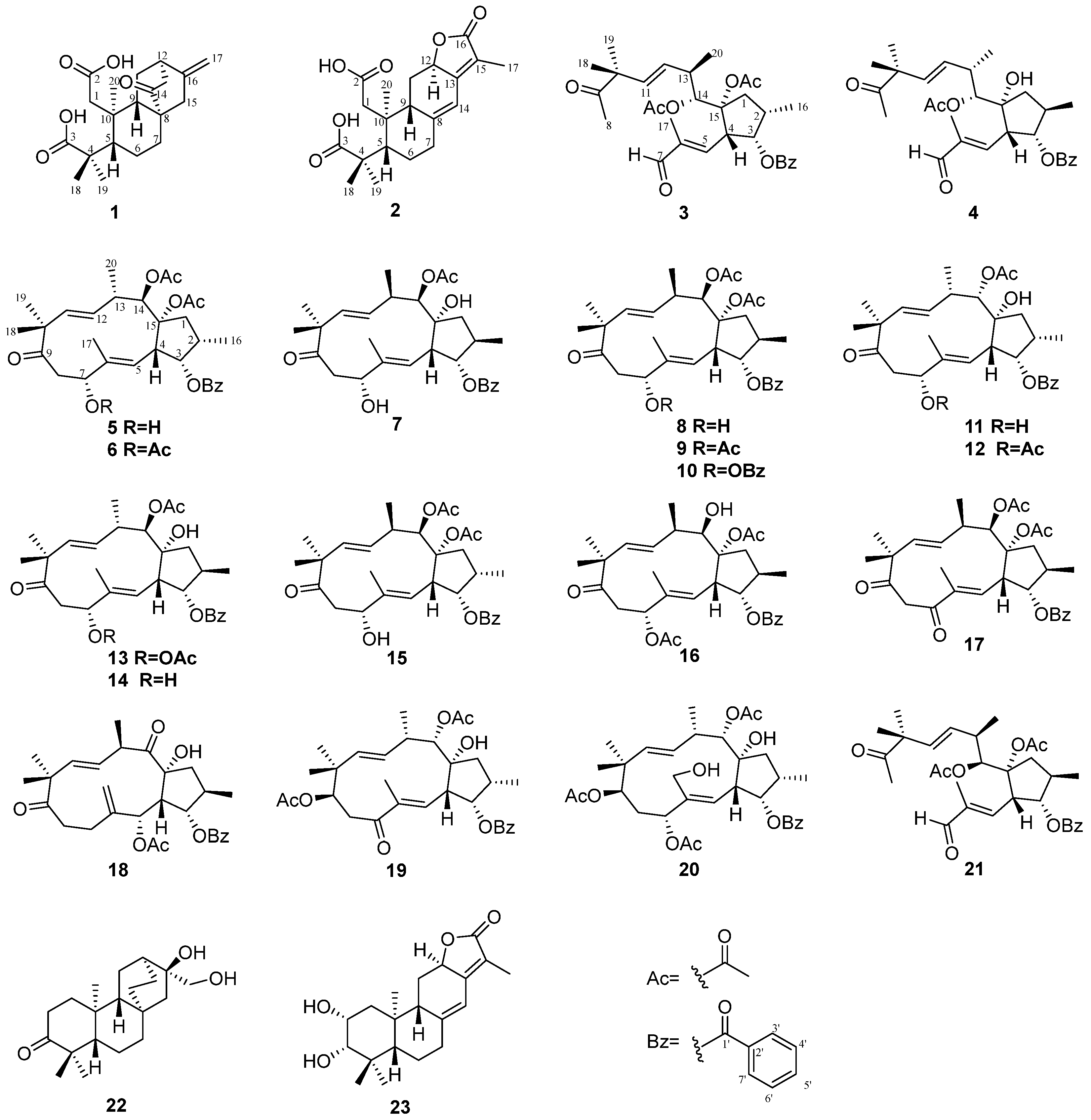
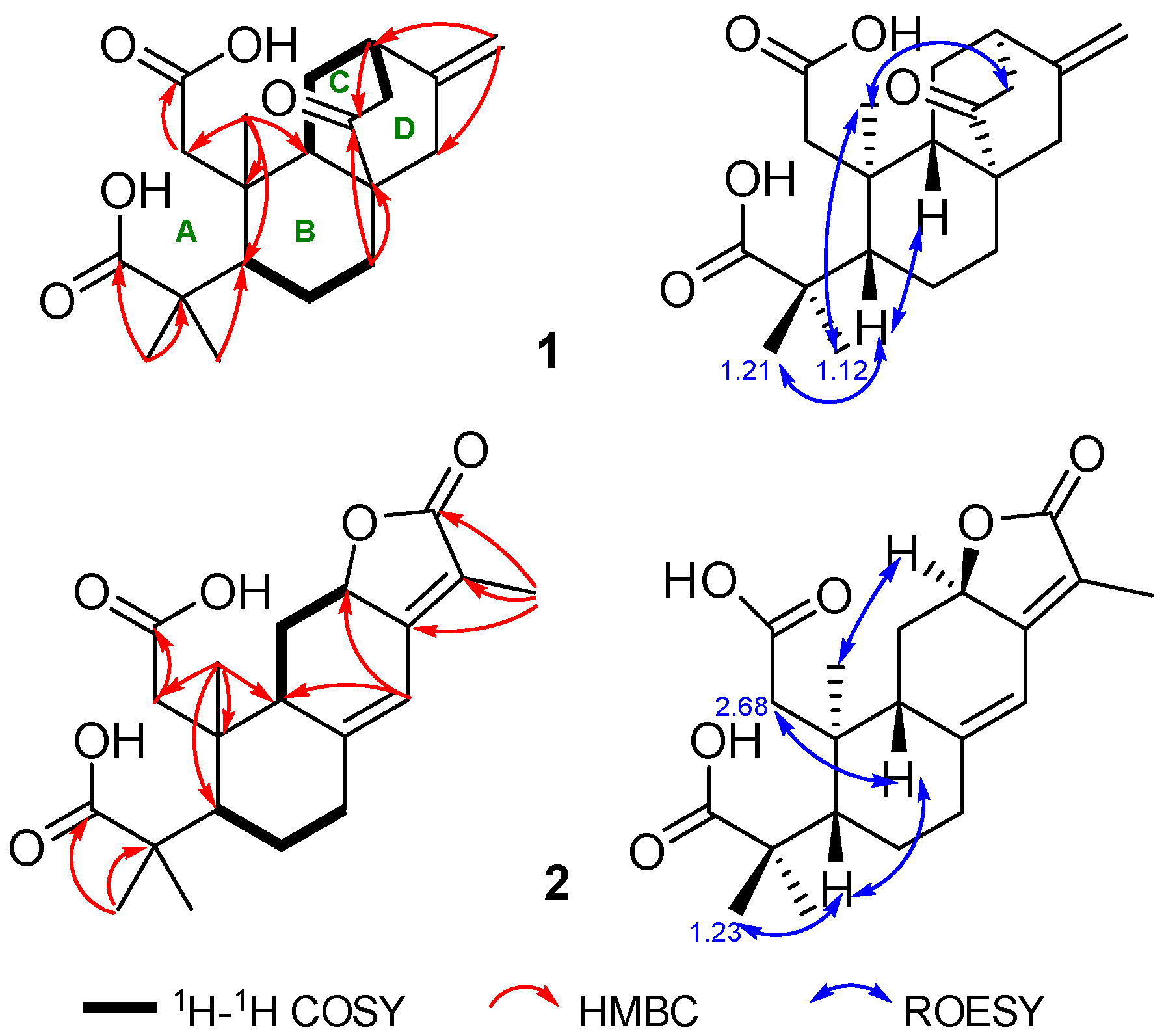
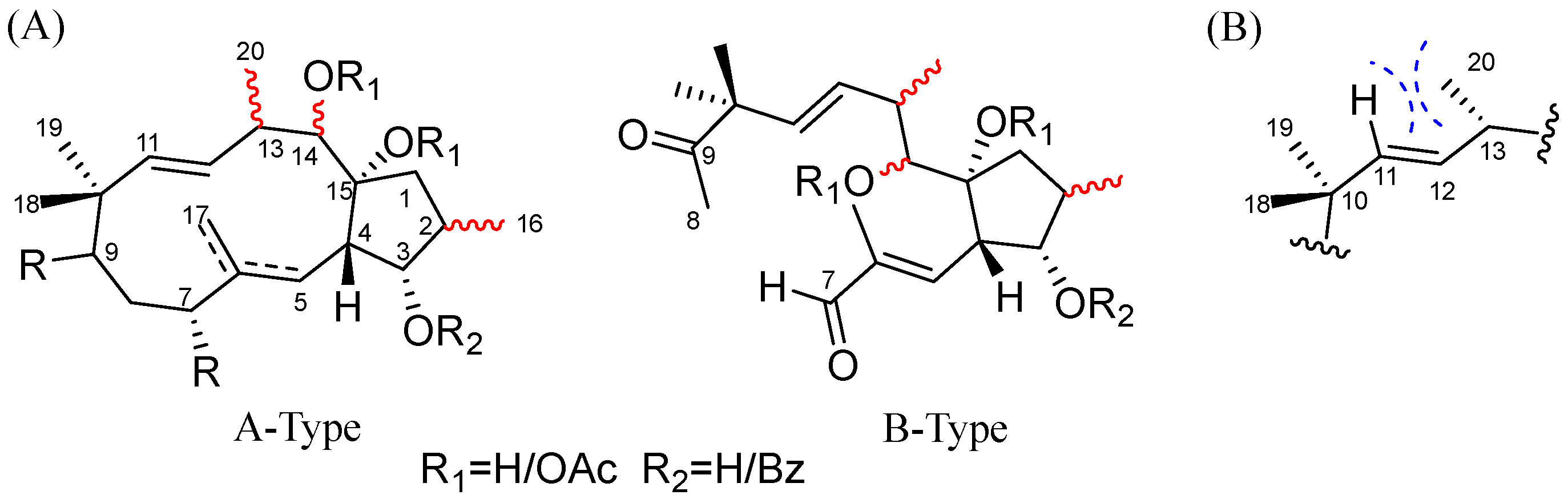
2.1. Structure Elucidation

Compound 1, isolated as white amorphous powder, has a molecular formula C20H28O5 deduced by HRESIMS ion peak at m/z 371.1808 [M+Na]+ (calcd for C20H28O5Na, 371.1829), appropriate for seven degrees of unsaturation. The absorptions at 1701 and 1721 cm−1 in the IR spectrum implied the presence of carboxyls. 1H NMR spectrum showed typical signals for a terminal olefine [δH 4.67 (s), 4.88 (s) H2-17), and three methyl singlets (δH 0.78, H3-20; 1.12, H3-19; 1.21, H3-18). 13C and DEPT NMR data displayed twenty carbons corresponding to three methyls (δC 17.4, C-20; 21.7, C-19; 29.5, C-18), six sp3 methenes (δC 19.5 C-6; 28.1, C-11; 31.2, C-7; 39.0, C-1; 42.9, C-15; 44.7, C-13), four sp3 methines (δC 38.6, C-12; 45.9, C-9; 48.3, C-5), two sp3 quaternary carbons (δC 41.3, C-10; δC 45.5, C-4; 47.7, C-8), a disubstituted terminal double bond (δC 107.3, C-17; 147.0, C-16), two carboxyl groups (δC 178.7, C-2; 187.5, C-3), and one ketone (δC 217.2, C-14) (Table 1). These data from 1 shared a certain similarity to the co-isolate, ent-16β,17-dihydroxyatisan-3-one (22) (Figure 1) [15]. However, as four degrees of the unsaturation accounted for two carboxyls, one double bond, and a ketone, the remaining three degrees of unsaturation required compound 1 to have a tricyclic system, so compound 1 may be a novel ent-seco-atisane diterpenoid. To verify this deduction and establish the specific structure of 1, a detailed analysis of 1H-1H COSY and HMBC correlations was proceeded subsequently (Figure 2). B/C/D rings with a double bond at C16-C17 and a ketone at C-14 were successfully constructed by the 1H-1H COSY correlations of H-5 (δH 2.54)/H2-6a (δH 1.61)/H2-7a (δH 2.32), and H-9 (δH 2.69)/H2-11 (δH 1.82, 1.56)/H-12 (δH 2.74)/H2-13 (δH 2.31), together with the HMBC correlations from H2-7 (δH 2.32, 0.98) to C-8 (δC 47.7), H2-17 to C-12 (δC 38.6)/C-15 (δC 42.9), H-12 to C-14, and H3-20 to C-5 (δC 48.3)/C-9 (δC 45.9)/C-10 (δC 41.3). For ring A, observed HMBC correlations from H3-20 to C-1, H2-1 to a carboxyl (C-2), and H3-18/19 to a carboxyl (C-3), together with unobserved correlations from H2-1 to C-3, highlighted an oxidative cleavage between C-2 and C-3 (Figure 2). Therefore, the planar structure of 1 was established, as depicted in Figure 1. Combining consideration of the rotating frame Overhauser effect spectroscopy (ROESY) spectrum and biosynthetic way, as well as comparing the experimental CD curve with the calculated ECD trend (Figure 2 and Figure 3) (Supplementary Materials), the absolute structure of 1 was identified as ent-2,3-seco-14-oxo-16-atisene-2,3-dioic acid.
The molecular formula of compound 2, C20H26O6, was determined based on HRESIMS and 13C NMR spectra, indicating eight degrees of unsaturation. The max absorption of 275.0 (4.54) in the UV spectrum and absorptions of 1735/1674 cm−1 in the IR spectrum indicated a large conjugated system and ketone groups in compound 2, respectively. 1D NMR data and HSQC spectrum displayed twenty carbon resources attributed to four tertiary methyls (δC/H 8.3/1.83, CH3-17; 29.5/1.23, CH3-18; 21.3/1.20, CH3-19; 21.0/1.01, CH3-20), four sp3methylenes, three sp3 methines (one oxygenated, δH/C 4.86/75.6, CH-12), two double bonds [δC/H 155.6, C-13; 116.9, C-15; 151.1, C-8; 114.5/6.30 (s), CH-14], two carboxyls (δC 178.6, C-2; 186.9, C-3) and a lactone carbonyl (δC 175.1, C-16) (Table 1). Signals of a five-membered carbon ring of α,β-unsaturated lactone, together with similar 1D NMR spectra data (Table 1) to those of helioscopinolide L, a chemical previously isolated from E. helioscopia [16,17], indicated that compound 2 possessed an abietanolide skeleton. However, as three carbonyls and two olefinic bonds accounted for the eight degrees of unsaturation, the remaining three degrees of unsaturation suggested that one ring was opened, so 2 is a tricyclic diterpenoid, too. Detailed analysis of the HMBC correlations from H2-1a [δH 2.68 (d, J = 20.3 HZ)] to a carboxyl (C-2) and H3-18/H3-19 to another carboxyl (C-3) revealed the break of C2-C3 bond in 2 as compound 1. Therefore, compound 2 is a novel 2,3-seco-abietanolide (Figure 2). So far, the planner structure has been constructed, as shown in Figure 1. Given ROESY correlations (Figure 2) and the similar biosynthetic way as other abietanolides from this plant [5,16], the absolute structure of compound 2 was deduced as ent-2,3-seco-2,3-dicarboxy-abieta-8(14),13(15)-dien-16,12-olide. This deduction was verified by comparing the experimental CD and calculated ECD spectra (Figure 3) (Supplementary Materials).
Based on the HRESIMS result and 13C NMR spectrum, the molecular formula of 3 was determined to be the same as the co-isolate secoheliosphane B (21) [11], C31H40O8. These 1D NMR data presented a set of rare but typical signals for a seco-jatropha triester: four methyl singlets, two methyl doublets, two acetoxys, two double bonds, one benzyloxy, one ketone, and a ketal (Table 2 and Table 3). Data from 3 were almost identical to those of 21, implying that the planer structure of compound 3 is the same as that of compound 21, which was supported by 1H-1H COSY and HMBC correlations (Figures S18 and S19, Supplementary Materials). The geometries of ∆5 and ∆11 double bonds were easily assigned as E for the large coupling constant (3JH-11, H-12 = 15.6 Hz) and the ROESY correlations (for ∆5: H-4/H3-17, H-5/H-7; for ∆11 H-11/H-13, H3-18/H-12) (Figure S20, Supplementary Materials). For all jatropha diterpenes, the orientations of the substitutions are usually fixed at C-3 (an α-oriented benzoate substituent), C-4 (a β-oriented hydrogen), and C-15 (an α-oriented hydroxy or acetoxy), but orientations of CH3-16 at C-2, CH3-20 at C-13, and OH(OAc)-14 at C-14 are usually changeable. Carefully analysis of 1H and 13C NMR data for compounds 3 and 21 both measured in CDCl3 revealed the relative configurations at C-2 and C-14 may be opposite for the differences observed in chemical shifts of relative carbons [3: δCH-2 39.4/2.15 (m), 21: δCH-2 38.2/2.46 (m); 3: δCH-14 74.7/5.82 (d, J = 9.5 Hz), 21: δCH-14 75.3/5.59 (d, J = 7.2 Hz); 3: δCH3-16 14.0/0.96 (d, J = 7.0 Hz), 21: δCH3-16 18.2/1.17 (d, J = 7.2 Hz)] (Table 2 and Table 3). The relative configurations of C-2 and C-14 in compound 3 were confirmed by the analysis below. A key ROESY correlation (Figure S20, Supplementary Materials) of H-2 (δH 2.15, m) and H-4 (δH 3.61, dd, J = 9.4, 4.0 Hz) indicated that the orientation of CH3-16 is α, leading to the obvious changes for J values of H-3 [3: δH 5.65 (t, J = 4.0 Hz), 21: δH 4.69 (t, J = 9.0 Hz)], and for chemical shifts of C-1, C-2, C-3, and C-16 (3: δC 46.8 C-1, 80.7 C-3; 21: δC 40.8 C-1, 82.5 C-3). Then, a large coupling constant between H-13 and H-14 (J = 9.5 Hz) indicated a trans-position for H-13 and H-14. Furthermore, the relative configuration of CH3-20 was deduced to be β-oriented for chemical shifts of H-11 (δH 5.38) larger than H-12 (δH 5.27) [12,13]. Therefore, the structure of 3 was defined as shown in Figure 1 and named secoheliosphane C.
HRESIMS ion peak at m/z 537.2459 [M+K]+ (calcd for 537.2449) and 13C NMR spectrum afforded compound 4 a molecular formula, C29H38O7, missing a mass unit of -CH2CO- compared with secoheliosphane B (21). A detailed analysis of 1D and 2D NMR data (Table 2 and Table 3) showed that the planner structure of compound 4 is also a seco-jatropha ester. The only difference between 4 and 21 is that OAc-15 in 21 is changed into OH-15 in 4, which was supported by 1H-1H COSY and HMBC correlations (Figures S25 and S26, Supplementary Materials). Subsequently, the relative configuration of 4 was determined by multiple methods, including ROESY (Figure S27, Supplementary Materials), J-based configuration analysis (JBCA), and a comparison of chemical shifts. First, the geometries of ∆5 and ∆11 double bonds were E based on the ROESY correlations and J values. Subsequently, JBCA of H-3 [δH 5.20 (dd, J = 8.5, 5.7 Hz)] together with a chemical shift of CH3-16 (δC 18.5), suggested a 16β-CH3; a small coupling constant between H-13 and H-14 (J = 3.1 Hz) indicated that CH3-20 and OAc-14 located co-facial. Moreover, compared with secoheliosphane A (δH-11 5.41, δH-12 5.46), secoheliosphane B (δH-11 5.46, δH-12 5.37) and compound 3 (δH-11 5.38, δH-12 5.27), for compound 4, the chemical shift of H-11 (δH 5.58), larger than that of H-12 (δH 5.64) suggests a 20α-CH3. Finally, the structure of 4 was defined as shown in Figure 1 and given a trivial name, secoheliosphane D.
Based on the known jatropha-skeleton diterpenoids with acetoxy groups from E. helioscopia [5], 1D NMR data of compounds 57 (Table 2 and Table 3) displayed a group of characteristic signals for jatropha ester framework corresponding to one benzyloxy group, one ketone, two methyl doublets, three methyl singlets, two double bonds, and one to three acetoxy groups. In addition, the remaining two degrees of unsaturation for compounds 57 revealed that they all are dicyclic jatropha esters.
Compound 5, C31H40O8, has the same molecular formula and closely similar 1D NMR with co-isolate 15 [13], implying that they share an identical planner structure (Table 2 and Table 3). Given JBCA, and the summarized correlations of the chemical shifts of the relevant carbons and the configurations of C-2, C-13, and C-14 (Figure 4 and Table 2) [12], the orientations of CH3-16, CH3-20, and OAc-14 were respectively deduced as α, α, β for coupling constants [δH-3 5.41 (t-like, J = 3.1 Hz), δH-14 5.90 (d, J = 10.3 Hz)] and chemical shifts (δC-16 13.9 < 15.0, δC-13 40.9 > 40.0, δH-11 5.15 < δH-12 5.25). Finally, the structure of 5 was established, as shown in Figure 1, and given a trivial name, heliosco-jatrophane A.
The molecular formula of heliosco-jatrophane B (6) is C33H42O9, implying that it may be an acetylated derivative of 5. Then, HMBC correlation from H-7 [δH 5.03 (dd, J = 8.1, 1.7 Hz)] to a carbonyl (δC 170.2) suggested that OH-7 in 5 was acetylated in 6 (Figure S40, Supplementary Materials). The relative configuration of 6 is the same as 5 for similar ROESY correlations (Figure S41, Supplementary Materials), coupling constants, and chemical shifts. Consequently, the structure of 6 was established, as shown in Figure 1.
The molecular formula of heliosco-jatrophane C (7), C29H38O7, implied one acetoxyl group in 5 was changed into a hydroxy in 7. In the 13C NMR spectrum (Table 3), an obvious difference was the chemical shift of a quaternary carbon (δC 83.6 in 7; δC-15 90.2 in 5), indicating that the hydroxy at C-15 was reserved in 7. Similarly, the orientations of CH3-16, OAc-14, and CH3-20 were speculated to be β, α and β by analysis of ROESY spectrum (Figure S48, Supplementary Materials), JBCA [δH-3 5.17 (dd, J = 8.8, 3.6 Hz), δH-14 (5.14, d, J = 9.0 Hz)] and chemical shifts (δC-16 19.2 > 15; δH-11 5.43 > δH-12 5.15).
The structural elucidation of natural products consistently challenges chemists despite the ongoing development of new methods, including applying derivatization with chiral reagents, using JBCA, examining the intensity of the nuclear Overhauser effect, and relying on quantum-chemical calculations or X-ray crystallographic analysis [12,13,18,19]. Among these, the NMR technique is a convenient and useful procedure for determining the relative configurations of organic molecules. For all jatropha diterpenes, the relative configurations of 3α-, 7α-, and 15α-oxygenated functionalities, as well as H-4β, are stable, while configurations at C-2, C-13, and C-14 positions vary. However, outcomes from nuclear Overhauser effect spectroscopy (NOESY) and ROESY experiments are often inconclusive because of the high flexibility of the 12-membered ring. Previously, Su et al. summarized empirical rules for deducing the relative configurations of C-2, C-13, and C-14 based on the chemical shifts of specific proximal carbons (CH3-16, C-4, C-13) [12]. When establishing the relative configurations of C-2, C-13, and C-14 for compounds 321, the chemical shifts of CH3-16 and C-13 are indeed correlated to the orientations of CH3-16 and H-13. However, once C-7 was oxygenated, leading to the chemical shift of C-4 exceeding 45 ppm, the orientation of OAc-14 does not always align with the empirical rules, such as euphoscopoid E (δC-4 45.0), epieuphoscopin B (δC-4 45.3), and euphornin H (δC-4 46.2) [13]. Therefore, searching for additional methods dealing with the stereochemical questions of jatropha diterpenoids remains an urgent priority. We summarized some efficient 1H NMR spectroscopic rules for assigning the relative configurations of C-2, C-13, and C-14 in (7,8-seco)/3α-benzyloxy-jatropha-11E-ene derivatives, providing a valuable complement to 13C NMR spectroscopic rules [12]. Based on the previous 13C NMR rules and literature research, the configurations of compounds 321 containing a (7,8-seco)/3α-benzyloxy-jatropha-11E-ene were successfully established. The relative configurations of C-2, C-13, and C-14 can be fully and readily assigned by analyzing 1H NMR information, including coupling constants and chemical shifts of H-11 and H-12 (Figure 4, Table 4 and Table 5). First of all, coupling constants of H-2/H-3 and H-13/H-14 determine whether the orientations of H-2/H-3 and CH3-20/14-OAc are the same or not. For instance, the signal of H-3 appeared as t-like (J ≈ 3.0–5.0 Hz), indicating a 16α-CH3, while the signal of H-3 appeared as a doublet of doublets (J ≈ 8.0, 3.0–5.0 Hz) indicating a 16β-CH3. Similarly, if the 3JH-13, H-14 values range from 1.0 to 4.0 Hz, the orientation of CH3-20 and 14-OAc/OH are the same, whereas they should adopt a different orientation. Moreover, if the chemical shift of H-11 was smaller than that of H-12, the CH3-20 group was α oriented; otherwise, the CH3-20 should be β oriented (Table 4 and Table 5).
Theoretically, these correlations are attributable to steric interactions within the molecules. For all compounds, 3J values indicative of the dihedral angle between adjacent protons serve as a practical means to infer their orientation relationship. In these analogs with a 3α-benzyloxy-jatropha-11E-ene system, the C-13 position includes a methyl substituent (CH3-20), and the C-12 position contains olefinic hydrogen: When CH3-20 is α-oriented, the gauche relationship between 20α-CH3 and H-11 induces a significant upfield shift in H-11, attributable to the γ-gauche effect (Figure 4); conversely, when CH3-20 is β-oriented, the increased distance between 20β-CH3 and H-11 does not result in a steric interaction between these groups. The above correlations align with NMR data from the reported jatropha diterpenes, whose structures have been confirmed by X-ray analysis [11,12,13,19,20,21,22,23]. These empirical 1H NMR spectrum rules are also suitable for 7,8-seco-jatrophanes with similar signals (Figure 4). Therefore, according to the empirical rules summarized in this and previous research [12], determining the relative configurations of C-2, C-13, and C-14 in jatropha diterpenoids can be efficiently achieved by analyzing their 1H and 13C NMR spectra.
Given the empirical rules, the structure of euphorbiapene C (13) required a correction of the orientation of OAc-14 (14β-OAc other than 14α-OAc) for coupling constant between H-13 and H-14 (J = 8.7 Hz) and chemical shift of C-4 (44.8 ppm < 45 ppm) [19,20]. The remaining fourteen known compounds were identified as euphoheliosnoid A (8) [24], euphoscopins B (9)/C (10) [25], euphornins F (11) [26]/G (12) [19], helioscopianoid A (14) [23], euphornin K (15) [26], euphoscopin J (16) [20], euphoscopin D (17) [20], euphornin C (18) [20,26], helioscopianoid M (19) [23], 14α-acetoxy-3α-benzyloxy-7α,9β,15α-trihydroxy jatropha-5E,11E-diene (20) [27], secoheliosphane B (21) [11], ent-16β,17-dihydroxyatlsan-3-one (22) [15], and 2α-hydroxy helioscopinolide B (23) [28] by comparison of their 1H and 13C NMR data with those reported in the literature.

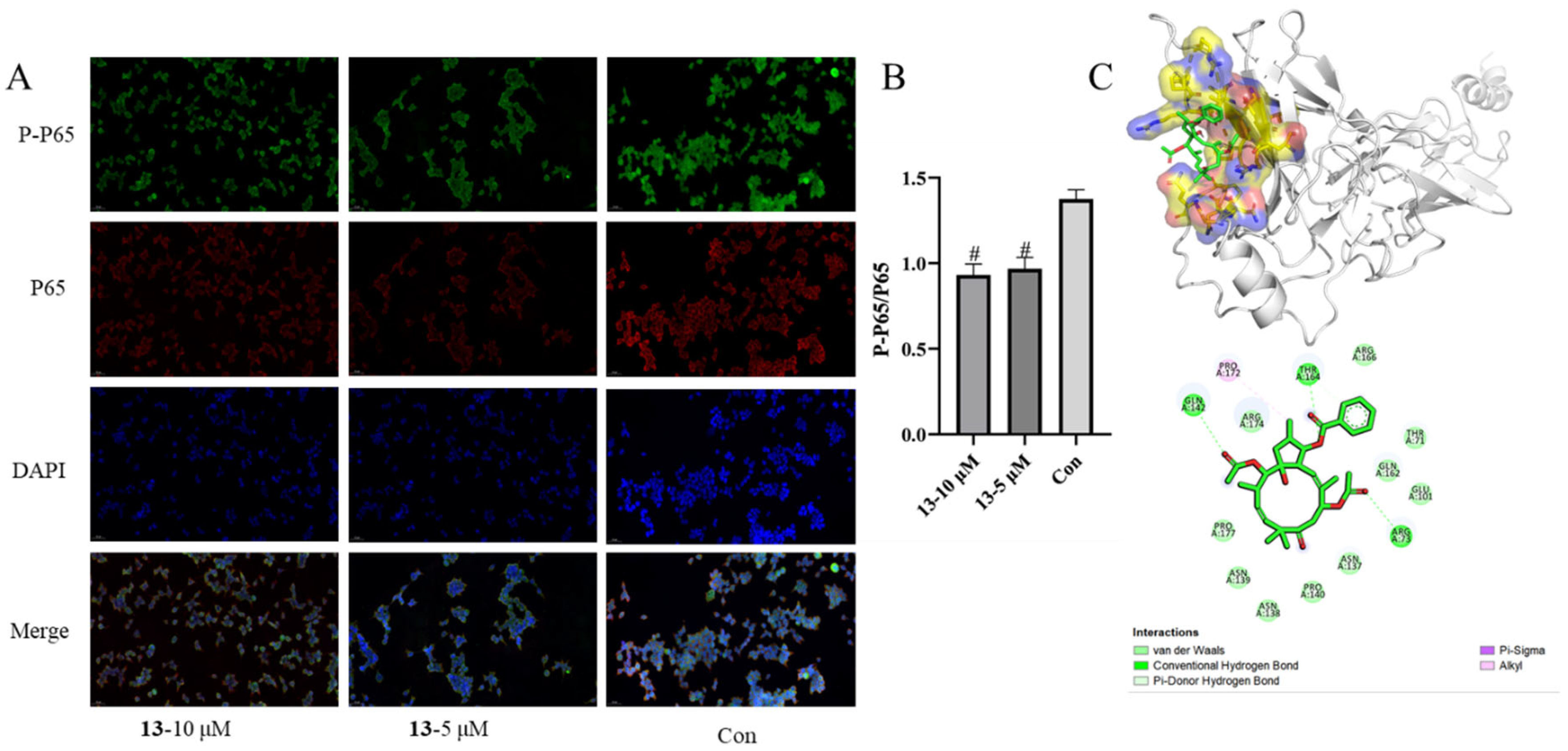
2.2. Biological Activity Results

In China, E. helioscopia is one of the main herbs to treat psoriasis [5,7,8,14], a skin disease characterized by the excessive proliferation of keratinocytes and lymphocytes. Therefore, compounds 123 were evaluated for their immunosuppressive activities against the proliferation of T/B lymphocyte cells and HaCaT cells (Human immortalized keratinocytes) in vitro. Preliminary results (Table 6) indicated that nearly all diterpenoids moderately inhibited concanavalin A (ConA)-induced T lymphocytes and/or lipopolysaccharide (LPS)-induced B lymphocytes proliferation (c 20.0 μM), as well as HaCaT cell proliferation (c 40.0 μM). Moreover, compounds 5 and 7 displayed significant immunosuppressive activities, with IC50 values of 17.6/10.2 and 6.7/11.5 μM (Table 7) against induced T and B cells. Compound 13 exhibited stronger anti-proliferative activities on HaCaT cells than the positive control, MTX (Table 8). Since compounds 7 and 21 in different jatropha-type families showed considerable immunosuppressive activity, they were also evaluated for their inhibitory effect by EdU experiments. Compared with the model or control controls, EdU experiments demonstrated that compounds 7/21 and 9/13 significantly inhibited T/B and HaCaT cells, respectively (Figure 5 and Figure 6) (Figure S54, Supplementary Materials).
In addition, compounds 7 and 21 were also evaluated for their inhibitory effect on the secretion of cytokine interferon (IFN) γ, interleukin (IL) 2, and IL-17A of mouse splenocytes, as previously described [29]. Lymphocytes were seeded in 96-well plates and treated with compounds 7 and 21 at concentrations of 20, 10, 5, 2.5, and 0 μM for 48 h. Cell viability was analyzed using a cell counting kit-8 (CCK-8) assay [30]. Since there were no obvious differences in cell viability between 7/21 (c = 2.5 μM) and the control group, inhibitory activity against the secretion of cytokines was evaluated at a concentration of 2.5 μM and 1 μM (Figure 5A). As shown in Figure 5B, after being stimulated with ConA, the levels of IFN-γ/IL-2/IL-17A increased significantly in the model group via the control group (p < 0.0001); when dealt with compounds 7 and 21, the secretions of IFN-γ (p < 0.0001) and IL-2 (p < 0.01) were significantly inhibited. Although compounds 7 and 21 reduced the secretion of IL-17A, there is no significant difference compared with the model group.
Nuclear factor-κB (NF-κB) is a family of heterodimeric proteins, including proteolytic processing of the p50 subunit as well as the P65 subunit. NF-κB activation results in the expression of genes associated with cellular proliferation, differentiation, and survival [31]. Therefore, the most promising compound 13 for psoriasis was chosen to evaluate its molecular mechanisms associated with the NF-κB pathway for the antiproliferative effect on HaCaT cells. As indicated in Figure 7, compound 13 attenuated the activation of NF-κB in a concentration-dependent manner (10 and 5 μM). Molecular docking analysis between 13 and P65 revealed that (i) the oxygen at C-3 and the carbonyl group of acetoxy groups at C-7 and C-14 formed hydrogen bonds with the residues THR164, ARG73, and GLN142, which make the structure of complex P65-13 stable; (ii) alkyl interaction was generated between 16-CH3 and the residue PRO172 (Figure 7). The binding energy of 13 with P65 is −7.3 kcal/mol. These results suggested that compound 13 inhibits the phosphorylation of NF-κB p65 in HaCaT cells.

3. Discussion

The structural elucidation of natural products remains a significant challenge for chemists. Developing new methods using JBCA, examining the intensity of the nuclear overhauser effect, relying on quantum-chemical calculations or X-ray crystallographic analysis, and summarizing empirical rules of NMR data are effective procedures for determining the relative configurations of organic molecules [12,13,18,19]. Recently, the relationships between the relative configurations of C-2/C-13/C-14 and 13C NMR chemical shifts (C-16, C-4, C-13) were summarized in specific jatropha diterpenes [12]. However, these rules did not encompass jatropha-11E-ene systems with (7,8-seco) 3α-benzyloxy (or hydroxy) derivatives or these hydroxy-substituted at C-3/C-14/C-15. Therefore, this work further analyzed the correlations between 3J values/chemical shifts of the relevant protons/carbons and the relative configuration of C-2, C-13, and C-14 in A/B-type jatropha diterpenes based on the previous study (Table 4 and Table 5) [12].
Psoriasis is a common immune-mediated skin disease with unclear cellular and molecular mechanisms; however, T cell-mediated hyperproliferation of keratinocytes is the key feature of the pathophysiology of psoriasis. Immunosuppressants, vitamin A acid, methotrexate, and glucocorticoids are traditional and classical clinical medicines [2]. Moreover, E. helioscopia is the predominant ingredient in the Chinese ZeQi Powder Preparation for treating psoriasis [5,14]. Kv1.3 ion channel is predominant in activated effector memory T (TEM) cells to further stimulate TEM cell proliferation and cytokine production [8]. Although some studies have found helioscopids A/O/euphohelioscoids A from E. helioscopia inhibited Kv1.3 activity in human embryonic kidney cells 293 (HEK293) cells [7,8], this activity only indirectly suggests the immunosuppressive potential of these compounds. Thus, more relevant bioassay experiments are needed to elucidate how and why compounds from E. helioscopia contribute to psoriasis treatment. Additionally, natural products often vary due to their different origins.
Lymphocytes, which are the most important immune cells, can be divided into T and B lymphocytes. During an immune response, T and B lymphocytes secrete various inflammatory factors (such as IL-2, IFN-γ, IL-17A, and so on) or antibodies to maintain internal environmental stability jointly. However, the overactivation of T or B lymphocytes can lead to various autoimmune diseases, such as psoriasis [32]. In vitro, the proliferation of T and B lymphocytes can be selectively stimulated by Con A and LPS, respectively. Meanwhile, the production level of cytokines and antibodies increases significantly when T and B lymphocytes are stimulated. Therefore, based on the specificity of these two mitogens and reference to the experimental model of splenic cell proliferation inhibition, this study evaluated the immunosuppressive activity of compounds from E. helioscopia using the previously established experimental model [33]. As depicted in Table 7 and Figure 5, most compounds displayed anti-proliferative effects on induced T and B cells. In addition, compounds 7 and 21 significantly reduced the secretions of IFN-γ (p < 0.0001) and IL-2 (p < 0.01). IL-2 and IFN-γ are closely associated with T cell proliferation [34]. Therefore, compounds 7 and 21 may exert anti-proliferative effects on T lymphocytes by reducing IFN-γ and IL-2 levels.
As previously reported, excessive proliferation and aberrant differentiation of keratinocytes are the main cellular events in psoriasis development [35]. Since the HaCaT cell line exhibits good stability and infinite proliferation ability, similar to the characteristics of the rapid proliferation of epidermal cells in psoriasis pathophysiological changes, it has been widely used in psoriasis models in vitro [35]. As shown in Table 8 and Figure 6, more than half of the compounds effectively inhibited HaCaT cells, particularly compound 13. NF-κB proteins are a family of transcription factors central to inflammation and immunity, and they also play crucial roles in development, cell growth, survival, proliferation, and various pathological conditions [36]. The mechanism of 13 was then explored, focusing on NF-κB P65. Compound 13 may bind with NF-κB P65, inhibiting its phosphorylation of NF-κB P65 and thereby reducing the proliferation of HaCaT cells (Figure 7).
Compounds 5/6, 8/9, 11/12, and 13/14 are four pairs of jatropha diterpenes differing only in their C-7 substitution (hydroxy or acetoxy). Analyzing the structure-activity relationship among these diterpenoid pairs, 7-OH is more beneficial than 7-OAc for the immunosuppressive activity of 5, 9, 12, and 13. However, 7-OAc is more effective than 7-OH in antiproliferative activity against HaCaT cells.

4. Materials and Methods

4.1. General Experimental Procedures

UV spectra were recorded on a Shimadzu UV-2401PC UV-visible recording spectrophotometer (Shimadzu, Kyoto, Japan). IR spectra (in CH3OH) were measured on a Bruker Tensor 27 spectrometer (Bruker, Karlsruhe, Germany). Rotations were measured using the APIV (Rudolph Research Analytical, Hackettstown, NJ, USA). A Chiral scan circular dichroism spectrometer (Applied Photophysics Ltd., Leatherhead, Surrey, UK) recorded the CD spectra. All the nuclear magnetic resonance (NMR) spectra were obtained on a Bruker Avance III 500 MHz spectrometer (Bruker Corporation, Karlsruhe, Germany). High-resolution electrospray ionization mass spectra (HRESIMS) were recorded using an AB Sciex Triple-TOF 6600 (AB SCIEX, Framingham, MA, USA). Sephadex LH-20 (Amersham Biosciences, Uppsala, Sweden) and silica gel (Qingdao Haiyang Chemical Co., Ltd., Qingdao, China) were used for column chromatography (CC). Medium-pressure liquid chromatography (MPLC) was performed on the Agela ODS flash column (C-18, 120 g, 40–60 mm, Tianjin, China) and the FL-H050G MPLC system (Agela Technologies, Tianjin, China). Preparative high-performance liquid chromatography (prep. HPLC) was performed on a SEP LC-52 equipped with a YMC-pack ODS-A column (dimensions 250 × i.d. 10 mm, particle size 5 μm, YMC, Tokyo, Japan) and a MWD UV detector (Separation Technology Co., Ltd., Beijing, China). All solvents used were analytical grade (TJshield Fine Chemicals Co., Ltd., Tianjin, China). All elution systems were described as volume ratio. Concanavalin A (ConA), lipopolysaccharide (LPS, Escherichia coli 055: B5), CCK-8, and RPMI 1640 medium were purchased from GibcoBRL, Life Technologies (Carlsbad, CA, USA). Fetal bovine serum (FBS) was purchased from HyClone Laboratories (Logan, UT, USA).

4.2. Plant Material

The whole plant, E. helioscopia L. (Euphorbiaceae), was collected from Kaifeng City, Henan Province, China, in May 2017 and identified by Prof. Cheng-Ming Dong (Henan University of Chinese Medicine), an expert in the taxonomic field of Chinese medicine. A voucher specimen (No. HZY201705) was deposited at the School of Pharmacy, Henan University of Chinese Medicine.

4.3. Extraction and Isolation

The whole air-dried and powdered E. helioscopia L. plant (6.0 kg) underwent a quadruple extraction with 95% ethanol. After evaporation under reduced pressure, the extract was partitioned between ethyl acetate and water four times, yielding a crude extract (420 g). The crude extract underwent normal column chromatography (CC) using a stepwise gradient of petroleum ether to acetone (from 10:1 to 1:1), resulting in three fractions (I–III). Fractions II (130 g) and III (120 g) were subjected to macroporous resin CC with gradient elution (EtOH/H2O: 20%, 40%, 60%, 80%, and 100%) each giving 14 (IIa–IIn) and 11 (IIIa–IIIk) subfractions. Subfractions IId, IIf, and IIg were further separated by Sephadex LH-20 CC (CH3OH), leading to fractions IId1–IId4, IIf1–IIf4, and IIg1–IIg7. Then, fraction IId3 was fractioned on silica gel CC (isocratic elution of ether/acetone, 2:1). Finally, compound 10 (82.6 mg, tR = 9.5 min) was purified from minor fraction IId3a by HPLC (CH3CN/H2O, 70:30 to 100:0, 30 min, 5 mL/min). Fraction IIf1 was separated on an MPLC equipped with an MCI CC (CH3OH/H2O: 20%, 40%, 60%, 80%, 100%) and 11 subfractions (IIf1a-IIf1k). Compound 22 (9.2 mg, tR = 25.3 min) was isolated from IIf1f via HPLC (CH3CN/H2O, 50:50 to 80:20, 30 min, 5 mL/min). After preparation by HPLC (CH3CN/H2O, 50:50 to 70:30, 30 min, 5 mL/min) of fraction IIf1g, 12 (11.7 mg, tR = 24.8 min), 20 (2.6 mg, tR = 27.8 min), 17 (4.2 mg, tR = 28.3 min), and a mixture IIf1g9. The mixture IIf1g9 was purified by HPLC (CH3CN/H2O, 66:34 to 70:30, 30 min, 5 mL/min) again, then compounds 5 (14.2 mg, tR = 18.8 min) and 16 (5.1 mg, tR = 21.9 min) were isolated. Subfraction IIf1h was divided into six fractions (IIf1h1-IIf1h6) by HPLC (CH3CN/H2O, 63:37 to 70:30, 35 min, 5 mL/min). Respectively, compounds 15 (17.4 mg, tR = 16.2 min; CH3CN/H2O, 68:32, 30 min, 5 mL/min) from subfraction IIf1h2, and 14 (7.4 mg, tR = 16.0 min; CH3CN/H2O, 66:34, 30 min, 5 mL/min) from subfraction IIf1h3 were obtained via HPLC once again. Fraction IIf1i gave compounds 6 (3.9 mg, tR = 23.6 min) and 13 (1.5 mg, tR = 39.0 min) via HPLC (CH3CN/H2O63:37 to 75:25, 40 min, 5 mL/min). Compounds 18 (2.1 mg, tR = 25.0 min, CH3CN/H2O 42:58 to 100:0, 40 min, 4 mL/min) from subfraction IIf2, 9 (4.6 mg, tR = 16.0 min, CH3CN/H2O 55:45 to 80:20, 30 min, 5 mL/min) from subfraction IIf3, and 3 (3.9 mg, tR = 13.2 min, CH3CN/H2O 60:40 to 100:0, 35 min, 5 mL/min) from subfraction IIfg1 were obtained through HPLC. After HPLC (CH3CN/H2O 60:40 to 80:20, 35 min, 5 mL/min), fraction IIg2 afforded 12 components (IIg2a-IIg2l). Subsequently, compounds 7 (1.3 mg, tR = 45.1 min CH3CN/H2O 46:54, 50 min, 5 mL/min) from subfraction IIg2a, 21 (9.2 mg, tR = 16.8 min; 60:40, 30 min, 5 mL/min) from subfraction IIg2b, 19 (3.3 mg, tR = 33.8 min CH3CN/H2O 55:45, 50 min, 5 mL/min) from subfraction IIg2c, 11 (11.7 mg, tR = 35.9 min; CH3CN/H2O 50:50, 50 min, 5 mL/min) from subfraction IIg2g, and 4 (4.7 mg, tR = 25.6 min; CH3CN/H2O 58:42, 50 min, 5 mL/min) from subfraction IIg2k via HPLC. Successively, components IIIh and IIIi were subjected to MPLC equipped with a C-18 column (CH3OH/H2O, 40%, 60%, 80%, and 100%), each affording four (IIIh1-IIIh14) and sixteen fractions (IIIi1-IIIi16). Fraction IIIh1 (CH3OH/H2O 48:52 to 100:0, 50 min, 10 mL/min) and IIIi8 (CH3OH/H2O 75:25 to 100:0, 40 min, 10 mL/min) gave six (IIIh1a-IIIh1f) and seven (IIIi8a-IIIi8g) fractions using HPLC. The compounds 1 (6.7 mg, tR = 26.2 min) and 2 (7.9 mg, tR = 27.2 min) were obtained from part IIIh1e purified by HPLC (CH3CN/H2O 42:58 to 70:30, 30 min, 4 mL/min) while compounds 23 (3.9 mg, tR = 12.6 min, CH3CN/H2O 50:50 to 80:20, 35 min, 4 mL/min) from subfraction IIIi8d, 8 (2.5 mg, tR = 32.8 min, CH3CN/H2O 55:45 to 80:20, 35 min, 4 mL/min) from subfraction IIIi8e were purified by HPLC.

4.4. Spectroscopic Data

Ent-2,3-seco-14-oxo-16-atisene-2,3-dioic acid (1): Amorphous powder, [α ] D 25 —34.7 (c 0.016, MeOH); UV (MeOH) λmax (logε): 295.0 (2.10), 200.0 nm (3.51); IR (MeOH) νmax/cm−1 1701, 1721, 1636, 1530, 1214, 1161; 1H NMR (500 MHz CDCl3) data see Table 1; 13C NMR (125 MHz CDCl3) data see Table 1; HRESIMS m/z 371.1808 [M+Na]+ (calcd for C20H28O5Na, 371.1829).
Ent-2,3-seco-2,3-dicarboxy-abieta-8(14),13(15)-dien-16,12-olide (2): Amorphous powder, [α ] D 25 + 130.1 (c 0.016, MeOH); UV (MeOH) λmax (logε): 275.0 (4.54), 205.0 nm (4.11); IR (MeOH) νmax/cm−1 3394, 1735, 1674, 1639, 1029; 1H NMR (500 MHz CDCl3) data see Table 1; 13C NMR (125 MHz CDCl3) data see Table 1; HRESIMS m/z 385.1613 [M+Na]+ (calcd for C20H26O6Na, 385.1622).
Secoheliosphane C (3): Amorphous powder, [α ] D 25 + 112.1 (c 0.02, MeOH), 1H NMR (500 MHz CDCl3) data see Table 2; 13C NMR (125 MHz CDCl3) data see Table 3; HRESIMS m/z 563.2564 [M+K]+ (calcd for C31H40O8K, 579.2354).
Secoheliosphane D (4): Colourless oil, [α ] D 25 + 128.1 (c 0.02, MeOH); 1H NMR (500 MHz CDCl3) data see Table 2; 13C NMR (125 MHz CDCl3) data see Table 3; HRESIMS m/z 537.2459 [M+K]+ (calcd for C29H38O7K, 537.2449).
Heliosco-jatrophane A (5): Colourless oil, [α ] D 25 + 98.6 (c 0.02, MeOH); 1H NMR (500 MHz CDCl3) data see Table 2; 13C NMR (125 MHz CDCl3) data see Table 3; HRESIMS m/z 563.2616 [M+Na]+ (calcd for C31H40O8Na, 563.2615.
Heliosco-jatrophane B (6): Amorphous powder, [α ] D 25 + 51.3 (c 0.02, MeOH) 1H NMR (500 MHz CDCl3) data see Table 2; 13C NMR (125 MHz CDCl3) data see Table 3; HRESIMS z m/z 605.2714 [M+Na]+ (calcd for C33H42O9Na, 605.2721.
Heliosco-jatrophane C (7): Colourless oil, [α ] D 25 + 100.1 (c 0.016, MeOH); 1H NMR (500 MHz CDCl3) data see Table 2; 13C NMR (125 MHz CDCl3) data see Table 3; HRESIMS m/z 521.2530 [M+Na]+ (calcd for C29H38O7Na, 521.2515).

4.5. Biological Activity Assays

Immunosuppressive activities assay and antiproliferative activity against HaCaT cells were performed according to the reported methods [33].

4.5.1. Immunosuppressive Activities Assay

Female or male Balb/c mice (6–8 weeks old) were sacrificed, and their spleens were removed aseptically. A single-cell suspension was prepared by pressing the spleens against the bottom of the Petri dish with a 5 mL syringe plunger, and cell debris and clumps were removed. Erythrocytes were depleted with ammonium chloride buffer solution, and the mononuclear cell suspensions were maintained in RPMI 1640 media containing 10% fetal bovine serum, penicillin (100 U/mL), and streptomycin (100 μg/mL). Then the mononuclear cell suspensions were dispensed into 96-well plates (2 × 10⁵ cells/well), in the absence or presence of compounds (c = 20.0 μM), were stimulated with ConA (5 μg/mL) or LPS (15 μg/mL) to induce T cell or B cell proliferative responses, respectively. Dexamethasone (Dex, c = 2.0 μM) was used as a positive control. In the 96-well plates, the wells cultured with cells and ConA/LPS were assigned as the modal group (Mod), the wells containing cells were described as the control group (Con), and the wells containing culture medium were described as the blank group. These 96-well plates were maintained under a humidified 5% CO2 atmosphere at 37 °C for 48 h. 10 μL of CCK-8 was added to each well at the final 4–5 h of culture, and the absorbance (OD) values were measured with a microplate reader (SpectraMax iD3, Molecular Devices, LLC.) at 450 nm. Stimulation Index (SI) = (ODsample−ODblank)/(ODCon−ODblank); Inhibition rate = (1−SIsample/SIMod) ×%. The IC50 value for each compound was calculated using the Reed and Muench method. All experiments were performed in triplicate.

4.5.2. Cell Viability Assays of Lymphocytes

Similarly, the mononuclear cell suspensions of lymphocytes (2 × 10⁵ cells/well) were cultured within 96-well plates in the absence or presence of compounds 7 or 21 (c = 20.0, 10, 5, 2.5, and 0 μM). In the 96-well plates, the wells containing cells were described as the control group (Con), and the wells containing culture medium were described as the blank group. These 96-well plates were maintained under a humidified 5% CO2 atmosphere at 37 °C for 48 h. 10 μL of CCK-8 was added to each well at the final 5 h of culture, and the OD values were measured with a microplate reader at 450 nm. Cell vialibity (%) = (ODsample−ODblank)/(ODCon−ODblank) × 100%.

4.5.3. Antiproliferative Activity against HaCaT Cells

The keratinocyte cell line was HaCaT (FH0186), bought from Fufeng Biology (Shanghai, China). HaCaT cells were cultured using the trypsin enzyme digesting technique and then passaged in vitro. The number of digested cells was counted by cell counter and were maintained in DMEM media containing 10% fetal bovine serum, penicillin (100 U/mL), and streptomycin (100 μg/mL) under a humidified 5% CO2 atmosphere at 37 °C. In brief, each well of a 96-well cell culture plate was seeded with 100 μL of cells (1 ×10⁵ cells/mL) and kept for 12 h for adherence and then added with test compounds (at a final concentration of 40 μM). The wells containing cells were described as the control group (Con), and the wells containing culture medium were described as the blank group. Methotrexate (MTX) was used as a positive control. After different concentrations of test compounds addition, each cell line was incubated for 48 h under a humidified 5% CO2 atmosphere at 37 °C. After the incubation, each well was treated with CCK-8 (10 μL), and incubation continued for 4 h at 37 °C. Then, the 96-well cell culture plates were subjected to a measure of optical density at 450 nm with a 96-well microplate reader. The IC50 value for each compound was calculated using the Reed and Muench method. All experiments were performed in triplicate.

4.5.4. EdU Assay

EdU fluorescence labeling for cell proliferation of induced T/B and HaCaT cells was performed as previously reported [37,38]. EdU (the Click-iT™ EdU Flow Cytometry Assay Kit, APExBIO, Houston, TX, USA) was added 24 h (final concentration 50 μM, T/B cells) and 4 h (final concentration 10 μM, HaCaT cells) before harvesting the cells. For the Click reaction, cells were fixed with 100 μL of 4% paraformaldehyde for 15 min. Cells were washed again and incubated with 100 μL of saponin-based permeabilization buffer for 15 min. After additional washing, cells were incubated with 100 μL Click-iT reaction buffer for 1 h and washed again with 200 μL permeabilization buffer. All procedures were performed according to the manufacturer’s instructions.

4.5.5. Cytokine Analysis by ELISA of Induced T Cells

The mononuclear cell suspensions (2 × 10⁵ cells/well) were cultured with ConA (5 μg/mL) in 96-well plates, and indicated concentrations of compounds were added simultaneously. After a 48-h culture period, cytokines in the supernatants were quantified using mouse IFN-γ, IL-2, and IL-17A ELISA kits (Mabtech, Stockholm, Sweden), following the manufacturer’s protocol [29].

4.5.6. Immunofluorescence Protocol (Cell Climbing Slides)

HaCaT cells (2.0 × 10⁵ cells/well) were plated onto 6-well plates with glass coverslips and allowed to adhere overnight (12 h) before compound addition. After treatment with compound 13 (final concentration 10/5 μM) for 36 h, the supernatant was removed, and the cells were washed twice with PBS, fixed with 4% paraformaldehyde for 15 min, and then washed with PBS three times. After blocking for 1 h with 4% bovine serum albumin (BSA) at 37 °C, the supernatant was discarded, washed three times, and incubated with NF-κB P65 and phosphorylated NF-κB P65 antibodies (1:200 diluted at 4% BSA; Servicebio, Wuhan, China) overnight at 4 °C. The initial incubation was followed by another incubation with secondary antibodies (Cy3 conjugated Goat Anti-Rabbit IgG, 1:300 diluted in 4% BSA; AlexaFluor®488-conjugated Goat Anti-Mouse IgG, 1:400 diluted in 4% BSA) (Servicebio, Wuhan, China) for another 50 min at room temperature. Then, cells were counterstained with 4,6-diamino-2-phenylindole (DAPI) for 5 min after washing three times with PBS. After the PBS washing, fluorescent seal liquid (PBS) was added, and the plate was monitored under an imaging system (Pannoramic MIDI, 3Dhistech, Budapest, Hungary). The nucleus is blue-labeled with DAPI. Positive cells are green (phosphorylation NF-κB P65 (P-P65) or red (NF-κB P65). The immunofluorescence areas for each indicator were analyzed in Image J 1.46r.

4.5.7. Statistics and Reproducibility

Data analyses were carried out using Prism 8.0, and One-way ANOVA was used to compare the differences between groups. LSD or Dunnett’s T3 test was used for pair comparison according to standard deviation values. p < 0.05 or p < 0.01 were considered to be statistically significant.

4.5.8. Ethic Statement

The animal study (No. DWLL202003116) was reviewed and approved by the Animal Welfare Ethics Committee of the Henan University of Chinese Medicine.

4.6. Molecular Docking Studies

The crystal structure of the inducible nitric oxide synthase (P65) (PDB: 1NFI) was downloaded from RCSB PDB (http://www.rcsb.org/, accessed on 6 June 2024), the structure of 13 was drawn by ChemDraw 14.0, and the 3D structure file was transformed by Chem3D 14.0. Before docking, the water molecules on the receptors were removed, and polar hydrogen atoms, charge, and magnetic field were added. AutoDockTools 1.5.6 was used to process ligands and receptors, and AutoDock vina was used for molecular docking. The 3D and 2D diagrams of the best-scored binding pose were visualized by Discovery Studio Visualizer v20 (http://www.discoverystudio.net/ accessed on 6 June 2024).

5. Conclusions

Phytochemical investigation of E. helioscopia resulted in isolating 23 diterpenoids, including four rare novel seco-diterpenoids (14) and three new jatropha diterpenes (57). Based on the NMR data obtained in this study and the previously summarized configuration rules, as well as those with crystallographic structures reported in the literature, the correlations between 3J values/chemical shifts of the relevant protons and the relative configuration of C-2, C-13, and C-14 in jatropha diterpenes were considered.
Bioassay results showed that almost all diterpenoids, especially the jatropha-type ones, simultaneously inhibited the proliferation of induced T/B, HaCaT cells, and the secretion of IFN-γ and IL-2. Immunofluorescence results and molecular docking studies suggested that compound 13 contributed, at least partially, to its antiproliferative effect on HaCaT cells by inactivating NF-κB through decreasing phosphorylation of P65. This work first explored the promising for psoriasis of jatropha diterpenoids from two aspects: T/B lymphocytes and HaCaT cells.

Supplementary Materials

The following supporting information can be downloaded at https://www.mdpi.com/article/10.3390/molecules29174104/s1, including the NMR, HRMS data, computational details, and other relevant information on compounds 17, 13, and 20 [39,40,41,42].

Author Contributions

Conceptualization and Supervision, W.-S.F. and X.-K.Z.; Methodology, Z.-Z.Z.; Investigation, X.-B.L.; Y.-S.Z. and L.-N.B.; Validation, G.-M.X.; Formal analysis, H.-J.H.; Data curation, Y.-J.S.; Writing–original draft, review and editing, Z.-Z.Z.; Funding acquisition, Z.-Z.Z. and H.C. All authors analyzed the data, discussed the results, and wrote the paper. All authors have read and agreed to the published version of the manuscript.

Funding

This work was financially supported by the National Natural Science Foundation of China (82003607), the Joint Fund of Provincial Science and Technology Plan of Henan Province (232301420094), Postdoctoral Research Funding of Henan Province (HN2022078), the Scientific and Technological Key Project in Henan Province (232102310426 and 242102310312), and Program for Innovative Research Team (in Science and Technology) in University of Henan Province (24IRTSTHN039).

Institutional Review Board Statement

Not applicable.

Informed Consent Statement

Not applicable.

Data Availability Statement

The original contributions presented in the study are included in the article/Supplementary Materials; further inquiries can be directed to the corresponding author.

Conflicts of Interest

The authors declare no conflict of interest.

References

  1. Gudjonsson, J.E.; Johnston, A.; Sigmundsdottir, H.; Valdimarsson, H. Immunopathogenic mechanisms in psoriasis. Clin. Exp. Immunol. 2004, 135, 1–8. [Google Scholar] [CrossRef]
  2. Rendon, A.; Schäkel, K. Psoriasis pathogenesis and treatment. Int. J. Mol. Sci. 2019, 20, 1475. [Google Scholar] [CrossRef]
  3. Christophers, E. Psoriasis-Epidemiology and clinical spectrum. Clin. Exp. Dermatol. 2001, 26, 314–320. [Google Scholar] [CrossRef] [PubMed]
  4. Shi, Q.W.; Su, X.H.; Kiyota, H. Chemical and pharmacological research of the plants in genus Euphorbia. Chem. Rev. 2008, 108, 4295–4327. [Google Scholar] [CrossRef] [PubMed]
  5. Yang, Y.; Chen, X.; Luan, F.; Wang, M.; Wang, Z.; Wang, J.; He, X. Euphorbia helioscopia L.: A phytochemical and pharmacological overview. Phytochemistry 2021, 184, 112649. [Google Scholar] [CrossRef] [PubMed]
  6. Wan, L.S.; Nian, Y.; Ye, C.J.; Shao, L.D.; Peng, X.R.; Geng, C.A.; Zuo, Z.L.; Li, X.N.; Yang, J.; Zhou, M.; et al. Three minor diterpenoids with three carbon skeletons from Euphorbia peplus. Org. Lett. 2016, 18, 2166–2169. [Google Scholar] [CrossRef] [PubMed]
  7. Zhou, C.G.; Xiang, Z.N.; Zhao, N.; Sun, X.; Hu, Z.F.; Wu, J.L.; Xia, R.F.; Chen, C.; Su, J.C.; Chen, J.C.; et al. Jatrophane diterpenoids with Kv1.3 ion channel inhibitory effects from Euphorbia helioscopia. J. Nat. Prod. 2022, 85, 815–827. [Google Scholar] [CrossRef] [PubMed]
  8. Xiang, Z.N.; Tong, Q.L.; Su, J.C.; Hu, Z.F.; Zhao, N.; Xia, R.F.; Wu, J.L.; Chen, C.; Chen, J.C.; Wan, L.S. Diterpenoids with rearranged 9(10→11)-abeo-10,12-cyclojatrophane skeleton and the first (15S)-jatrophane from Euphorbia helioscopia: Structural elucidation, biomimetic conversion, and their immunosuppressive effects. Org. Lett. 2022, 24, 697–701. [Google Scholar] [CrossRef]
  9. Cai, Q.; Zha, H.J.; Yuan, S.Y.; Sun, X.; Lin, X.; Zheng, X.Y.; Qian, Y.X.; Xia, R.F.; Luo, Y.S.; Shi, Z.; et al. Diterpenoids from Euphorbia fischeriana with Kv1.3 inhibitory activity. J. Nat. Prod. 2023, 86, 2379–2390. [Google Scholar] [CrossRef]
  10. Kim, S.J.; Jang, Y.W.; Hyung, K.E.; Lee, D.K.; Hyun, K.H.; Park, S.Y.; Park, E.S.; Hwang, K.W. Therapeutic effects of methanol extract from Euphorbia kansui Radix on imiquimod-induced psoriasis. J. Immunol. Res. 2017, 2017, 7052560. [Google Scholar] [CrossRef]
  11. Mai, Z.P.; Ni, G.; Liu, Y.F.; Li, Y.H.; Li, L.; Li, J.Y.; Yu, D.Q. Secoheliosphanes A and B and secoheliospholane A, three diterpenoids with unusual seco-jatrophane and seco-jatrophane skeletons from Euphorbia helioscopia. J. Org. Chem. 2018, 83, 167–173. [Google Scholar] [CrossRef] [PubMed]
  12. Su, J.C.; Cheng, W.; Song, J.G.; Zhong, Y.L.; Huang, X.J.; Jiang, R.W.; Li, Y.L.; Li, M.M.; Ye, W.C.; Wang, Y. Macrocyclic diterpenoids from Euphorbia helioscopia and their potential anti-inflammatory activity. J. Nat. Prod. 2019, 82, 2818–2827. [Google Scholar] [CrossRef] [PubMed]
  13. Zhou, D.; Zhang, F.; Kikuchi, T.; Yao, M.; Otsuki, K.; Chen, G.; Li, W.; Li, N. Lathyrane and jatrophane, diterpenoids from Euphorbia helioscopia evaluated for cytotoxicity against a paclitaxel-resistant A549 human lung cancer cell line. J. Nat. Prod. 2022, 85, 1174–1179. [Google Scholar] [CrossRef] [PubMed]
  14. Shi, L.L.; Liu, T.F. The effect of compound Zeqi granules on the serum content of MMP-2, MMP-9 and IL-18 in patients with psoriasis vulgaris. J. Dermatol. Venereol. 2014, 36, 68–69. [Google Scholar] [CrossRef]
  15. Lal, A.R.; Cambie, R.C.; Rutledge, P.S.; Woodgate, P.D. Ent-atisane diterpenes from Euphorbia fidjiana. Phytochemistry 1990, 29, 1925–1935. [Google Scholar] [CrossRef]
  16. Crespi-Perellino, N.; Garofano, L.; Arlandini, E.; Pinciroli, V. Identification of new diterpenoids from Euphorbia calyptrata cell cultures. J. Nat. Prod. 1996, 59, 773–776. [Google Scholar] [CrossRef]
  17. Wang, Y.; Liang, X.B.; Zhao, Z.Z. Diterpenoids from the whole herb of Euphorbia helioscopia. Chin. Tradit. Herb. Drugs 2022, 53, 4625–4633. [Google Scholar] [CrossRef]
  18. Zhou, L.; Guo, R.; Zhang, H.; Lu, L.W.; Du, Y.Q.; Liu, Q.B.; Huang, X.X.; Song, S. Rapid approaches for assignment of the relative configuration in 1-oxygenated 1,2-diarylpropan-3-ols by 1H NMR spectroscopy. J. Nat. Prod. 2021, 84, 20–25. [Google Scholar] [CrossRef]
  19. Chen, H.; Wang, H.; Yang, B.; Jin, D.Q.; Yang, S.; Wang, M.; Xu, J.; Ohizumi, Y.; Guo, Y. Diterpenes inhibiting NO production from Euphorbia helioscopia. Fitoterapia 2014, 95, 133–138. [Google Scholar] [CrossRef]
  20. Li, J.; Li, H.H.; Wang, W.Q.; Song, W.B.; Wang, Y.P.; Xuan, L.J. Jatrophane diterpenoids from Euphorbia helioscopia and their lipid-lowering activities. Fitoterapia 2018, 128, 102–111. [Google Scholar] [CrossRef]
  21. Kúsz, N.; Orvos, P.; Bereczki, L.; Fertey, P.; Bombicz, P.; Csorba, A.; Tálosi, L.; Jakab, G.; Hohmann, J.; Rédei, D. Diterpenoids from Euphorbia dulcis with potassium ion channel inhibitory activity with selective G protein-activated inwardly rectifying ion channel (GIRK) blocking effect. J. Nat. Prod. 2018, 81, 2483–2492. [Google Scholar] [CrossRef] [PubMed]
  22. Esposito, M.; Nothias, L.F.; Nedev, H.; Gallard, J.F.; Leyssen, P.; Retailleau, P.; Costa, J.; Roussi, F.; Iorga, B.I.; Paolini, J.; et al. Euphorbia dendroides Latex as a source of jatrophane esters, isolation, structural analysis, conformational study, and anti-CHIKV activity. J. Nat. Prod. 2016, 79, 2873–2882. [Google Scholar] [CrossRef] [PubMed]
  23. Mai, Z.P.; Ni, G.; Liu, Y.F.; Zhang, Z.; Li, L.; Chen, N.H.; Yu, D.Q. Helioscopianoids A–Q, bioactive jatrophane diterpenoid esters from Euphorbia helioscopia. Acta Pharm. Sin. B 2018, 8, 805–817. [Google Scholar] [CrossRef] [PubMed]
  24. Liu, C.; Liao, Z.X.; Liu, S.J.; Qu, Y.B.; Wang, H.S. Two new diterpene derivatives from Euphorbia lunulata Bge and their anti-proliferative activities. Fitoterapia 2014, 96, 33–38. [Google Scholar] [CrossRef] [PubMed]
  25. Tao, H.W.; Hao, X.J.; Liu, P.P.; Zhu, W.M. Cytotoxic macrocyclic diterpenoids from Euphorbia helioscopia. Arch. Pharmacal Res. 2008, 31, 1547–1551. [Google Scholar] [CrossRef]
  26. Yamamura, S.; Shizuri, Y.; Kosemura, S.; Ohtsuka, J.; Tayama, T.; Ohba, S.; Ito, M.; Saito, Y.; Terada, Y. Diterpenes from Euphorbia helioscopia. Phytochemistry 1989, 28, 3421–3436. [Google Scholar] [CrossRef]
  27. Lu, Z.Q.; Guan, S.H.; Li, X.N.; Chen, G.T.; Zhang, J.Q.; Huang, H.L.; Liu, X.; Guo, D.A. Cytotoxic diterpenoids from Euphorbia helioscopia. J. Nat. Prod. 2008, 71, 873–876. [Google Scholar] [CrossRef]
  28. Zhang, W.; Guo, Y.W. Chemical studies on the constituents of the Chinese medicinal herb Euphorbia helioscopia L. Chem. Pharm. Bull. 2006, 54, 1037–1039. [Google Scholar] [CrossRef]
  29. Jing, S.X.; Fu, R.; Li, C.H.; Zhou, T.T.; Liu, Y.C.; Liu, Y.; Luo, S.H.; Li, X.N.; Zeng, F.; Li, S.H. Immunosuppresive sesterterpenoids and norsesterterpenoids from Colquhounia coccinea var. mollis. J. Org. Chem. 2021, 86, 11169–11176. [Google Scholar] [CrossRef]
  30. He, H.; Cao, L.; Wang, Z.; Wang, Z.; Miao, J.; Li, X.M.; Miao, M. Sinomenine relieves airway remodeling by inhibiting epithelial-mesenchymal transition through downregulating TGF-β1 and smad3 expression in vitro and in vivo. Front. Immunol. 2021, 12, 736479. [Google Scholar] [CrossRef]
  31. Hexum, J.K.; Tello-Aburto, R.; Struntz, N.B.; Harned, A.M.; Harki, D.A. Bicyclic cyclohexenones as inhibitors of NF-κB signaling. ACS Med. Chem. Lett. 2012, 3, 459–464. [Google Scholar] [CrossRef] [PubMed]
  32. Ogawa, E.; Sato, Y.; Minagawa, A.; Okuyama, R. Pathogenesis of psoriasis and development of treatment. J. Dermatol. 2018, 45, 264–272. [Google Scholar] [CrossRef] [PubMed]
  33. Zhao, Z.Z.; Zhang, F.; Ji, B.Y.; Zhou, N.; Chen, H.; Sun, Y.J.; Feng, W.S.; Zheng, X.K. Pyrrole alkaloids from the fruiting bodies of edible mushroom Lentinula edodes. RSC Adv. 2023, 13, 18223–18228. [Google Scholar] [CrossRef] [PubMed]
  34. Abbas, A.K.; Trotta, E.; R Simeonov, D.; Marson, A.; Bluestone, J.A. Revisiting IL-2: Biology and therapeutic prospects. Sci. Immunol. 2018, 3, eaat1482. [Google Scholar] [CrossRef]
  35. Jiang, B.W.; Zhang, W.J.; Wang, Y.; Tan, L.P.; Bao, Y.L.; Song, Z.B.; Yu, C.L.; Wang, S.Y.; Liu, L.; Li, Y.X. Convallatoxin induces HaCaT cell necroptosis and ameliorates skin lesions in psoriasis-like mouse models. Biomed. Pharmacother. 2020, 121, 109615. [Google Scholar] [CrossRef]
  36. Morgan, M.J.; Liu, Z.G. Crosstalk of reactive oxygen species and NF-κB signaling. Cell Res. 2011, 21, 103–115. [Google Scholar] [CrossRef]
  37. Biram, A.; Shulman, Z. Evaluation of B Cell proliferation in vivo by EdU incorporation assay. Bio-Protocol 2020, 10, e3602. [Google Scholar] [CrossRef]
  38. Yu, Y.; Arora, A.; Min, W.; Roifman, C.M.; Grunebaum, E. EdU incorporation is an alternative non-radioactive assay to [3H]thymidine uptake for in vitro measurement of mice T-cell proliferations. J. Immunol. Methods 2009, 350, 29–35. [Google Scholar] [CrossRef]
  39. Goto, H.; Osawa, E. Corner flapping: A simple and fast algorithm for exhaustive generation of ring conformations. J. Am. Chem. Soc. 1989, 111, 8950–8951. [Google Scholar] [CrossRef]
  40. Goto, H.; Osawa, E. Application of a three-carbon ring expansion process to bridged bicyclic systems. J. Chem. Soc. Perkin Trans. 1993, 2, 187–198. [Google Scholar]
  41. Frisch, M.J.; Trucks, G.W.; Schlegel, H.B.; Scuseria, G.E.; Robb, M.A.; Cheeseman, J.R.; Scalmani, G.; Barone, V.; Mennucci, B.; Petersson, G.A.; et al. Gaussian 16, Revision C.01; Gaussian, Inc.: Wallingford, CT, USA, 2016. [Google Scholar]
  42. Bruhn, T.; Schaumlöffel, A.; Hemberger, Y.; Bringmann, G. SpecDis, Version 1.71; University of Würzburg: Berlin, Germany, 2012.
Figure 1. Structures of compounds 123 isolated from E. helioscopia L.
Molecules 29 04104 g001
Figure 2. Key 2D NMR correlations of compounds 1 and 2 (four rings numbered as A–D).
Molecules 29 04104 g002
Figure 3. Experimental CD spectra and calculated ECD spectra of 1 (σ = 0.22 eV, UV shift—7 nm) and 2 (σ = 0.25 eV, UV shift—4 nm).
Molecules 29 04104 g003
Figure 4. (A) Types A (3α-benzyloxy-jatropha-11E-ene) and B (8-seco-3α-benzyloxy-jatropha-11E-ene), whose relative configurations of C-2, C-14, and C-13 can be determined by analysis 1D NMR data. (B) γ-Gauche relationship of 20α-CH3 with H-11.
Molecules 29 04104 g004
Figure 5. (A) Cell viability (% of Control) of compounds 7 and 21 in different concentrations (vs. Con #### p < 0.0001, ### p < 0.001, ## p < 0.01). (B) Concentrations of IFN-γ/IL-2/IL-17A of induced T cells (Con A 5 μg/mL) in different groups (vs. Con #### p < 0.0001; vs. Mod, **** p < 0.0001, ** p < 0.01, * p < 0.05). (C) Inhibitory activities of compounds 7 (c = 10 μM), 21 (c = 10 μM), and Dex (c = 2/1 μM) against induced T cells (Con A 5 μg/mL) and B (LPS, 15 μg/mL) measured by EdU.
Molecules 29 04104 g005
Figure 6. Inhibitory activities of compounds 9 (c = 20 μM), 13 (c = 5 μM), and MTX (c = 20 μM) against HaCaT cells measured by EdU.
Molecules 29 04104 g006
Figure 7. (A) Immunofluorescence images: Effects of compound 13 (c = 10/5 μM) on P-NF-κB P65 (P-P65) and NF-κB P65 (P65) expression levels in HaCaT cells. (B) Ratios of P-P65/P65 of HaCaT cells in different groups (vs. Con, # p < 0.05). (C) Molecular docking analysis for predicted lowest-energy binding mode of P65 in 3-dimensional and 2-dimensional figures with 13 (Affinity −7.3 Kcal/mol); for ligand, the carbon and oxygen are highlighted in green and red, respectively; for residents of proteins, the yellow, red and blue stand for C, O, and N, respectively.
Molecules 29 04104 g007
Table 1. 13C (125 MHz) and 1H (500 MHz) NMR data of compounds 1 and 2 (δ in ppm, J in Hz, measured in CDCl3).
No.12
δCδHδCδH
139.02.79, d (19.7)39.53.21, d (20.3)
2.32, d (19.7)2.68, d (20.3)
2178.7 178.6
3187.5 186.9
445.5. C 45.5
548.32.54, d (12.4)48.12.84, d (12.0)
619.51.61, m23.32.03, m
1.03, overlapped1.53, overlapped
731.22.32, overlapped36.12.55, d (13.8)
0.98, overlapped2.34, m, overlapped
847.7 151.1
945.92.69 (m, overlap)45.93.32, d (7.2)
1041.3 43.6
1128.11.82, m28.02.38, m
1.56, m1.59, m
1238.62.74, m, overlapped75.64.86 dd (13.0, 5.7)
1344.72.31, m, overlapped155.6
14217.2 114.56.30, s
1542.92.27, m, overlapped116.9
1.76, m, overlapped
16147.0 175.1
17107.34.88, s8.31.83, s
4.67, s
1829.51.21, s29.51.23, s
1921.71.12, s21.31.20, s
2017.40.78, s21.01.01, s
Table 2. 1H (500 MHz) NMR data of compounds 37 (δ in ppm, measured in CDCl3, J in Hz).
No.34567
12.57, dd (12.5, 5.2)
2.12, overlapped
2.20, overlapped
1.53, dd (13.4, 11.1)
2.51, dd (14.0, 6.1)
1.95, t (14.0)
2.54, dd (13.6, 6.0)
1.99, d (13.6)
2.13, dd (14.5, 8.2)
1.51, dd (14.5, 8.6)
22.15, m2.75, m2.06, m2.06, m2.43, m
35.65, t-like (4.0)5.20, dd (8.5, 5.7)5.41, t-like (4.5)5.45, t-like (4.4)5.17, dd (6.8, 3.6)
43.61, dd (9.4, 4.0)3.28, dd (9.8, 8.5)3.26, dd (10.4, 4.5)3.25, dd (9.9, 4.4)3.16, dd (8.8, 3.6)
56.82, dd (9.3, 1.1)6.77, brs (9.8, 1.3)5.55, d (10.4)5.69, d (9.9)5.60, dd (8.8, 1.7)
79.36, s9.32, s4.16, t (7.5)5.03, dd (8.1, 1.7)4.39, dd (10.8, 4.5)
82.06, s2.16, s3.01, dd (14.5, 1.7)
2.44, dd (14.5, 6.9)
2.77, dd (13.6, 2.2)
2.86, dd (13.6, 8.4)
2.95, dd (15.4, 10.5)
2.69, dd (15.5, 4.6)
115.38, d (15.6) 5.58, d (15.9)5.15, d (15.4) 5.16, d (15.4) 5.43, d (16.1)
125.27, dd (15.6, 8.8)5.64, dd (15.9, 8.6) 5.25, dd (15.4, 8.8)5.25, dd (15.4, 8.7)5.15, dd (16.1, 9.0)
132.20, m2.68, m2.36, m2.37, m2.95, m
145.82, d (9.5) 4.94, d (3.1) 5.90, d (9.7) 5.93, d (9.9) 5.14, d (1.7)
160.96, d (7.0)1.22, d (6.9)0.92, d (6.6)0.92, d (6.4)1.08, d (5.9)
171.94, d (1.1)1.68, d (1.4)1.72, d (0.8)1.81, br. s1.81, d (1.5)
181.15, s1.29, s1.20, s1.12, s1.09, s
191.16, s1.29, s1.10, s1.14, s1.21, s
200.98, d (6.9)0.99, d (6.9)0.95, d (6.7)0.97, d (6.7)0.93, d (7.2)
3′, 7′7.99, dd (7.5, 1.3)7.93, dd (7.5, 1.3)7.95, dd (7.5, 1.3)7.97, d (7.5,1.3)7.94, dd (7.5, 1.2)
4′, 6′7.46, t (7.5)7.39, t (7.5)7.42, t (7.5)7.43, t (7.5)7.44, t (7.5)
5′7.60, t (7.5)7.52, t (7.5)7.54, t (7.5)7.55, t (7.5)7.56, t (7.5)
OAc-142.17, s1.90, s2.16, s2.14, s2.16, s
OAc-152.05, s 2.14, s2.20, s
OAc-7 1.31, s
Table 3. 13C NMR (125 MHz) data of compounds 37 (δ in ppm, measured in CDCl3).
No.34567
146.845.046.746.848.3
239.438.738.838.636.4
380.782.780.980.885.8
447.951.346.246.343.5
5147.3149.6120.1123120.6
6141.1140.7136.1133.7140.6
7194.719573.273.871.9
825.525.338.839.245.5
9211.2212.8212.2206.7210.0
1050.150.250.851.049.2
11135.2135.7130.3132.4130.1
12132.1130.1132.6130.7134.5
1340.139.640.941.437.1
1474.781.275.875.878.2
1590.484.390.290.483.6
1614.018.513.913.819.2
1710.59.516.217.118.9
1824.323.924.625.025.7
192424.919.620.221.2
2017.919.121.120.922.7
1′165.9166166.1165.4166.3
2′129.8133.2130.5130.3133.4
3′, 7′129.6129.6129.6129.6129.5
4′, 6′128.7128.5128.5128.6128.8
5′133.5131.7133133.1133.5
OAc-14169.3170.6169.8170.0170.7
22.320.822.321.124.8
OAc-15169.5 170.0169.5
21.2 20.922.4
OAc-7 170.2
20.2
Table 4. Correlations between 1H NMR data signals and the relative configuration of C-2, C-14, and C-13 in types A or B.
1H NMR DataConfigurationsSuitable Type
H-3, t-like, J = 3.0–5.0 Hz16α-CH3A, B
H-3, dd, J ≈ 8.0, 3.0–5.0 Hz16β-CH3A, B
H-14, d, J = 1.0–4.0 HzCH3-20/OR-14 same orientationA
H-14, d, J = 1.0–4.0 HzCH3-20/OR-14 same orientationB
H-14, d, J > 8.0 HzCH3-20/OR-14 different orientationA, B
δH-11 > δH-12 20β-CH3A, B
δH-11 < δH-12 20α-CH3A, B
Table 5. Correlations between 13C NMR data signals and the relative configuration of C-2, C-14, and C-13 in types A or B.
13C NMR DataConfigurationsSuitable Type
When OBz-3, δC-16 < 1516α-CH3A, B
When OBz-3, δC-16 > 1516β-CH3A, B
When OAc-14, δC-4 < 4514β-OAcA
When OAc-14, δC-4 > 4514α-OAc or 14β-OAc A
When OAc-14, δC-20 > 22CH3-20/OR-14 same orientationA
when OAc-14, δC-20 > 18.5 CH3-20/OR-14 same orientationB
When OAc-15, δC-13 < 4020β-CH3A, B
Table 6. Inhibition rate of compounds 123 (c 20.0 μM) and Dex (c 2.0 μM) against the ConA-induced proliferation of T and/or LPS-induced proliferation of B cells.
CompoundInhibition Rate (%)CompoundInhibition Rate (%)
T CellsB CellsT CellsB Cells
123.7 ± 4.9<101315.0 ± 2.727.2 ± 4.5
223.7 ± 6.8<101446.5 ± 8.328.2 ± 4.8
334.5 ± 5.420.0 ± 9.51525.4 ± 3.517.1 ± 7.3
423.8 ± 4.526.6 ± 9.71644.4 ± 8.626.6 ± 5.4
558.2 ± 1.464.9 ± 4.61735.6 ± 1.1<10
634.8 ± 7.4<101837.5 ± 5.633.5 ± 2.8
783.8 ± 2.592.4 ± 9.61942.5 ± 7.548.3 ± 8.8
849.8 ± 6.943.1 ± 8.82048.5 ± 7.146.6 ± 1.7
932.2 ± 2.927.1 ± 2.42149.8 ± 7.848.2 ± 6.4
1048.4 ± 3.248.7 ± 5.722<10<10
1146.0 ± 3.217.6 ± 2.42333.0 ± 8.9<10
1234.0 ± 3.314.2 ± 4.6Dex46.7 ± 4.379.3 ± 7.7
Table 7. IC50 values of compounds 5 and 7 against the induced proliferation of T cells (ConA) and B cells (LPS).
CompoundIC50 (μM)
T CellsB Cells
517.6 ± 2.710.3 ± 1.3
76.7 ± 1.8 11.4 ± 1.5
Dex1.6 ± 0.30.8 ± 0.05
Table 8. IC50 values of active compounds against the proliferation of HaCaT cells.
CompoundIC50 (μM)CompoundIC50 (μM)
731.0 ± 3.71625.9 ± 2.4
826.0 ± 2.21731.5 ± 3.3
919.7 ± 2.01829.8 ± 2.9
1031.3 ± 1.72128.5 ± 3.0
1223.7 ± 2.52229.6 ± 2.3
136.9 ± 0.82330.4 ± 3.0
1523.4 ± 1.6MTX18.1 ± 2.1
Disclaimer/Publisher’s Note: The statements, opinions and data contained in all publications are solely those of the individual author(s) and contributor(s) and not of MDPI and/or the editor(s). MDPI and/or the editor(s) disclaim responsibility for any injury to people or property resulting from any ideas, methods, instructions or products referred to in the content.

