
Application of Intelligent Response Fluorescent Probe in Breast Cancer

Abstract
1. Introduction
2. Use of Different Functionalized Fluorescent Probes in Breast Cancer
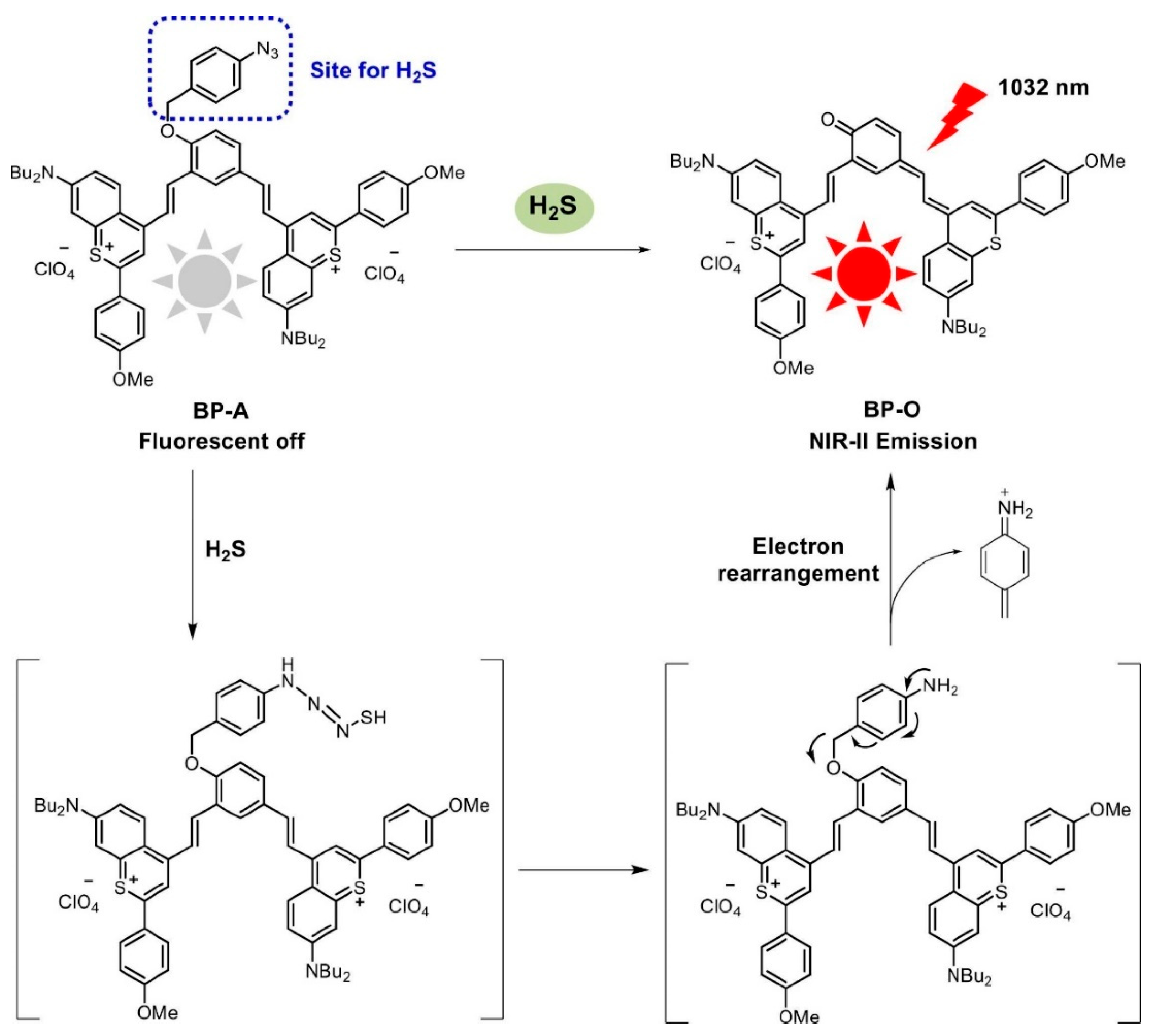
2.1. Synthesis of Near-Infrared Responsive Fluorescent Probes and Their Application in Breast Cancer
2.2. Synthesis of pH-Responsive Fluorescent Probes and Their Application in Breast Cancer
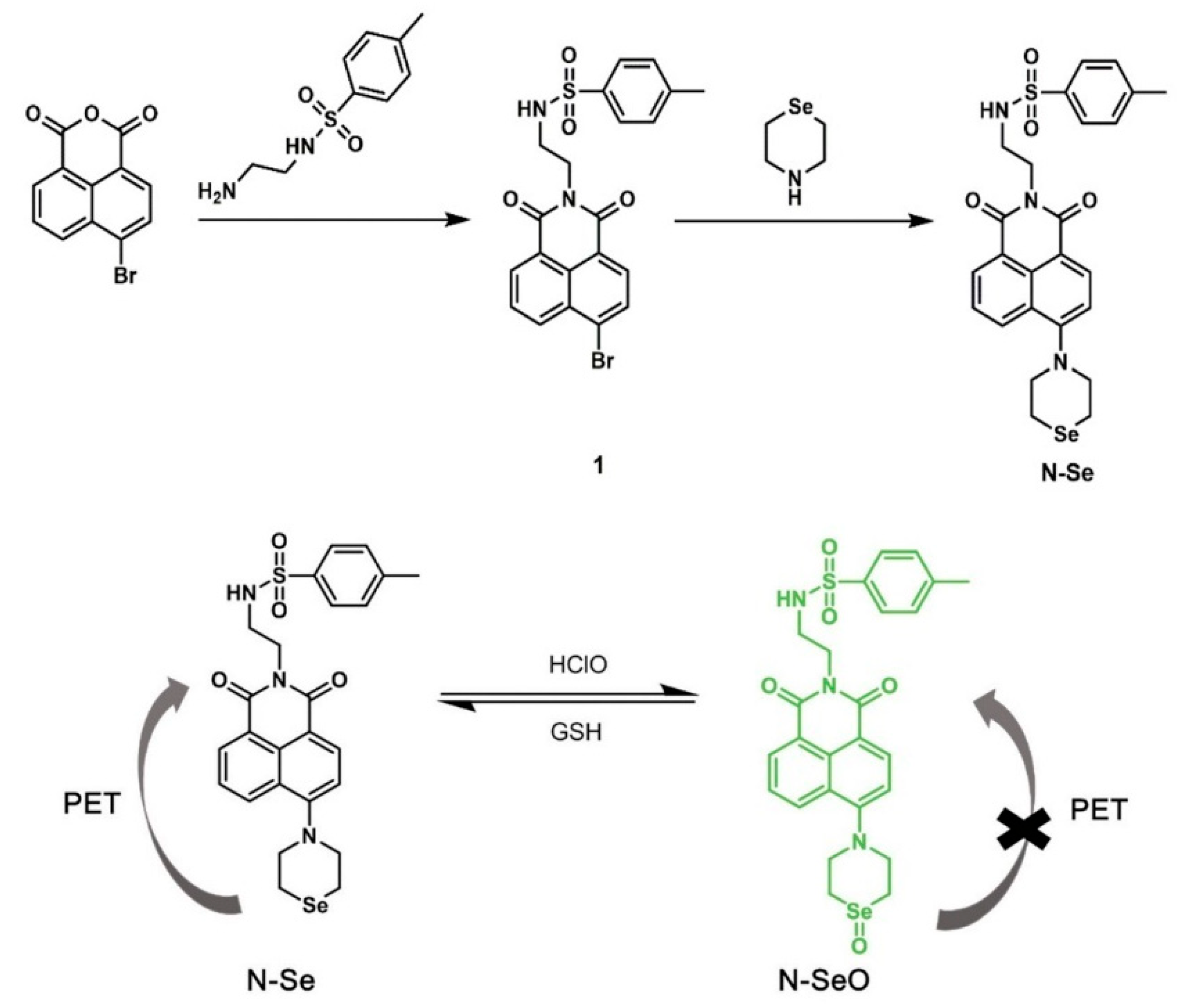
2.3. Synthesis of Redox Fluorescent Probes and Their Application in Breast Cancer
2.4. Synthesis of Enzyme-Responsive Fluorescent Probes and Their Application in Breast Cancer
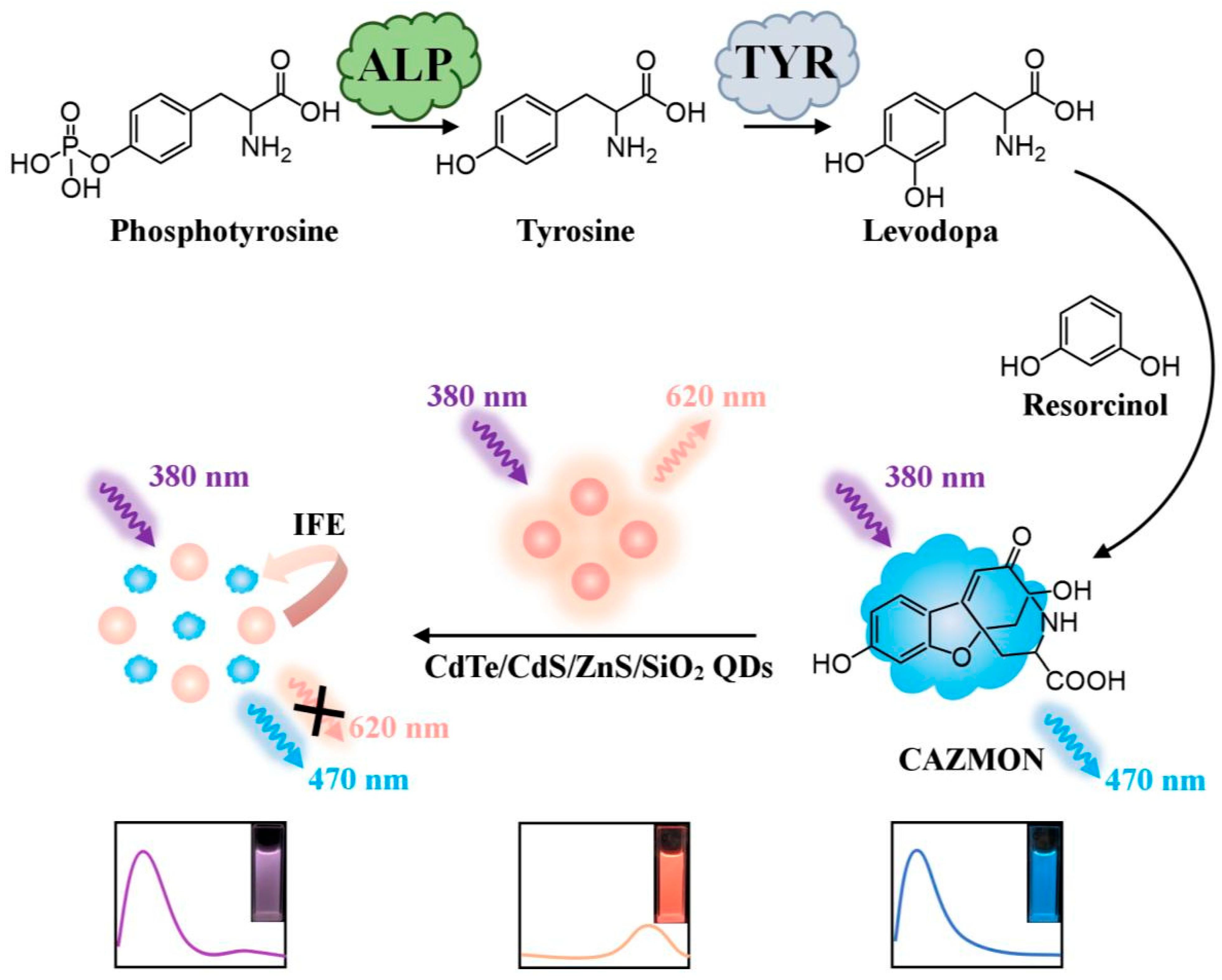
2.4.1. Alkaline Phosphatase Fluorescent Probe
2.4.2. Histonease
3. Diagnostic Integration of Fluorescent Probes in Breast Cancer
4. Summary and Outlook
Author Contributions
Funding
Institutional Review Board Statement
Informed Consent Statement
Data Availability Statement
Conflicts of Interest
References
- Yang, R.Z.; Han, Y.; Yi, W.J.; Long, Q. Autoantibodies as biomarkers for breast cancer diagnosis and prognosis. Front. Immunol. 2022, 13, 17. [Google Scholar] [CrossRef] [PubMed]
- Liu, M.; Li, Z.Y.; Yang, J.J.; Jiang, Y.; Chen, Z.; Ali, Z.; Wang, Z. Cell-specific biomarkers and targeted biopharmaceuticals for breast cancer treatment. Cell Prolif. 2016, 49, 409–420. [Google Scholar] [CrossRef]
- Lu, B.Y.; Natarajan, E.; Raghavendran, H.R.B.; Markandan, U.D. Molecular Classification, Treatment, and Genetic Biomarkers in Triple-Negative Breast Cancer: A Review. Technol. Cancer Res. Treat. 2023, 22, 10. [Google Scholar] [CrossRef] [PubMed]
- Wu, Y.Y.; Feng, Y.Q.; Li, X. Classification of breast cancer by a gold nanoparticle based multicolor fluorescent aptasensor. J. Colloid. Interface Sci. 2022, 611, 287–293. [Google Scholar] [CrossRef] [PubMed]
- Harbeck, N.; Gnant, M. Breast cancer. Lancet 2017, 389, 1134–1150. [Google Scholar] [CrossRef]
- Harbeck, N.; Penault-Llorca, F.; Cortes, J.; Gnant, M.; Houssami, N.; Poortmans, P.; Ruddy, K.; Tsang, J.; Cardoso, F. Breast cancer. Nat. Rev. Dis. Primers 2019, 5, 66. [Google Scholar] [CrossRef]
- Xu, D.Z.; Li, L.; Chu, C.C.; Zhang, X.Y.; Liu, G. Advances and perspectives in near-infrared fluorescent organic probes for surgical oncology. Wiley Interdiscip. Rev.—Nanomed. Nanobiotechnol. 2020, 12, 17. [Google Scholar] [CrossRef]
- Wen, Y.; Jing, N.; Huo, F.J.; Yin, C.X. Rational design of a turn-on fluorescent probe for visualization of GRP78 protein in tumor models. Chin. Chem. Lett. 2023, 34, 4. [Google Scholar] [CrossRef]
- Pola, R.; Böhmová, E.; Filipová, M.; Pechar, M.; Pankrác, J.; Vetvicka, D.; Olejár, T.; Kabesová, M.; Poucková, P.; Sefc, L.; et al. Targeted Polymer-Based Probes for Fluorescence Guided Visualization and Potential Surgery of EGFR-Positive Head-and-Neck Tumors. Pharmaceutics 2020, 12, 31. [Google Scholar] [CrossRef]
- Zhu, W.L.; Li, Q.H.; Gong, S.Y.; Feng, G.Q. Cell membrane targetable NIR fluorescent polarity probe for selective visualization of cancer cells and early tumor. Anal. Chim. Acta 2023, 1278, 341748. [Google Scholar] [CrossRef]
- Luo, C.X.; Zhang, Q.; Sun, S.G.; Li, H.J.; Xu, Y.Q. Research progress of auxiliary groups in improving the performance of fluorescent probes. Chem. Commun. 2023, 59, 2199–2207. [Google Scholar] [CrossRef] [PubMed]
- Wang, Q.Y.; Li, Z.; Hao, Y.T.; Zhang, Y.; Zhang, C.X. Near-Infrared Fluorescence Probe with a New Recognition Moiety for Specific Detection and Imaging of Aldehyde Dehydrogenase Expecting the Identification and Isolation of Cancer Stem Cells. Anal. Chem. 2022, 94, 17328–17333. [Google Scholar] [CrossRef] [PubMed]
- Jia, D.L.; Li, Z.; Ma, H.Y.; Ji, H.Y.; Qi, H.L.; Zhang, C.X. Near-Infrared Fluorescence Probe with a New Recognition Moiety for the Specific Detection of Cysteine to Study the Corresponding Physiological Processes in Cells, Zebrafish, and Arabidopsis thaliana. Anal. Chem. 2024, 96, 6030–6036. [Google Scholar] [CrossRef] [PubMed]
- Smith, A.M.; Mancini, M.C.; Nie, S.M. BIOIMAGING Second window for in vivo imaging. Nat. Nanotechnol. 2009, 4, 710–711. [Google Scholar] [CrossRef]
- Tung, C.H.; Han, M.S.; Shen, Z.H.; Gray, B.D.; Pak, K.Y.; Wang, J.G. Near-Infrared Fluorogenic Spray for Rapid Tumor Sensing. ACS Sens. 2021, 6, 3657–3666. [Google Scholar] [CrossRef]
- Kaibori, M.; Kosaka, H.; Matsui, K.; Ishizaki, M.; Matsushima, H.; Tsuda, T.; Hishikawa, H.; Okumura, T.; Sekimoto, M. Near-Infrared Fluorescence Imaging and Photodynamic Therapy for Liver Tumors. Front. Oncol. 2021, 11, 11. [Google Scholar] [CrossRef]
- Wang, H.; Mu, X.Y.; Yang, J.; Liang, Y.Y.; Zhang, X.D.; Ming, D. Brain imaging with near-infrared fluorophores. Coord. Chem. Rev. 2019, 380, 550–571. [Google Scholar] [CrossRef]
- Wu, Y.; Zhang, W.; Xu, D.; Ding, L.; Ma, R.; Wu, J.Z.; Tang, J.H. A novel Met-IR-782 near-infrared probe for fluorescent imaging-guided photothermal therapy in breast cancer. Lasers Med. Sci. 2018, 33, 1601–1608. [Google Scholar] [CrossRef]
- Liu, H.Y.; Wu, P.J.; Kuo, S.Y.; Chen, C.P.; Chang, E.H.; Wu, C.Y.; Chant, Y.H. Quinoxaline-Based Polymer Dots with Ultrabright Red to Near-Infrared Fluorescence for In Vivo Biological Imaging. J. Am. Chem. Soc. 2015, 137, 10420–10429. [Google Scholar] [CrossRef]
- Zhang, X.N.; Li, S.S.; Ma, H.Z.; Wang, H.; Zhang, R.P.; Zhang, X.D. Activatable NIR-II organic fluorescent probes for bioimaging. Theranostics 2022, 12, 3345–3371. [Google Scholar] [CrossRef]
- Ruiu, R.; Cossu, C.; Iacoviello, A.; Conti, L.; Bolli, E.; Ponzone, L.; Magri, J.; Rumandla, A.; Calautti, E.; Cavallo, F. Cystine/glutamate antiporter xCT deficiency reduces metastasis without impairing immune system function in breast cancer mouse models. J. Exp. Clin. Cancer Res. 2023, 42, 16. [Google Scholar] [CrossRef] [PubMed]
- Yuan, S.; Shen, X.G.; Kevil, C.G. Beyond a Gasotransmitter: Hydrogen Sulfide and Polysulfide in Cardiovascular Health and Immune Response. Antioxid. Redox Signal. 2017, 27, 634–653. [Google Scholar] [CrossRef] [PubMed]
- Li, H.A.; Xu, F.X.; Gao, G.; Gao, X.; Wu, B.; Zheng, C.; Wang, P.; Li, Z.L.; Hua, H.M.; Li, D.H. Hydrogen sulfide and its donors: Novel antitumor and antimetastatic therapies for triple-negative breast cancer. Redox Biol. 2020, 34, 101564. [Google Scholar] [CrossRef] [PubMed]
- Yang, L.; Jiang, L.; Xu, F.F.; Zheng, H.Y.; Liu, M.M.; Shi, P.F.; Zhang, S.S.; Song, X.Z. Hydrogen sulfide activatable NIR-II fluorescent probe for highly specific imaging of breast cancer. Sens. Actuator B—Chem. 2023, 379, 133251. [Google Scholar] [CrossRef]
- Chen, H.; Guo, S.; Liu, Y.; Jiang, H.; Liao, Y.X.; Shen, J.L.; Song, W.; Hou, J.T. A stable NIR fluorescent probe for imaging lipid droplets in triple-negative breast cancer. Sens. Actuator B—Chem. 2024, 398, 9. [Google Scholar] [CrossRef]
- Wei, Z.X.; Duan, G.X.; Huang, B.X.; Qiu, S.S.; Zhou, D.D.; Zeng, J.F.; Cui, J.B.; Hu, C.H.; Wang, X.M.; Wen, L.; et al. Rapidly liver-clearable rare-earth core-shell nanoprobe for dual-modal breast cancer imaging in the second near-infrared window. J. Nanobiotechnol. 2021, 19, 14. [Google Scholar] [CrossRef]
- Wu, Y.M.; Chen, Z.J.; Shen, D.; He, Z.Q.; Lv, J.J.; Li, H.Y.; Yang, M.Y.; Tan, J.; Yuan, J.R.; Gao, J.; et al. A Lysosome-Targeted Near-Infrared Fluorescent Probe with Excellent Water Solubility for Surgery Navigation in Breast Cancer. ACS Omega 2023, 8, 12481–12488. [Google Scholar] [CrossRef]
- Wu, Q.; Zhou, Q.H.; Li, W.; Ren, T.B.; Zhang, X.B.; Yuan, L. Evolving an Ultra-Sensitive Near-Infrared β-Galactosidase Fluorescent Probe for Breast Cancer Imaging and Surgical Resection Navigation. ACS Sens. 2022, 7, 3829–3837. [Google Scholar] [CrossRef]
- Webb, B.A.; Chimenti, M.; Jacobson, M.P.; Barber, D.L. Dysregulated pH: A perfect storm for cancer progression. Nat. Rev. Cancer 2011, 11, 671–677. [Google Scholar] [CrossRef]
- Guo, Z.Q.; Park, S.; Yoon, J.; Shin, I. Recent progress in the development of near-infrared fluorescent probes for bioimaging applications. Chem. Soc. Rev. 2014, 43, 16–29. [Google Scholar] [CrossRef]
- Dong, B.L.; Song, X.Z.; Wang, C.; Kong, X.Q.; Tang, Y.H.; Lin, W.Y. Dual Site-Controlled and Lysosome-Targeted Intramolecular Charge Transfer-Photoinduced Electron Transfer-Fluorescence Resonance Energy Transfer Fluorescent Probe for Monitoring pH Changes in Living Cells. Anal. Chem. 2016, 88, 4085–4091. [Google Scholar] [CrossRef] [PubMed]
- Wei, Y.F.; Cheng, D.; Ren, T.B.; Li, Y.H.; Zeng, Z.B.; Yuan, L. Design of NIR Chromenylium-Cyanine Fluorophore Library for “Switch-ON” and Ratiometric Detection of Bio-Active Species In Vivo. Anal. Chem. 2016, 88, 1842–1849. [Google Scholar] [CrossRef] [PubMed]
- Wu, L.J.; Yan, Y.Q.; Gao, P.Y.; Huang, S.S. Recognition of MCF-7 human breast carcinoma cells using silica-encapsulated fluorescent nanoparticles modified with aminophenylboronic acid. Microchim. Acta 2016, 183, 1115–1122. [Google Scholar] [CrossRef]
- Kim, Y.J.; Jang, M.; Roh, J.; Lee, Y.J.; Moon, H.J.; Byun, J.; Wi, J.; Ko, S.K.; Tae, J. Rhodamine-Based Cyclic Hydroxamate as Fluorescent pH Probe for Imaging of Lysosomes. Int. J. Mol. Sci. 2023, 24, 15073. [Google Scholar] [CrossRef] [PubMed]
- Cao, Z.W.; Li, W.; Liu, R.; Li, C.X.; Song, Y.R.; Liu, G.Z.; Chen, Y.W.; Lu, C.; Lu, A.P.; Liu, Y.Y. pH-Responsive Fluorescence Enhanced Nanogel for Targeted Delivery of AUR and CDDP against Breast Cancer. Int. J. Nanomed. 2020, 15, 8369–8382. [Google Scholar] [CrossRef]
- Xia, S.; Wang, J.B.; Bi, J.H.; Wang, X.; Fang, M.X.; Phillips, T.; May, A.; Conner, N.; Tanasova, M.; Luo, F.T.; et al. Fluorescent probes based on π-conjugation modulation between hemicyanine and coumarin moieties for ratiometric detection of pH changes in live cells with visible and near-infrared channels. Sens. Actuator B—Chem. 2018, 265, 699–708. [Google Scholar] [CrossRef]
- Yu, Q.H.; Ding, F.; Shen, J.L.; He, X.J. A newly nitrobenzoxadiazole (NBD)-fused reversible fluorescence probe for pH monitoring and application in bioimaging. Talanta 2021, 228, 122218. [Google Scholar] [CrossRef]
- Zhang, C.L.; Nie, S.R.; Shang, L.B.; Liu, C.; Zhang, Y.P.; Zhang, Y.; Guo, J.H. A novel fluorescent probe based on naphthalimide and nile blue for selective recognition of Cu2+ and pH. J. Mol. Struct. 2023, 1294, 136541. [Google Scholar] [CrossRef]
- Lee, H.; Akers, W.; Bhushan, K.; Bloch, S.; Sudlow, G.; Tang, R.; Achilefu, S. Near-Infrared pH-Activatable Fluorescent Probes for Imaging Primary and Metastatic Breast Tumors. Bioconjugate Chem. 2011, 22, 777–784. [Google Scholar] [CrossRef]
- Jin, X.L.; Gao, J.K.; Xie, P.; Yu, M.C.; Wang, T.; Zhou, H.W.; Ma, A.J.; Wang, Q.; Leng, X.; Zhang, X.H. Dual-functional probe based on rhodamine for sequential Cu2+ and ATP detection in vivo. Spectroc. Acta Part A—Molec. Biomolec. Spectr. 2018, 204, 657–664. [Google Scholar] [CrossRef]
- Dong, Z.P.; Jin, J.; Zhao, W.F.; Geng, H.M.; Zhao, P.; Li, R.; Ma, J.T. Quinoline group grafted carbon nanotube fluorescent sensor for detection of Cu2+ ion. Appl. Surf. Sci. 2009, 255, 9526–9530. [Google Scholar] [CrossRef]
- Yue, S.; Sun, X.T.; Wang, N.; Wang, Y.N.; Wang, Y.; Xu, Z.R.; Chen, M.L.; Wang, J.H. SERS-Fluorescence Dual-Mode pH-Sensing Method Based on Janus Microparticles. Acs Appl. Mater. Interfaces 2017, 9, 39699–39707. [Google Scholar] [CrossRef] [PubMed]
- Wang, T.N.; Douglass, E.F.; Fitzgerald, K.J.; Spiegel, D.A. A “Turn-On” Fluorescent Sensor for Methylglyoxal. J. Am. Chem. Soc. 2013, 135, 12429–12433. [Google Scholar] [CrossRef] [PubMed]
- Wang, S.T.; Lin, Y.Y.; Molly, C.D.S.; Stevens, M.M. Bio-inspired Maillard-Like reactions enable a simple and sensitive assay for colorimetric detection of methylglyoxal. Chem. Commun. 2015, 51, 11026–11029. [Google Scholar] [CrossRef]
- Liu, H.J.; Hu, F.Y.; Cao, Z.H.; Qu, Y.; Wen, H.M.; Wang, X.Z.; Li, W. High-contrast NIR fluorescent probes for selective detection of NQO1 in breast cancer. Spectroc. Acta Part A—Molec. Biomolec. Spectr. 2024, 311, 123898. [Google Scholar] [CrossRef]
- Yue, D.F.; Wang, M.L.; Deng, F.; Yin, W.T.; Zhao, H.D.; Zhao, X.M.; Xu, Z.C. Biomarker-targeted fluorescent probes for breast cancer imaging. Chin. Chem. Lett. 2018, 29, 648–656. [Google Scholar] [CrossRef]
- Zhao, L.H.; Chu, H.Y.; Zhang, S.Q.; Xu, L.L.; Yang, B.; Ma, P.Y.; Wu, Q.; Song, D.Q. A novel probe for identifying breast cancer cells based on fluorescence response of the cascade process of biothiol and viscosity. Sens. Actuator B—Chem. 2023, 375, 132883. [Google Scholar] [CrossRef]
- Tang, Y.J.; He, S.; Guo, X.F.; Wang, H. A redox reversible endoplasmic reticulum-targeted fluorescent probe for revealing the redox status of living cells. Analyst 2021, 146, 7740–7747. [Google Scholar] [CrossRef]
- Li, B.A.; He, Z.S.; Zhou, H.X.; Zhang, H.; Cheng, T.Y. Reversible fluorescent probes for chemical and biological redox process. Chin. Chem. Lett. 2017, 28, 1929–1934. [Google Scholar] [CrossRef]
- Pourmadadi, M.; Ghaemi, A.; Khanizadeh, A.; Yazdian, F.; Mollajavadi, Y.; Arshad, R.; Rahdar, A. Breast cancer detection based on cancer antigen 15-3; emphasis on optical and electrochemical methods: A review. Biosens. Bioelectron. 2024, 260, 116425. [Google Scholar] [CrossRef]
- Yao, F.D.; Wang, Z.G.; Liu, S.L.; Wang, H.Z.; Zhu, J.; He, R.; Yang, X.F.; Liu, X.Y.; Wu, Q.N.; Wu, J.K. Purified fluorescent nanohybrids based on quantum dot-HER2-antibody for breast tumor target imaging. Talanta 2023, 260, 124560. [Google Scholar] [CrossRef] [PubMed]
- Wang, K.; Liu, C.Y.; Zhu, H.C.; Zhang, Y.; Su, M.J.; Wang, X.; Liu, M.Y.; Rong, X.D.; Zhu, B.C. Recent advances in small-molecule fluorescent probes for diagnosis of cancer cells/tissues. Coord. Chem. Rev. 2023, 477, 214946. [Google Scholar] [CrossRef]
- Wang, M.L.; Yue, D.F.; Qiao, Q.L.; Miao, L.; Zhao, H.D.; Xu, Z.C. Aptamer based fluorescent probe for serum HER2-ECD detection: The clinical utility in breast cancer. Chin. Chem. Lett. 2018, 29, 703–706. [Google Scholar] [CrossRef]
- Singh, H.; Tiwari, K.; Tiwari, R.; Pramanik, S.K.; Das, A. Small Molecule as Fluorescent Probes for Monitoring Intracellular Enzymatic Transformations. Chem. Rev. 2020, 120, 4254–4255. [Google Scholar] [CrossRef] [PubMed]
- Liu, H.W.; Chen, L.L.; Xu, C.Y.; Li, Z.; Zhang, H.Y.; Zhang, X.B.; Tan, W.H. Recent progresses in small-molecule enzymatic fluorescent probes for cancer imaging. Chem. Soc. Rev. 2018, 47, 7140–7180. [Google Scholar] [CrossRef]
- Ramu, V.; Gautam, S.; Kondaiah, P.; Chakravarty, A.R. Diplatinum(II) Catecholate of Photoactive Boron-Dipyrromethene for Lysosome-Targeted Photodynamic Therapy in Red Light. Inorg. Chem. 2019, 58, 9067–9075. [Google Scholar] [CrossRef]
- Hoylaerts, M.F.; Ding, L.; Narisawa, S.; Van Kerckhoven, S.; Millán, J.L. Mammalian alkaline phosphatase catalysis requires active site structure stabilization via the N-terminal amino acid microenvironment. Biochemistry 2006, 45, 9756–9766. [Google Scholar] [CrossRef]
- Sharma, U.; Pal, D.; Prasad, R. Alkaline phosphatase: An overview. Indian J. Clin. Biochem. IJCB 2014, 29, 269–278. [Google Scholar] [CrossRef]
- Haarhaus, M.; Brandenburg, V.; Kalantar-Zadeh, K.; Stenvinkel, P.; Magnusson, P. Alkaline phosphatase: A novel treatment target for cardiovascular disease in CKD. Nat. Rev. Nephrol. 2017, 13, 429–442. [Google Scholar] [CrossRef]
- Liu, H.W.; Hu, X.X.; Zhu, L.M.; Li, K.; Rong, Q.M.; Yuan, L.; Zhang, X.B.; Tan, W.H. In vivo imaging of alkaline phosphatase in tumor-bearing mouse model by a promising near-infrared fluorescent probe. Talanta 2017, 175, 421–426. [Google Scholar] [CrossRef]
- Cheng, X.; Chai, Y.Y.; Xu, J.; Wang, L.; Wei, F.D.; Xu, G.H.; Sun, Y.; Hu, Q.; Cen, Y. Enzyme cascade reaction-based ratiometric fluorescence probe for visual monitoring the activity of alkaline phosphatase. Sens. Actuator B—Chem. 2020, 309, 7. [Google Scholar] [CrossRef]
- Olson, O.C.; Joyce, J.A. Cysteine cathepsin proteases: Regulators of cancer progression and therapeutic response. Nat. Rev. Cancer 2015, 15, 712–729. [Google Scholar] [CrossRef] [PubMed]
- Gocheva, V.; Wang, H.W.; Gadea, B.B.; Shree, T.; Hunter, K.E.; Garfall, A.L.; Berman, T.; Joyce, J.A. IL-4 induces cathepsin protease activity in tumor-associated macrophages to promote cancer growth and invasion. Genes Dev. 2010, 24, 241–255. [Google Scholar] [CrossRef] [PubMed]
- Suurs, F.V.; Qiu, S.Q.; Yim, J.J.; Schröder, C.P.; Timmer-Bosscha, H.; Bensen, E.S.; Santini, J.T.; de Vries, E.G.E.; Bogyo, M.; van Dam, G.M. Fluorescent image-guided surgery in breast cancer by intravenous application of a quenched fluorescence activity-based probe for cysteine cathepsins in a syngeneic mouse model. EJNMMI Res. 2020, 10, 10. [Google Scholar] [CrossRef]
- Xu, H.R.; Zhao, Y.; Gao, X.; Wang, F.; Gu, Y.Q. An innovative fluorescent probe targeting IGF1R for breast cancer diagnosis. Eur. J. Med. Chem. 2021, 219, 12. [Google Scholar] [CrossRef]
- Wang, B.H.; Fan, J.L.; Wang, X.W.; Zhu, H.; Wang, J.Y.; Mu, H.Y.; Peng, X.J. A Nile blue based infrared fluorescent probe: Imaging tumors that over-express cyclooxygenase-2. Chem. Commun. 2015, 51, 792–795. [Google Scholar] [CrossRef]
- Shi, X.; Liu, P.; Ma, Y.Y.; Li, M.Y.; Zhang, Z.Y.; Zhang, X.Y.; Shi, D.H.; Si, X.X. Identification of a 2-phenylthiazole derivative acetylcholinesterase modulator with in vitro antitumor activity in breast cancer cells. Chem. Biol. Drug Des. 2024, 103, 13. [Google Scholar] [CrossRef]
- Fan, J.L.; Guo, S.G.; Wang, S.Z.; Kang, Y.; Yao, Q.C.; Wang, J.; Gao, X.; Wang, H.J.; Du, J.J.; Peng, X.J. Lighting-up breast cancer cells by a near-infrared fluorescent probe based on KIAA1363 enzyme-targeting. Chem. Commun. 2017, 53, 4857–4860. [Google Scholar] [CrossRef]
- Sun, Y.Y.; Zhou, X.N.; Sun, L.Y.; Zhao, X.X.; He, Y.R.; Gao, G.; Han, W.N.; Zhou, J. Lysosome-targeting red fluorescent probe for broad carboxylesterases detection in breast cancer cells. Chin. Chem. Lett. 2022, 33, 4229–4232. [Google Scholar] [CrossRef]
- He, Y.B.; Wang, M.Z.; Fu, M.; Yuan, X.; Luo, Y.L.; Qiao, B.; Cao, J.; Wang, Z.G.; Hao, L.; Yuan, G.B. Iron(II) phthalocyanine Loaded and AS1411 Aptamer Targeting Nanoparticles: A Nanocomplex for Dual Modal Imaging and Photothermal Therapy of Breast Cancer. Int. J. Nanomed. 2020, 15, 5927–5949. [Google Scholar] [CrossRef]
- Li, Y.; Zhang, J.; Zhu, L.J.; Jiang, M.; Ke, C.S.; Long, H.X.; Lin, R.Y.; Ye, C.H.; Zhou, X.; Jiang, Z.X.; et al. All-in-One Heptamethine Cyanine Amphiphiles for Dual Imaging-Guided Chemo-Photodynamic-Photothermal Therapy of Breast Cancer. Adv. Healthc. Mater. 2023, 12, 11. [Google Scholar] [CrossRef] [PubMed]
- Sharma, A.; Verwilst, P.; Li, M.L.; Ma, D.D.; Singh, N.; Yoo, J.; Kim, Y.; Yang, Y.; Zhu, J.H.; Huang, H.Q.; et al. Theranostic Fluorescent Probes. Chem. Rev. 2024, 124, 2699–2804. [Google Scholar] [CrossRef] [PubMed]
- Li, J.; Huang, J.; Yang, X.H.; Yang, Y.J.; Quan, K.; Xie, N.L.; Wu, Y.A.; Ma, C.B.; Wang, K.M. Gold nanoparticle-based 2′-O-methyl modified DNA probes for breast cancerous theranostics. Talanta 2018, 183, 11–17. [Google Scholar] [CrossRef] [PubMed]
- Castilho, M.L.; Jesus, V.P.S.; Vieira, P.F.A.; Hewitt, K.C.; Raniero, L. Chlorin e6-EGF conjugated gold nanoparticles as a nanomedicine based therapeutic agent for triple negative breast cancer. Photodiagnosis Photodyn. Ther. 2021, 33, 7. [Google Scholar] [CrossRef]
- Rubtsova, N.I.; Hart, M.C.; Arroyo, A.D.; Osharovich, S.A.; Liebov, B.K.; Miller, J.; Yuan, M.; Cochran, J.M.; Chong, S.; Yodh, A.G.; et al. NIR Fluorescent Imaging and Photodynamic Therapy with a Novel Theranostic Phospholipid Probe for Triple-Negative Breast Cancer Cells. Bioconjugate Chem. 2021, 32, 1852–1863. [Google Scholar] [CrossRef] [PubMed]
- Barman, S.; Ghosh, S.; Roy, R.; Gupta, V.; Ghosh, S.; Ghosh, S. A potent estrogen receptor and microtubule specific purine-benzothiazole-based fluorescent molecular probe induces apoptotic death of breast cancer cells. Sci. Rep. 2022, 12, 17. [Google Scholar] [CrossRef]
- Zeng, L.Y.; Pan, Y.W.; Zou, R.F.; Zhang, J.C.; Tian, Y.; Teng, Z.G.; Wang, S.J.; Ren, W.Z.; Xiao, X.S.; Zhang, J.C.; et al. 808 nm-excited upconversion nanoprobes with low heating effect for targeted magnetic resonance imaging and high-efficacy photodynamic therapy in HER2-overexpressed breast cancer. Biomaterials 2016, 103, 116–127. [Google Scholar] [CrossRef]
- Zhang, X.J.; Wei, P.F.; Wang, Z.; Zhao, Y.; Xiao, W.K.; Bian, Y.; Liang, D.; Lin, Q.; Song, W.L.; Jiang, W.; et al. Herceptin-Conjugated DOX-Fe3O4/P(NIPAM-AA-MAPEG) Nanogel System for HER2-Targeted Breast Cancer Treatment and Magnetic Resonance Imaging. Acs Appl. Mater. Interfaces 2022, 14, 15956–15969. [Google Scholar] [CrossRef]
- Tang, S.Y.; Li, R.H.; Luo, T.; Huang, T.H.; Lu, X.T.; Wu, X.Y.; Dong, Y.L.; Wu, C.Y.; Xu, K.; Wang, Y. Preparation of Gd-doped AuNBP@mSiO2 nanocomposites for the MR imaging, drug delivery and chemo-photothermal synergistic killing of breast cancer cells. RSC Adv. 2023, 13, 23976–23983. [Google Scholar] [CrossRef]
- Huang, Y.; Yu, L.; Lu, P.P.; Wei, Y.H.; Wang, X.Y.; Chen, L.X. Evaluate the inhibition of cytochrome P450 1A1 for enhancing breast cancer chemotherapy with a turn-on fluorescent probe. Sens. Actuator B—Chem. 2021, 344, 8. [Google Scholar] [CrossRef]
- Zhan, Y.X.; Ma, W.J.; Zhang, Y.X.; Mao, C.C.; Shao, X.R.; Xie, X.P.; Wang, F.; Liu, X.G.; Li, Q.; Lin, Y.F. DNA-Based Nanomedicine with Targeting and Enhancement of Therapeutic Efficacy of Breast Cancer Cells. Acs Appl. Mater. Interfaces 2019, 11, 15354–15365. [Google Scholar] [CrossRef] [PubMed]
- Fu, Y.P.; Zhang, H.M.; Ye, J.H.; Chen, C.R.; Yang, Y.X.; Wu, B.J.; Yin, X.; Shi, J.J.; Zhu, Y.; Zhao, C.; et al. An “all-in-one” treatment and imaging nanoplatform for breast cancer with photothermal nanoparticles. Nanoscale Adv. 2024, 6, 1423–1435. [Google Scholar] [CrossRef] [PubMed]


| Fluorescent Probe Type | Probe Molecular Structure | Wavelength | Application | Ref. |
|---|---|---|---|---|
| Organic Semiconductor Polymers |  | 681 nm | Detection of mouse breast cancer cells in homozygous mice for evaluation of the therapeutic effect of cisplatin. | [25] |
| Rare Earths | 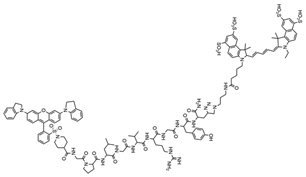 | 1060 nm and 1340 nm | It can be rapidly eliminated from the hepatobiliary pathway and has been successfully used as a contrast agent for NIR-II in vivo imaging and magnetic resonance imaging (MRI), enabling precise detection of the location of breast cancer lesions. | [26] |
| Cyanide | 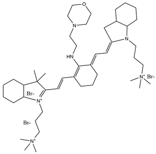 | 759 nm | Observation of in situ breast tumors and improved surgical accuracy. | [27] |
| NIR-β-galactosidase (NIR-β-gal-2) |  | 700 nm | Successful removal of an in situ breast tumor by “spraying in situ” and monitoring post-operative recovery. | [28] |
| Fluorescent Probe Type | Probe Molecular Structure | Application | Ref. |
|---|---|---|---|
| Rhodamine |  | Identification of sialic acid in breast cancer cells and cellular imaging of MCF-7 human cancers. | [33] |
| Coumarins | 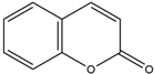 | Precision imaging and cell-targeted therapy for breast cancer. | [35] |
| Benzoxazole |  | It enhances fluorescence in acidic environments and can be used for cellular imaging. | [37] |
| Naphthylimides | 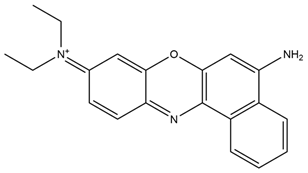 | For breast cancer MCF-7 cells and determined Cu2+ under neutral conditions. | [38] |
| pH-Sensitive dye 1 | 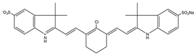 | For primary and metastatic breast tumor imaging. | [39] |
| Fluorescent Probe Type | Biomarker | Application | Ref. |
|---|---|---|---|
| Dihydroacetylacetone-based fluorescent probes | DAP-1 and DAP-4 | It can be used to monitor levels of ROS in breast cancer cells to help assess the efficacy of anti-cancer drugs. | [50] |
| Redox fluorescent probes for imidazole–phosphorus–copper analogues | Cu-ImPy and Cu-ImPho | It can be used to monitor levels of ROS in breast cancer cells to help assess the efficacy of anti-cancer drugs. | [51] |
| Thiol-based probes | AQC, mBBr, and OPA | It can detect GSH and other levels in breast cancer cells, determine drug efficacy, and optimize therapeutic regimens. | [52] |
| Fluorescent Probe Type | Probe Molecular Structure | Names of Enzymes and Receptors | Application | Ref. |
|---|---|---|---|---|
| YQ-L | 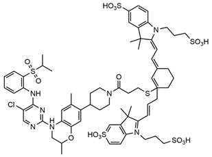 | Insulin-like growth factor type 1 receptor (IGF1R) | For the detection of breast cancer. | [65] |
| Indomethacin | 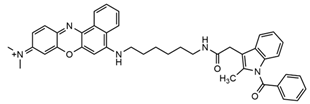 | Cyclooxygenase-2 (COX-2) | Tumor imaging applied to mouse models. | [66] |
| Acetylcholinesterase inhibitors (AchEIs) | 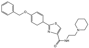 | Acetylcholinesterase (AChE), a hydrolyzing enzyme in blood plasma | For identification and inhibition of breast cancer cells. | [67] |
| Inhibitors AX11890 | 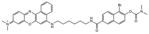 | KIAA1363 enzyme | For ultrafast differentiation of breast cancer cells from normal cells in fluorescence imaging with applications to in vivo tissue and tumor imaging. | [68] |
| CES-Lyso | 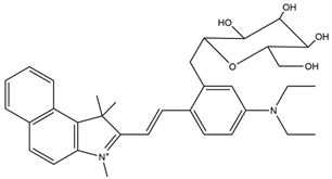 | Carboxylesterase (chemistry) | Carboxylesterase activity for differentiating breast cancer cells of different origins and monitoring anti-cancer drug therapy. | [69] |
| Type of Drugs | Name of Drug | Synthesis and Applications | Ref. |
|---|---|---|---|
| Antibiotics | Adriamycin (DOX) | A microemulsion polymerization in situ loading of Adriamycin (DOX) in iron tetroxide/P (NIPAM-AA-MAPEG) nanogels (MNLs) was designed and prepared with the ability to improve the cell penetration of the drug to achieve controlled release of the drug, to prolong the time of release of the drug, to reduce the toxicity and side effects, and to enable magnetic resonance imaging (MRI), thus enabling the integration of diagnostic and therapeutic treatments. | [78] |
| A mSiO2-coated AuNBP with a core–shell structure (AuNBP@mSiO2) was prepared. The surface was modified with Gd3+ chelated with 3-aminopropyl (trimethoxyphenyl) diethylenetriamine tetra-acetic acid (SiDTTA). DOX was subsequently incorporated into the nanoprobe, resulting in MRI-imageable and chemo-photothermally killable breast cancer cells, AuNBP@mSiO2-Gd-DTTA@DOX. | [79] | ||
| Natural medicine | Salvia divinorum | Inhibition of cytochrome P450 1A1 to enhance breast cancer chemotherapy. | [80] |
| Antimetabolites | 5-FU | A new tumor-targeting nanomedicine (AS1411-T-5-FU) was synthesized using DNA aptamer (AS1411) to modify DNA tetrahedra as a DNA delivery system for loading the anti-tumor drug 5-FU and labelling the DNA tetrahedra with the fluorescent probe Cyanine 5 (Cy5), which enhances the photostability with minimal impact on its basic function, to improve the breast cancer therapeutic efficacy and targeting. | [81] |
| Natural medicine | Paclitaxel | An integrated nanoplatform for therapeutics and imaging is proposed, which was constructed by co-encapsulation of the photothermal therapeutic agent IR780, the passive targeting drug OA@Fe3O4, and the chemotherapeutic drug paclitaxel. The nanoparticles exhibited improved photothermal–chemotherapeutic synergistic effects under magnetic targeting guidance, and showed anti-tumor effects in both in vitro and in vivo experiments. | [82] |
Disclaimer/Publisher’s Note: The statements, opinions and data contained in all publications are solely those of the individual author(s) and contributor(s) and not of MDPI and/or the editor(s). MDPI and/or the editor(s) disclaim responsibility for any injury to people or property resulting from any ideas, methods, instructions or products referred to in the content. |
© 2024 by the authors. Licensee MDPI, Basel, Switzerland. This article is an open access article distributed under the terms and conditions of the Creative Commons Attribution (CC BY) license (https://creativecommons.org/licenses/by/4.0/).
Share and Cite
Sheng, A.; Zhang, H.; Li, Q.; Chen, S.; Wang, Q. Application of Intelligent Response Fluorescent Probe in Breast Cancer. Molecules 2024, 29, 4294. https://doi.org/10.3390/molecules29184294
Sheng A, Zhang H, Li Q, Chen S, Wang Q. Application of Intelligent Response Fluorescent Probe in Breast Cancer. Molecules. 2024; 29(18):4294. https://doi.org/10.3390/molecules29184294
Chicago/Turabian StyleSheng, Anqi, Hao Zhang, Qing Li, Shu Chen, and Qingshuang Wang. 2024. "Application of Intelligent Response Fluorescent Probe in Breast Cancer" Molecules 29, no. 18: 4294. https://doi.org/10.3390/molecules29184294
APA StyleSheng, A., Zhang, H., Li, Q., Chen, S., & Wang, Q. (2024). Application of Intelligent Response Fluorescent Probe in Breast Cancer. Molecules, 29(18), 4294. https://doi.org/10.3390/molecules29184294