Article

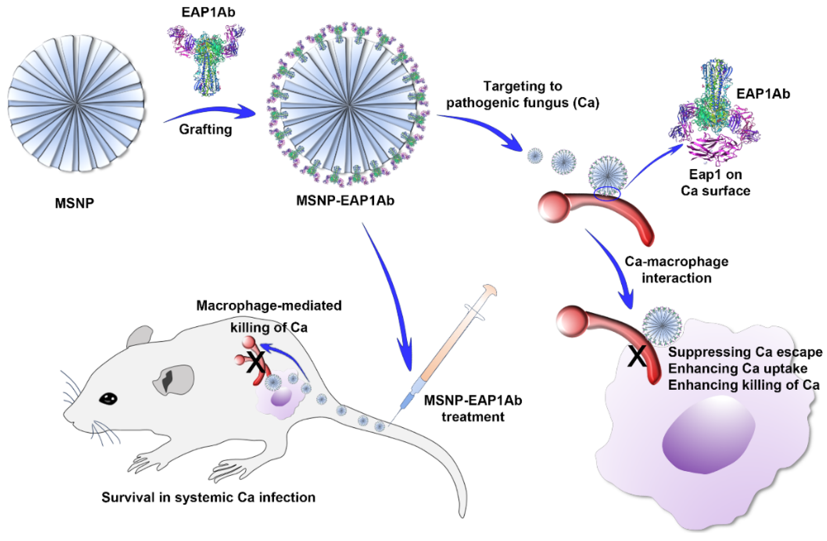
Adhesin Antibody-Grafted Mesoporous Silica Nanoparticles Suppress Immune Escape for Treatment of Fungal Systemic Infection

1
National Key Laboratory of Intelligent Tracking and Forecasting for Infectious Diseases, College of Life Sciences, Nankai University, Tianjin 300071, China
2
Key Laboratory of Molecular Microbiology and Technology, Ministry of Education, Department of Microbiology, College of Life Sciences, Nankai University, Tianjin 300071, China
*
Author to whom correspondence should be addressed.
Molecules 2024, 29(19), 4547; https://doi.org/10.3390/molecules29194547
Submission received: 19 August 2024 / Revised: 12 September 2024 / Accepted: 19 September 2024 / Published: 25 September 2024
(This article belongs to the Special Issue Chemical Biology of Antimicrobial Resistance, 2nd Edition)

Abstract

:
Life-threatening systemic fungal infections caused by Candida albicans are significant contributors to clinical mortality, particularly among cancer patients and immunosuppressed individuals. The evasion of the immune response facilitated by fungal surface components enables fungal pathogens to evade macrophage attacks and to establish successful infections. This study developed a mesoporous silica nanoplatform, i.e., MSNP-EAP1Ab, which is composed of mesoporous silica nanoparticles grafted with the antibody of C. albicans surface adhesin Eap1. The activity of MSNP-EAP1Ab against C. albicans immune escape and infection was then evaluated by using the cell interaction model and the mouse systemic infection model. During interaction between C. albicans cells and macrophages, MSNP-EAP1Ab significantly inhibited fungal immune escape, leading to the enhanced phagocytosis of fungal cells by macrophages, with phagocytosis rates increasing from less than 8% to 14%. Furthermore, after treatment of the C. albicans-infected mice, MSNP-EAP1Ab drastically prolonged the mouse survival time and decreased the kidney fungal burden from >30,0000 CFU/g kidney to <100 CFU/g kidney, indicating the rapid recognition and killing of the pathogens by immune cells. Moreover, MSNP-EAP1Ab attenuated kidney tissue inflammation, with remarkable attenuation of renal immune cell accumulation. This study presents an innovative nanoplatform that targets the C. albicans adhesin, offering a promising approach for combatting systemic fungal infections.

Graphical Abstract

1. Introduction

Approximately 1.5 million individuals succumb to invasive fungal infections annually on a global scale, with the prevalent culprit being various species of Candida. Candida albicans (C. albicans) stands out as the primary contributor to candidiasis worldwide [1]. Invasive candidiasis encompasses a variety of disorders, may affect any organ, and refers to deep-seated or disseminated infections (systemic candidiasis), which usually affect patients with impaired host defense mechanisms [2]. Candida bloodstream infections (BSIs) represent the most serious manifestation of candidiasis. The murine model of hematogenously disseminated candidiasis is most commonly used to study systemic candidiasis and to evaluate the efficacy of antifungal therapy. Following intravenous infection, C. albicans initially infects almost all organs [3,4], however, the fungal burden in the kidneys increases progressively over time [5], indicating that the kidney acts as the major site of fungal replication [6].
During the inflammatory process, macrophages and neutrophils are crucial effector immune cells involved in fungal eradication and the generation of inflammatory mediators [7,8,9], e.g., tumor necrosis factor α (TNF-α), interleukin-1 (IL-1), and interleukin-6 (IL-6) [10]. The inflammatory factors have the potential to stimulate adaptive immune responses, generate CD4+ and CD8+ T cells, and bolster antifungal immune activity [11,12,13,14]. While the increased activity of inflammatory monocytes and neutrophils results in hyper-inflammation and lethal kidney pathology [15]. Therefore, effective strategies for alleviating systemic fungal infections involve inhibiting the hyperinflammatory response, controlling fungal proliferation, and maintaining the body’s antifungal activity [16,17,18,19,20]. However, pathogenic fungi evolve complicated immune escape mechanisms, such as the production of surface polysaccharides and proteins to deceive immune cells, and the secretion of proteases to degrade immune response-related molecules [21]. The presence of fungal immune escape frequently compromises the efficiency of immune cells against fungal infections. Up to now, there has been no promising strategy to inhibit fungal immune response.
Mesoporous silica nanoparticles (MSNs) have emerged as a versatile imaging platform in recent years, offering customizable size and morphology, diverse surface chemistry, biocompatibility, and other advantageous physicochemical properties [22,23,24]. Simultaneously, extensive research has been conducted on MSN-based gating systems to integrate pore-capping, drug-loading, and targeting abilities (Table S1) [25,26,27,28]. Nevertheless, few MSN delivery platforms are tailored for targeting C. albicans, underscoring the critical necessity to develop MSN platforms that are responsive to fungal infections in order to combat these pathogens.
Eap1 is a novel adhesin of C. albicans, capable of mediating adhesion to both polystyrene and epithelial cells. Its expression is under the regulation of the transcription factor Efg1p, which is essential for hyphal formation, adhesion to and invasion of multilayered human epidermal tissue, and virulence in a murine model [29,30]. Importantly, in both hyphal and yeast cells, Eap1 could be highly expressed and exposed on the surface of C. albicans cells, endowing it with an ideal surface antigen for the specific targeting of this pathogen. However, up to now, there has been no study focusing on it as a target of antifungal strategies.
In this study, to avoid the immune escape of the life-threatening C. albicans, we developed a mesoporous silica nanoparticle platform (MSNP) grafted by the adhesin antibody for treatment of fungus-induced systemic infections (Figure 1). The MSNP was firstly prepared by the surfactant-mediated formation of silica pores, and then grafted by the C. albicans adhesin Eap1 antibody EAP1Ab to obtain MSNP-EAP1Ab. During the interaction between the pathogenic fungus C. albicans (Ca) and the macrophages, MSNP-EAP1Ab strongly suppressed the immune escape of Ca, leading to the enhanced recognition of Ca and consequent efficient killing of Ca by macrophages. During the systemic infection by Ca, the MSNP-EAP1Ab treatment efficiently causes the macrophage-mediated killing of Ca, leading to the prolonged survival of mice with the life-threatening Ca infection. This study provided a fungal antibody-grafted nanoplatform for avoiding fungal immune escape to treat dangerous fungal systemic infections.

2. Results

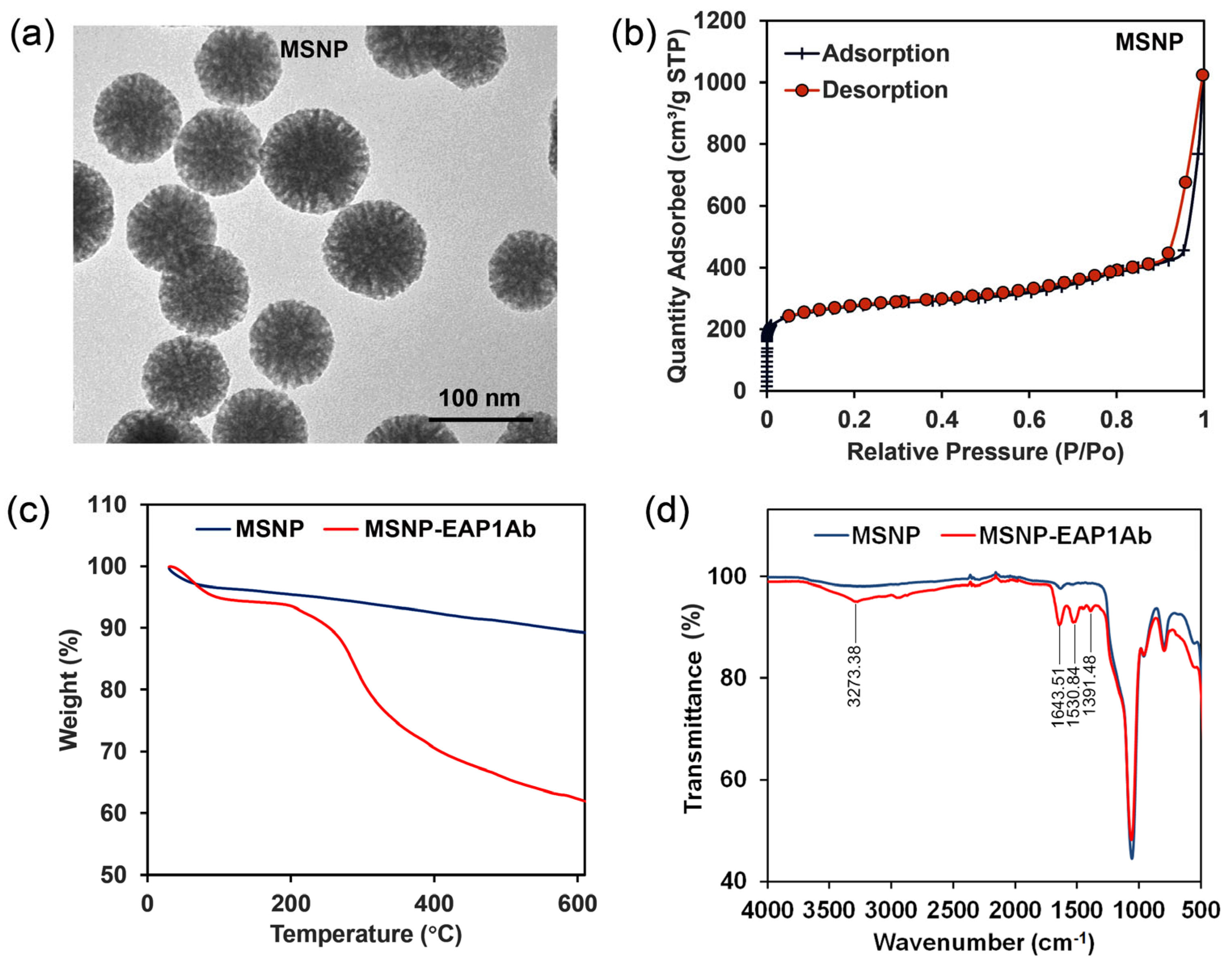
2.1. Characterization of MSNP and MSNP-EAP1Ab

As revealed by the TEM image, the initial MSNP had regular round-like morphology, with the radically distributed pores in the nanoparticles (Figure 2a). The adsorption–desorption curve revealed a sudden change of adsorbed quantity when the relative pressure increased to 0.95 (Figure 2b), with the average pore diameter at 6.239 nm. TGA analysis showed that the weight of the MSNP nanoparticles decreased to 90%, while that of MSNP-EAP1Ab decreased to 62% when the temperature reached 600 °C (Figure 2c). This indicated that approximately 28% of the antibody was grafted onto the MSNP. In addition, as shown in the FT-IR spectra, the MSNP-EAP1Ab nanoparticles had differential absorption peaks of 3723.38 cm−1, 1643.51 cm−1, 1530.84 cm−1, and 1391.48 cm−1 (Figure 2d), confirming the presence of -CO-NH- in MSNP-EAP1Ab. This indicated that EAP1Ab was successfully grafted onto the MSNP.

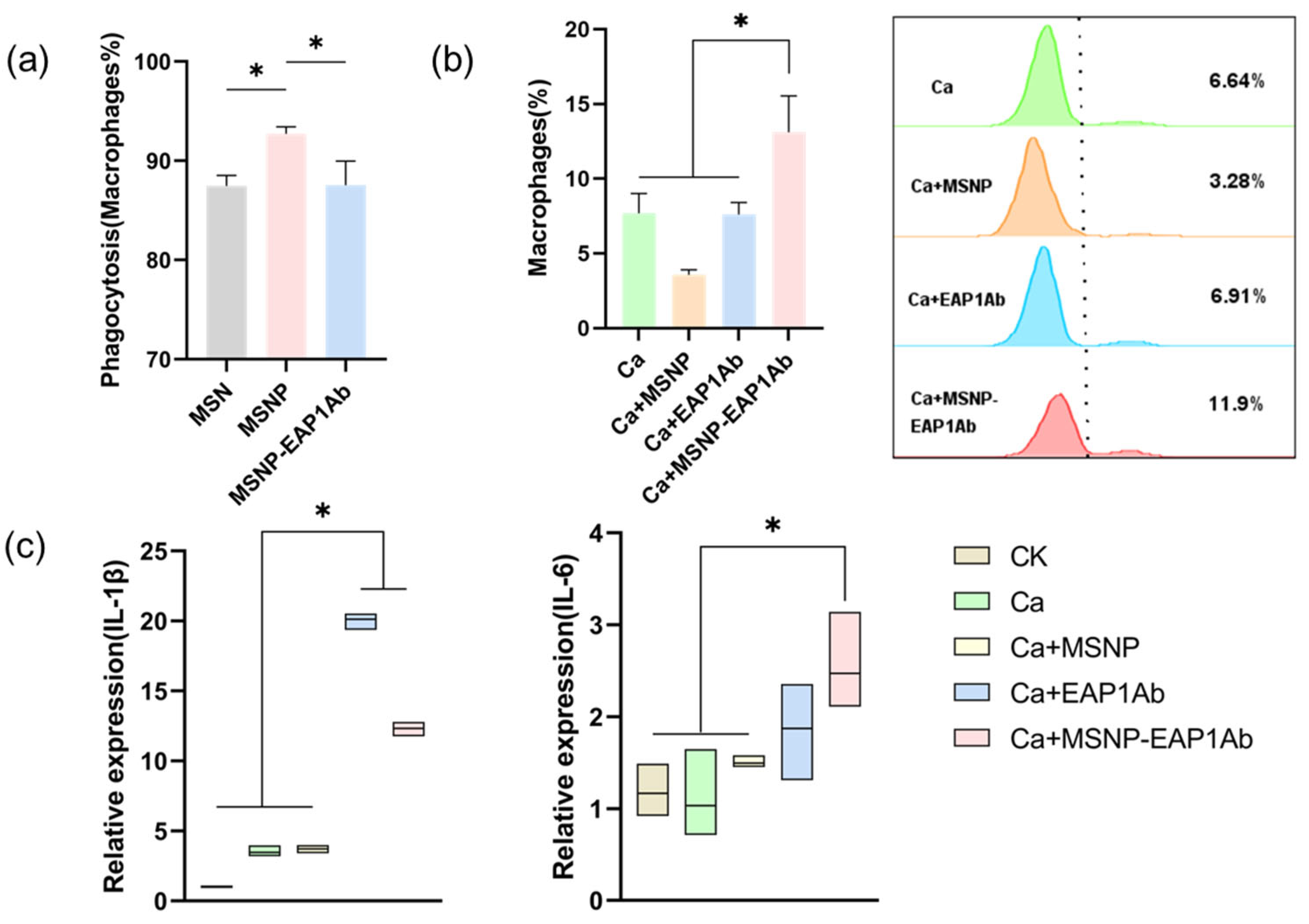
2.2. MSNP-EAP1Ab Enhances Both Phagocytosis and Cytokine Secretion in RAW264.7 Cells

Macrophages are vital players in the host’s innate immune response. Phagocytosis, defined as the uptake of particles greater than 0.5 μm, is a significant process in the innate immune response [31]. After co-culturing with FITC-labeled MSN, MSNP, MSNP-EAP1Ab, and RAW264.7 cells, the macrophages’ uptake of nanomaterials was assessed via flow cytometry (Figure 3a). The endocytosis rate of MSN exceeded 85%, and, following P modification, MSNP-EAP1Ab showed over 90% endocytosis. However, the endocytosis rate of MSNP-EAP1Ab in RAW264.7 cells decreased to 85%–90%. Subsequent experiments will be conducted using MSNP and MSNP-EAP1Ab. Based on the aforementioned co-culture of nanomaterials with RAW264.7 cells, we infected them with GFP-labeled C. albicans to assess the impact of nanomaterials on macrophage phagocytosis using flow cytometry. As shown in Figure 3b, it is evident that MSNP significantly decreased macrophage phagocytosis, whereas MSNP-EAP1Ab notably increased it.
Macrophages play a critical role in inflammation by producing cytokines like IL-1β and IL-6 to facilitate fungal killing. Following the infection of macrophages and nanomaterial co-cultures by C. albicans, we evaluated IL-1β, IL-6 and TNF-α transcription levels using RT-qPCR (Figure 3c and Figure S1). Our findings indicated that MSNP-EAP1Ab demonstrated the ability to enhance the expresssion of these cytokines, promoting C. albicans elimination. Conversely, upon C. albicans infection, EAP1Ab resulted in excessive IL-1β transcription, triggering heightened cellular inflammation and increasing the likelihood of pyroptosis.

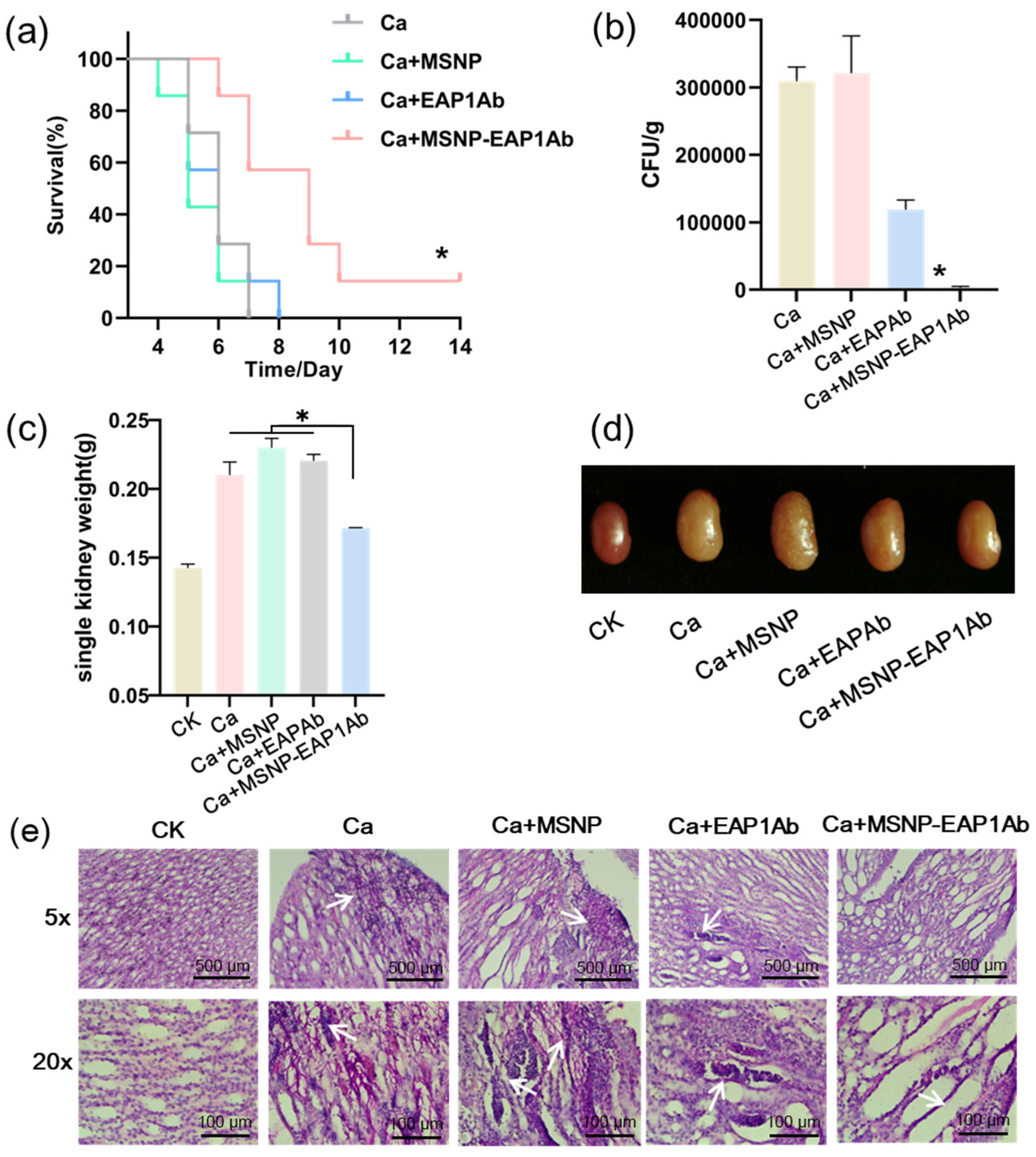
2.3. MSNP-EAP1Ab Suppresses Mouse Death and Kidney Damage Induced by Systemic C. albicans Infection

In a mouse model of systemic infection, the kidney emerges as the primary site of infection, underscoring the significance of mitigating inflammation within the kidney for the effective treatment of C. albicans systemic infection [32,33]. Twenty-four hours following systemic infection of mice by C. albicans, administering MSNP-EAP1Ab via a tail vein injection significantly prolonged the survival time of the mice (Figure 4a). On the fifth day post-infection, the mice underwent kidney weight measurements. The data presented in Figure 4b revealed that the group treated with MSNP-EAP1Ab exhibited a more vibrant kidney color and reduced swelling, whereas the group treated with MSNP alone displayed pronounced kidney swelling and renal nodules. Simultaneously, the kidneys were trisected and revealed a reduced fungal burden in the kidneys of mice treated with MSNP-EAP1Ab (Figure 4c). Analysis of kidney sections through Periodic Acid-Schiff staining revealed that mice treated with MSNP exhibited a considerable presence of hyphae at the renal pelvis site, leading to the disruption of their tissue morphology. In contrast, mice treated with EAP1Ab showed a substantial accumulation of immune cells, causing severe local kidney damage, but without notable hyphal growth. Mice receiving the MSNP-EAP1Ab treatment maintained relatively intact tissue morphology, with an enhanced and evenly distributed population of immune cells (Figure 4d).

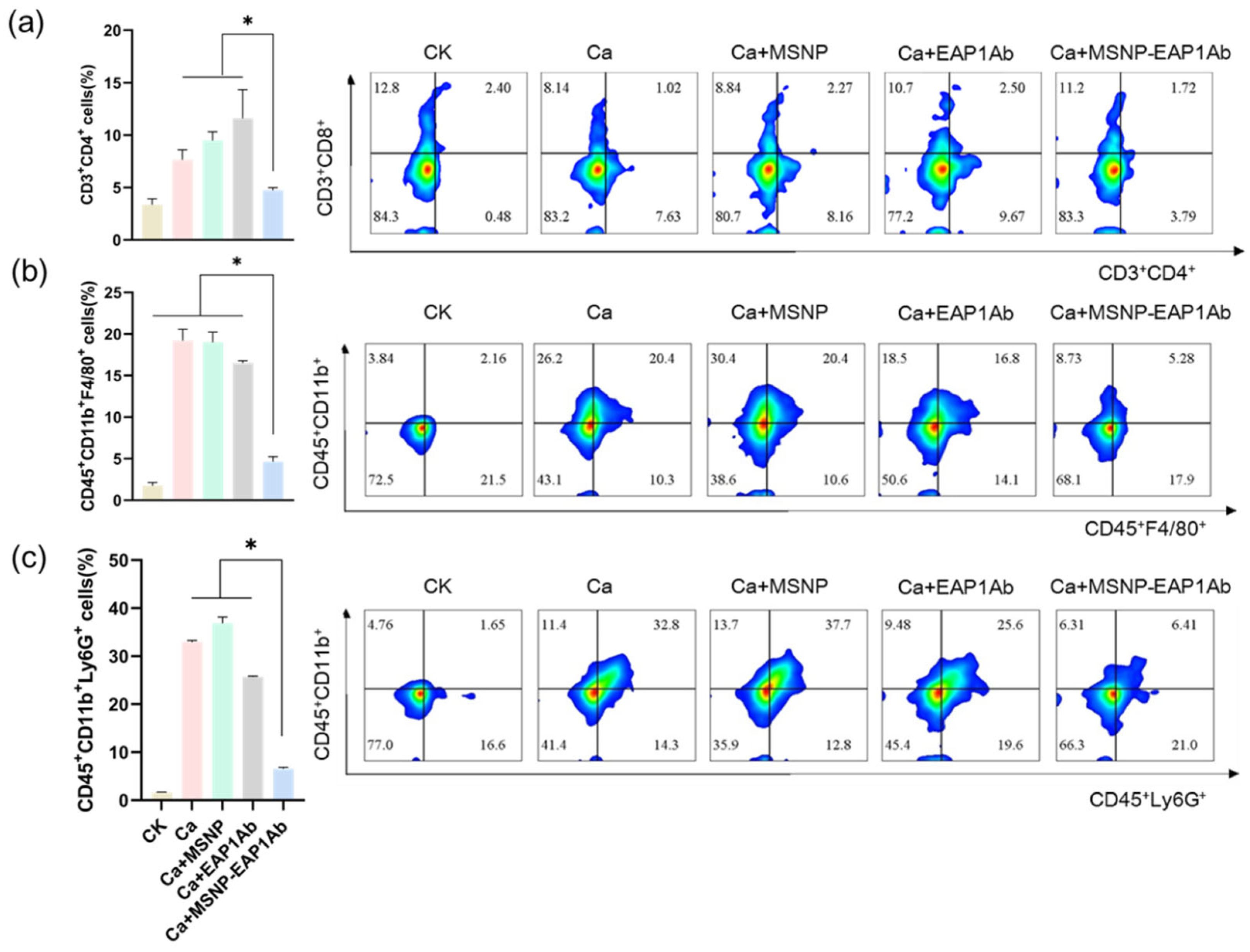
2.4. MSNP-EAP1Ab Enhances the Cytotoxicity of Renal Immune Cells without Inducing Excessive Inflammation

Insufficient early inflammatory cell-mediated response impairs the ability of mice to control fungal growth. Conversely, an excessive fungal growth elicits a heightened inflammatory response in mice, ultimately culminating in multi-organ failure [34]. In this study, flow cytometry was employed to evaluate the levels of CD3+CD4+ T cells (Figure 5a), CD45+CD11b+F4/80+ macrophages (Figure 5b), and CD45+CD11b+Ly6G+ neutrophils (Figure 5c) in the kidney. Excessive proliferation of these immune cells can result in an intensified inflammatory response, thereby causing damage and renal failure. MSNP-EAP1Ab effectively suppressed the proliferation of inflammatory cells and demonstrated a notable fungicidal impact when compared to the control group.
Immunofluorescent staining of neutrophils from the kidneys was then performed to further detect the distribution of immune cells. As revealed by observation using confocal microscopy, while the groups of Ca, Ca+MSNP, and Ca+EAP1Ab exhibited dense distribution of the pro-inflammatory immune cells (Ly6G+, CD45+ and CD11b+) in the kidney tissues, the MSNP-EAP1Ab only had gentle distribution of these immune cells (Figure 6). These results are consistent with the findings obtained through flow cytometry analysis (Figure 5). Hence, the treatment of MSNP-EAP1Ab severely attenuated the accumulation of pro-inflammatory immune cells, leading to the alleviation of inflammation in the infected kidneys.

3. Discussion

The human fungal pathogen C. albicans causes invasive candidiasis, a condition marked by fatal organ failure resulting from widespread fungal proliferation and inflammatory harm. Among peripheral organs, the kidneys offer the most conducive environment for fungal growth and the transition to a hyphal form [35,36]. An efficient inflammatory response is crucial for combating infections, however, the usual reaction to C. albicans results in tissue damage, intensifying the pathological consequences of the infection. Therefore, there is a requirement to evolve sophisticated and improved treatment strategies.
Research on intelligent drug delivery systems in the biomedical sector is anticipated to improve drug effectiveness and mitigate side effects at the sites of diseases. The distinctive characteristics of mesoporous silica nanoparticles (MSNs), including their capacity for stable covalent attachment to recognition groups like antibodies or aptamers, offer a myriad of possibilities for developing drug delivery platforms and biosensors. Currently, numerous drug delivery systems are available, such as PEG-MSN-Stattic, Chl-MSN, and MSN-NH2/RC [37,38,39,40]. Targeted delivery involves the following mechanism: the carrier containing the drug enters the bloodstream, circulates through the body, and accumulates exclusively in the area of the lesion [41]. Consequently, the nanoplatform incorporating EAP1Ab demonstrates superior delivery efficiency and targeting capabilities.

4. Materials and Methods

4.1. Materials

The Periodic Acid-Schiff Staining Kits (C0142M) were purchased from Beyotime, Shanghai, China. The primers for RT-qPCR were synthesized by Sangon Biotechnology (Sangon Biotech Co., Ltd., Shanghai, China). The 4′, 6 diamidino-2-phenylindole (DAPI, C1002) was purchased from Beyotime, China. The PE Anti-Mouse CD45 Antibody (E-AB-F1136D) was purchased from Elabscience, Wuhan, China. The FITC anti-mouse F4/80 (123107), APC anti-mouse IFN-γ (505809), FITC anti-mouse Ly-6G (127605), APC anti-mouse/human CD11b (101211), and APC anti-mouse TNF-α (506307) were purchased from BioLegend (Biotechnology Co., Ltd., San Diego, CA, USA). The wild-type C. albicans strain SC5314 was purchased from ATCC, Manassas, VA, USA. The GFP-labeled C. albicans strain CaGFP was constructed by transformation of the wild-type C. albicans strain BWP17 with the Erg6-localizing plasmid pErg6-GFP. The RAW264.7 macrophages were purchased from the Cell Resource Center, Beijing, China. The antibody against the C. albicans adhesin Eap1 (i.e., EAP1Ab) was prepared by immunization of the rabbits with the purified the Eap1 antigen, followed by serum collection and antibody purification.

4.2. Preparation and Characterization of the Nanoparticles

The initial MSNP was prepared by using tetraethyl orthosilicate (TEOS) as the silica source, and hexadecyltrimethylammonium bromide (CTAB) as the template agent. Briefly, 400 mg of CTAB was dissolved in 100 mL of distilled water. Under constantly magnetic stirring, 448 μL of the 100 mg/mL NH4Cl solution, 100 μL of the 2 mol/L NaOH solution, and 1.828 mL of mesitylene were then added into the CTAB solution. The mixture was stirred at 8000 rpm and 78 °C for 30 min, followed by the addition of 1.5 mL TEOS and further stirring for another 1 h. A total of 100 μL of (3-aminopropyl)triethoxysilane (APTES) were then added into the reaction solution, and the mixture was further stirred for 30 min. The nanoparticles were centrifuged at 12,000 rpm for 10 min, and washed by ethanol and distilled water, obtaining the initial MSNP.
To graft the MSNP by EAP1Ab, 50 mg of the MSNP were suspended in 50 mL of distilled water, and then 100 μL of glutaric dialdehyde were added into the suspension. The mixture was magnetically stirred at 750 rpm for 6 h. The activated nanoparticles were harvested by centrifugation at 12,000 rpm for 10 min, and washed by distilled water three times. The nanoparticles were further suspended in 50 mL of PBS (20 mM, pH = 7.2) containing 50 mg EAP1Ab. The mixture was also stirred at 800 rpm for 12 h under 4 °C for 12 h, followed by centrifugation with the same condition. The nanoparticles were washed three times in distilled water and dried by a freeze vacuum dried, obtaining MSNP-EAP1Ab.
The morphology and pore sizes of MSNP were characterized by using a transmission electron microscope (TEM, Tecnai T12, FEI, Hillsboro, OR, USA) and a Brunauer-Emmett-Teller (BET) pore size analyzer (ASAP 2460, Micromeritics, Norcross, GA, USA), respectively. The thermogravimetric analysis (TGA) of MSNP and MSNP-EAP1Ab was performed using a TG/DTA Instrument (STA 8000, PerkinElmer, Waltham, MA, USA) under the nitrogen flow. The Fourier Transform Infrared Spectoscopy (FT-IR) spectra of the nanoparticles were performed by using the FT-IR analyzer (Nicolet iS20, Thermo Fisher Scientific, Waltham, MA, USA).

4.3. Animals

All animal experiments were approved by the Animal Care and Use Committee of Nankai University. Fifty 5-week-old ICR pathogen-free (SPF) female mice, weighing between 18 g and 20 g, were chosen and randomly divided into groups of 10 replicates. The mice were maintained in a 12-h light-dark cycle with ad libitum access to food and water.

4.4. Cell Culture

Dulbecco’s Modified Eagle’s Medium (DMEM) with high glucose (HyClone, Logan, UT, USA), enriched with 10% heat-inactivated fetal bovine serum (FBS) (HyClone, Logan, UT, USA) was used for the growth of RAW264.7 cells in an atmosphere containing 5% CO2 at 37 °C without any penicillin or streptomycin, and 5 × 10⁶ cells were inoculated into a fresh 10-cm cell-culture dish (Corning-Costar, Corning, NY, USA). Upon 80% confluence, RAW 264.7 cells were sub-cultured serially to be used for the following experiments.

4.5. Cellular Phagocytosis

The day before the experiment, RAW264.7 cells were seeded onto a 24-well plate. Once the cells adhered to the surface, MSNP and MSNP-EAP1Ab were sequentially added. After a 6-h incubation period, the supernatant was aspirated, and the cells were washed twice with PBS. Subsequently, Erg6−/−-GFP C. albicans (MOI 5) was added, followed by a 40-min co-incubation at 37 °C with 5% CO2. The cells were then harvested, and the phagocytic activity was assessed by measuring the fluorescence signal of GFP using flow cytometry after appropriate steps of washing, centrifugation, and resuspension.

4.6. MSN Endocytosis Efficiency

The RAW264.7 cells were plated onto a 24-well plate and incubated with FITC-labeled MSNP and MSNP-EAP1Ab overnight on a shaker. Subsequently, the supernatant was discarded, and the cells were resuspended after centrifugation at 12,000 rpm for 2 min. The cells were then grouped and added back onto a 24-well plate for a 6-h incubation period. Following this, the cells were collected by centrifugation at 1200 rpm, washed, and resuspended in PBS. The fluorescence signal intensity of macrophages was later detected using flow cytometry.

4.7. Reverse Transcriptase PCR

The macrophages (2.0 × 10⁵ cells/well) were seeded overnight in a six-well plate. Total RNA was extracted from treated cells according to the manufacturer’s instructions (TransGen Biotech, Beijing, China) and used for cDNA synthesis (One-Step gDNA Removal, TransGen Biotech). PCR was then performed using primer pairs specific for TNFα, IL-1β, IL-6, and β-actin (the primer sequences are shown in Table 1). The amplified products were electrophoresed using 1.5% agarose gel and stained with ethidium bromide before visualization under a UV lamp.

4.8. Systemic Infection

To evaluate the therapeutic effect of MSNP-EAP1Ab, the mice (except for the control group) were infected by the fresh C. albicans SC5314 cells with intravenous inoculation at the tail vein on day 0. After 24 h, the mice were injected with MSNP, EAP1Ab, and MSNP-EAP1Ab via the tail vein based on their respective group assignments. The survival of the infected mice was recorded from day 0 to day 14. On day 4, the kidneys of three mice in each group were sampled and weighed. The kidneys were further broken by Dounce homogenizers, and the numbers of the fungal cells in the obtained homogenates were determined using colony-forming unit assays on plates of yeast extract-peptone-dextrose medium.

4.9. Flow Cytometry Assays

On the fifth day of systemic infection, the kidneys of mice were harvested. The kidney tissues were digested into a cell suspension, which was subsequently washed twice with phosphate-buffered saline (PBS), and then resuspended. The cells were diluted for staining with a fluorescent dye at a ratio of 1:1000, incubated in the dark at 4 °C for 30 min, followed by resuspension after two washes with PBS for detection.

4.10. Paraffin Immunofluorescence

On the fifth day of systemic infection, the mouse kidneys were harvested, fixed in 4% paraformaldehyde for 24 h, and subsequently processed through dehydration, transparency, and embedding in paraffin blocks. The resulting sections were cut into slices of 5 to 10 microns thickness, followed by deparaffinization and rehydration using xylene and ethanol gradients after a 2-h baking period. A citrate antigen retrieval solution (pH 6.0) was prepared, and antigen retrieval was conducted in a microwave oven. Following natural cooling, the tissues were washed three times with PBS buffer for 5 min each. A dye, diluted at a ratio of 1:500, was then meticulously added to the tissue and left to stain in darkness for 30 min before observation under a confocal microscope.

4.11. Statistical Analysis

Most of the experiments were performed with three replicates (n = 3), except the experiment of systemic infection (n = 10). The data were shown with the means ± the standard errors. The differences between the groups were analyzed by the ANOVA test and the Student’s t-test (p < 0.05) via SPSS 22 software (IBM, Armonk, NY, USA).

5. Conclusions

In conclusion, this study developed a novel adhesin antibody-grafted nanoplatform for treatment of the body from life-threatening systemic fungal infections. MSNP was synthesized through the surfactant-mediated creation of silica pores, followed by the grafting of Msnp-EAP1Ab with the Eap1 antibody specific to the C. albicans adhesin EAP1Ab. Upon interaction with macrophages, MSNP-EAP1Ab effectively suppressed the immune escape of C. albicans, leading to the increased recognition and killing of the pathogen. In the systemic C. albicans infection model, treatment with MSNP-EAP1Ab successfully triggered the macrophage-mediated killing of C. albicans, resulting in the prolonged survival time of mice facing life-threatening fungal infections and attenuation of kidney inflammation. In conclusion, this study introduced a novel nanoplatform utilizing a fungal antibody grafted nanoplatform to counteract fungal immune evasion against severe systemic fungal infections.

Supplementary Materials

The following supporting information can be downloaded at: https://www.mdpi.com/article/10.3390/molecules29194547/s1, Figure S1. The impact of MSNP, and MSNP-EAP1Ab on the transcription levels of TNFα following C. albicans infection; Table S1. The diverse applications of MSNs as drug delivery carriers across various fields.

Author Contributions

Conceptualization, Q.Y. and M.C.; methodology, Q.Y., M.C. and S.L.; software, Q.Y. and M.C.; validation, Q.Y., M.C., S.L. and M.Z.; formal analysis, Q.Y. and M.C.; investigation, Q.Y., M.C., S.L. and M.Z.; resources, Q.Y., M.C. and M.L.; data curation, Q.Y., M.C., S.L., M.Z. and M.L.; writing—original draft preparation, Q.Y. and M.C.; writing—review and editing, Q.Y., M.C. and M.L.; visualization, Q.Y. and M.C.; supervision, Q.Y.; project administration, Q.Y.; funding acquisition, Q.Y. All authors have read and agreed to the published version of the manuscript.

Funding

This work was supported by the National Natural Science Foundation of China, China (32170102), the Natural Science Foundation of Tianjin (21JCYBJC01420), the National Key Laboratory of Intelligent Tracking and Forecasting for Infectious Diseases (2024NITFID401), and the Fundamental Research Funds for the Central Universities (63243120).

Institutional Review Board Statement

Not applicable.

Informed Consent Statement

Not applicable.

Data Availability Statement

The original contributions presented in the study are included in the article/Supplementary Material.

Acknowledgments

The authors would like to thank the editors and the reviewers for constructive suggestions.

Conflicts of Interest

The authors declare no conflicts of interest.

References

  1. Lu, H.; Hong, T.; Jiang, Y.; Whiteway, M.; Zhang, S. Candidiasis: From Cutaneous to Systemic, New Perspectives of Potential Targets and Therapeutic Strategies. Adv. Drug Deliv. Rev. 2023, 199, 114960. [Google Scholar] [CrossRef] [PubMed]
  2. Lass-Flörl, C.; Kanj, S.S.; Govender, N.P.; Thompson, G.R.; Ostrosky- Zeichner, L.; Govrins, M.A. Invasive Candidiasis. Nat. Rev. Dis. Primers 2024, 10, 20. [Google Scholar] [CrossRef] [PubMed]
  3. Duggan, S.; Leonhardt, I.; Hünniger, K.; Kurzai, O. Host Response to Candida Albicans Bloodstream Infection and Sepsis. Virulence 2015, 6, 316–326. [Google Scholar] [CrossRef] [PubMed]
  4. Hebecker, B.; Vlaic, S.; Conrad, T.; Bauer, M.; Brunke, S.; Kapitan, M.; Linde, J.; Hube, B.; Jacobsen, I.D. Dual-Species Transcriptional Profiling during Systemic Candidiasis Reveals Organ-Specific Host-Pathogen Interactions. Sci. Rep. 2016, 6, 36055. [Google Scholar] [CrossRef]
  5. MacCallum, D.M.; Castillo, L.; Brown, A.J.P.; Gow, N.A.R.; Odds, F.C. Early-Expressed Chemokines Predict Kidney Immunopathology in Experimental Disseminated Candida Albicans Infections. PLoS ONE 2009, 4, e6420. [Google Scholar] [CrossRef]
  6. Lionakis, M.S.; Lim, J.K.; Lee, C.-C.R.; Murphy, P.M. Organ-Specific Innate Immune Responses in a Mouse Model of Invasive Candidiasis. J. Innate Immun. 2011, 3, 180–199. [Google Scholar] [CrossRef]
  7. Chu, C.; Rung, S.; Wang, Y.; Qu, Y.; Man, Y. Comment on “In Situ Mannosylated Nanotrinity-Mediated Macrophage Remodeling Combats Candida Albicans Infection”. ACS Nano 2021, 15, 3541–3543. [Google Scholar] [CrossRef]
  8. Gazendam, R.P.; Van Hamme, J.L.; Tool, A.T.J.; Van Houdt, M.; Verkuijlen, P.J.J.H.; Herbst, M.; Liese, J.G.; Van De Veerdonk, F.L.; Roos, D.; Van Den Berg, T.K.; et al. Two Independent Killing Mechanisms of Candida Albicans by Human Neutrophils: Evidence from Innate Immunity Defects. Blood 2014, 124, 590–597. [Google Scholar] [CrossRef]
  9. Li, L.; Huang, L.; Vergis, A.L.; Ye, H.; Bajwa, A.; Narayan, V.; Strieter, R.M.; Rosin, D.L.; Okusa, M.D. IL-17 Produced by Neutrophils Regulates IFN-γ–Mediated Neutrophil Migration in Mouse Kidney Ischemia-Reperfusion Injury. J. Clin. Investig. 2010, 120, 331–342. [Google Scholar] [CrossRef]
  10. Zhao, Y.; Liu, S.; Shi, Z.; Zhu, H.; Li, M.; Yu, Q. Pathogen Infection-Responsive Nanoplatform Targeting Macrophage Endoplasmic Reticulum for Treating Life-Threatening Systemic Infection. Nano Res. 2022, 15, 6243–6255. [Google Scholar] [CrossRef]
  11. Tham, E.L.; Freeley, S.J.; Bearder, S.; Barros, F.F.; Cragg, M.S.; Mócsai, A.; Robson, M.G. VISTA Deficiency Protects from Immune Complex-Mediated Glomerulonephritis by Inhibiting Neutrophil Activation. J. Autoimmun. 2020, 113, 102501. [Google Scholar] [CrossRef] [PubMed]
  12. Kretschmar, M.; Hein, A.; Geginat, G.; Mueller, C.; Hof, H.; Nichterlein, T. Inefficient T Cell Memory in the Brain of Mice Infected with Candida Albicans. J. Neuroimmunol. 2000, 105, 161–168. [Google Scholar] [CrossRef] [PubMed]
  13. Pernomian, L.; Do Prado, A.F.; Gomes, M.S.; Pernomian, L.; Da Silva, C.H.T.P.; Gerlach, R.F.; De Oliveira, A.M. MAS Receptors Mediate Vasoprotective and Atheroprotective Effects of Candesartan upon the Recovery of Vascular Angiotensin-Converting Enzyme 2–Angiotensin-(1-7)–MAS Axis Functionality. Eur. J. Pharmacol. 2015, 764, 173–188. [Google Scholar] [CrossRef] [PubMed]
  14. Wu, Y.; Zeng, Z.; Guo, Y.; Song, L.; Weatherhead, J.E.; Huang, X.; Zeng, Y.; Bimler, L.; Chang, C.-Y.; Knight, J.M.; et al. Candida Albicans Elicits Protective Allergic Responses via Platelet Mediated T Helper 2 and T Helper 17 Cell Polarization. Immunity 2021, 54, 2595–2610.e7. [Google Scholar] [CrossRef]
  15. Majer, O.; Bourgeois, C.; Zwolanek, F.; Lassnig, C.; Kerjaschki, D.; Mack, M.; Müller, M.; Kuchler, K. Type I Interferons Promote Fatal Immunopathology by Regulating Inflammatory Monocytes and Neutrophils during Candida Infections. PLoS Pathog. 2012, 8, e1002811. [Google Scholar] [CrossRef]
  16. Hu, L.; Bai, G.; Xu, Q.; Zhao, G.; Jiang, N.; Yao, H.; Liu, X.; Du, Z. Candidalysin Amplifies the Immune Inflammatory Response in Candida Albicans Keratitis through the TREM-1/DAP12 Pathway. Int. Immunopharmacol. 2023, 119, 110195. [Google Scholar] [CrossRef]
  17. Liu, J.; Lai, X.; Yu, R.; Ding, H.; Bai, H.; Yang, Z.; Yin, Y.; Xu, F.; Cao, J. Progranulin Aggravates Lethal Candida Albicans Sepsis by Regulating Inflammatory Response and Antifungal Immunity. PLoS Pathog. 2022, 18, e1010873. [Google Scholar] [CrossRef]
  18. Lv, Q.-Z.; Li, D.-D.; Han, H.; Yang, Y.-H.; Duan, J.-L.; Ma, H.-H.; Yu, Y.; Chen, J.-Y.; Jiang, Y.-Y.; Jia, X.-M. Priming with FLO8-Deficient Candida Albicans Induces Th1-Biased Protective Immunity against Lethal Polymicrobial Sepsis. Cell. Mol. Immunol. 2021, 18, 2010–2023. [Google Scholar] [CrossRef]
  19. Qin, Y.; Zhang, L.; Xu, Z.; Zhang, J.; Jiang, Y.; Cao, Y.; Yan, T. Innate Immune Cell Response upon Candida Albicans Infection. Virulence 2016, 7, 512–526. [Google Scholar] [CrossRef]
  20. Xu, M.; Li, L.; Pan, W.; Zheng, H.; Wang, M.; Peng, X.; Dai, S.; Tang, Y.; Zeng, K.; Huang, X. Zinc Oxide Nanoparticles Prime a Protective Immune Response in Galleria Mellonella to Defend Against Candida Albicans. Front. Microbiol. 2021, 12, 766138. [Google Scholar] [CrossRef]
  21. Jiménez-López, C.; Lorenz, M.C. Fungal Immune Evasion in a Model Host–Pathogen Interaction: Candida Albicans versus Macrophages. PLoS Pathog. 2013, 9, e1003741. [Google Scholar] [CrossRef] [PubMed]
  22. Manzano, M.; Vallet-Regí, M. Mesoporous Silica Nanoparticles in Nanomedicine Applications. J. Mater. Sci. Mater. Med. 2018, 29, 65. [Google Scholar] [CrossRef] [PubMed]
  23. Yuan, D.; Ellis, C.M.; Davis, J.J. Mesoporous Silica Nanoparticles in Bioimaging. Materials 2020, 13, 3795. [Google Scholar] [CrossRef] [PubMed]
  24. Möller, K.; Bein, T. Talented Mesoporous Silica Nanoparticles. Chem. Mater. 2017, 29, 371–388. [Google Scholar] [CrossRef]
  25. Chen, X.; Sun, H.; Hu, J.; Han, X.; Liu, H.; Hu, Y. Transferrin Gated Mesoporous Silica Nanoparticles for Redox-Responsive and Targeted Drug Delivery. Colloids Surf. B Biointerfaces 2017, 152, 77–84. [Google Scholar] [CrossRef]
  26. De La Torre, C.; Coll, C.; Ultimo, A.; Sancenón, F.; Martínez-Máñez, R.; Ruiz-Hernández, E. In Situ-Forming Gels Loaded with Stimuli-Responsive Gated Mesoporous Silica Nanoparticles for Local Sustained Drug Delivery. Pharmaceutics 2023, 15, 1071. [Google Scholar] [CrossRef]
  27. Sha, L.; Zhao, Q.; Wang, D.; Li, X.; Wang, X.; Guan, X.; Wang, S. “Gate” Engineered Mesoporous Silica Nanoparticles for a Double Inhibition of Drug Efflux and Particle Exocytosis to Enhance Antitumor Activity. J. Colloid Interface Sci. 2019, 535, 380–391. [Google Scholar] [CrossRef] [PubMed]
  28. Zhang, Y.; Wang, W.; Zhang, Y.; Cheng, M.; Wu, Q.; Yuan, Z. “Three-in-One” Multifunctional Gatekeeper Gated Mesoporous Silica Nanoparticles for Intracellular pH-Activated Targeted Cancer Therapy. ACS Appl. Bio Mater. 2018, 1, 572–580. [Google Scholar] [CrossRef] [PubMed]
  29. Li, F.; Palecek, S.P. EAP1, a Candida Albicans Gene Involved in Binding Human Epithelial Cells. Eukaryot. Cell 2003, 2, 1266–1273. [Google Scholar] [CrossRef]
  30. Zhao, Z.; Sun, Y.; Li, M.; Yu, Q. Construction of Candida Albicans Adhesin-Exposed Synthetic Cells for Preventing Systemic Fungal Infection. Vaccines 2023, 11, 1521. [Google Scholar] [CrossRef]
  31. Onyishi, C.U.; Desanti, G.E.; Wilkinson, A.L.; Lara-Reyna, S.; Frickel, E.-M.; Fejer, G.; Christophe, O.D.; Bryant, C.E.; Mukhopadhyay, S.; Gordon, S.; et al. Toll-like Receptor 4 and Macrophage Scavenger Receptor 1 Crosstalk Regulates Phagocytosis of a Fungal Pathogen. Nat. Commun. 2023, 14, 4895. [Google Scholar] [CrossRef] [PubMed]
  32. Navarathna, D.H.M.L.P.; Pathirana, R.U.; Lionakis, M.S.; Nickerson, K.W.; Roberts, D.D. Candida Albicans ISW2 Regulates Chlamydospore Suspensor Cell Formation and Virulence In Vivo in a Mouse Model of Disseminated Candidiasis. PLoS ONE 2016, 11, e0164449. [Google Scholar] [CrossRef]
  33. Nguyen, N.Z.N.; Tran, V.G.; Baek, J.; Kim, Y.; Youn, E.H.; Na, S.W.; Park, S.J.; Seo, S.-K.; Kwon, B. IL-33 Coordinates Innate Defense to Systemic Candida Albicans Infection by Regulating IL-23 and IL-10 in an Opposite Way. J. Immunol. 2022, 208, 660–671. [Google Scholar] [CrossRef] [PubMed]
  34. Thompson, A.; Davies, L.C.; Liao, C.-T.; Da Fonseca, D.M.; Griffiths, J.S.; Andrews, R.; Jones, A.V.; Clement, M.; Brown, G.D.; Humphreys, I.R.; et al. The Protective Effect of Inflammatory Monocytes during Systemic, C. Albicans Infection Is Dependent on Collaboration between C-Type Lectin-like Receptors. PLoS Pathog. 2019, 15, e1007850. [Google Scholar] [CrossRef] [PubMed]
  35. Carpino, N.; Naseem, S.; Frank, D.M.; Konopka, J.B. Modulating Host Signaling Pathways to Promote Resistance to Infection by Candida Albicans. Front. Cell. Infect. Microbiol. 2017, 7, 481. [Google Scholar] [CrossRef]
  36. Swidergall, M.; Khalaji, M.; Solis, N.V.; Moyes, D.L.; Drummond, R.A.; Hube, B.; Lionakis, M.S.; Murdoch, C.; Filler, S.G.; Naglik, J.R. Candidalysin Is Required for Neutrophil Recruitment and Virulence During Systemic Candida Albicans Infection. J. Infect. Dis. 2019, 220, 1477–1488. [Google Scholar] [CrossRef]
  37. Massahi Khosrowshahi, F.; Ebrahimi-Hosseinzadeh, B.; Hatamian-Zarmi, A. Sumac (Rhus Coriaria) Extract Loaded-Mesoporous Silica Nanoparticle Efficiently as a Controlled Drug Delivery System for the Treatment of Atherosclerosis. J. Inorg. Organomet. Polym. Mater. 2024, 34, 793–803. [Google Scholar] [CrossRef]
  38. Adnane, F.; Soliman, S.M.A.; ElZayat, E.; Abdelsalam, E.M.; Fahmy, H.M. Evaluation of Chlorophyll-Loaded Mesoporous Silica Nanoparticles for Photodynamic Therapy on Cancer Cell Lines. Lasers Med. Sci. 2024, 39, 45. [Google Scholar] [CrossRef]
  39. Xu, C.; Amna, N.; Shi, Y.; Sun, R.; Weng, C.; Chen, J.; Dai, H.; Wang, C. Drug-Loaded Mesoporous Silica Nanoparticles Enhance Antitumor Immunotherapy by Regulating MDSCs. Molecules 2024, 29, 2436. [Google Scholar] [CrossRef]
  40. Mladenović, M.; Jarić, S.; Mundžić, M.; Pavlović, A.; Bobrinetskiy, I.; Knežević, N.Ž. Biosensors for Cancer Biomarkers Based on Mesoporous Silica Nanoparticles. Biosensors 2024, 14, 326. [Google Scholar] [CrossRef]
  41. Kovtareva, S.; Kusepova, L.; Tazhkenova, G.; Mashan, T.; Bazarbaeva, K.; Kopishev, E. Surface Modification of Mesoporous Silica Nanoparticles for Application in Targeted Delivery Systems of Antitumour Drugs. Polymers 2024, 16, 1105. [Google Scholar] [CrossRef]
Figure 1. A scheme illustrating the preparation of MSNP-EAP1Ab, suppression of C. albicans (Ca) immune escape, and enhancement of macrophage-mediated Ca killing against systemic fungal infection.
Molecules 29 04547 g001
Figure 2. Characterization of the prepared MSNP and MSNP-EAP1Ab. (a) The TEM image of MSNP. (b) The adsorption–desorption curve of MSNP. (c) TGA curves of MSNP and MSNP-EAP1Ab. (d) FT-IR spectra of MSNP and MSNP-EAP1Ab.
Molecules 29 04547 g002
Figure 3. MSNP-EAP1Ab enhances both phagocytosis and cytokine secretion in RAW264.7 cells. (a) Endocytosis of MSN, MSNP, and MSNP-EAP1Ab was observed (6 h). (b) The impact of MSNP and MSNP-EAP1Ab on macrophage phagocytosis post-C. albicans infection. (c) The impact of MSNP and MSNP-EAP1Ab on the transcription levels of IL-1β and IL-6 following C. albicans infection. The asterisk indicates a significant difference between the two groups (p < 0.05).
Molecules 29 04547 g003
Figure 4. MSNP-EAP1Ab mitigates kidney damage induced by systemic C. albicans infection. (a) Survival curves of the mice (n = 10 mice/group). The mice were infected by the fungal cells (5 × 10⁶ cells/mouse) via tail intravenous injection on day 0, mice were divided into groups and treated with MSNP, EAP1Ab, or MSNP-EAP1Ab on day 1, the survival of mice was monitored for 14 days. (b) Fungal burdens at day 5 post-infection were presented per kidney or gram basis (n = 6 kidneys/group). (c) Weight of the kidneys at day 5 post-infection (n = 6 kidneys/group). (d) The morphology of kidneys at day 5 post-infection. (e) Histopathologic findings: PAS staining. The asterisk indicates a significant difference between the two groups (p < 0.05).
Molecules 29 04547 g004
Figure 5. MSNP-EAP1Ab attenuates the accumulation of renal immune cells in Ca-infected mice. (a) The CD3+CD4+ T cell levels in the kidneys at day 5 post-infection (n = 4/group). (b) The CD45+CD11b+F4/80+ macrophage levels in the kidneys at day 5 post-infection (n = 4/group). (c) The CD45+CD11b+Ly6G+ neutrophils levels in the kidneys at day 5 post-infection (n = 4/group). The asterisk indicates a significant difference between the two groups (p < 0.05).
Molecules 29 04547 g005
Figure 6. MSNP-EAP1Ab attenuates the in situ localization of pro-inflammatory immune cells in Ca-infected kidneys. The white arrows indicated the representative sites displaying both FITC-Ly6G, PE-CD45 and APC-CD11b.
Molecules 29 04547 g006
Table 1. Nucleotide primer sequences used for PCR amplification.
PrimerSequence
SenseAntisense
Mouse-TNFαGGTGCCTATGTCTCAGCCTCTTGCCATAGAACTGATGAGAGGGAG
Mouse-IL-1βTGGACCTTCCAGGATGAGGACAGTTCATCTCGGAGCCTGTAGTG
Mouse-IL-6TACCACTTCACAAGTCGGAGGCCTGCAAGTGCATCATCGTTGTTC
Mouse-β-actinCATTGCTGACAGGATGCAGAAGGTGCTGGAAGGTGGACAGTGAGG
Disclaimer/Publisher’s Note: The statements, opinions and data contained in all publications are solely those of the individual author(s) and contributor(s) and not of MDPI and/or the editor(s). MDPI and/or the editor(s) disclaim responsibility for any injury to people or property resulting from any ideas, methods, instructions or products referred to in the content.

