Article

Design, Synthesis of Hydrogen Peroxide Response AIE Fluorescence Probes Based on Imidazo [1,2-a] Pyridine

1
Department of Veterinary Medicine, Shanxi Agricultural University, Jinzhong 030801, China
2
Department of Mining Engineering, Shanxi Institute of Technology, Yangquan 045000, China
3
Yangquan Technology Innovation Center of Carbon Dioxide Capture, Utilization and Storage, Shanxi Institute of Technology, Yangquan 045000, China
*
Authors to whom correspondence should be addressed.
Molecules 2024, 29(4), 882; https://doi.org/10.3390/molecules29040882
Submission received: 24 January 2024 / Revised: 6 February 2024 / Accepted: 7 February 2024 / Published: 16 February 2024
(This article belongs to the Section Organic Chemistry)

Abstract

:
Hydrogen peroxide (H2O2), a significant member of reactive oxygen species, plays a crucial role in oxidative stress and cell signaling. Abnormal levels of H2O2 in the body can induce damage or even impair body function, leading to the development of certain diseases. Therefore, real-time monitoring of H2O2 in living cells is very important. In this work, the aggregation-induced emission fluorescence probe 2-(2-((4-(4,4,5,5-tetramethyl-1,3,2-dioxaborolan-2-yl) benzyl) oxy) phenyl) imidazo [1,2-a] pyridine (B2) was designed and synthesized, which enables the long-term tracing of H2O2 in living cells. The addition of H2O2 to probe B2 results in a dramatic fluorescence enhancement around 500 nm. Notably, B2 can visualize both exogenous and endogenous H2O2 in living cells. The synthesis method for B2 is simple, has a high yield, and utilizes readily available materials. It exhibits advantages such as low toxicity, photostability, and good biocompatibility. Consequently, the developed fluorescent probe in this study has great potential as a reliable tool for determining H2O2 in living cells.

1. Introduction

Reactive oxygen species (ROS), which include H2O2, O2, OH, O3, and 1O2, possess higher redox activity compared to ordinary oxygen atoms [1]. Hydrogen peroxide (H2O2) is produced when NADPH oxidase complexes are activated by cytokines, neurotransmitters, and peptides that promote cell growth. It acts as a second messenger in signal transduction during host defense, immune response, and pathogen invasion [2]. However, abnormal production or accumulation of H2O2 can disrupt protein structure and function, resulting in oxidative cell damage and causing diseases such as cancer, cardiovascular disease, and neurological disorders. There are numerous ways to detect H2O2 today, including chemiluminescence [3], electrochemistry [4], and spectrometry analysis [5]. When compared to traditional detection methods, fluorescent probes paired with optical imaging technology offer the advantages of simplicity, high sensitivity, and non-invasiveness. Consequently, this approach has gained widespread use, such as detecting macromolecules, screening drugs, identifying tumors, and studying gene expression in various applications [6,7,8,9,10,11,12,13].
Currently, there are seven types of recognition groups used for the detection of H2O2. These groups are based on aryl borate [14], benzoyl [15], sulfonate [16], selenium oxide [17], acetyl [18], Payne/Dakin reaction [19], and methenonitrile [20]. Boric acid or borate esters are commonly utilized as superior components in H2O2 reactions. In 2004, Chang’s group [21] reported the H2O2 probe PF1; they introduced a pinacol borate into a fluorescent probe. It was a new type of optical probe for intracellular imaging of H2O2 in living biological samples at the 3′ and 6′ positions of a xanthenone scaffold containing two boronic ester groups as a response moiety. It is worth mentioning that PF1 utilizes a boronate deprotection mechanism to provide unprecedented selectivity and optical dynamic range for detecting H2O2 in an aqueous solution. Furthermore, the probe PF1 was applied to the fluorescence imaging of exogenous H2O2 in HEK293T cells, and good results were obtained. In 2016 and 2017, Yibing Zhao’s group [22] designed two lysosomal-targeted H2O2 probes. The probe is a pH-switchable fluorescent H2O2 probe that responds sequentially to intracellular H2O2. The amphiphilic and nucleophilic nature of the interaction between H2O2 and the borate probe enables advantageous hydrolysis. Compared to other ROS probes with non-specific oxidation mechanisms, the specific interaction between H2O2 and borate exhibits higher selectivity [23,24,25].
Numerous fluorescent probes based on small-molecule fluorophores have been developed for the detection of H2O2, including rhodamine [26], coumarin [27], naphthalimide [28], and BODIPY [29]. In 2014, Shijun Shao’s group [30] reported a case of the BODIPY derivatives probe, which was designed and synthesized via the N-alkylation reaction of meso-(4-pyridinyl)-substituted BODIPY and showed a novel water-soluble “turn-on” fluorescent probe for the discrimination of H2O2. In 2020, Tian’s group [31] connected a naphthalimide derivative to a rhodamine derivative and developed a two-photon fluorescence probe TFP-based lifetime, which offered real-time imaging and the simultaneous determination of mitochondrial H2O2 and ATP changes in two well-separated fluorescence channels without spectral crosstalk. However, these probes exhibit small Stokes shifts. Conversely, fluorescent probes with large Stokes shifts minimize crosstalk between excitation light and emission light, leading to a significant improvement in the signal-to-noise ratio [32].
According to the application requirements of the next-generation probes, fluorescent probes with large Stokes shifts can realize accurate imaging and sensing. Unlike traditional fluorescence, which exhibits aggregation-induced quenching (ACQ), molecules with aggregation-induced emission (AIE) have the advantage of almost no emission in a weak solution but high emission in an aggregation state [33]. AIE serves as a selective “turn-on” fluorescent probe for bioanalysis [34,35,36,37] and enables long-term monitoring of biological processes due to its exceptional properties, including significant Stokes shifts, photostability, and accurate signal output at high concentrations [38]. Thus, developing fluorescent probes with AIE properties is very important due to the significant academic value and biological applications of AIE materials.
This study aimed to capitalize on the properties of AIE by developing a fluorescent probe that consisted of an imidazole [1,2-a] pyridine group as the fluorescent moiety and a borate ester as the reactive group for H2O2. The newly constructed probe, 2-(2-((4-(4,4,5,5-tetramethyl-1,3,2-dioxaborolan-2-yl) benzyl) oxy) phenyl) imidazo[1,2-a] pyridine (B2), was designed for the selective detection of H2O2, allowing for on-site sensing and long-term tracking. The incorporation of the borate ester moiety in B2 enables highly sensitive detection of H2O2. Upon reaction with H2O2, the boronate group of B2 undergoes cleavage, leading to the formation of the B2-OH fluorophore. The fluorescent probe exhibits a pronounced fluorescence turn-on signal, reliably detecting H2O2 in the fluorescence imaging of A549 cells. Based on the collective findings, this probe demonstrates great potential as a sensor for living-cells detection of H2O2 and serves as a valuable reference for the development of highly sensitive fluorescent probes.

2. Results and Discussion

2.1. Synthesis of Probe B2

The synthesis of probe B2 is outlined in Scheme 1. The reaction of 2-hydroxyacetophenone 2-aminopyridine morpholine in EtOH was refluxed to obtain compound 1. In the second step, compound 1 is reacted with 4-(Bromomethyl) benzene boronic acid pinacol ester in anhydrous acetonitrile, resulting in the formation of probe B2 with a yield of 73.3%. In this work, imidazole [1,2-a] pyridine is synthesized and discovered as a fluorophore for the first time. The boronic ester group is selected as the recognition moiety to detect H2O2 because it exhibits a “turn-on” fluorescent signal that can be easily distinguished, which is illustrated in Figure 1. The characterizations of compound 1 and probe B2 were confirmed using 1H NMR, 13C NMR, and a mass spectrum (MS), as shown in Figures S1–S6 in the supporting information. The CheckCIF report of compound 1 is shown in the supporting information. Furthermore, the probe is soluble in common solvents such as ethyl acetate, dichloromethane, methanol, and acetonitrile.

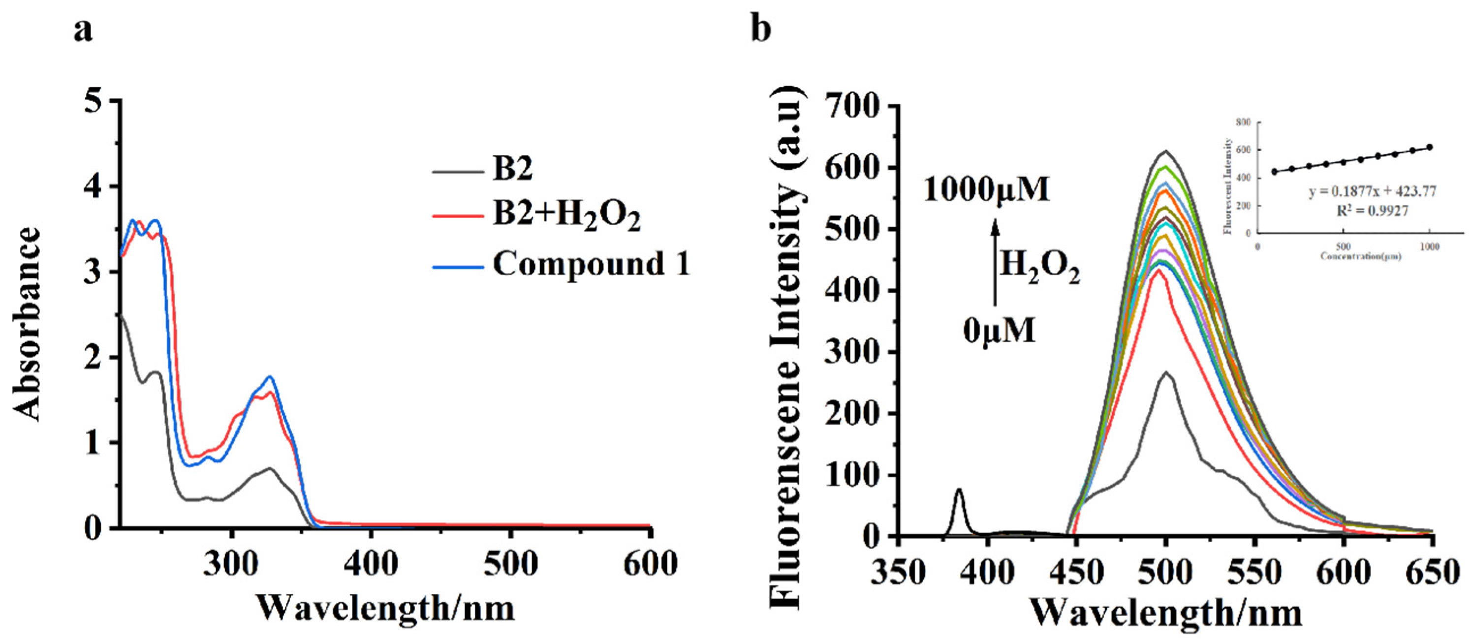
2.2. Optical Characterizations of Probe B2
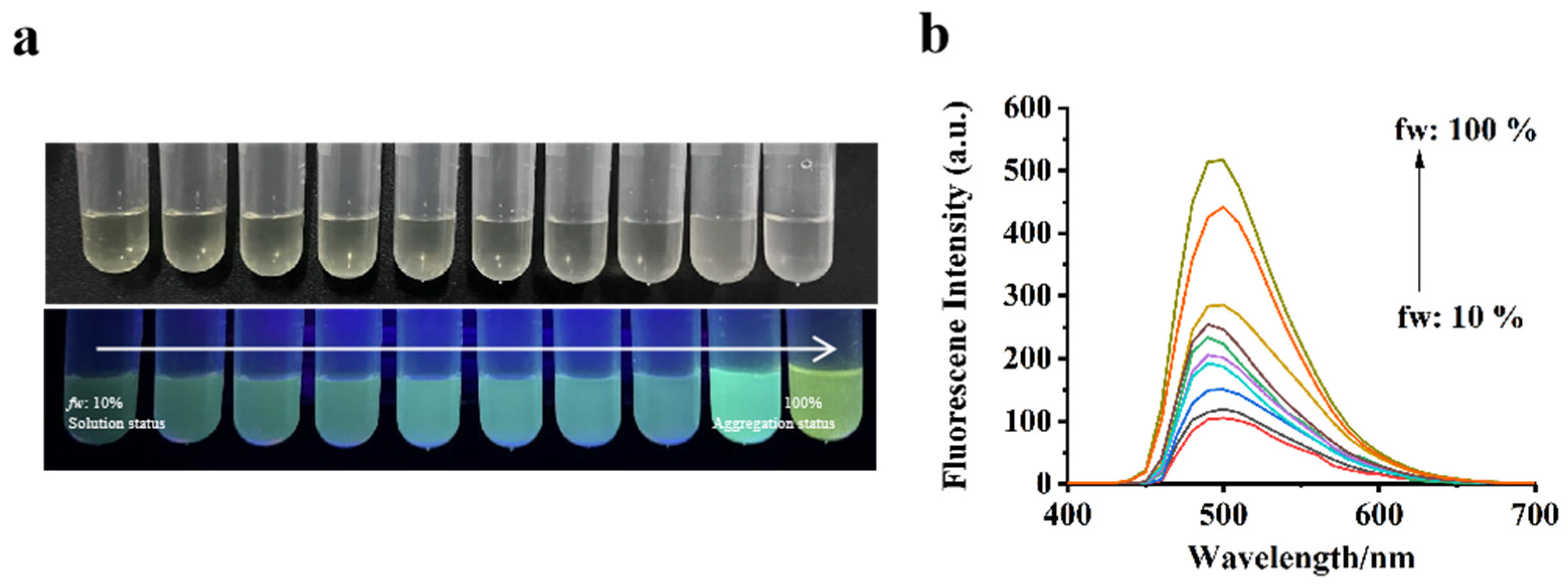
At first, the absorption spectra of B2 before and after adding H2O2 were measured. As described in Figure 2a, in the UV–Vis absorption, which was studied in the absence of H2O2, an absorption of B2 at 325 nm is seen; however, after adding H2O2, the absorption peaked at a 325 nm strength. As shown in Figure S7, B2 was weak when excited at 384 nm (Φ = 0.09), while a significant increase in fluorescence was observed at 500 nm after adding H2O2. The B2 did not emit fluorescence at 500 nm, suggesting that the phenyl boronic acid pinacol ester group of the probe B2 quenched the fluorescence of compound 1 (Φ = 0.17) fluorophore. While H2O2 was added into the B2 system, the system emitted strong fluorescence at 500 nm.
The molar absorption coefficient and Stokes shift of B2 and compound 1 had been studied in different solvents (Table S1). In organic solvents such as DCM, MeOH, MeCN, DMSO, DMF, and THF, B2 and compound 1 showed a high molar absorption coefficient and large Stokes shift in MeCN; thus, MeCN was chosen as the mixture for further spectroscopic study. As shown in Figure 2b, the B2 concentration was 5 µM, and the tested H2O2 concentrations ranged from 0 µM to 1000 µM. As the concentration of H2O2 increases, the fluorescence intensity at 500 nm gradually increases. Moreover, a linear relationship exists between the absorbance values at 500 nm (R2 = 0.9927). The LOD was calculated to be 49.74 nM, comparable to the reported detection methods for peroxynitrite (Table S3). These preliminary in vitro experiments indicated that probe B2 acted as a good “turn-on” response sensor for determining H2O2.

2.3. AIE Property

To further investigate the AIE property of compound 1, the water/MeCN mixtures were used to estimate the influence of the water’s volume fraction (fw) on the fluorescence. The photo of compound 1 in different proportions of the water/MeCN solution under white light and ultraviolet light is shown in Figure 3a. When fw was increased, a distinct enhancement of fluorescence was observed, and the compound 1 solution emitted bright green fluorescence under a 365 nm UV lamp. The fluorescence intensity at 500 nm (λex = 325 nm) increased significantly with the increase in fw (Figure 3b). The change in fluorescent emission with the water fraction indicated that compound 1 has AIE characteristics.

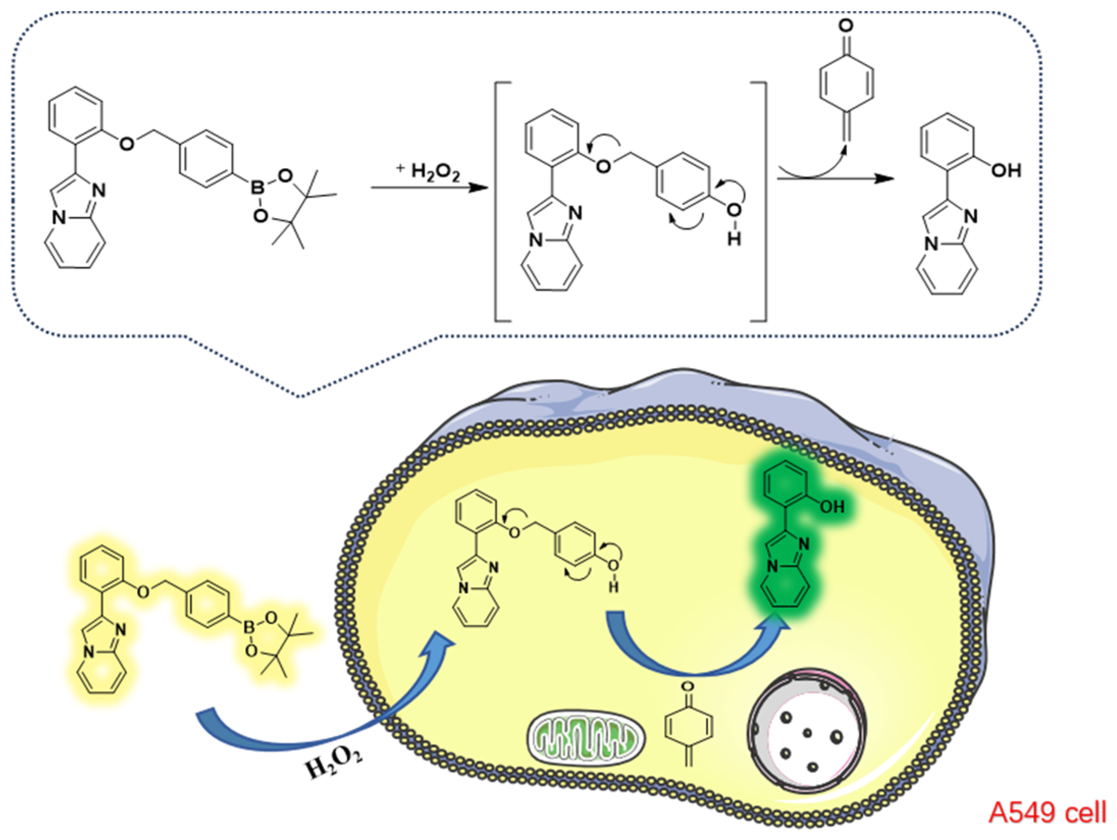
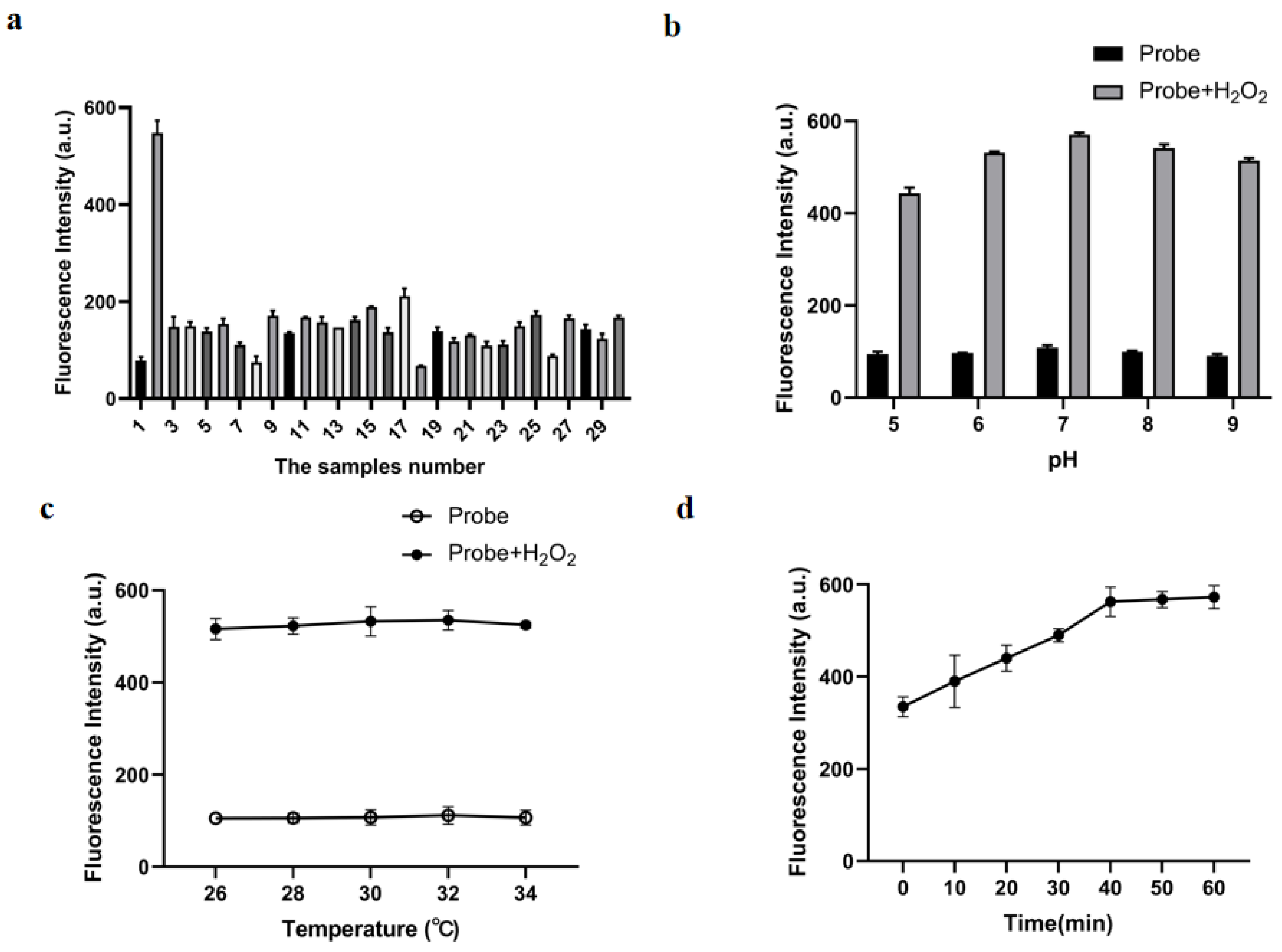
2.4. Characteristics of H2O2 Sensing via Probe B2

In addition to H2O2, various active oxygen species and oxidants were present in organisms. To investigate the selectivity and anti-interference capability of probe B2, this study performed interference experiments on B2 by introducing other interfering substances, including various ROS (H2O2, ONOO, O2, HOCl, 1O2, GSH, and TBHP), Al3+, Fe3+, Fe2+, Cu2+, Ba2+, Ca2+, Mg2+, K+, Na+, F, N3, HPO42−, HSO4, SO42−, HCO3, CO32−, CH3COO, OH, CN, NO3, NO2, Cl, and a blank. Results are shown in Figure 4a. These similar interferences did not cause significant fluorescence changes in probe B2 and did not affect the H2O2 recognition of probe B2. Only the incubation with H2O2 could induce the significant fluorescence enhancement of probe B2, while under the same conditions, no fluorescence increase could be detected for other potential interference.
The acid–base conditions may affect the stability and responsiveness of probe B2 towards H2O2. Therefore, this experiment detected the fluorescence emission of probe B2 before and after H2O2 addition at pH 5 to 9. As shown in Figure 4b, with the addition of H2O2, the probe B2 fluorescence intensity was enhanced at pH 5 to 9, and the fluorescence intensity was maximal at pH 7. Given that the physiological pH value is about 7.4 and probe B2 showed a favorable fluorescence response to H2O2 at pH 7, probe B2 could be utilized for detecting H2O2 in living systems. Furthermore, Figure 4c illustrates the temperature-dependent fluorescence changes of the probe. The results showed that, with the addition of H2O2, the probe reached a plateau in the range of 23 to 36 °C. The fluorescent probe has good stability. Hence, the synthetic probe B2, developed in this experiment, holds promise for detecting H2O2 in the internal environment of living cells.
The time dependence of probe B2 for detecting H2O2 was evaluated by measuring the fluorescence of B2. The time response of probe B2 (5.0 μM) for H2O2 (200 μM) was performed (Figure 4d). After adding 200 μM of H2O2, the fluorescence intensity of probe B2 increased with time until it reached a maximum at 40 min and remained stable. The experimental results showed that B2 had the ability to detect H2O2 in real-time and could complete the detection within 40 min, while the fluorescence intensity almost no longer changed over time.

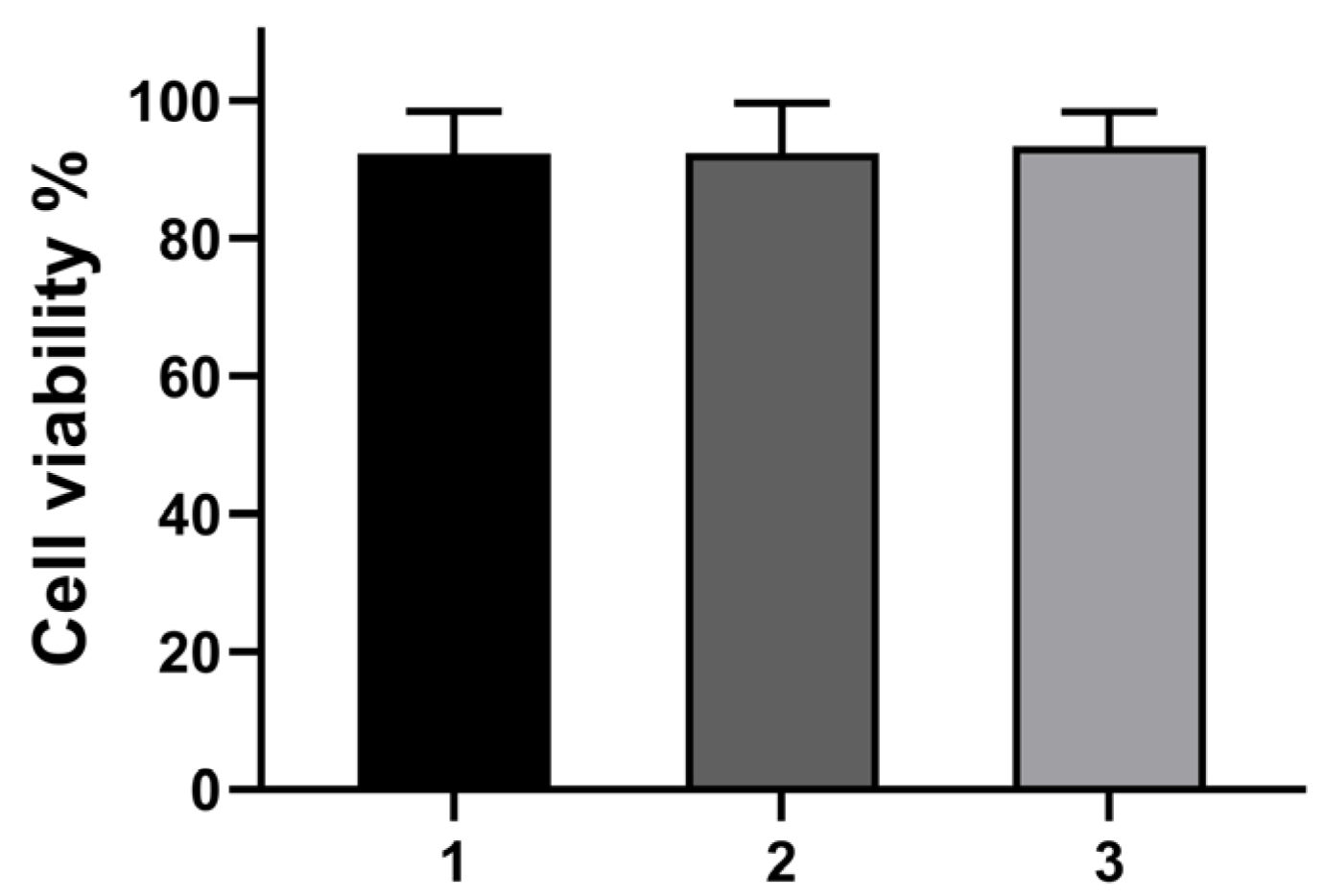
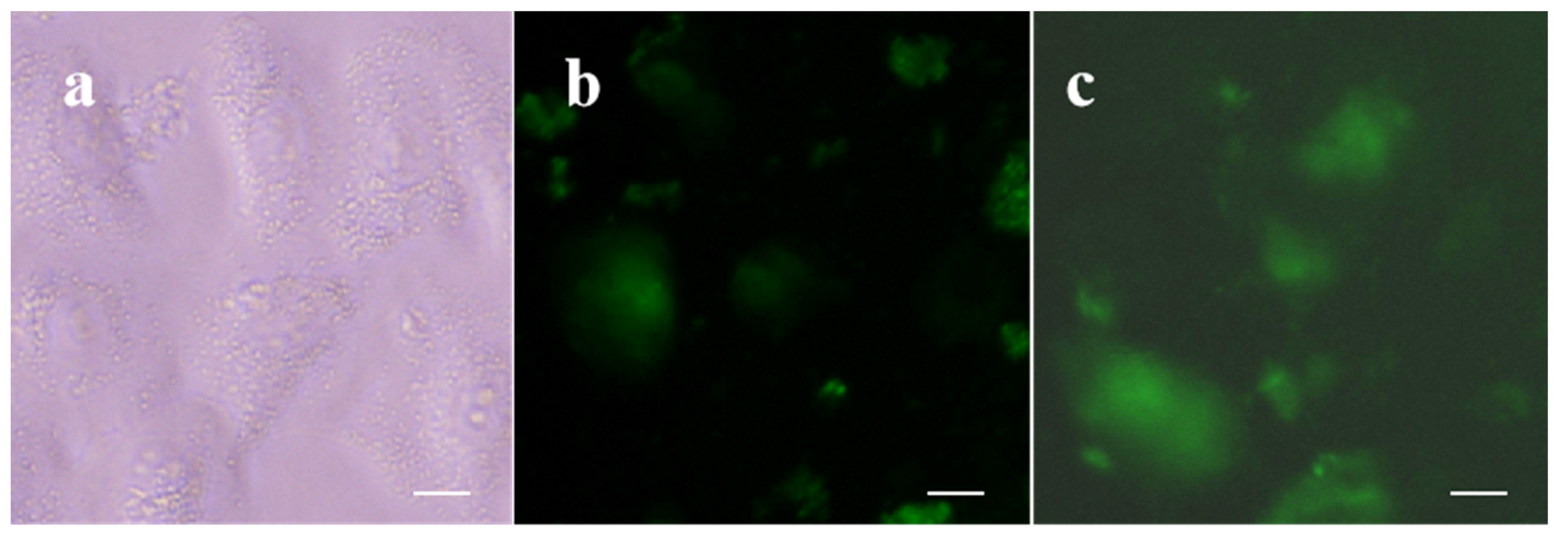
2.5. Cytotoxicity and Cell Imaging of B2

The cytotoxicity of the probe was evaluated via the MTT assay using A549 cells. The test results are shown in Figure 5. At a probe B2 concentration of 30 μM, the cell survival rate exceeded 90%. The IC50 values of the probe on A549 cells were all above 30 μM, indicating that they had no toxicity to normal cells at this concentration. Cells were seeded onto 96-well cell-culture plates and then irradiated for 5 min under the blue channel. The survival of cells was measured using a microplate reader, and the cell survival rate was 90%, 92%, and 98%, respectively. The cytotoxicity test confirmed the safety and reliability of the compounds obtained as chemical diagnostic drugs to a certain extent and established a foundation for subsequent experiments.
Investigating the physical properties of probe B2, further evaluations of its potential applications for localizing hydrogen peroxide in living cells have been conducted in the experiment below. Based on in vitro experiments and cytotoxicity assessments, the optimal conditions were a temperature of 37 °C and a probe concentration of 30 µM was established. This experiment employed probe B2 to detect H2O2 under physiological conditions in live A549 cells. As shown in Figure 6a, A549 cells themselves had no fluorescence (blank group). As shown in Figure 6b, a conspicuous green fluorescence signal was observed upon treatment with PMA for 30 min and subsequent incubation with the fluorescent probe B2 for 30 min. The above results showed that probe B2 could detect endogenous H2O2. Figure 6c shows that these cells exhibited substantial fluorescence emission upon the addition of exogenous H2O2. Fluorescence imaging confirmed the ability of the probe B2 fluorescence probe to detect both endogenous and exogenous H2O2.

3. Materials and Methods

3.1. Chemicals and Instruments

The 1H NMR and 13C NMR spectra were acquired at 25 °C in a nuclear magnetic resonance spectrophotometer (DRX—400, Bruker, Germany) at 400 MHz for 1H NMR in DMSO-d6 and 100 MHz for 13C NMR in DMSO-d6. Thin-layer chromatography (TLC) was performed on silica gel (GF254, Qingdao Haiyang Chemical Co., Ltd., Qingdao, China) with a visualization of the spots under UV light at 254 nm. Fluorescence spectra used a fluorescence spectrophotometer (F-2710, Hitachi, Hiroshima-shi, Japan). Absorption measurements were conducted using a UV–Vis spectrophotometer (UH5300, Hitachi, Japan) within a 190–1100 nm wavelength range.

3.2. Synthesis of the Fluorescence Probe B2

3.2.1. Synthesis of (E)-2-(Imidazole[1,2-a] Pyridin-2-yl) Phenol (1)

NaHCO3 (126 mg, 1.5 mmol) was added to the ethanol solution (15 mL) of a solution of 2-hydroxyacetophenone (642.0 mg, 3.0 mmol) and 2-aminopyridine (282.2 mg, 3.0 mmol). The resulting mixture was refluxed for 2 h, and the reaction was monitored using thin-layer chromatography (TLC). After completion of the reaction, the formed precipitate was filtrated and washed with 15 mL of acetone. The mixture was extracted with dichloromethane (3 × 20 mL). The combined organic layer was dried over MgSO4, and dichloromethane was evaporated under reduced pressure to give compound 1 (458.6 mg, 72.8%). 1H NMR (400 MHz, DMSO-d6) δ 12.05 (s, 1H, OH), 8.62 (dd, 1H, J = 4.0 Hz, ArH), 8.50 (s, 1H, ArH), 7.89 (dd, 1H, J = 8.0 Hz, J = 4.0 Hz, ArH), 7.68 (d, 1H, J = 8.0 Hz, ArH), 7.35 (t, 1H, J = 8.0 Hz, ArH), 7.20 (1H, t, J = 8.0 Hz, ArH), 7.00 (t, 1H, J = 8.0 Hz, ArH), 6.93 (d, 1H, J = 8.0 Hz, ArH), and 6.89 (d, 1H, J = 8.0 Hz, ArH). 13C NMR (100 Hz, DMSO-d6) δ 156.6, 143.5, 143.4, 129.6, 127.3, 127.0, 126.2, 119.6, 116.4, 113.4, and 109.8. HRMS (ESI-TOF) C13H10N2O [M + H]+ 211.0827, found 211.0876.

3.2.2. Synthesis of (E)-2-(2-((4-(4,4,5,5-tetramethyl-1,3,2-dioxaborolan-2-yl) Benzyl) oxy) Phenyl) Imidazo [1,2-a] Pyridine (B2)

K2CO3 (239.8 mg, 2.2 mmol) was added to the anhydrous acetonitrile (10 mL) solution of 1 (420 mg, 2.0 mmol) and 4-(Bromomethyl) benzene boronic acid pinacol ester (651.2 mg, 2.2 mmol). The mixture was heated to reflux for 6 h and then cooled to room temperature. A white solid was produced, filtered, and recrystallized with DMC and hexyl hydride to isolate the purified product B2 (624.8 mg, 73.3%). 1H NMR (400 MHz, DMSO-d6) δ 8.49 (dd, 1H, J = 8.0 Hz, J = 1.6 Hz, ArH), 8.16 (s, 1H, ArH), 8.02 (d, 1H, J = 8.0 Hz, ArH), 7.88 (d, 2H, J = 8.0 Hz, ArH), 7.61 (d, 1H, J = 8.0 Hz, ArH), 7.53 (d, 2H, J = 8.0 Hz, ArH), 7.28–7.32 (m, 1H, ArH), 7.13–7.17 (m, 1H, ArH), 7.05 (d, 1H, J = 8.0 Hz ArH), 6.73 (t, 1H, J = 8.0 Hz, ArH), 5.25 (2H, s, ArH), and 1.39 (s, 12H, CH3). 13C NMR (100 Hz, DMSO-d6) δ 155.6, 144.0, 141.0, 140.6, 135.2, 128.9, 127.5, 127.2, 125.4, 122.9, 121.3, 116.8, 113.3, 113.0, 112.3, 83.9, 70.0, and 24.9. HRMS (ESI-TOF) C26H27BN2O3 [M + H]+ 426.3230, found 427.2193.

3.3. Spectroscopic Materials and Methods

The fluorescence intensity of fluorescent probe B2 measurement experiments was conducted in a 10% MeCN solution (MeCN/H2O = 1:9, v/v). The fluorescent emission spectra were recorded at 350 to 650 nm.

3.4. Measurement of the Detection Limit

The detection limit was obtained from the fluorescence titration curve and was calculated according to the following equations: Detection limit = 3σ/k. Here, σ represents the deviation of the blank, and k is the slope of the linear regression equation.

3.5. Cytotoxic Assay

Dulbecco’s modified Eagle’s medium (DMEM) was used to culture A549 cells at 37 °C. The MTT assay was used to measure the cytotoxicity of probe B2 towards A549 cells. About 5 × 10⁴ cells/well were seeded onto 96-well cell-culture plates for 24 h. Then, different concentrations of probes were added to the wells. After another 12 h, the MTT (100 µL) was added to each well and incubated at 37 °C under 5% CO2 for 4 h. The MTT solution was removed, purple precipitates (formazan) observed in plates were further dissolved in DMSO (100 µL), and a microplate reader was used to measure the absorbance for each well.

3.6. Cell Image Experiments

The A549 cells were cultured in a DMEM culture medium supplemented with 1% (vol/vol) penicillin and 10% (vol/vol) fetal bovine serum at 37 °C in a 5% CO2 atmosphere for 24 h. Before the experiment, the cell culture medium was poured off, washed three times with PBS, and changed into a new medium. For the imaging of cellular endogenous H2O2, A549 cells were incubated with 100 µM H2O2 for 30 min in the control group. In the experimental group, one group of A549 cells was treated with the probe alone for 30 min. The ROS levels in A549 cells were upregulated using the activator PMA (phorbol-12-myristate-13-acetate, 1 μM) for 30 min. In the other group, A549 cells were pretreated with PMA for 30 min and then incubated with the probe for an additional 30 min. To detect exogenous H2O2, cells were first incubated with H2O2 for 30 min, and then A549 cells were treated with a probe B2 for 30 min. Finally, 1 mL of paraformaldehyde was added to each group of cells and fixed for 5 min. After three washes with PBS, 1 mL DAPI dye (1 µg/mL) was added to each well, nuclear staining for 1–5 min, and finally washed three times with PBS. Finally, a fluorescence inverted microscope was applied to fluorescence imaging.

4. Conclusions

In summary, the 2-(2-((4-(4,4,5,5-tetramethyl-1,3,2-dioxaborolan-2-yl) benzyl) oxy) phenyl) imidazo[1,2-a] pyridine (B2) had been designed and synthesized successfully for the first time to detect H2O2 in living cells. It can react with H2O2 selectively, thus producing a green fluorescence signal at 500 nm. The probe B2 demonstrates high selectivity, sensitivity, and anti-interference ability. Importantly, the synthesis process of B2 was simple, and the yield was high. This study has successfully employed probe B2 to visualize H2O2 in A549 cells, allowing clear and visual monitoring of both exogenous and endogenous changes in H2O2 levels. It is expected that B2 will become a promising tool to detect H2O2 and exhibit important potential applications in the diagnosis of H2O2-related diseases.

Supplementary Materials

The following supporting information can be downloaded at: https://www.mdpi.com/article/10.3390/molecules29040882/s1, Figures S1–S6: 1H NMR, 13C NMR and MS of all products; Figure S7: UV–Vis response of B2 towards H2O2; Table S1: The photoluminescence properties of B2 and compound 1; Table S2: The microscope set-up (for the imaging experiments); Table S3: Comparing the limit of detection with other reported methods for H2O2 detection. CheckCIF report of compound 1.

Author Contributions

Conceptualization, L.T., M.Z. and J.J.; data curation, Y.Y. and L.Z.; formal analysis, L.T., J.T. and B.S.; funding acquisition, M.Z. and J.J.; investigation, C.S. and M.Q.; methodology, C.S. and Y.Y.; project administration, M.Z. and J.J.; resources, J.T., M.Z. and J.J.; software, L.T.; supervision, M.Z. and J.J.; validation, L.T., Y.Y. and F.Y.; writing—original draft, L.T.; writing—review and editing, M.Z. and J.J. All authors have read and agreed to the published version of the manuscript.

Funding

Project supported by the Fundamental Research Program of Shanxi Province [202103021224335], Yangquan Key Research and Development Project [2022JH067], and Supported by Scientific and Technological Innovation Programs of Higher Education Institutions in Shanxi [2022L595].

Institutional Review Board Statement

Not applicable.

Informed Consent Statement

Not applicable.

Data Availability Statement

Data are contained within the article.

Conflicts of Interest

The authors declare no conflicts of interest.

References

  1. Murrant, C.L.; Reid, M.B. Detection of reactive oxygen and reactive nitrogen species in skeletal muscle. Microsc. Res. Tech. 2001, 55, 236–248. [Google Scholar] [CrossRef]
  2. Dilly, S.; Romero, M.; Solier, S.; Feron, O.; Dessy, C.; Slama Schwok, A. Targeting M2 macrophages with a novel NADPH oxidase inhibitor. Antioxidants 2023, 12, 440. [Google Scholar] [CrossRef] [PubMed]
  3. Song, X.; Bai, S.; He, N.; Wang, R.; Xing, Y.; Lv, C.; Yu, F. Real-time evaluation of hydrogen peroxide injuries in pulmonary fibrosis mice models with a mitochondria-targeted near-infrared fluorescent probe. ACS Sens. 2021, 6, 1228–1239. [Google Scholar] [CrossRef] [PubMed]
  4. Lu, D.; Cagan, A.; Munoz, R.A.; Tangkuaram, T.; Wang, J. Highly sensitive electrochemical detection of trace liquid peroxide explosives at a Prussian-blue ‘artificial-peroxidase’ modified electrode. Analyst 2006, 131, 1279–1281. [Google Scholar] [CrossRef] [PubMed]
  5. Victoria, T.; Heikes, B.G.; Mcneill, A.S.; Silwal, I.K.C.; Sullivan, D.W. Measurement of formic acid, acetic acid and hydroxyacetaldehyde, hydrogen peroxide, and methyl peroxide in air by chemical ionization mass spectrometry: Airborne method development. Atmos. Meas. Tech. 2018, 11, 1901–1920. [Google Scholar]
  6. Ying, A.; Ying, L. A new hydrocyanine probe for imaging reactive oxygen species in the mitochondria of live cells. Bioorganic Med. Chem. Lett. 2023, 87, 129262. [Google Scholar] [CrossRef]
  7. Lian, J.; Wang, Y.; Sun, X.; Shi, Q.; Meng, F. Progress on multifunction enzyme-activated organic fluorescent probes for bioimaging. Front. Chem. 2022, 10, 935586. [Google Scholar] [CrossRef]
  8. Chen, J.; Wu, T.; Liang, W.; Ciou, J.; Lai, C. Boronates as hydrogen peroxide-reactive warheads in the design of detection probes, prodrugs, and nanomedicines used in tumors and other diseases. Drug Deliv. Transl. Res. 2023, 13, 1305–1321. [Google Scholar] [CrossRef]
  9. Zhong, H.; Yu, S.; Li, B.; He, K.; Li, D.; Wang, X.; Wu, Y. Two-photon fluorescence and MR bio-imaging of endogenous H2O2 in the tumor microenvironment using a dual-mode nanoprobe. Chem. Commun. 2021, 57, 6288–6291. [Google Scholar] [CrossRef]
  10. Liu, M.; Cao, J.; Tu, Y.; Huang, C.; Zhang, M.; Zheng, J. An ultra-sensitive near-infrared fluorescent probe based on triphenylamine with high selectivity detecting the keratin. Anal. Biochem. 2022, 646, 114638. [Google Scholar] [CrossRef]
  11. Li, Y.; Ren, L.; Gao, T.; Chen, T.; Han, J.; Wang, Y. A coumarin-based fluorescent probe for sensitive monitoring H2O2 in water and living cells. Tetrahedron Lett. 2023, 114, 154291. [Google Scholar] [CrossRef]
  12. Zhou, J.; Geng, Y.; Wang, Z. Fluorescent molecular probes for imaging and detection of oxidases and peroxidases in biological samples. Methods 2023, 210, 20–35. [Google Scholar] [CrossRef] [PubMed]
  13. Pinzi, M.; Galvan, S.; Rodriguez, Y.; Baena, F. The Adaptive Hermite Fractal Tree (AHFT): A novel surgical 3D path planning approach with curvature and heading constraints. Int. J. Comput. Assist. Radiol. Surg. 2019, 14, 659–670. [Google Scholar] [CrossRef] [PubMed]
  14. Miller, E.W.; Albers, A.E.; Pralle, A.; Isacoff, E.Y.; Chang, C.J. Boronate-based fluorescent probes for imaging cellular hydrogen peroxide. J. Am. Chem. Soc. 2005, 127, 16652–16659. [Google Scholar] [CrossRef] [PubMed]
  15. Lavis, L.D.; Raines, R.T. Bright building blocks for chemical biology. ACS Chem. Biol. 2014, 9, 855–866. [Google Scholar] [CrossRef] [PubMed]
  16. Su, J.; Zhang, S.; Wang, C.; Li, M.; Wang, J.; Su, F.; Wang, Z. A fast and efficient method for detecting H2O2 by a dual-locked model chemosensor. ACS Omega 2021, 6, 14819–14823. [Google Scholar] [CrossRef] [PubMed]
  17. Zhao, J.; Pan, X.; Zhu, J.; Zhu, X. Novel AIEgen-functionalized diselenide-crosslinked polymer gels as fluorescent probes and drug release carriers. Polymers 2020, 12, 551. [Google Scholar] [CrossRef]
  18. Han, H.; He, X.; Wu, M.; Huang, Y.; Zhao, L.; Xu, L.; Ma, P.; Sun, Y.; Song, D.; Wang, X. A novel colorimetric and near-infrared fluorescence probe for detecting and imaging exogenous and endogenous hydrogen peroxide in living cells. Talanta 2020, 21, 121000. [Google Scholar] [CrossRef]
  19. Wu, F.; Yu, H.; Wang, Q.; Zhang, J.; Li, Z.; Yang, F. Development of a coumarin-based fluorescent probe for hydrogen peroxide based on the Payne/Dakin tandem reaction. Dye. Pigment. 2021, 190, 335. [Google Scholar] [CrossRef]
  20. Wei, Y.; Wang, X.; Shi, W.; Chen, R.; Zheng, L.; Wang, Z.; Chen, K.; Gao, L. A novel methylenemalononitrile -BODIPY-based fluorescent probe for highly selective detection of hydrogen peroxide in living cells. Eur. J. Med. Chem. 2021, 226, 113828. [Google Scholar] [CrossRef]
  21. Chang, M.C.; Pralle, A.; Isacoff, E.Y.; Chang, C.J. A selective, cell-permeable optical probe for hydrogen peroxide in living cells. J. Am. Chem. Soc. 2004, 126, 15392–15393. [Google Scholar] [CrossRef] [PubMed]
  22. Song, Z.; Kwok, R.T.; Ding, D.; Nie, H.; Lam, J.W.; Liu, B.; Tang, B.Z. An AIE-active fluorescence turn-on bioprobe mediated by hydrogen-bonding interaction for highly sensitive detection of hydrogen peroxide and glucose. Chem. Commun. 2016, 52, 10076–10079. [Google Scholar] [CrossRef] [PubMed]
  23. Lippert, A.R.; Van de Bittner, G.C.; Chang, C.J. Boronate oxidation as a bioorthogonal reaction approach for studying the chemistry of hydrogen peroxide in living systems. Acc. Chem. Res. 2011, 44, 793–804. [Google Scholar] [CrossRef]
  24. de Gracia Lux, C.; Joshi-Barr, S.; Nguyen, T.; Mahmoud, E.; Schopf, E.; Fomina, N.; Almutairi, A. Biocompatible polymeric nanoparticles degrade and release cargo in response to biologically relevant levels of hydrogen peroxide. J. Am. Chem. Soc. 2012, 134, 15758–15764. [Google Scholar] [CrossRef] [PubMed]
  25. Franks, A.T.; Franz, K.J. A prochelator with a modular masking group featuring hydrogen peroxide activation with concurrent fluorescent reporting. Chem. Commun. 2014, 50, 11317–11320. [Google Scholar] [CrossRef] [PubMed]
  26. Gu, T.; Mo, S.; Mu, Y.; Huang, X.; Hu, L. Detection of endogenous hydrogen peroxide in living cells with para -nitrophenyl oxoacetyl rhodamine as turn-on mitochondria-targeted fluorescent probe. Sens. Actuators B. 2020, 309, 127731. [Google Scholar] [CrossRef]
  27. Wang, K.; Yao, T.; Xue, J.; Guo, Y.; Xu, X. A novel fluorescent probe for the detection of hydrogen peroxide. Biosensors 2023, 13, 658. [Google Scholar] [CrossRef] [PubMed]
  28. Liu, C.; Shen, Y.; Yin, P.; Li, L.; Liu, M.; Zhang, Y.; Li, H.; Yao, S. Sensitive detection of acetylcholine based on a novel boronate intramolecular charge transfer fluorescence probe. Anal. Biochem. 2014, 465, 172–178. [Google Scholar] [CrossRef]
  29. Wen, D.; Deng, X.; Xu, G.; Wu, H.; Yu, Y. A novel FRET fluorescent probe based on BODIPY-rhodamine system for Hg2+ imaging in living cells. J. Mol. Struct. 2021, 1236, 130323. [Google Scholar] [CrossRef]
  30. Xu, J.; Li, Q.; Yue, Y.; Guo, Y.; Shao, S. A water-soluble BODIPY derivative as a highly selective “Turn-On” fluorescent sensor for H2O2 sensing in vivo. Biosens. Bioelectron. 2014, 56, 58–63. [Google Scholar] [CrossRef]
  31. Wu, Z.; Liu, M.; Liu, Z.; Tian, Y. Real-Time Imaging and Simultaneous Quantification of Mitochondrial H2O2 and ATP in Neurons with a Single Two-Photon Fluorescence-Lifetime-Based Probe. J. Am. Chem. Soc. 2020, 142, 7532–7541. [Google Scholar] [CrossRef] [PubMed]
  32. Chen, Q.; Cheng, K.; Wang, W.; Yang, L.; Xie, Y.; Feng, L.; Zhang, J.; Zhang, H.; Sun, H. A pyrene-based ratiometric fluorescent probe with a large Stokes shift for selective detection of hydrogen peroxide in living cells. J. Pharm. Anal. 2020, 10, 490–497. [Google Scholar] [CrossRef] [PubMed]
  33. Liu, Y.; Du, W.; Liu, G.; Zhou, W.; Gao, X.; Xing, G. Assembly of water-soluble AIE-active fluorescent organic nanoparticles for ratiometric detection of hypochlorite in living cells. Chem.-Asian J. 2021, 16, 277–281. [Google Scholar] [CrossRef] [PubMed]
  34. Xu, L.; Zhai, J.; Han, X.; Shang, W.; Chen, M.; Xu, Y.; Cui, X.; Zu, G.; Sang, F.; Zhang, B. A new strategy of transforming an ACQ compound into an AIE theranostic system for bacterial imaging and photodynamic antibacterial therapy. Luminescence 2023, 38, 497–504. [Google Scholar] [CrossRef] [PubMed]
  35. Li, Y.; Huang, Y.; Sun, X.; Zhong, K.; Tang, L. An AIE mechanism-based fluorescent probe for relay recognition of HSO3/H2O2 and its application in food detection and bioimaging. Talanta 2023, 258, 124412. [Google Scholar] [CrossRef] [PubMed]
  36. Cui, W.; Wang, M.; Yang, Y.; Wang, J. Effect of different substituents on the fluorescence properties of precursors of synthetic GFP analogues and a polarity-sensitive lipid droplet probe with AIE properties for imaging cells and zebrafish. Org. Biomol. Chem. 2023, 21, 2960–2967. [Google Scholar] [CrossRef]
  37. Das, R.; Bej, S.; Hirani, H.; Banerjee, P. Trace-level humidity sensing from commercial organic solvents and food products by an AIE/ESIPT-triggered piezochromic luminogen and ppb-level “OFF-ON-OFF” sensing of Cu2+: A combined experimental and theoretical outcome. ACS Omega 2021, 6, 14104–14121. [Google Scholar] [CrossRef]
  38. Shen, J.; Fan, Z. Ce3+-induced fluorescence amplification of copper nanoclusters based on aggregation-induced emission for specific sensing 2,6-pyridine dicarboxylic acid. J. Fluoresc. 2023, 33, 135–144. [Google Scholar] [CrossRef]
Scheme 1. (a) Anhydrous ethanol, sodium bicarbonate, reflux, 2 h, 72.8%; (b) potassium carbonate, anhydrous acetonitrile, reflux, 6 h, 73.3%.
Molecules 29 00882 sch001
Figure 1. The principle of ESIPT-based fluorescent probe B2 detecting H2O2.
Molecules 29 00882 g001
Figure 2. (a) UV–Vis absorption spectra of compound 1 and B2 and B2 after addition of H2O2; (b) fluorescence emission spectra of probe B2 (5 μM) following incubation with an increasing concentration of H2O2 (30, 50, 100, 150, 200, 300, 450, 500, 600, 700, 800, 900, and 1000 µM). The experiments were repeated three times, and the data are shown as mean (±S.D.).
Molecules 29 00882 g002
Figure 3. (a) Photographs of compound 1 in water/MeCN mixtures taken under natural light and UV illumination; (b) fluorescence spectra of compound 1 in the water/MeCN mixture. The experiments were repeated three times, and the data are shown as mean (±S.D.).
Molecules 29 00882 g003
Figure 4. (a) Comparison of the fluorescence intensity of probe B2 (5 μM) at 500 nm after incubation with (1) blank, (2) H2O2, (3) ONOO, (4) O2, (5) HOCl, (6) 1O2, (7) GSH, (8) TBHP, (9) Al3+, (10) Fe3+, (11) Fe2+, (12) Cu2+, (13) Ba2+, (14) Ca2+, (15) Mg2+, (16) K+, (17) Na+, (18) F, (19) N3, (20) HPO42−, (21) HSO4, (22) SO42−, (23) HCO3, (24) CO32−, (25) CH3COO, (26) OH, (27) CN, (28) NO3, (29) NO2, and (30) Cl. Interfering substances were at a concentration of 500 μM, H2O2 (200 μM); (b) fluorescence spectral changes of B2 at 500 nm before and after the addition of H2O2 at different pH; (c) B2 fluorescence intensity with different temperatures; and (d) time-dependent curve of probe B2 at 500 nm toward H2O2. The experiments were repeated three times, and the data are shown as mean (±S.D.).
Molecules 29 00882 g004
Figure 5. The cell survival rate of A549 cells. (1) After 5 min of irradiation with blue light; (2) after adding 30 µM of probe B2; and (3) after irradiation under blue light for 5 min and the addition of probe B2. The experiments were repeated three times, and the data are shown as mean (±S.D.).
Molecules 29 00882 g005
Figure 6. (a) Image of endogenous H2O2 in A549 cells untreated; (b) images of the probe and the PMA in A549 cells; and (c) images of probe B2 and the exogenous H2O2 in A549 cells. Scale bar = 40 μm.
Molecules 29 00882 g006
Disclaimer/Publisher’s Note: The statements, opinions and data contained in all publications are solely those of the individual author(s) and contributor(s) and not of MDPI and/or the editor(s). MDPI and/or the editor(s) disclaim responsibility for any injury to people or property resulting from any ideas, methods, instructions or products referred to in the content.

