
Discovery of a Novel Chemo-Type for TAAR1 Agonism via Molecular Modeling


 ,
,  ,
,  ,
,  ,
,  ,
, 

Abstract
1. Introduction
2. Results
2.1. Molecular Docking Studies of Oxazoline-Based TAAR1 Agonists
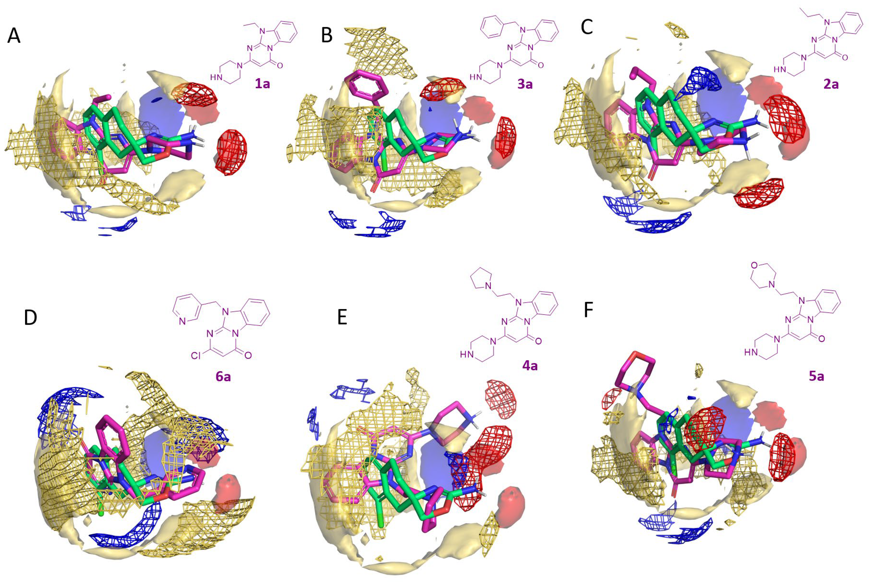
2.2. In Silico Evaluation of Pyrimidinone-Benzimidazole-Based Compounds as TAAR1 Agonists
2.3. Biological Evaluation of Compounds 1a–10a as Novel TAAR1 Agonists
3. Discussion
4. Materials and Methods
4.1. Computational Studies
4.2. Chemicals
4.3. Biological Evaluation of Compounds 1a–10a as TAAR1 Agonists
Bioluminescence Resonance Energy Transfer (BRET) Measurement
5. Conclusions
Supplementary Materials
Author Contributions
Funding
Institutional Review Board Statement
Informed Consent Statement
Data Availability Statement
Conflicts of Interest
Correction Statement
References
- Gainetdinov, R.R.; Hoener, M.C.; Berry, M.D. Trace Amines and Their Receptors. Pharmacol. Rev. 2018, 70, 549–620. [Google Scholar] [CrossRef]
- Dedic, N.; Dworak, H.; Zeni, C.; Rutigliano, G.; Howes, O.D. Therapeutic Potential of TAAR1 Agonists in Schizophrenia: Evidence from Preclinical Models and Clinical Studies. Int. J. Mol. Sci. 2021, 22, 13185. [Google Scholar] [CrossRef] [PubMed]
- Hart, M.E.; Suchland, K.L.; Miyakawa, M.; Bunzow, J.R.; Grandy, D.K.; Scanlan, T.S. Trace Amine-Associated Receptor Agonists: Synthesis and Evaluation of Thyronamines and Related Analogues. J. Med. Chem. 2006, 49, 1101–1112. [Google Scholar] [CrossRef] [PubMed]
- Rutigliano, G.; Accorroni, A.; Zucchi, R. The Case for TAAR1 as a Modulator of Central Nervous System Function. Front. Pharmacol. 2017, 8, 987. [Google Scholar] [CrossRef]
- Xu, Z.; Li, Q. TAAR Agonists. Cell. Mol. Neurobiol. 2020, 40, 257–272. [Google Scholar] [CrossRef] [PubMed]
- Halff, E.F.; Rutigliano, G.; Garcia-Hidalgo, A.; Howes, O.D. Trace Amine-Associated Receptor 1 (TAAR1) Agonism as a New Treatment Strategy for Schizophrenia and Related Disorders. Trends Neurosci. 2023, 46, 60–74. [Google Scholar] [CrossRef] [PubMed]
- Guo, L.; Dai, W.; Xu, Z.; Liang, Q.; Miller, E.T.; Li, S.; Gao, X.; Baldwin, M.W.; Chai, R.; Li, Q. Evolution of Brain-Expressed Biogenic Amine Receptors into Olfactory Trace Amine-Associated Receptors. Mol. Biol. Evol. 2022, 39, msac006. [Google Scholar] [CrossRef] [PubMed]
- Guo, L.; Cheng, J.; Lian, S.; Liu, Q.; Lu, Y.; Zheng, Y.; Zhu, K.; Zhang, M.; Kong, Y.; Zhang, C.; et al. Structural Basis of Amine Odorant Perception by a Mammal Olfactory Receptor. Nature 2023, 618, 193–200. [Google Scholar] [CrossRef]
- Espinoza, S.; Sukhanov, I.; Efimova, E.V.; Kozlova, A.; Antonova, K.A.; Illiano, P.; Leo, D.; Merkulyeva, N.; Kalinina, D.; Musienko, P.; et al. Trace Amine-Associated Receptor 5 Provides Olfactory Input Into Limbic Brain Areas and Modulates Emotional Behaviors and Serotonin Transmission. Front. Mol. Neurosci. 2020, 13, 18. [Google Scholar] [CrossRef]
- Efimova, E.V.; Kozlova, A.A.; Razenkova, V.; Katolikova, N.V.; Antonova, K.A.; Sotnikova, T.D.; Merkulyeva, N.S.; Veshchitskii, A.S.; Kalinina, D.S.; Korzhevskii, D.E.; et al. Increased Dopamine Transmission and Adult Neurogenesis in Trace Amine-Associated Receptor 5 (TAAR5) Knockout Mice. Neuropharmacology 2021, 182, 108373. [Google Scholar] [CrossRef] [PubMed]
- Efimova, E.V.; Kuvarzin, S.R.; Mor, M.S.; Katolikova, N.V.; Shemiakova, T.S.; Razenkova, V.; Ptukha, M.; Kozlova, A.A.; Murtazina, R.Z.; Smirnova, D.; et al. Trace Amine-Associated Receptor 2 Is Expressed in the Limbic Brain Areas and Is Involved in Dopamine Regulation and Adult Neurogenesis. Front. Behav. Neurosci. 2022, 16, 847410. [Google Scholar] [CrossRef] [PubMed]
- Borowsky, B.; Adham, N.; Jones, K.A.; Raddatz, R.; Artymyshyn, R.; Ogozalek, K.L.; Durkin, M.M.; Lakhlani, P.P.; Bonini, J.A.; Pathirana, S.; et al. Trace Amines: Identification of a Family of Mammalian G Protein-Coupled Receptors. Proc. Natl. Acad. Sci. USA 2001, 98, 8966–8971. [Google Scholar] [CrossRef] [PubMed]
- Bunzow, J.R.; Sonders, M.S.; Arttamangkul, S.; Harrison, L.M.; Zhang, G.; Quigley, D.I.; Darland, T.; Suchland, K.L.; Pasumamula, S.; Kennedy, J.L.; et al. Amphetamine, 3,4-Methylenedioxymethamphetamine, Lysergic Acid Diethylamide, and Metabolites of the Catecholamine Neurotransmitters Are Agonists of a Rat Trace Amine Receptor. Mol. Pharmacol. 2001, 60, 1181–1188. [Google Scholar] [CrossRef] [PubMed]
- Lindemann, L.; Meyer, C.A.; Jeanneau, K.; Bradaia, A.; Ozmen, L.; Bluethmann, H.; Bettler, B.; Wettstein, J.G.; Borroni, E.; Moreau, J.-L.; et al. Trace Amine-Associated Receptor 1 Modulates Dopaminergic Activity. J. Pharmacol. Exp. Ther. 2008, 324, 948–956. [Google Scholar] [CrossRef]
- John, J.; Kukshal, P.; Bhatia, T.; Chowdari, K.V.; Nimgaonkar, V.L.; Deshpande, S.N.; Thelma, B.K. Possible Role of Rare Variants in Trace Amine Associated Receptor 1 in Schizophrenia. Schizophr. Res. 2017, 189, 190–195. [Google Scholar] [CrossRef] [PubMed]
- Mühlhaus, J.; Dinter, J.; Jyrch, S.; Teumer, A.; Jacobi, S.F.; Homuth, G.; Kühnen, P.; Wiegand, S.; Grüters, A.; Völzke, H.; et al. Investigation of Naturally Occurring Single-Nucleotide Variants in Human TAAR1. Front. Pharmacol. 2017, 8, 807. [Google Scholar] [CrossRef] [PubMed]
- Imbriglio, T.; Alborghetti, M.; Bruno, V.; Battaglia, G.; Nicoletti, F.; Cannella, M. Up-Regulation of the Trace Amine Receptor, TAAR-1, in the Prefrontal Cortex of Individuals Affected by Schizophrenia. Schizophr. Bull. 2023, 50, 374–381. [Google Scholar] [CrossRef] [PubMed]
- De Gregorio, D.; Posa, L.; Ochoa-Sanchez, R.; McLaughlin, R.; Maione, S.; Comai, S.; Gobbi, G. The Hallucinogen D-Lysergic Diethylamide (LSD) Decreases Dopamine Firing Activity through 5-HT1A, D2 and TAAR1 Receptors. Pharmacol. Res. 2016, 113, 81–91. [Google Scholar] [CrossRef]
- Liberles, S.D. Trace Amine-Associated Receptors: Ligands, Neural Circuits, and Behaviors. Curr. Opin. Neurobiol. 2015, 34, 1–7. [Google Scholar] [CrossRef]
- Polini, B.; Ricardi, C.; Bertolini, A.; Carnicelli, V.; Rutigliano, G.; Saponaro, F.; Zucchi, R.; Chiellini, G. T1AM/TAAR1 System Reduces Inflammatory Response and β-Amyloid Toxicity in Human Microglial HMC3 Cell Line. Int. J. Mol. Sci. 2023, 24, 11569. [Google Scholar] [CrossRef]
- Frycz, B.A.; Nowicka, K.; Konopka, A.; Hoener, M.C.; Bulska, E.; Kaczmarek, L.; Stefaniuk, M. Activation of Trace Amine-associated Receptor 1 (TAAR1) Transiently Reduces Alcohol Drinking in Socially Housed Mice. Addict. Biol. 2023, 28, e13285. [Google Scholar] [CrossRef] [PubMed]
- Berry, M.D.; Gainetdinov, R.R.; Hoener, M.C.; Shahid, M. Pharmacology of Human Trace Amine-Associated Receptors: Therapeutic Opportunities and Challenges. Pharmacol. Ther. 2017, 180, 161–180. [Google Scholar] [CrossRef] [PubMed]
- Jia, L.; Li, S.; Dai, W.; Guo, L.; Xu, Z.; Scott, A.M.; Zhang, Z.; Ren, J.; Zhang, Q.; Dexheimer, T.S.; et al. Convergent Olfactory Trace Amine-Associated Receptors Detect Biogenic Polyamines with Distinct Motifs via a Conserved Binding Site. J. Biol. Chem. 2021, 297, 101268. [Google Scholar] [CrossRef] [PubMed]
- Cisneros, I.E.; Ghorpade, A.; Borgmann, K. Methamphetamine Activates Trace Amine Associated Receptor 1 to Regulate Astrocyte Excitatory Amino Acid Transporter-2 via Differential CREB Phosphorylation During HIV-Associated Neurocognitive Disorders. Front. Neurol. 2020, 11, 593146. [Google Scholar] [CrossRef]
- Freyberg, Z.; Saavedra, J.M. Trace Amines and Trace Amine-Associated Receptors: A New Frontier in Cell Signaling. Cell. Mol. Neurobiol. 2020, 40, 189–190. [Google Scholar] [CrossRef]
- Zhai, L.; Xiao, H.; Lin, C.; Wong, H.L.X.; Lam, Y.Y.; Gong, M.; Wu, G.; Ning, Z.; Huang, C.; Zhang, Y.; et al. Gut Microbiota-Derived Tryptamine and Phenethylamine Impair Insulin Sensitivity in Metabolic Syndrome and Irritable Bowel Syndrome. Nat. Commun. 2023, 14, 4986. [Google Scholar] [CrossRef]
- Zheng, Y.; Yasuda, M.; Yamao, M.; Gokan, T.; Sejima, Y.; Nishikawa, T.; Katayama, S. Fermented Soybean Foods (Natto) Ameliorate Age-Related Cognitive Decline by Hippocampal TAAR1-Mediated Activation of the CaMKII/CREB/BDNF Signaling Pathway in Senescence-Accelerated Mouse Prone 8 (SAMP8). Food Funct. 2023, 14, 10097–10106. [Google Scholar] [CrossRef] [PubMed]
- Liu, J.; Wu, R.; Li, J.-X. TAAR1 as an Emerging Target for the Treatment of Psychiatric Disorders. Pharmacol. Ther. 2024, 253, 108580. [Google Scholar] [CrossRef]
- Cichero, E.; Tonelli, M. Targeting Species-Specific Trace Amine-Associated Receptor 1 Ligands: To Date Perspective of the Rational Drug Design Process. Future Med. Chem. 2017, 9, 1507–1527. [Google Scholar] [CrossRef]
- Wainscott, D.B.; Little, S.P.; Yin, T.; Tu, Y.; Rocco, V.P.; He, J.X.; Nelson, D.L. Pharmacologic Characterization of the Cloned Human Trace Amine-Associated Receptor1 (TAAR1) and Evidence for Species Differences with the Rat TAAR1. J. Pharmacol. Exp. Ther. 2007, 320, 475–485. [Google Scholar] [CrossRef]
- Reese, E.A.; Bunzow, J.R.; Arttamangkul, S.; Sonders, M.S.; Grandy, D.K. Trace Amine-Associated Receptor 1 Displays Species-Dependent Stereoselectivity for Isomers of Methamphetamine, Amphetamine, and Para-Hydroxyamphetamine. J. Pharmacol. Exp. Ther. 2007, 321, 178–186. [Google Scholar] [CrossRef] [PubMed]
- Tan, E.S.; Miyakawa, M.; Bunzow, J.R.; Grandy, D.K.; Scanlan, T.S. Exploring the Structure−Activity Relationship of the Ethylamine Portion of 3-Iodothyronamine for Rat and Mouse Trace Amine-Associated Receptor 1. J. Med. Chem. 2007, 50, 2787–2798. [Google Scholar] [CrossRef] [PubMed]
- Guariento, S.; Tonelli, M.; Espinoza, S.; Gerasimov, A.S.; Gainetdinov, R.R.; Cichero, E. Rational Design, Chemical Synthesis and Biological Evaluation of Novel Biguanides Exploring Species-Specificity Responsiveness of TAAR1 Agonists. Eur. J. Med. Chem. 2018, 146, 171–184. [Google Scholar] [CrossRef]
- Tonelli, M.; Espinoza, S.; Gainetdinov, R.R.; Cichero, E. Novel Biguanide-Based Derivatives Scouted as TAAR1 Agonists: Synthesis, Biological Evaluation, ADME Prediction and Molecular Docking Studies. Eur. J. Med. Chem. 2017, 127, 781–792. [Google Scholar] [CrossRef]
- Francesconi, V.; Cichero, E.; Kanov, E.V.; Laurini, E.; Pricl, S.; Gainetdinov, R.R.; Tonelli, M. Novel 1-Amidino-4-Phenylpiperazines as Potent Agonists at Human TAAR1 Receptor: Rational Design, Synthesis, Biological Evaluation and Molecular Docking Studies. Pharmaceuticals 2020, 13, 391. [Google Scholar] [CrossRef] [PubMed]
- Galley, G.; Stalder, H.; Goergler, A.; Hoener, M.C.; Norcross, R.D. Optimisation of Imidazole Compounds as Selective TAAR1 Agonists: Discovery of RO5073012. Bioorg. Med. Chem. Lett. 2012, 22, 5244–5248. [Google Scholar] [CrossRef] [PubMed]
- Simmler, L.D.; Rickli, A.; Schramm, Y.; Hoener, M.C.; Liechti, M.E. Pharmacological Profiles of Aminoindanes, Piperazines, and Pipradrol Derivatives. Biochem. Pharmacol. 2014, 88, 237–244. [Google Scholar] [CrossRef]
- Galley, G.; Beurier, A.; Décoret, G.; Goergler, A.; Hutter, R.; Mohr, S.; Pähler, A.; Schmid, P.; Türck, D.; Unger, R.; et al. Discovery and Characterization of 2-Aminooxazolines as Highly Potent, Selective, and Orally Active TAAR1 Agonists. ACS Med. Chem. Lett. 2016, 7, 192–197. [Google Scholar] [CrossRef] [PubMed]
- Kotańska, M.; Marcinkowska, M.; Kuder, K.J.; Walczak, M.; Bednarski, M.; Siwek, A.; Kołaczkowski, M. Metabolic and Cardiovascular Benefits and Risks of 4-Hydroxy Guanabenz Hydrochloride: A2-Adrenoceptor and Trace Amine-Associated Receptor 1 Ligand. Pharmacol. Rep. 2023, 75, 1211–1229. [Google Scholar] [CrossRef] [PubMed]
- Gowtham, L.; Halder, N.; Angmo, D.; Singh, S.B.; Jayasundar, R.; Dada, T.; Velpandian, T. Untargeted Metabolomics in the Aqueous Humor Reveals the Involvement of TAAR Pathway in Glaucoma. Exp. Eye Res. 2023, 234, 109592. [Google Scholar] [CrossRef]
- Dedic, N.; Jones, P.G.; Hopkins, S.C.; Lew, R.; Shao, L.; Campbell, J.E.; Spear, K.L.; Large, T.H.; Campbell, U.C.; Hanania, T.; et al. SEP-363856, a Novel Psychotropic Agent with a Unique, Non-D2 Receptor Mechanism of Action. J. Pharmacol. Exp. Ther. 2019, 371, 1–14. [Google Scholar] [CrossRef]
- Achtyes, E.D.; Hopkins, S.C.; Dedic, N.; Dworak, H.; Zeni, C.; Koblan, K. Ulotaront: Review of Preliminary Evidence for the Efficacy and Safety of a TAAR1 Agonist in Schizophrenia. Eur. Arch. Psychiatry Clin. Neurosci. 2023, 273, 1543–1556. [Google Scholar] [CrossRef] [PubMed]
- Millan, M.J.; Dekeyne, A.; Newman-Tancredi, A.; Cussac, D.; Audinot, V.; Milligan, G.; Duqueyroix, D.; Girardon, S.; Mullot, J.; Boutin, J.A.; et al. S18616, a Highly Potent, Spiroimidazoline Agonist at Alpha(2)-Adrenoceptors: I. Receptor Profile, Antinociceptive and Hypothermic Actions in Comparison with Dexmedetomidine and Clonidine. J. Pharmacol. Exp. Ther. 2000, 295, 1192–1205. [Google Scholar] [PubMed]
- Pérez-Peña, H.; Abel, A.-C.; Shevelev, M.; Prota, A.E.; Pieraccini, S.; Horvath, D. Computational Approaches to the Rational Design of Tubulin-Targeting Agents. Biomolecules 2023, 13, 285. [Google Scholar] [CrossRef]
- Dorahy, G.; Chen, J.Z.; Balle, T. Computer-Aided Drug Design towards New Psychotropic and Neurological Drugs. Molecules 2023, 28, 1324. [Google Scholar] [CrossRef]
- Bon, C.; Chern, T.-R.; Cichero, E.; O’Brien, T.E.; Gustincich, S.; Gainetdinov, R.R.; Espinoza, S. Discovery of Novel Trace Amine-Associated Receptor 5 (TAAR5) Antagonists Using a Deep Convolutional Neural Network. Int. J. Mol. Sci. 2022, 23, 3127. [Google Scholar] [CrossRef]
- Galati, S.; Di Stefano, M.; Bertini, S.; Granchi, C.; Giordano, A.; Gado, F.; Macchia, M.; Tuccinardi, T.; Poli, G. Identification of New GSK3β Inhibitors through a Consensus Machine Learning-Based Virtual Screening. Int. J. Mol. Sci. 2023, 24, 17233. [Google Scholar] [CrossRef] [PubMed]
- Galati, S.; Sainas, S.; Giorgis, M.; Boschi, D.; Lolli, M.L.; Ortore, G.; Poli, G.; Tuccinardi, T. Identification of Human Dihydroorotate Dehydrogenase Inhibitor by a Pharmacophore-Based Virtual Screening Study. Molecules 2022, 27, 3660. [Google Scholar] [CrossRef]
- Poli, G.; Di Stefano, M.; Estevez, J.A.; Minutolo, F.; Granchi, C.; Giordano, A.; Parisi, S.; Mauceri, M.; Canzonieri, V.; Macchia, M.; et al. New PIN1 Inhibitors Identified through a Pharmacophore-Driven, Hierarchical Consensus Docking Strategy. J. Enzym. Inhib. Med. Chem. 2022, 37, 145–150. [Google Scholar] [CrossRef]
- Baselious, F.; Hilscher, S.; Robaa, D.; Barinka, C.; Schutkowski, M.; Sippl, W. Comparative Structure-Based Virtual Screening Utilizing Optimized AlphaFold Model Identifies Selective HDAC11 Inhibitor. Int. J. Mol. Sci. 2024, 25, 1358. [Google Scholar] [CrossRef]
- Liao, S.; Pino, M.J.; Deleon, C.; Lindner-Jackson, M.; Wu, C. Interaction Analyses of HTAAR1 and MTAAR1 with Antagonist EPPTB. Life Sci. 2022, 300, 120553. [Google Scholar] [CrossRef]
- Cichero, E.; Espinoza, S.; Franchini, S.; Guariento, S.; Brasili, L.; Gainetdinov, R.R.; Fossa, P. Further Insights Into the Pharmacology of the Human Trace Amine-Associated Receptors: Discovery of Novel Ligands for TAAR1 by a Virtual Screening Approach. Chem. Biol. Drug Des. 2014, 84, 712–720. [Google Scholar] [CrossRef] [PubMed]
- Cichero, E.; Espinoza, S.; Gainetdinov, R.R.; Brasili, L.; Fossa, P. Insights into the Structure and Pharmacology of the Human Trace Amine-Associated Receptor 1 (HTAAR1): Homology Modelling and Docking Studies. Chem. Biol. Drug Des. 2013, 81, 509–516. [Google Scholar] [CrossRef] [PubMed]
- Reese, E.A.; Norimatsu, Y.; Grandy, M.S.; Suchland, K.L.; Bunzow, J.R.; Grandy, D.K. Exploring the Determinants of Trace Amine-Associated Receptor 1′s Functional Selectivity for the Stereoisomers of Amphetamine and Methamphetamine. J. Med. Chem. 2014, 57, 378–390. [Google Scholar] [CrossRef]
- Jumper, J.; Evans, R.; Pritzel, A.; Green, T.; Figurnov, M.; Ronneberger, O.; Tunyasuvunakool, K.; Bates, R.; Žídek, A.; Potapenko, A.; et al. Highly Accurate Protein Structure Prediction with AlphaFold. Nature 2021, 596, 583–589. [Google Scholar] [CrossRef] [PubMed]
- Varadi, M.; Anyango, S.; Deshpande, M.; Nair, S.; Natassia, C.; Yordanova, G.; Yuan, D.; Stroe, O.; Wood, G.; Laydon, A.; et al. AlphaFold Protein Structure Database: Massively Expanding the Structural Coverage of Protein-Sequence Space with High-Accuracy Models. Nucleic Acids Res. 2022, 50, D439–D444. [Google Scholar] [CrossRef]
- Tunyasuvunakool, K.; Adler, J.; Wu, Z.; Green, T.; Zielinski, M.; Žídek, A.; Bridgland, A.; Cowie, A.; Meyer, C.; Laydon, A.; et al. Highly Accurate Protein Structure Prediction for the Human Proteome. Nature 2021, 596, 590–596. [Google Scholar] [CrossRef]
- Kosoglu, K.; Aydin, Z.; Tuncbag, N.; Gursoy, A.; Keskin, O. Structural Coverage of the Human Interactome. Brief. Bioinform. 2023, 25, bbad496. [Google Scholar] [CrossRef]
- Di Braccio, M.; Grossi, G.; Signorello, M.G.; Leoncini, G.; Cichero, E.; Fossa, P.; Alfei, S.; Damonte, G. Synthesis, in Vitro Antiplatelet Activity and Molecular Modelling Studies of 10-Substituted 2-(1-Piperazinyl)Pyrimido[1,2-a]Benzimidazol-4(10H)-Ones. Eur. J. Med. Chem. 2013, 62, 564–578. [Google Scholar] [CrossRef] [PubMed]
- Baroni, M.; Cruciani, G.; Sciabola, S.; Perruccio, F.; Mason, J.S. A Common Reference Framework for Analyzing/Comparing Proteins and Ligands. Fingerprints for Ligands And Proteins (FLAP): Theory and Application. J. Chem. Inf. Model. 2007, 47, 279–294. [Google Scholar] [CrossRef]
- Cross, S.; Baroni, M.; Goracci, L.; Cruciani, G. GRID-Based Three-Dimensional Pharmacophores I: FLAPpharm, a Novel Approach for Pharmacophore Elucidation. J. Chem. Inf. Model. 2012, 52, 2587–2598. [Google Scholar] [CrossRef] [PubMed]
- Cichero, E.; Francesconi, V.; Casini, B.; Casale, M.; Kanov, E.; Gerasimov, A.S.; Sukhanov, I.; Savchenko, A.; Espinoza, S.; Gainetdinov, R.R.; et al. Discovery of Guanfacine as a Novel TAAR1 Agonist: A Combination Strategy through Molecular Modeling Studies and Biological Assays. Pharmaceuticals 2023, 16, 1632. [Google Scholar] [CrossRef] [PubMed]
- Barak, L.S.; Salahpour, A.; Zhang, X.; Masri, B.; Sotnikova, T.D.; Ramsey, A.J.; Violin, J.D.; Lefkowitz, R.J.; Caron, M.G.; Gainetdinov, R.R. Pharmacological Characterization of Membrane-Expressed Human Trace Amine-Associated Receptor 1 (TAAR1) by a Bioluminescence Resonance Energy Transfer CAMP Biosensor. Mol. Pharmacol. 2008, 74, 585–594. [Google Scholar] [CrossRef] [PubMed]
- Moro, S.; Bacilieri, M.; Cacciari, B.; Bolcato, C.; Cusan, C.; Pastorin, G.; Klotz, K.-N.; Spalluto, G. The Application of a 3D-QSAR (AutoMEP/PLS) Approach as an Efficient Pharmacodynamic-Driven Filtering Method for Small-Sized Virtual Library: Application to a Lead Optimization of a Human A3 Adenosine Receptor Antagonist. Bioorg. Med. Chem. 2006, 14, 4923–4932. [Google Scholar] [CrossRef] [PubMed]
- Cichero, E.; Menozzi, G.; Spallarossa, A.; Mosti, L.; Fossa, P. Exploring the Binding Features of Rimonabant Analogues and Acyclic CB1 Antagonists: Docking Studies and QSAR Analysis. J. Mol. Model. 2008, 14, 1131–1145. [Google Scholar] [CrossRef] [PubMed]
- Nair, P.C.; Chalker, J.M.; McKinnon, R.A.; Langmead, C.J.; Gregory, K.J.; Bastiampillai, T. Trace Amine-Associated Receptor 1 (TAAR1): Molecular and Clinical Insights for the Treatment of Schizophrenia and Related Comorbidities. ACS Pharmacol. Transl. Sci. 2022, 5, 183–188. [Google Scholar] [CrossRef]
- Chemical Computing Group ULC Molecular Operating Environment (MOE2019.01) 2021. Available online: http://www.chemcomp.com/ (accessed on 15 February 2024).
- Dulsat, J.; López-Nieto, B.; Estrada-Tejedor, R.; Borrell, J.I. Evaluation of Free Online ADMET Tools for Academic or Small Biotech Environments. Molecules 2023, 28, 776. [Google Scholar] [CrossRef] [PubMed]
- Parodi, A.; Righetti, G.; Pesce, E.; Salis, A.; Tasso, B.; Urbinati, C.; Tomati, V.; Damonte, G.; Rusnati, M.; Pedemonte, N.; et al. Discovery of Novel VX-809 Hybrid Derivatives as F508del-CFTR Correctors by Molecular Modeling, Chemical Synthesis and Biological Assays. Eur. J. Med. Chem. 2020, 208, 112833. [Google Scholar] [CrossRef]
- Advanced Chemistry Development, Inc. ACD/Percepta Platform; Advanced Chemistry Development, Inc.: Toronto, Canada, 2015. [Google Scholar]
- Gfeller, D.; Michielin, O.; Zoete, V. Shaping the Interaction Landscape of Bioactive Molecules. Bioinformatics 2013, 29, 3073–3079. [Google Scholar] [CrossRef]
- Chai, Y.; Zhang, E.; Cai, Z.; Xu, D.; Zhu, C.; Sun, B. Isolation, Synthesis and Identification of Degraded Impurities in Letermovir. J. Pharm. Biomed. Anal. 2023, 236, 115691. [Google Scholar] [CrossRef]
- Vezenkov, L.T.; Tsekova, D.S.; Kostadinova, I.; Mihaylova, R.; Lozanov, V.; Vassilev, N.G.; Danchev, N.; Tsakovska, I.; Pajeva, I. Synthesis and Biological Study of 4-Aminopyridine-Peptide Derivatives Designed for the Treatment of Neurodegenerative Disorders. Curr. Alzheimer Res. 2023, 20, 120–129. [Google Scholar] [CrossRef] [PubMed]
- Maciejewska, K.; Czarnecka, K.; Kręcisz, P.; Niedziałek, D.; Wieczorek, G.; Skibiński, R.; Szymański, P. Novel Cyclopentaquinoline and Acridine Analogs as Multifunctional, Potent Drug Candidates in Alzheimer’s Disease. Int. J. Mol. Sci. 2022, 23, 5876. [Google Scholar] [CrossRef] [PubMed]
- Chen, F.; Yin, X.; Wang, Y.; Lv, Y.; Sheng, S.; Ouyang, S.; Zhong, Y. Pharmacokinetics, Tissue Distribution, and Druggability Prediction of the Natural Anticancer Active Compound Cytisine N-Isoflavones Combined with Computer Simulation. Biol. Pharm. Bull. 2020, 43, 976–984. [Google Scholar] [CrossRef] [PubMed]
- Huang, C.; Zhang, Y.; Xu, Y.; Wei, S.; Yang, T.; Wang, S.; Li, C.; Lin, H.; Li, X.; Zhao, S.; et al. Prepared Radix Polygoni Multiflori and Emodin Alleviate Lipid Droplet Accumulation in Nonalcoholic Fatty Liver Disease through MAPK Signaling Pathway Inhibition. Aging 2024, 16, 2362–2384. [Google Scholar] [CrossRef] [PubMed]
- Bepari, A.K.; Shatabda, S.; Reza, H.M. Virtual Screening of Flavonoids as Potential RIPK1 Inhibitors for Neurodegeneration Therapy. PeerJ 2024, 12, e16762. [Google Scholar] [CrossRef]
- Ahmad, V.; Khan, M.I.; Jamal, Q.M.S.; Alzahrani, F.A.; Albiheyri, R. Computational Molecular Docking and Simulation-Based Assessment of Anti-Inflammatory Properties of Nyctanthes Arbor-Tristis Linn Phytochemicals. Pharmaceuticals 2023, 17, 18. [Google Scholar] [CrossRef] [PubMed]
- Veber, D.F.; Johnson, S.R.; Cheng, H.-Y.; Smith, B.R.; Ward, K.W.; Kopple, K.D. Molecular Properties That Influence the Oral Bioavailability of Drug Candidates. J. Med. Chem. 2002, 45, 2615–2623. [Google Scholar] [CrossRef] [PubMed]
- Lipinski, C.A.; Lombardo, F.; Dominy, B.W.; Feeney, P.J. Experimental and Computational Approaches to Estimate Solubility and Permeability in Drug Discovery and Development Settings. Adv. Drug Deliv. Rev. 2012, 64, 4–17. [Google Scholar] [CrossRef]
- Hou, C.; Wen, X.; Yan, S.; Gu, X.; Jiang, Y.; Chen, F.; Liu, Y.; Zhu, Y.; Liu, X. Network-Based Pharmacology-Based Research on the Effect and Mechanism of the Hedyotis Diffusa–Scutellaria Barbata Pair in the Treatment of Hepatocellular Carcinoma. Sci. Rep. 2024, 14, 963. [Google Scholar] [CrossRef] [PubMed]
- Mao, J.; Tang, L.; Fang, L.; Tian, C.; Zhu, Z.; Li, Y. Systematic Pharmacology-Based Strategy to Explore the Mechanism of Semen Strychni for Treatment of Papillary Thyroid Carcinoma. Sci. Rep. 2023, 13, 18492. [Google Scholar] [CrossRef] [PubMed]
- Wu, X.; Zheng, X.; Tang, H.; Zhao, L.; He, C.; Zou, Y.; Song, X.; Li, L.; Yin, Z.; Ye, G. A Network Pharmacology Approach to Identify the Mechanisms and Molecular Targets of Curcumin against Alzheimer Disease. Medicine 2022, 101, e30194. [Google Scholar] [CrossRef]
- Yang, X.; Guan, Y.; Yan, B.; Xie, Y.; Zhou, M.; Wu, Y.; Yao, L.; Qiu, X.; Yan, F.; Chen, Y.; et al. Evidence-based Complementary and Alternative Medicine Bioinformatics Approach through Network Pharmacology and Molecular Docking to Determine the Molecular Mechanisms of Erjing Pill in Alzheimer’s Disease. Exp. Ther. Med. 2021, 22, 1252. [Google Scholar] [CrossRef] [PubMed]
- Asproni, B.; Catto, M.; Loriga, G.; Murineddu, G.; Corona, P.; Purgatorio, R.; Cichero, E.; Fossa, P.; Scarano, N.; Martínez, A.L.; et al. Novel Thienocycloalkylpyridazinones as Useful Scaffolds for Acetylcholinesterase Inhibition and Serotonin 5-HT6 Receptor Interaction. Bioorg. Med. Chem. 2023, 84, 117256. [Google Scholar] [CrossRef] [PubMed]
- Djeujo, F.M.; Ragazzi, E.; Urettini, M.; Sauro, B.; Cichero, E.; Tonelli, M.; Froldi, G. Magnolol and Luteolin Inhibition of α-Glucosidase Activity: Kinetics and Type of Interaction Detected by In Vitro and In Silico Studies. Pharmaceuticals 2022, 15, 205. [Google Scholar] [CrossRef] [PubMed]
- Poongavanam, V.; Kongsted, J. Virtual Screening Models for Prediction of HIV-1 RT Associated RNase H Inhibition. PLoS ONE 2013, 8, e73478. [Google Scholar] [CrossRef]
- Zheng, C.; Javitch, J.A.; Lambert, N.A.; Donthamsetti, P.; Gurevich, V.V. In-Cell Arrestin-Receptor Interaction Assays. Curr. Protoc. 2023, 3, e890. [Google Scholar] [CrossRef] [PubMed]
- Ti, R.; Pang, B.; Yu, L.; Gan, B.; Ma, W.; Warshel, A.; Ren, R.; Zhu, L. Fine-tuning activation specificity of G-protein-coupled receptors via automated path searching. Proc. Natl. Acad. Sci. USA 2024, 121, e2317893121. [Google Scholar] [CrossRef] [PubMed]
- Calabretta, M.M.; Michelini, E. Current advances in the use of bioluminescence assays for drug discovery: An update of the last ten years. Expert. Opin. Drug Discov. 2024, 19, 85. [Google Scholar] [CrossRef] [PubMed]
- Compan, V.; Pelegrín, P. Measuring IL-1β Processing by Bioluminescence Sensors: Using a Bioluminescence Resonance Energy Transfer Biosensor. Methods Mol. Biol. 2023, 2696, 47. [Google Scholar] [CrossRef]
- Janicot, R.; Park, J.C.; Garcia-Marcos, M. Detecting GPCR Signals with Optical Biosensors of Gα-GTP in Cell Lines and Primary Cell Cultures. Curr. Protoc. 2023, 3, e796. [Google Scholar] [CrossRef]
- Cho, K.F.; Javier, N.; Choi, K. BRET measurement on CCD camera-based microtiter plate readers. SLAS Discov. 2022, 27, 413. [Google Scholar] [CrossRef] [PubMed]
- Mujawar, A.; De, A. In Vivo Assessment of Protein-Protein Interactions Using BRET Assay. Methods Mol. Biol. 2022, 2525, 239. [Google Scholar] [CrossRef]


| Comp. | Chemical Structure | EC50 (nM) | S | Comp. | Chemical Structure | EC50 (nM) | S |
|---|---|---|---|---|---|---|---|
| 1 |  | 15 | −6.8545 | 12 |  | 59 | −6.1163 |
| 2 |  | 154 | −6.2525 | 13 | 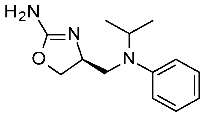 | 140 | −5.1443 |
| 3 | 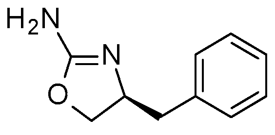 | 330 | −6.1549 | 14 |  | 230 | −6.3802 |
| 4 |  | 2900 | −6.7972 | 15 | 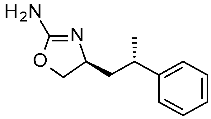 | 1540 | −4.6935 |
| 5 | 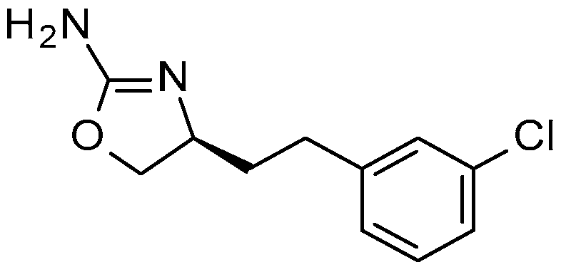 | 18 | −6.8864 | 16 | 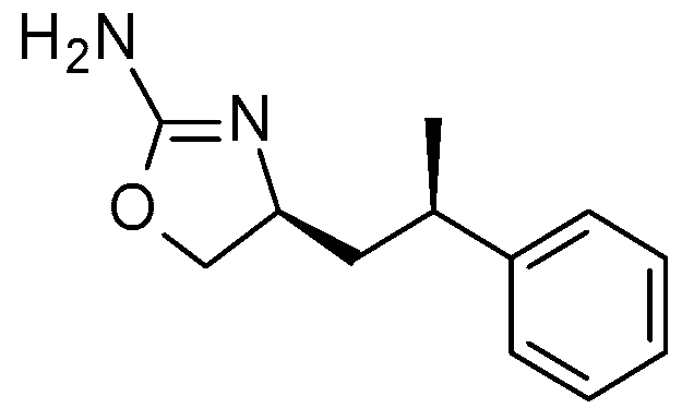 | 730 | −7.2651 |
| 6 | 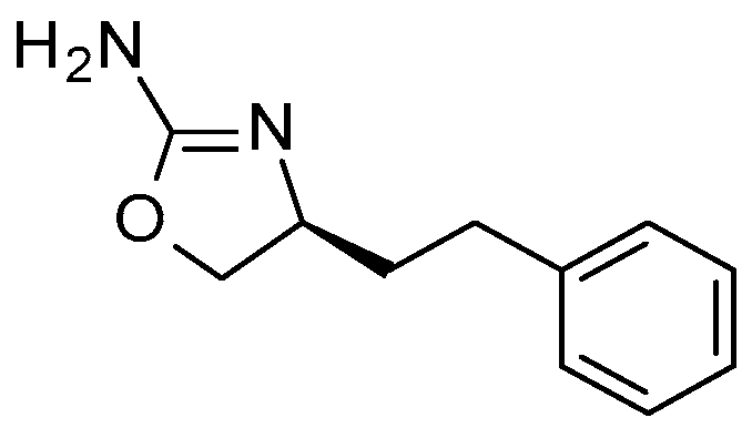 | 27 | −7.1321 | 17 | 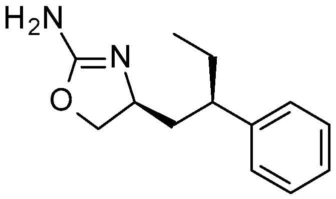 | 2260 | −5.9924 |
| 7 | 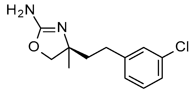 | 330 | −5.6156 | 18 |  | 18 | −5.8995 |
| 8 | 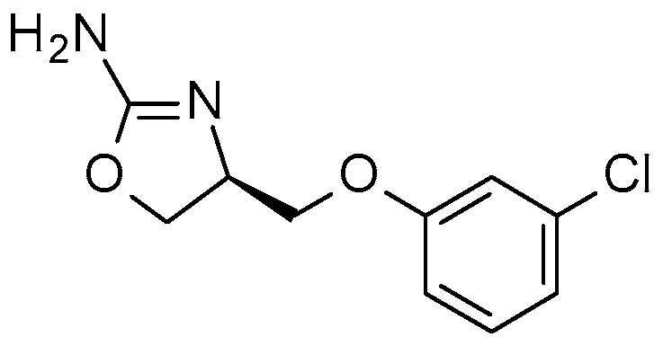 | 270 | −6.1963 | 19 |  | 27 | −5.8673 |
| 9 | 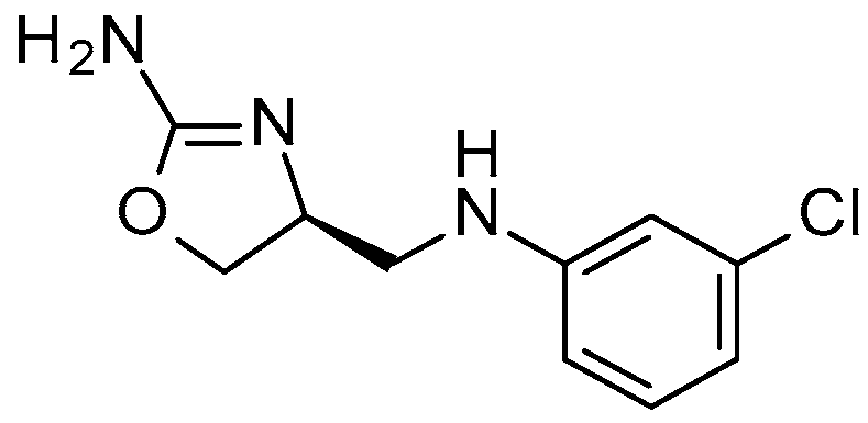 | 580 | −4.9034 | 20 |  | 360 | −5.1471 |
| 10 | 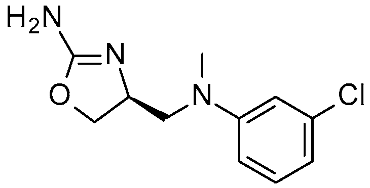 | 27 | −6.5934 | 21 |  | 9 | −7.1321 |
| 11 |  | 29 | −6.5403 |
| Comp. | Chemical Structure | EC50 (nM) | S | Comp. | Chemical Structure | EC50 (nM) | S |
|---|---|---|---|---|---|---|---|
| 22 | 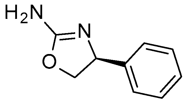 | 67 | −6.2967 | 30 | 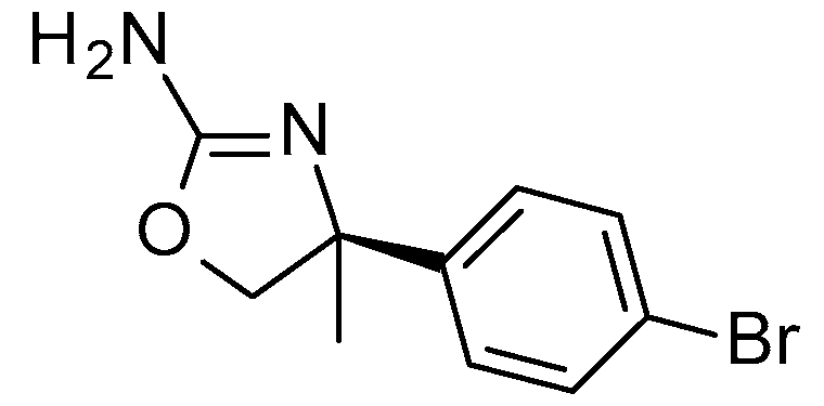 | 41 | −5.7542 |
| 23 | 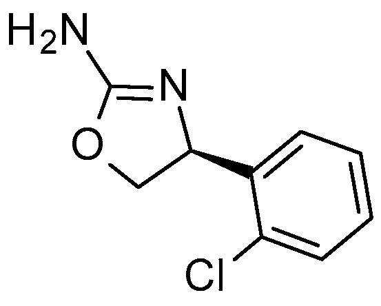 | 23 | −6.6376 | 31 |  | 2670 | −5.2125 |
| 24 | 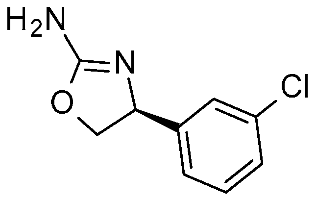 | 21 | −6.7660 | 32 |  | 490 | −5.6820 |
| 25 | 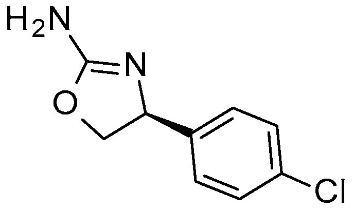 | 143 | −6.0587 | 33 | 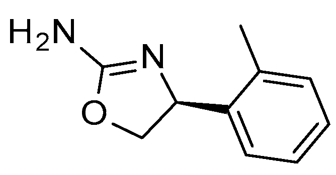 | 67 | −6.7540 |
| 26 |  | 31 | −6.5172 | 34 | 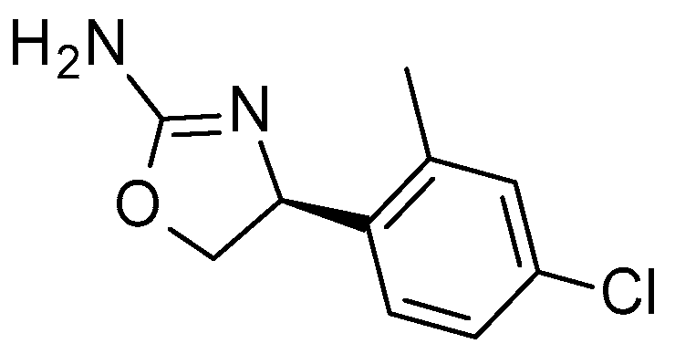 | 11 | −6.9372 |
| 27 | 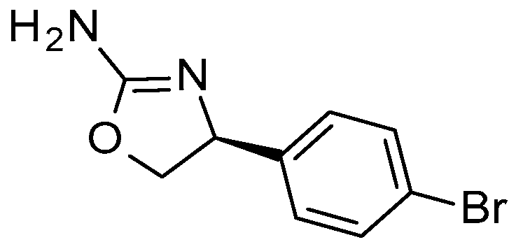 | 150 | −5.9241 | 35 | 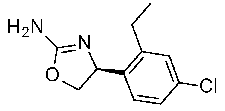 | 26 | −6.4499 |
| 28 | 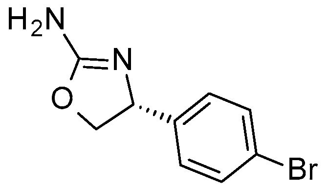 | >10,000 | −5.0876 | 36 | 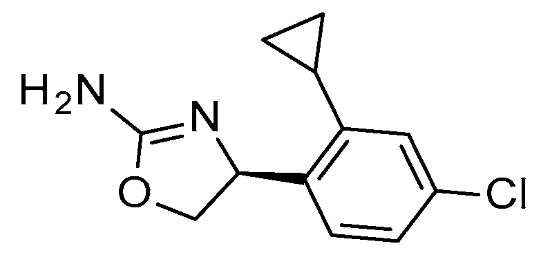 | 12 | −6.0151 |
| 29 | 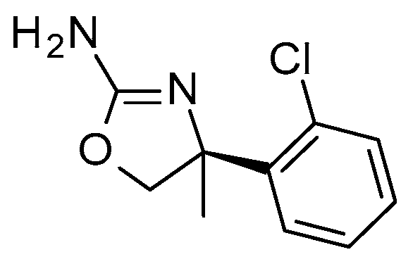 | 165 | −5.5681 | 37 | 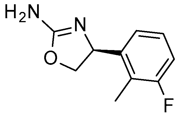 | 17 | −6.6828 |
| Comp. | Chemical Structure | EC50 (nM) | S | Comp. | Chemical Structure | EC50 (nM) | S |
|---|---|---|---|---|---|---|---|
| 1a | 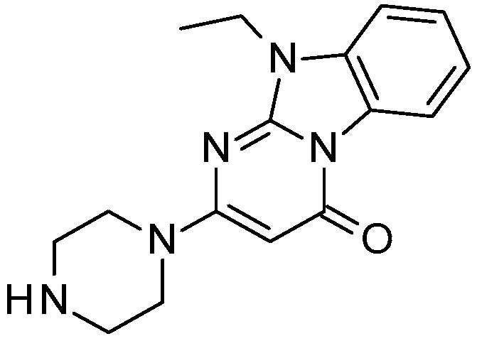 | 526 | −3.1695 | 6a | 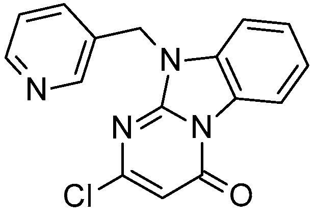 | 1430 | −2.9174 |
| 2a | 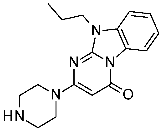 | 657 | −3.1254 | 7a | 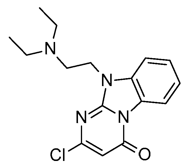 | inactive | 3.9286 |
| 3a | 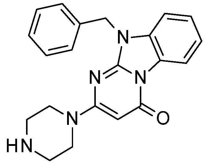 | 756 | −3.1408 | 8a | 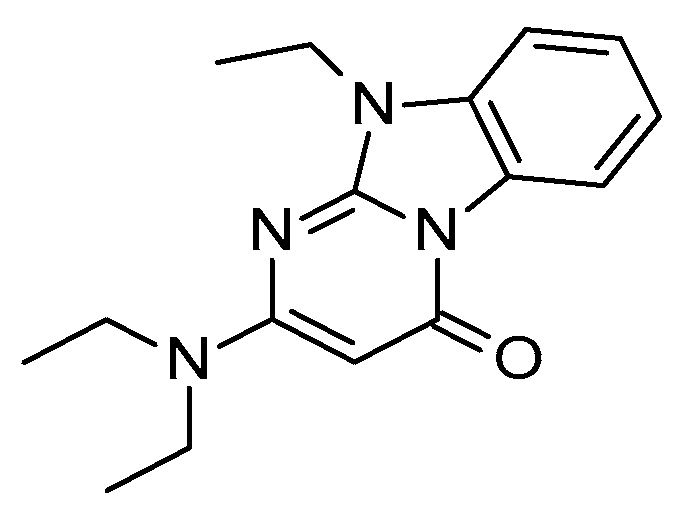 | inactive | 0.4922 |
| 4a | 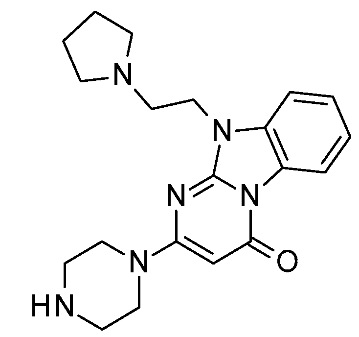 | 1060 | −2.6083 | 9a | 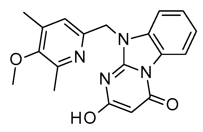 | inactive | −1.4568 |
| 5a | 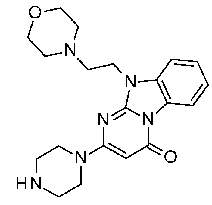 | 1010 | −3.1141 | 10a | 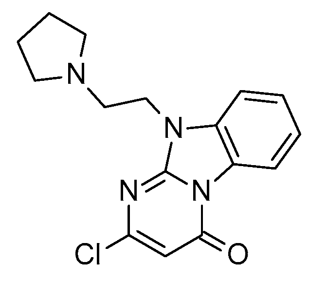 | inactive | 0.4922 |
| Comp. | MW a | N. H-Bond Acceptor b | N. H-Bond Donor c | N. Rotatable Bonds d | CLogP e (R.I. ≥ 0.45) | TPSA f | LogBB g | LogPS h | HIA (%) i | Vd (L/kg) j | %PPB k (R.I. ≥ 0.40) | LogKa HAS l (R.I. ≥ 0.30) | F% m (Oral) |
|---|---|---|---|---|---|---|---|---|---|---|---|---|---|
| 1a | 297.36 | 6 | 1 | 2 | 0.87 | 51.18 | −0.58 | −3.1 | 46 | 2.1 | 84.09 | 4.48 | 27.1 |
| 2a | 311.38 | 6 | 1 | 3 | 1.21 | 51.18 | −0.51 | −2.9 | 52 | 2.3 | 84.54 | 4.49 | 30.7 |
| 3a | 359.42 | 6 | 1 | 3 | 2.21 | 51.18 | −0.53 | −2.5 | 95 | 2.8 | 95.79 | 4.95 | 80.8 |
| 4a | 366.46 | 7 | 1 | 4 | 0.82 | 54.42 | −0.46 | 3.3 | 17 | 4.6 | 77.89 | 4.32 | 8.9 |
| 5a | 382.46 | 8 | 1 | 4 | 0.52 | 63.65 | −0.48 | −3.5 | 29 | 3.2 | 75.84 | 4.23 | 15.7 |
| 6a | 310.74 | 5 | 0 | 2 | 2.53 | 48.80 | −0.77 | −1.3 | 100 | 1.7 | 98.25 | 5.29 | 37.7 |
| S18616 | 236.70 | 3 | 2 | 0 | 3.11 | 47.61 | 0.56 | −1.4 | 100 | 3.2 | 71.82 | 3.49 | 99.3 |
| Chemical structure | |||||||||||||
1a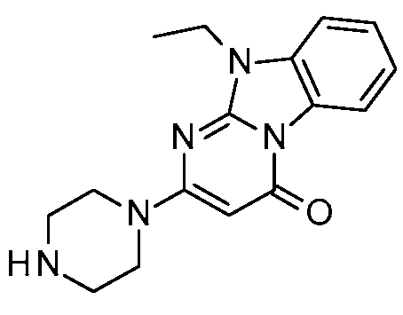 EC50 = 526 nM | 2a EC50 = 657 nM | 3a EC50 = 756 nM | 4a EC50 = 1060 nM | 5a EC50 = 1010 nM | 6a EC50 = 1430 nM | S18616 EC50 = 15 nM | |||||||
Disclaimer/Publisher’s Note: The statements, opinions and data contained in all publications are solely those of the individual author(s) and contributor(s) and not of MDPI and/or the editor(s). MDPI and/or the editor(s) disclaim responsibility for any injury to people or property resulting from any ideas, methods, instructions or products referred to in the content. |
© 2024 by the authors. Licensee MDPI, Basel, Switzerland. This article is an open access article distributed under the terms and conditions of the Creative Commons Attribution (CC BY) license (https://creativecommons.org/licenses/by/4.0/).
Share and Cite
Grossi, G.; Scarano, N.; Musumeci, F.; Tonelli, M.; Kanov, E.; Carbone, A.; Fossa, P.; Gainetdinov, R.R.; Cichero, E.; Schenone, S. Discovery of a Novel Chemo-Type for TAAR1 Agonism via Molecular Modeling. Molecules 2024, 29, 1739. https://doi.org/10.3390/molecules29081739
Grossi G, Scarano N, Musumeci F, Tonelli M, Kanov E, Carbone A, Fossa P, Gainetdinov RR, Cichero E, Schenone S. Discovery of a Novel Chemo-Type for TAAR1 Agonism via Molecular Modeling. Molecules. 2024; 29(8):1739. https://doi.org/10.3390/molecules29081739
Chicago/Turabian StyleGrossi, Giancarlo, Naomi Scarano, Francesca Musumeci, Michele Tonelli, Evgeny Kanov, Anna Carbone, Paola Fossa, Raul R. Gainetdinov, Elena Cichero, and Silvia Schenone. 2024. "Discovery of a Novel Chemo-Type for TAAR1 Agonism via Molecular Modeling" Molecules 29, no. 8: 1739. https://doi.org/10.3390/molecules29081739
APA StyleGrossi, G., Scarano, N., Musumeci, F., Tonelli, M., Kanov, E., Carbone, A., Fossa, P., Gainetdinov, R. R., Cichero, E., & Schenone, S. (2024). Discovery of a Novel Chemo-Type for TAAR1 Agonism via Molecular Modeling. Molecules, 29(8), 1739. https://doi.org/10.3390/molecules29081739