Article

Phytochemical Composition and Antimicrobial Properties of New Lavandula angustifolia Ecotypes

1
Institute of Wood Sciences and Furniture, Warsaw University of Life Sciences—SGGW, 159 Nowoursynowska St., 02-776 Warsaw, Poland
2
Department of Food Technology and Human Nutrition, Institute of Food Technology and Nutrition, College of Natural Sciences, University of Rzeszów, 4 Zelwerowicza St., 35-601 Rzeszów, Poland
3
Department of Chemistry and Food Toxicology, University of Rzeszów, 1a Ćwiklińskiej St., 35-601 Rzeszów, Poland
4
Department of Nanobiotechnology, Institute of Biology, Warsaw University of Life Sciences, 8 Ciszewskiego St., 02-786 Warsaw, Poland
5
Department of Plant Production and Food Safety, Carpathian State College in Krosno, 38-400 Krosno, Poland
6
Breeding Department, Małopolska Plant Breeding Company sp. z o. o., 4 Zbożowa St., 30-002 Kraków, Poland
*
Authors to whom correspondence should be addressed.
Molecules 2024, 29(8), 1740; https://doi.org/10.3390/molecules29081740
Submission received: 29 January 2024 / Revised: 3 April 2024 / Accepted: 8 April 2024 / Published: 11 April 2024

Abstract

:
The purpose of this study was to characterize ethanol extracts from leaves and flowers of two ecotypes (PL—intended for industrial plantations and KC—intended for cut flowers) of Lavandula angustifolia Mill. The plant was cultivated in 2019 in southern Poland as part of a long-term research plan to develop new varieties resistant to difficult environmental conditions. The collected leaves and flowers were used to prepare ethanol extracts, which were then analyzed in terms of phytochemical composition and antioxidant, bactericidal, and fungicidal properties. Using UPLC techniques, 22 compounds belonging to phenolic acids and flavonoids were identified. UPLC test results indicated that ethanol extracts from leaves and flowers differ in phytochemical composition. Lower amounts of phenolic acids and flavonoids were identified in leaf extracts than in flower extracts. The predominant substances in the flower extracts were rosmarinic acid (829.68–1229.33 µg/g), ferulic acid glucoside III (810.97–980.55 µg/g), and ferulic acid glucoside II (789.30–885.06 µg/g). Ferulic acid glucoside II (3981.95–6561.19 µg/g), ferulic acid glucoside I (2349.46–5503.81 µg/g), and ferulic acid glucoside III (1303.84–2774.17 µg/g) contained the highest amounts in the ethanol extracts of the leaves. The following substances were present in the extracts in trace amounts or at low levels: apigenin, kaempferol, and caftaric acid. Leaf extracts of the PL ecotype quantitatively (µg/g) contained more phytochemicals than leaf extracts of the KC ecotype. The results obtained in this study indicate that antioxidant activity depends on the ecotype. Extracts from the PL ecotype have a better ability to eliminate free radicals than extracts from the KC ecotype. At the same time, it was found that the antioxidant activity (total phenolic content, ABTS•+, DPPH, and FRAP) of PL ecotype leaf extracts was higher (24.49, 177.75, 164.88, and 89.10 μmol (TE)/g) than that determined in flower extracts (15.84, 125.05, 82.35, and 54.64 μmol (TE)/g). The test results confirmed that leaf and flower extracts, even at low concentrations (0.313–0.63%), significantly inhibit the growth of selected Gram-negative and Gram-positive bacteria and Candida yeasts. Inhibition of mold growth was observed at a dose extract of at least 1 mL/100 mL.

1. Introduction

The genus Lavandula includes about 50 species cultivated around the world as an ornamental and medicinal plant [1]. This plant comes from the Mediterranean area, the Middle East, North Africa, and the Republic of Cape Verde. It is also successfully cultivated in Asia, Australia, and the Americas. In Europe, the main production of lavender is concentrated in the regions of France, Bulgaria, Spain, and Ukraine. French lavender, and in fact, the raw material obtained from it, is an important item in the economic balance of this country [2].
The first plantations of this species in the climatic and soil conditions of southern Poland were already carried out in the 1920s [3,4]. The currently observed changes in weather conditions, especially the occurrence of mild winters, enable the cultivation of lavender with a longer flowering period, which may last until the end of October. One of the most frequently chosen lavender species for cultivation in Poland is Lavandula angustifolia Mill. Its numerous foreign varieties are valued, such as Siesta, Beate, Essence, Blue Scent, and Hidcote Blue Strain [5]. Most of these varieties show relatively high resistance to low temperatures, so they can be successfully cultivated in Poland, unlike Lavandula stoechas L., which does not tolerate negative temperatures and freezes in winter in field cultivation [6]. Despite numerous plantations in various regions of Poland, there is still no Polish breeding variety.
The most valuable raw material, Lavandula angustifolia Mill. is flowers containing essential oil that is valuable for the cosmetic and pharmaceutical industry. Shi [7] indicated that essential oil contains over 100 individual components with aromatic and biological effects. The dominant substances in the oil are linalool, linalyl acetate, cineole, β-ocimene, β-lavender acetate, lavender alcohol, terpene-4-alcohol, and camphor [7,8]. At the same time, significant variability in the composition of the oil was demonstrated, both among different varieties of lavender and the method of obtaining it [9]. Adaszyńska et al. [10] compared the chemical composition of the essential oil of 5 varieties is L. angustifolia Mill. originating from controlled crops, and they observed significant differences in the quantitative composition of the main components of the oil and chemical substances occurring in small concentrations. Moreover, the method of extraction and the selection of raw materials (leaves, flowers, and inflorescence stalks) allows for the attainment of oils of different quality [11]. For example, in the research conducted by Dvorackova et al. [12] and Bajkacz et al. [13], it was shown that the leaves of L. angustifolia Mill. are richer in hydroxycinnamic acids than the flowers of this plant. In turn, Dobros et al. [14] showed that the extraction method plays an important role in the final content of bioactive substances and antioxidant properties.
In addition to terpenoid components, lavender contains numerous polyphenols, coumarins, sterols, and tannins [15,16,17]. Undoubtedly, important chemical compounds present in lavender flowers are phenolic compounds, which are believed to have an antioxidant role. Yadikar et al. [18] proved the strong antioxidant effect of polyphenolic compounds such as lavandunat, lavandufurandiol, lavanduflu-oren, lavandupyrones, lavandudiphenyls, 4-(1-hydroxy-1-methylethyl) benzoic acid, methyl 3-(3,4-dihydroxyphenyl) propanoate, 3,4,α-trihydroxyl-ethyl phenylpropionate, rosmarinic acid, and isosalvianolic acid, isolated from the flowers of L. angustifolia Mill. in the steam distillation process.
The composition and proportions of bioactive ingredients in extracts obtained from lavender raw materials determine its biological functions and potential applications. Due to its proven antibacterial [19], antifungal [20], antioxidant [21], and anti-inflammatory [22] properties, this plant is very popular among both breeders and potential recipients of the raw material. Numerous scientific publications devoted to the chemical analysis of the raw material also demonstrate great interest [23,24]. Lavender is therefore becoming more and more popular in cultivation, as well as in regions where it was rarely found before. New growing conditions also make it possible to obtain new varieties that will give high yields in climatic and soil conditions previously considered unfavorable for the plant’s vegetation. At the same time, the chemical characteristics of the raw material and its pharmacopoeial significance are important, as they will show that the raw material obtained from a cold temperate climate is characterized by broad biological activity, which may constitute an alternative to the raw material obtained from regions typical for its cultivation.
The aim of the study was to assess the phytochemical composition and antioxidant and antimicrobial activity of ethanol extracts from flowers and leaves of two ecotypes of Lavandula angustifolia Mill. (marked with PL and KC symbols) cultivated in southern Poland since 2019. The presented lavender ecotypes were selected for the first time and adapted to the soil and climatic conditions of Poland. So far, no one has tested the antioxidant, antifungal, and antibacterial properties of the presented ecotypes. The cultivation of narrow-leaved lavender in the form of ecotypes of a medium-high plant and a bouquet plant is intended in the future to lead to the separation of a new variety, adapted to cultivation in regions dominated by low temperatures in winter. However, for lavender to be a good raw material that meets quality standards for the cosmetics and pharmaceutical industry, the composition of biologically active substances must be thoroughly characterized, and its properties have to be examined.

2. Results

2.1. Phytochemical Compounds

UPLC analysis showed that eight phenolic acids and two flavonoid compounds were identified in leaf extracts (PL1 and KC1). As many as 14 phenolic acids and 6 flavonoid compounds have been identified in flower extracts (PL2 and KC2). Although leaf extracts are less diverse in phenolic compounds, it should be noted that, quantitatively, these compounds significantly exceed the content of phenolic compounds identified in flower extracts. The content of ferulic acid glucose II in the extracts from PL1 leaves was almost ten times higher than in the extracts from PL2 flowers. Similarly, the content of substances such as caftaric acid, isorhamnetin-3-O-rhamnoside, and ferulic acid was much higher in leaf extracts than in lavender flower extracts (Table 1). Substances such as glucoside I, caffeic acid, chicoric acid, kaempferol, salvinic acid B, apigenin glucoside C, and apigenin glucoside C were not determined in trace amounts in leaf extracts. These substances were identified in flower extracts. Ferulic acid glucoside I and isorhamnetin 3-O-rutinoside were not determined in trace amounts in flower extracts. These substances were identified in leaf extracts in amounts of 243.81 µg/g (KC1) and 621.76 µg/g (PL1). The tests performed showed that extracts from ecotype PL contained higher amounts of phenolic compounds than extracts from ecotype KC (Table 1).
GC-MS analysis showed qualitative and quantitative differences in the composition of volatile substances between ecotypes. Decane, lavandulyl acetate, and β-farnesene were identified only in extracts of the KC ecotype (Table 2), while myrcene was only identified in the PL ecotype (Table 2). Linalool and linalyl acetate, the main components of the essential oil, were identified only in flower extracts, with the KC ecotype containing much larger amounts of the indicated substances than the PL ecotype. The number of volatile substances in flower extracts was almost twice as high as in leaf extracts.

2.2. Antioxidant Assay

The analysis of the antioxidant properties of lavender leaf and flower extracts was carried out based on the ability of the extracts to reduce radicals (ABTS•+ and DPPH) and iron (FRAP). The total content of phenolic compounds (TPC) was also measured. Based on the analyses performed, it was found that the extract from the PL ecotype leaves has a strong antioxidant effect (Table 3). There were no statistically significant differences in antioxidant properties between flower extracts. The leaf extract of the KC ecotype showed the weakest ability to reduce the DPPH radical, iron and it contained a relatively small amount of TPC.
In all cases (total phenolic content, ABTS•+, DPPH, and FRAP), the influence of the analyzed factors (ecotype and L/F extract) and the interactions between them showed a statistically significant effect (p < 0.05). Only in the case of ABTS•+ was the effect of the interaction between ecotype and L/F extract statistically insignificant. By analyzing the influence of individual factors, it should be concluded that in relation to total phenolic content, DPPH, and FRAP, the greatest influence is exerted by the ecotype (38.0–47.7%) and the interaction between the ecotype and the type of extract (46.3–57.0%). The type of extract is important (61.6%) only in relation to ABTS•+; in other cases, the influence of this factor is much lower (0.4–14.5%). It should also be emphasized that in all cases, there was only a slight impact of factors not taken into account in these studies (error 0.1–0.5%) (Table 4).

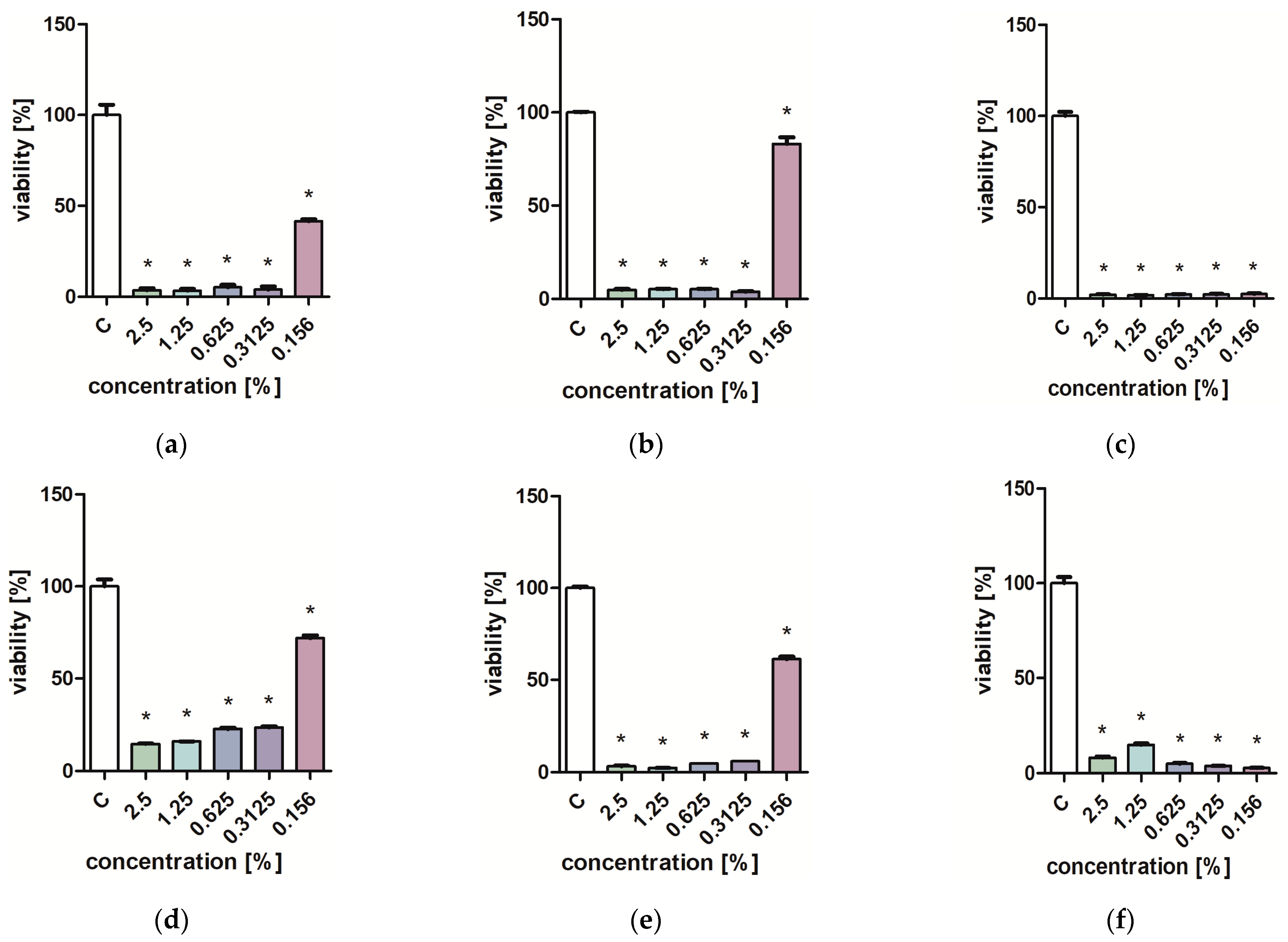
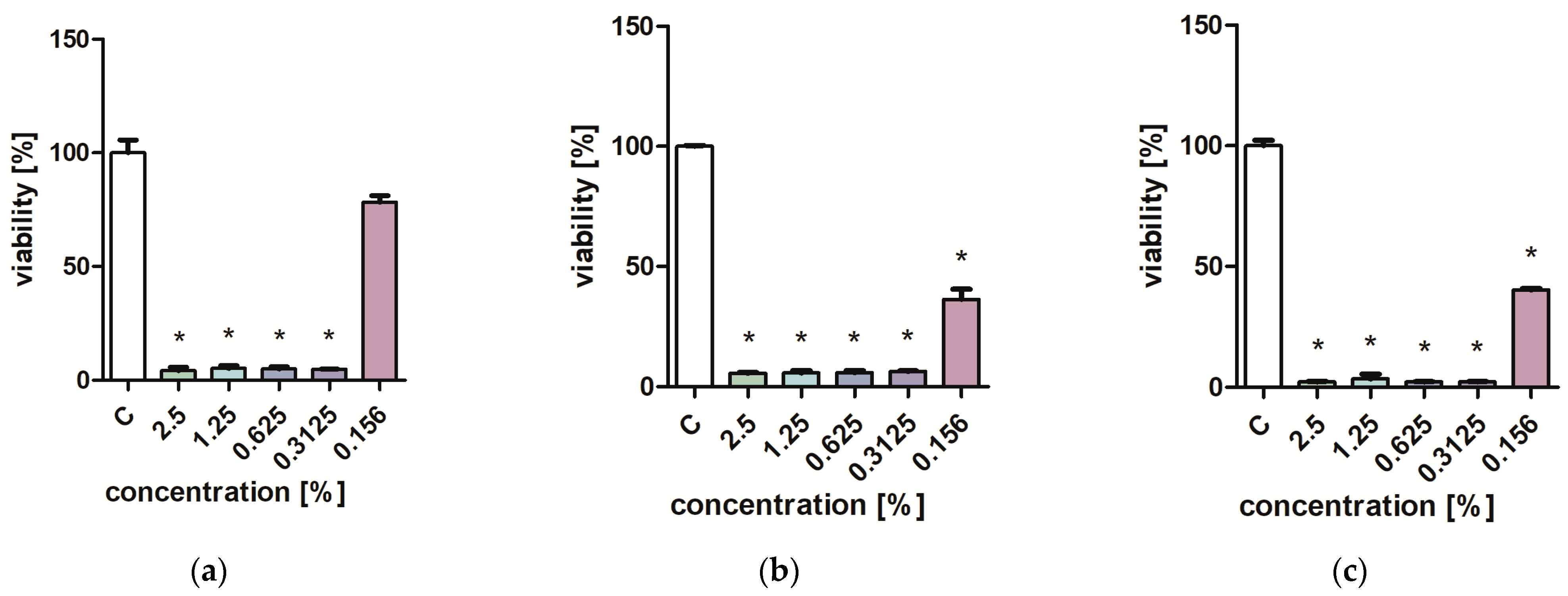
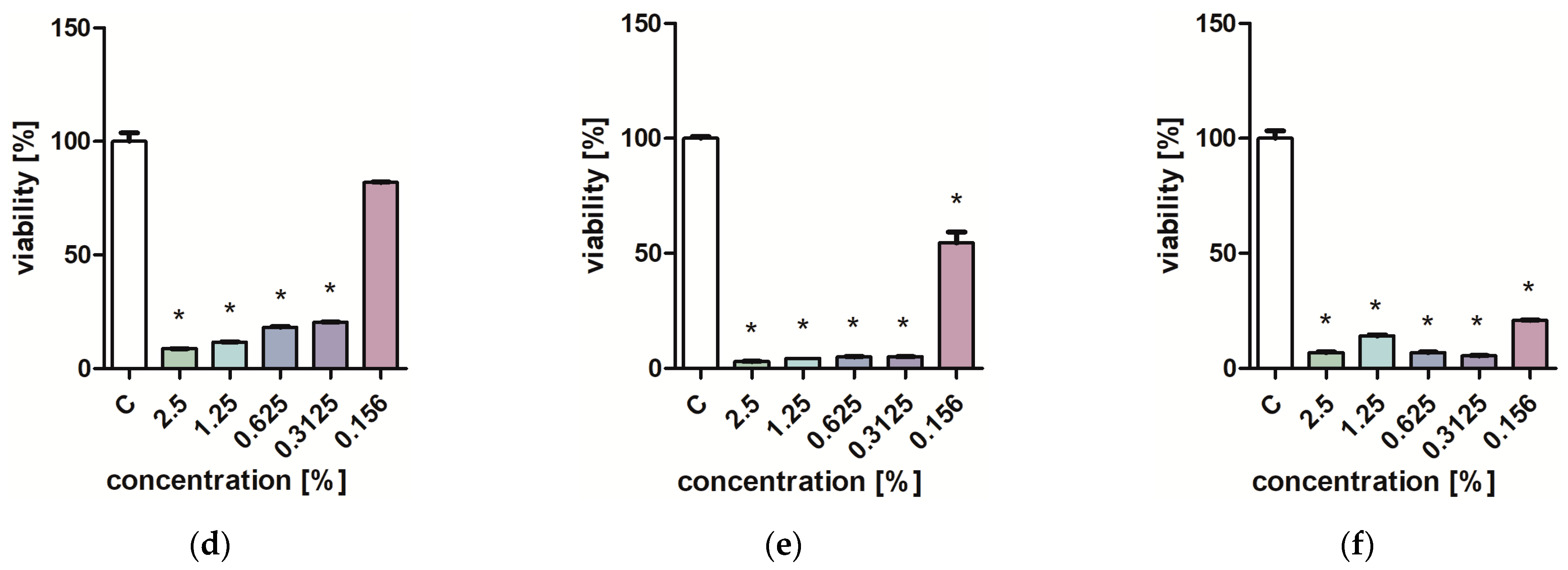
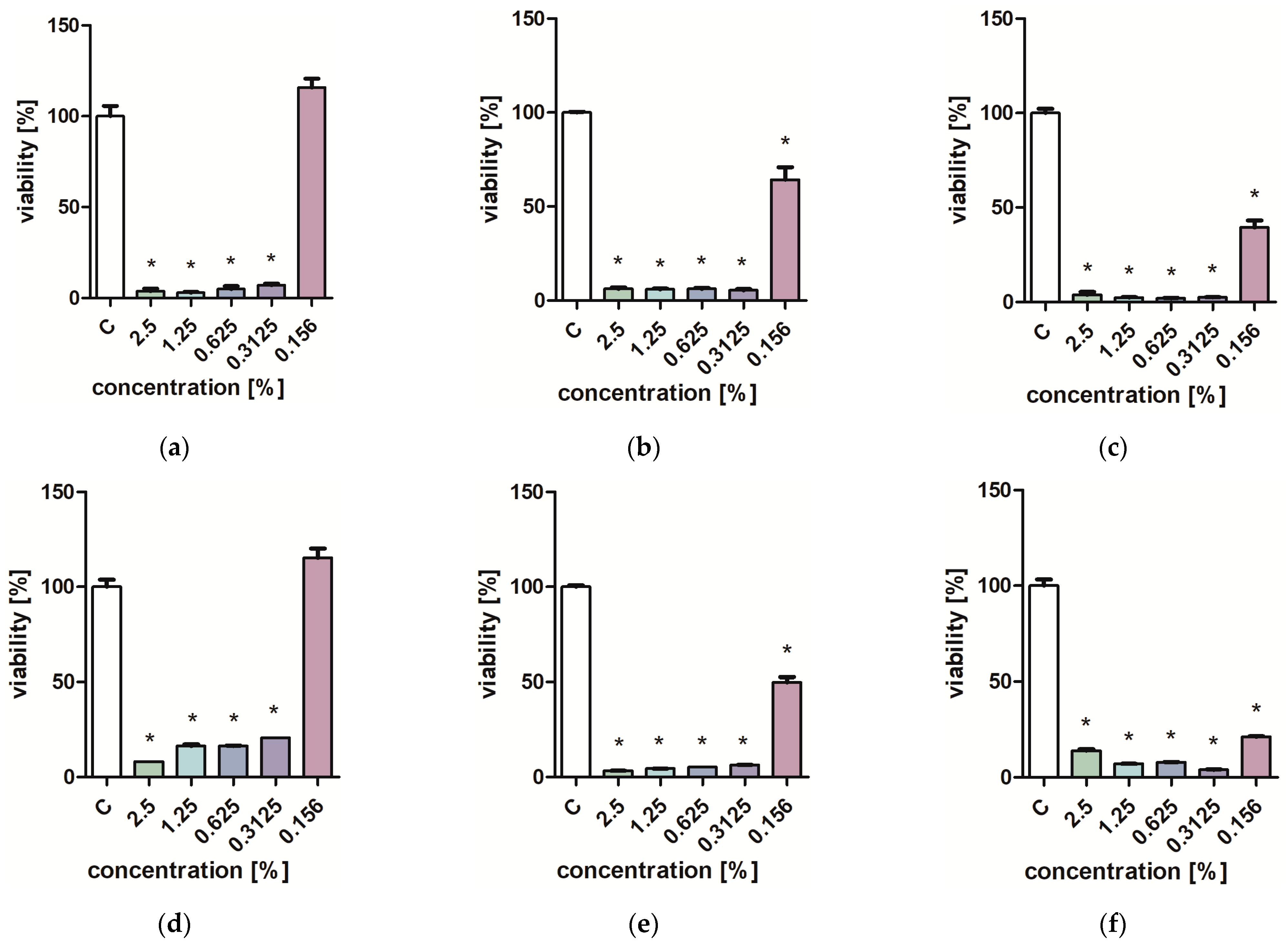
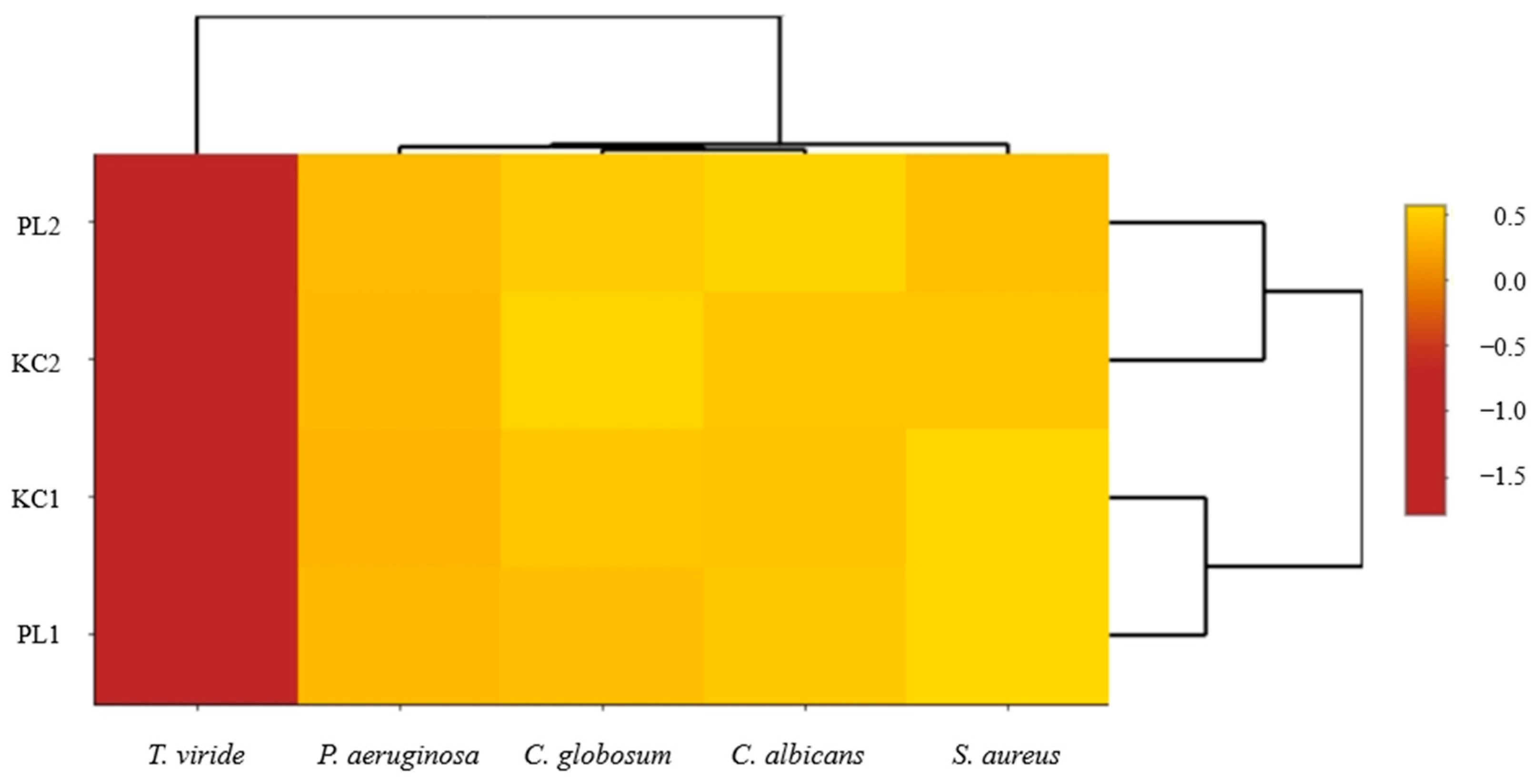
2.3. Measurement of the Growth of Mold Fungi and Bacterial Viability

Table 5 and Table 6 show the effect of extracts on the growth of mold fungi. Studies have shown that leaf extracts at a concentration of 1 mL/100 mL completely inhibit the growth of Trichoderma viride and Chaetomium globosum. Lavender flower extracts had a weaker fungicidal effect, especially against T. viride.
Studies on the influence of extracts on the viability of Pseudomonas aeruginosa, Staphylococcus aureus, and Candida albicans yeast cells showed that low concentrations of extracts (0.313–0.625%) inhibit the growth of microorganisms (Figure 1, Figure 2, Figure 3 and Figure 4). Particularly strong antibacterial activities of leaf extracts (PL1 and KC1) were demonstrated in the PrestoBlue test (Figure 1 and Figure 2). A percentage of 0.156% leaf extract of the PL ecotype turned out to be a strong inhibitor of the growth of S. aureus (Figure 1). Flower extracts (PL2 and KC2) also showed a strong effect on microbial viability (Figure 3 and Figure 4). Ethanol used in analogous concentrations did not inhibit the tested mold fungi.

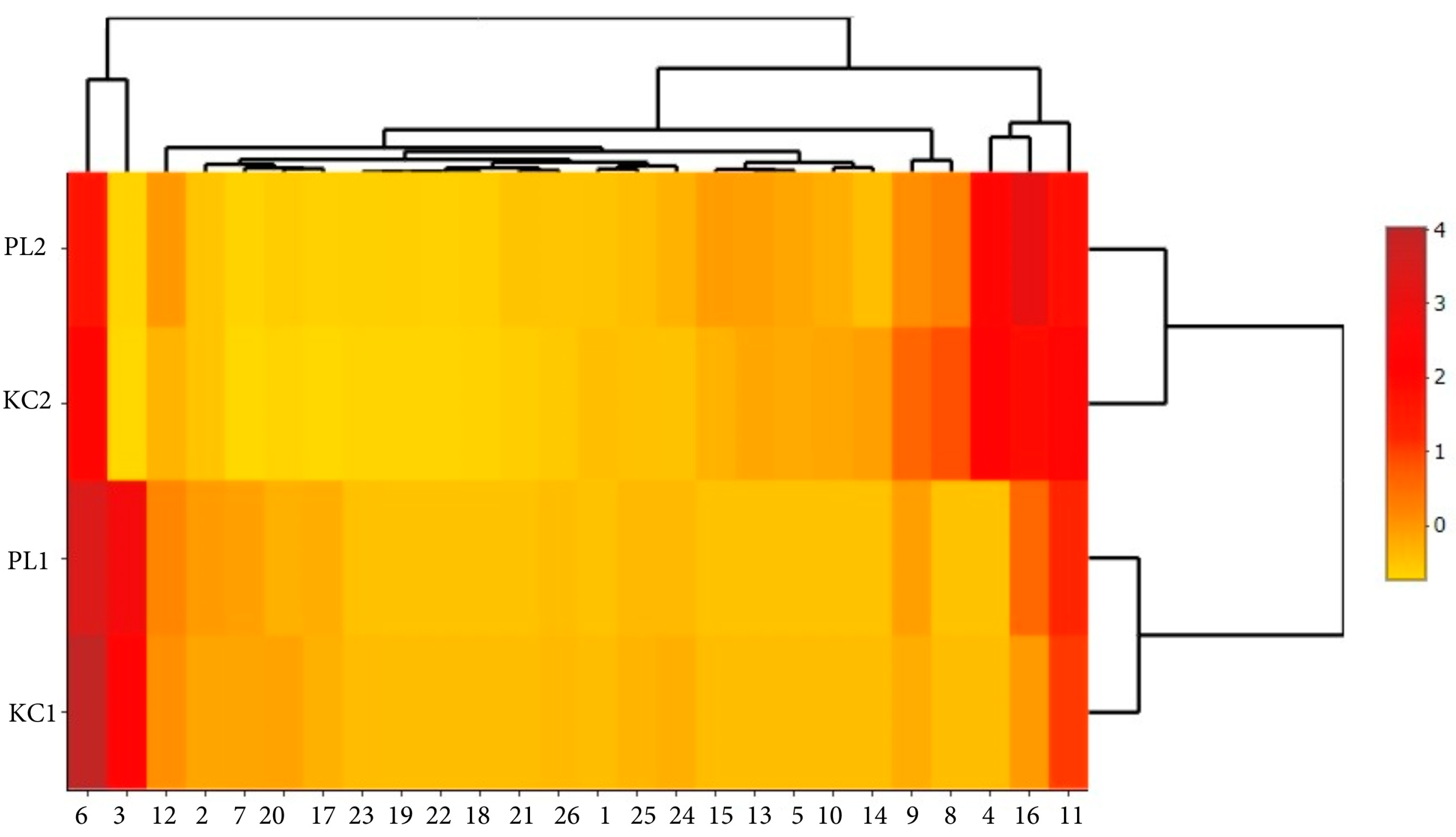
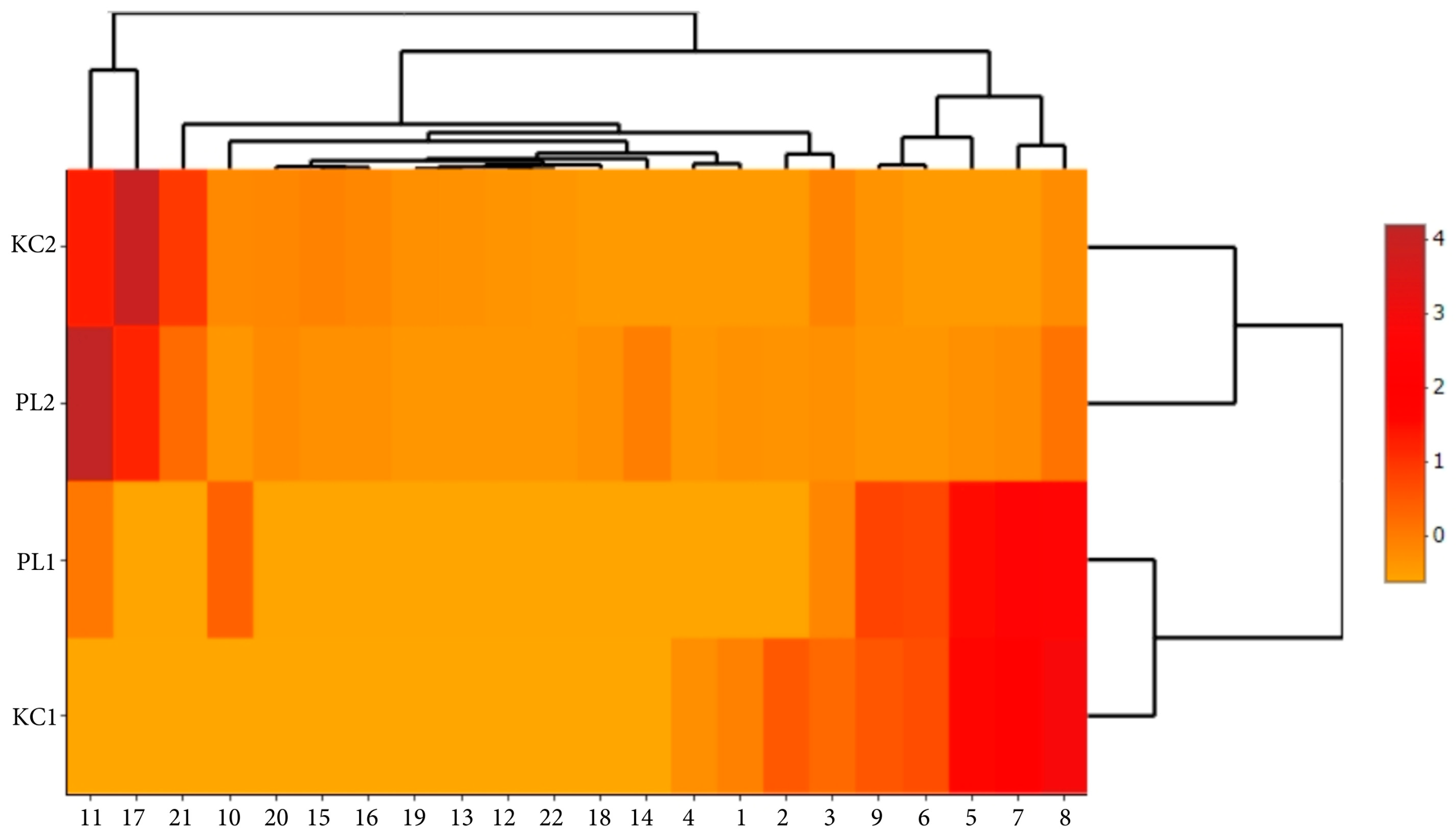
2.4. Graphical Interpretation of Results

Mapping the data obtained from the tested material using scaled heat maps shows a clear differentiation of qualitative and quantitative characteristics of two lavender ecotypes KC2, PL2 to KC1, and PL1. This differentiation is clearly visible in the case of polyphenolic compounds (Figure 5), volatile compounds (Figure 6), and antimicrobial activity (Figure 7).
The use of a heat map with a scaling function enabled a more accurate presentation of the relationships between the analyzed factors within ecotypes and specific extracts. It is clearly visible that leaf extracts (KC1 and PL1) are characterized by greater activity of ferulic acid glucoside I, caftaric acid, and isorhamnetin 3-O-rutinoside (Figure 5). Flower extracts (KC2 and PL2) are richer in volatile substances such as ocimene isomers mix, linalyl acetate, and β-caryophyllene. In turn, lavender leaf extracts have a greater ability to eliminate free radicals (Figure 6). The heat map analysis showed that the microbiological activity within ecotypes and extracts is similar (Figure 7). The tested ecotypes showed very strong moldicidal activity against T. viride. Lavender leaf extracts had a weaker effect on the viability of S. aureus than lavender flower extracts.

3. Discussion

Lavender is a plant of high pharmacopoeial value. Thanks to numerous bioactive ingredients, it is an important raw material for the pharmaceutical, cosmetics, and food industries. The quality of the raw material depends largely on the species but also on the climatic and soil conditions in which it is grown. Due to the observed changes in the environment related to climate warming, it has become possible to grow and process Lavandula angustifolia Mill. in regions that were not favorable for its cultivation until recently. The economic value of the raw material depends on its quality related to the expected properties. The quality of the lavender raw material, understood as the quality of the crop, giving a high content of essential oil and bioactive ingredients with biological properties, is the subject of numerous analyses [25,26,27]. Crişan et al. [28] indicated that the phytochemical profile determines the functional potential of lavender. Da Porto and Decorti [29] showed that in extracts from L. angustifolia Mill. originating from cultivation in Italian regions, the dominant ingredients are linalyl acetate, linalool, and a low percentage of camphor. The characteristic ingredients of Iranian lavender are linalool, linalyl acetate, lavandulyl acetate, α-terpineol, and geranyl acetate [30]. This study showed that L. angustifolia Mill. grown in southern Poland is dominated by 1-octen-3-yl acetate, ocimene isomers mix, γ-cadinene, and limonene. In addition to the main components of the oil, lavender extracts contain antioxidant compounds. The conducted analyses identified 22 compounds belonging to phenolic acids and flavonoids. In research conducted by Tundis et al. [31], the dominant polyphenolic compounds in water–ethanol extracts from L. angustifolia Mill. were rosmarinic acid, ferulic acid glucoside, morin, and caffeic acid. The results of our research confirm the reports of other researchers. Rosmarinic acid was the dominant component in leaf and flower extracts. Apigenin, kaempferol, and caftaric acid were present in the extracts in trace amounts or at low levels. In studies by other authors, substances found in trace amounts were chlorogenic acid and ferulic acid [32].
In our own research, extracts from L. angustifolia Mill. flowers were characterized by a greater variety of bioactive ingredients than leaf extracts. Differences in qualitative composition were also observed between ecotypes. Bioactive compounds, especially polyphenols, determine the antioxidant potential [33]. Scientific research indicates that extracts obtained from raw lavender materials can prevent the damage of oxidative cells [34]. The presented research results proved that the antioxidant activity depends on the ecotype. The PL ecotype has a better ability to eliminate free radicals than the KC ecotype. Additionally, it can be concluded that the obtained results of antioxidant properties are at a satisfactory level. The best antioxidant properties were demonstrated for ethanol extracts from leaves and flowers of the PL ecotype. The antioxidant potential of extracts from the PL ecotype was in the range of 125.05–177.75 μmol(TE)/g (ABTS•+), 82.35–164.88 mol(TE)/g (DPPH), and 54.64–89.10 μmol(TE)/g (FRAP). The scientific literature contains extensive data on the antioxidant properties of various L. angustifolia extracts. Caser et al. [1] showed that the antioxidant potential of fresh and dried flowers of L. angustifolia Mill. assessed in the ABTS method ranges from 17.89 to 44.81 μmol TE/g and in the DPPH method from 16.06 to 47.36 μmol TE/g. In turn, Robu et al. [35] showed that the highest antioxidant value of water–ethanol extracts does not exceed 110.36 µg/mL.
The antimicrobial properties of various lavender extracts have been documented in numerous scientific studies [36,37,38]. de Rapper et al. [39] demonstrated the synergistic effect of combinations of essential oil with selected antibiotics against representative bacterial cells (Staphylococcus aureus and Pseudomonas aeruginosa) and yeast (Candida albicans). A comprehensive review of the properties of L. angustifolia Mill. extracts. conducted by Salehi et al. [40] indicates the crucial importance of lavender as an antibacterial, antifungal, and antiseptic raw material. These tests also confirmed high biocidal properties of low concentrations of extracts against selected Gram-negative and Gram-positive bacteria, fungi of the Candida genus, and mold fungi.
The conducted research demonstrated the relationship between ecotypes and plant parts and the identified phytochemical composition and biological properties. Therefore, it can be assumed that there is a relationship between the phytochemical composition of specific extracts and ecotypes and their biological properties. The results presented in the form of heat maps may suggest that certain groups of substances contained in ecotypes and in specific parts of plants may have an effect on the viability of bacteria and mold fungi. Substances such as ferulic acid glucoside I, ferulic acid glucoside II, ferulic acid glucoside III, or rosmarinic may have antimicrobial effects in flower and leaf extracts. These assumptions seem to be correct from the point of view of the results of other researchers, who point to the enormous antimicrobial potential of these substances [41]. Similar conclusions can be drawn by analyzing the impact of ecotypes and the type of extract on antioxidant activities. Heat map analysis indicates that the antioxidant properties of the PL ecotype may be related to a different phytochemical composition than the antioxidant properties of the KC ecotype.
Lavender ecotypes grown in the Polish climate are plants with good antioxidant and biological potential, which may translate into their high use in various industries where such properties are desired. The presented work showed that extracts prepared from lavenders are not rich in linalool, which dominates in lavenders from Mediterranean countries [42]. Research presented by Despinasse et al. [43] indicates the division of Mediterranean lavenders into three chemotypes, differing in the dominance of phytochemical components. The tested PL ecotype was dominated by volatile components such as m-cymene, limonene, and ocimene isomers mix. In turn, the KC ecotype contained the highest percentage of volatile components, such as linalyl acetate and limonene. Therefore, it can be concluded that the PL ecotype differs significantly from the most frequently cultivated French varieties, which are dominated by linalool and linalyl acetate. In turn, the dominance of limonene in the KC ecotype suggests that its origin in French lavenders can be ruled out. The ISO3515:2004 standard characterizes the composition of French L. angustifolia, in which the content of limonene in the oil fraction does not exceed 0.5% [44]. It should also be mentioned that French lavenders are very difficult to grow in the difficult climate of winter seasons in Poland; therefore, breeding varieties for which the climatic conditions prevailing in southern Poland allow for growth and are additionally characterized by a rich phytochemical composition is extremely important. From an agronomic point of view, it is very important.
Comparing the biological properties of new lavender ecotypes with varieties grown in the Mediterranean region, it should be concluded that they are comparable [45]. Similarly to lavenders from the Mediterranean regions, the oxygenated monoterpenes and phenolic compounds contained in them are responsible for the antimicrobial properties against pathogens such as P. aeruginosa and S. aureus [46].
The quantitative and qualitative composition of phytochemical components of the tested lavender ecotypes provides extremely valuable knowledge about the quality of the raw material. It is believed that the Polish climate, especially the temperature and humidity conditions prevailing in the winter and spring, are a factor influencing the high content of selected volatile fractions, but for these data to be confirmed as reliable, many years of agronomic research are required. It should be additionally mentioned that the phytochemical composition of two lavender ecotypes analyzed in the presented work may also have a huge impact on its sensory properties, which should be assessed in subsequent research tasks.

4. Materials and Methods

4.1. Characteristics of the Research Material

The research material included two ecotypes of lavender (Lavandula angustifolia Mill.): a medium-height plant form intended for cultivation as a flower on production plantations for the pharmaceutical, cosmetics, or perfume industry (PL) and a bouquet form, developing long-stemmed inflorescences, elongated racemes, and less frequently set pseudo circles (KC). The field experiment was carried out in Polanowice (50°19′ N 20°07′ E) at the Plant Breeding Station belonging to the Małopolska Plant Breeding Station. The research was carried out on chernozem soil (bonite class 1) with a limestone substrate (pH 6.8). Mineral fertilization was applied in spring before planting. Seedlings obtained generatively (from seeds) were planted on 19 June 2019 in 9-point strips with a spacing of 0.5 × 0.5 m. A total of 91 rows. Three samples of flowers and leaves, each weighing 100 g, were collected from three-year-old plants (7 July 2021) of each ecotype.
Weather conditions varied during the growing season in 2019–2021. The years 2019 and 2020 were similar in terms of rainfall (646.3 and 652.75 mm, respectively), and in 2021, 200 mm more was recorded (845 mm). Their distribution was also different. In 2019, most rain occurred in May and August; in 2020, in May and June; and in 2021, in July and August. In turn, the warmest year was 2019, and the highest average temperatures occurred in June, July, and August.
The leaves and flowers were divided into portions of 10 g each and then placed in vessels to which 200 cm3 of 60% ethanol was added. The whole thing was left to macerate for 72 h. After this time, the extracts were separated from the leaves and flowers and purified using syringe filters with a pore diameter of 0.22 µm. Leaf extracts were given the symbols PL1 and KC1, and flower extracts PL2 and KC2. Extracts for testing were stored at 2–3 °C.

4.2. Phytochemicals Analysis

4.2.1. Phenolic Compound Identification

The determination of polyphenolic compounds was carried out using the ultra-performance liquid chromatography (UPLC-PDA-MS/MS) Waters ACQUITY system (Waters, Milford, MA, USA), according to the previous article [47]. The UPLC system (UPLC-PDA-MS/MS) was equipped with a binary pump manager, column manager, sample manager, photodiode array (PDA) detector, and tandem quadrupole mass spectrometer (TQD) with an electrospray ionization (ESI) source. Separation of polyphenols was performed using a 1.7 µm, 100 mm × 2.1 mm UPLC BEH RP C18 column (Waters, Milford, MA, USA). For separation, the mobile phase consisted of 0.1% formic acid in water, v/v (solvent A), and 0.1% formic acid in 40% acetonitrile, v/v (solvent B). The flow rate was kept constant at 0.35 mL/min for a total run time of 8 min. The system was run with the following gradient program: from 0 min 5% B, from 0 to 8 min linear to 100% B, and from 8 to 9.5 min for washing and back to initial conditions. The injection volume of the samples was 5 µL, and the column was supported at 50 °C. The following TQD parameters were used: cone voltage of 30 V, capillary voltage of 3500 V, source and desolvation temperature of 120 °C and 350 °C, respectively, and desolvation gas flow rate of 800 L/h. Characterization of the individual polyphenolic compounds was performed on the basis of the retention time, mass-to-charge ratio, fragment ions, and comparison with data obtained with commercial standards and literature findings. The obtained data were processed in Waters MassLynx v.4.1 software (Waters, Milford, MA, USA). The method was validated for parameters such as linearity, accuracy (relative error, RE), limit of detection (LOD), limit of quantification (LOQ), and precision (relative standard deviation, RSD). Quantification was determined by the injection of solutions of known concentrations ranging from 0.05 to 5 mg mL−1 (R 2 ≤ 0.999) of the following phenolic compounds as standards: caffeic acid, caftaric acid, p-coumaric acid, ferulic acid, rosmarinic acid, apigenin 8-C-glucoside, (vitexin), kaempferol 3-O-glucoside (Extrasynthese, Genay Cedex, France). Stock standard solutions of polyphenols were prepared using methanol. Six calibrators established the peak area ratio of each polyphenol versus the nominal concentration. The regression equation was obtained by a weighted (1/c2) least-squares linear regression. The LOD was determined as a signal-to-noise ratio (S/N) of 3:1, and the LOQ was determined as an S/N of >10. An acceptable RE within ±20% and the intra- and inter-day variations were determined using relative standard deviation (RSD) values, which were determined using relative standard deviation (RSD) values, which were <3.5% for all the analyzed compounds.

4.2.2. Analysis of Volatile Components

The qualitative and quantitative analysis of volatile substances was performed using the HS-SPME solid-phase microextraction method of compound isolation using 100 µm polydimethylsiloxane (PDMS) fiber from Supelco Ltd. (Bellefonte, PA, USA). Next, the analytes were separated and identified using the GC-MS method with the protocol described previously [47]. Briefly, the analyzed material was placed in a 100 mL conical flask equipped with an aluminum membrane. The fiber exposure was carried out using the headspace method for 30 min at 20 °C. Then, the SPME holder was transferred to the gas chromatograph injector (temp. 250 °C). Using a gas chromatograph (GC-MS, Varian 450GC coupled with 240 MS detector, the composition of compounds desorbed from the SPME fiber was examined. Separation of the analytes was carried out using a 30 m × 0.25 mm × 0.25 µm capillary column with a moderately polar HP-5 stationary phase. The column oven temperature program was as follows: start 50 °C for 5 min isotherm, then set to a temperature gradient of 10 °C/min to 300 °C (5 min isotherm). Based on NIST.08 and the Willey database, compounds found in the extracts were identified. GC-MS analysis was performed in duplicate.

4.3. Antioxidant Activity and Total Phenolic Content

The antioxidant activity of the extracts was assessed in the test: (a) ferric ion reduction (FRAP), (b) scavenging of ABTS•+ and DPPH radicals. The reduction in ferric ions in the FRAP test was determined by the method described by Benzie and Strain [48]. An amount of 3 mL of FRAP solution was added to 0.5 mL of the sample. After 10 min of reaction, the absorbance was measured at a wavelength of 593 nm. Absorbance measurement was performed using a UV-VIS spectrometer (Type UV2900, Hitachi, Tokyo, Japan). The scavenging activity of the extracts against ABTS•+ radicals was determined by the method described by Re et al. [49]. An amount of 3 mL ABTS•+ solution (diluted to an absorbance of 0.7) was added to 0.03 mL of the lavender leaf and flower extracts. After 6 min of incubation, the absorbance at 734 nm was measured using a spectrophotometer. The results are expressed as µmol Trolox equivalent (TE)/g. The ability of the extracts to scavenge DPPH˙ radicals was performed based on the method presented by Blois [50]. To the extracts, 2 mL of methanol DPPH solution was mixed. After 10 min of incubation, the absorbance was measured at 517 nm. Results are expressed as µmol Trolox equivalent (TE)/g.
The total phenolic content (TPC) was determined using the method described by Gao et al. [51]. Measurements of 2 mL distilled water, 0.2 mL Folin–Ciocalteau reagent, and 1 mL 20% sodium carbonate solution were added to the 0.1 mL lavender leaf and flower extracts. After 60 min of incubation, the absorbance was measured at 765 nm. Results are expressed in mg gallic acid (GAE)/g. The results of all analyses were performed in triplicate.

4.4. Assessment of the Growth of Mold Fungi

The test was performed on the fungi Trichoderma viride Pers., strain A-102, and Chaetomium globosum Kunze, strain A-141 (ATCC 6205). Extracts in amounts of 0.5, 1.0, 2.5, and 5 cm3 were added to a sterile Petri dish and then poured with an appropriate portion of the microbiological medium so that the total volume of the mixture was 10 cm3. An amount of 2.5% maltose–agar medium was used (Bio-Maxima, Lublin, Poland). After 24 h, 5 mm of fungal inoculum was placed in the central part of the Petri dish. The culture was performed in a Thermolyne Type 42000 thermal incubator (ThermoFisher Scientific, Waltham, MA, USA) under temperature and relative humidity conditions of 26 ± 2 °C and 65 ± 2%, respectively. At 48 h intervals, measurements of the growth diameter of the fungus were made in two perpendicular directions. The tests were completed on the day when the Petri dish was completely covered with control samples. In control samples, 5.0 cm3 of 60% ethanol was used instead of the extract. Each test was performed in triplicate.

4.5. Assessment of the Viability of Microorganisms

The following strains of microorganisms were used for research: P. aeruginosa (ATCC 27853), S. aureus (ATCC 25923), and C. albicans (ATCC 10231). The microorganisms were cultured in Mueller–Hinton broth (Bio-Maxima, Lublin, Poland) and incubated in a shaking incubator at 37 °C overnight. Prior to experiments, microbial cells were brought to a dedicated concentration by dilution in sterile distilled saline based on the McFarland scale. Lavender ethanol extracts at a concentration of 5% were diluted in a 24-well plate in culture medium to concentrations of 2.5, 1.25, 0.625, 0.3125, and 0.156%. Then, 10 µL of the suspension of the tested microorganisms (OD = 0.5 on the McFarland scale) was added to each well. After 24 h of incubation at 37 °C in a rotary incubator, cells from each well were collected and centrifuged at 2000 rpm for 10 min. The supernatant was discarded, and the microorganisms were suspended in 1 mL of PBS and pipetted into a 96-well plate. Two viability tests were performed on the microorganism suspensions prepared in this way: XTT (Cell Proliferation Kit II, Merck, Darmstadt, Germany) and Presto Blue (PrestoBlue™ Cell Viability Reagent, Invitrogen, Waltham, MA, USA). The tests were carried out in accordance with the manufacturer’s recommendations. Each test was performed in triplicate.

4.6. Statistical Analysis

Statistical analysis of the results was carried out in Statistica version 13 (TIBCO Software Inc., Palo Alto, CA, USA). Analysis of variance (ANOVA) was used to test (ɑ = 0.05) for significant differences between factors. A comparison of the means was performed by Tukey test, with ɑ = 0.05. In order to multidimensional imaging of data, heat maps were created with the use of R Studio program.

5. Conclusions

Based on the obtained research results, that the following conclusions should be made:
  • L. angustifolia ecotypes grown in southern Poland are characterized by good biological activity, expressed in terms of effects on microbial growth and viability and antioxidant activity.
  • The leaves and flowers of harvested lavender are a good raw material for many industries, including cosmetics and pharmaceuticals.
  • The obtained ecotypes can be a good alternative to L. angustifolia cultivars grown in regions with Mediterranean climates.
  • The dominant phytochemical components differ from those that are standardly determined in lavenders of French origin.
It should also be noted that the presented research is an introduction to the characterization of the raw material. In the experiments planned for the future, it would be necessary to assess the influence of climatic and soil conditions on the variability of the analyzed biological characteristics, as well as their verification, allowing the selection of the most effective methods of isolating bioactive substances from the harvested raw material. The possibility of growing lavender in climatic conditions other than those in the Mediterranean region, as well as lavender having good antioxidant and antimicrobial properties, is an important signal for plant breeders. Lavender is one of the raw plant materials highly desired by the cosmetics, perfume, and pharmaceutical industries. Therefore, new ecotypes of lavender harvested in southern Poland can be a good alternative to raw materials from Mediterranean regions.

Author Contributions

Conceptualization, I.B.; methodology, I.B., T.C., N.Ż., S.J., A.L., M.K., B.A., I.K., M.B. and A.K.; software, I.B., T.C. and B.A.; validation, T.C. and P.B. (Piotr Borysiuk); formal analysis, I.B. and T.C.; investigation, I.B.; resources, I.B.; data curation, I.B., T.C. and S.J.; writing—original draft preparation, I.B.; writing—review and editing, E.P., P.B. (Piotr Boruszewski) and B.K.-M.; visualization, I.B.; supervision, I.B.; project administration, P.B. (Piotr Borysiuk) and I.B.; funding acquisition, P.B. (Piotr Borysiuk), P.B. (Piotr Boruszewski) and I.B. All authors have read and agreed to the published version of the manuscript.

Funding

This research received no external funding.

Institutional Review Board Statement

Not applicable.

Informed Consent Statement

Not applicable.

Data Availability Statement

No new data were created or analyzed in this study. Data sharing is not applicable to this article.

Acknowledgments

The publication was (co)financed by the Science development fund of the Warsaw University of Life Sciences—SGGW.

Conflicts of Interest

The author Agnieszka Kidacka is from the Breeding Department at the Małopolska Plant Breeding Company. The remaining authors declare that the research was conducted in the absence of any commercial relationships that could be construed as a potential conflict of interest. The authors declare no conflicts of interest.

References

  1. Caser, M.; Falla, N.M.; Demasi, S.; Scariot, V. From Fresh to Dried Lavender Flower: Changes in Phytochemical Profile According to Drying Method. Horticulturae 2023, 9, 700. [Google Scholar] [CrossRef]
  2. Osińska, E.; Formal-Pieniak, B. Uprawa i Przetwórstwo Lawendy, 1st ed.; Centrum Doradztwa Rolniczego w Brwinowie: Brwinów, Poland, 2022; pp. 1–60. [Google Scholar]
  3. Pisulewska, E.; Janeczko, Z. Krajowe Rośliny Olejkowe; Know-How: Kraków, Poland, 2008; p. 137. [Google Scholar]
  4. Pisulewska, E.; Puchalska, H.; Zaleski, T. Uprawa Lawendy Wąskolistnej (Lavandula angustifolia Mill) na Wyżynie Miechowskiej; Akademia Rolnicza w Krakowie: Kraków, Poland, 2004; pp. 1–39. [Google Scholar]
  5. Kacprzak, M. Lawendowa mapa Polski. Polska Lawenda 2014, 1, 4–7. [Google Scholar]
  6. Pawlak, D.; Pawlak, T. Jak Uprawiać Lawendę dla Przyjemności i Zysku; AD REM: Jelenia Góra, Poland, 2009; pp. 4–40. [Google Scholar]
  7. Shi, Y.G. Lavender Volatile Composition Analysis and Its Quality Control. Xinjiang University: Ürümqi, China, 2012. [Google Scholar]
  8. Danh, L.T.; Triet, N.D.A.; Han, L.T.N.; Zhao, J.; Mammucari, R.; Foster, N. Antioxidant activity, yield and chemical composition of lavender essential oil extracted by supercritical CO2. J. Supercrit. Fluids 2012, 70, 27–34. [Google Scholar] [CrossRef]
  9. Dalda-Şekerci, A.; Çetin, N.; Beyzi, E.; Karaman, K.; Gülşen, O. Drying Methods Affect the Drying Kinetics, Bioactive Characteristics and Essential Oil Composition of Lavender (Lavandula angustifolia Mill.) and Lavandin (Lavandula hybrida). J. Essent. Oil-Bear. Plants 2013, 26, 143–160. [Google Scholar] [CrossRef]
  10. Adaszyńska, M.; Swarcewicz, M.; Dzięcioł, M.; Dobrowolska, A. Comparison of chemical composition and antibacterial activity of lavender varieties from Poland. Nat. Prod. Res. 2013, 27, 1497–1501. [Google Scholar] [CrossRef]
  11. Śmigielski, K.; Prusinowska, R.; Raj, A.; Sikora, M.; Wolińska, K.; Gruska, R. Effect of drying on the composition of essential oil from Lavandula angustifolia. J. Essent. Oil-Bear. Plants 2011, 14, 532–542. [Google Scholar] [CrossRef]
  12. Dvorackova, E.; Snóblová, M.; Hrdlicka, P. Content of phenolic compounds in herbs used in the Czech Republic. Int. Food Res. J. 2014, 21, 1495–1500. [Google Scholar]
  13. Bajkacz, S.; Baranowska, I.; Buszewski, B.; Kowalski, B.; Ligor, M. Determination of Flavonoids and Phenolic Acids in Plant Materials Using SLE-SPE-UHPLC-MS/MS Method. Food Anal. Methods 2018, 11, 3563–3575. [Google Scholar] [CrossRef]
  14. Dobros, N.; Zawada, K.; Paradowska, K. Phytochemical Profile and Antioxidant Activity of Lavandula angustifolia and Lavandula x intermedia Cultivars Extracted with Different Methods. Antioxidants 2022, 11, 711. [Google Scholar] [CrossRef]
  15. Hawrył, A.; Hawrył, M.; Waksmundzka-Hajnos, M. Liquid chromatography fingerprint analysis and antioxidant activity of selected lavender species with chemometric calculations. PLoS ONE 2019, 14, e0218974. [Google Scholar] [CrossRef]
  16. Najafian, S.; Afshar, M.; Radi, M. Annual Phytochemical Variations and Antioxidant Activity within the Aerial Parts of Lavandula angustifolia, an Evergreen Medicinal Plant. Chem. Biodivers. 2022, 19, e202200536. [Google Scholar] [CrossRef] [PubMed]
  17. Boufellous, M.; El Haoud, H.; Lrhorfi, L.A.; Berrani, A.; Bikri, S.; Zaher, A.; Bengueddour, R. Phenolic compounds identification, antioxidant activity (in vitro) and acute oral toxicity in wistar rat of the Moroccan Lavandula stoechas plant. Asia Life Sci. 2020, 10, 751–766. [Google Scholar]
  18. Yadikar, N.; Bobakulov, K.; Li, G.; Aisa, H.A. Seven new phenolic compounds from Lavandula angustifolia. Phytochem. Lett. 2018, 23, 149–154. [Google Scholar] [CrossRef]
  19. El-Naggar, N.E.A.; Eltarahony, M.; Hafez, E.E.; Bashir, S.I. Green fabrication of chitosan nanoparticles using Lavendula angustifolia, optimization, characterization and in-vitro antibiofilm activity. Sci. Rep. 2023, 13, 11127. [Google Scholar] [CrossRef] [PubMed]
  20. Caprari, C.; Fantasma, F.; Monaco, P.; Divino, F.; Iorizzi, M.; Ranalli, G.; Fasano, F.; Saviano, G. Chemical Profiles, In Vitro Antioxidant and Antifungal Activity of Four Different Lavandula angustifolia L. EOs. Molecules 2023, 28, 392. [Google Scholar] [CrossRef]
  21. Prusinowska, R.; Śmigielski, K.; Stobiecka, A.; Kunicka-Styczyńska, A. Hydrolates from Lavender (Lavandula angustifolia)—Their Chemical Composition as Well as Aromatic, Antimicrobial and Antioxidant Properties. Nat. Prod. Res. 2016, 30, 386–393. [Google Scholar] [CrossRef]
  22. Hashem-Dabaghian, F.; Azimi, S.A.; Bahrami, M.; Latifi, S.-A.; Enayati, A.; Qaraaty, M. Effect of Lavender (Lavandula angustifolia L.) syrup on olfactory dysfunction in COVID-19 infection: A pilot controlled clinical trial. Avicenna J. Phytomed. 2022, 12, 1–7. [Google Scholar]
  23. Jianu, C.; Pop, G.; Gruia, A.T.; Horhat, F.G. Chemical Composition and Antimicrobial Activity of Essential Oils of Lavender (Lavandula angustifolia) and Lavandin (Lavandula x intermedia) Grown in Western Romania. Int. J. Agric. Biol. 2013, 15, 772–776. [Google Scholar]
  24. Miastkowska, M.; Kantyka, T.; Bielecka, E.; Kałucka, U.; Kamińska, M.; Kucharska, M.; Kilanowicz, A.; Cudzik, D.; Cudzik, K. Enhanced Biological Activity of a Novel Preparation of Lavandula angustifolia Essential Oil. Molecules 2021, 26, 2458. [Google Scholar] [CrossRef]
  25. Flores, C.R.; Pennec, A.; Nugier-Chauvin, C.; Daniellou, R.; Herrera-Estrella, L.; Chauvina, A.L. Chemical Composition and Antibacterial Activity of Essential Oils Extracted from Plants Cultivated in Mexico. Chem. Soc. 2014, 58, 452–455. [Google Scholar]
  26. Todorowa, V.; Ivanov, K.; Georgieva, Y.; Karcheva-Bahchevanska, D.; Ivanova, S. Comparison between the Chemical Composition of Essential Oil from Commercial Products and Biocultivated Lavandula angustifolia Mill. Int. J. Anal. Chem. 2023, 1997157. [Google Scholar]
  27. Hassiotis, C.N.; Ntana, F.; Lazari, D.M.; Poulios, S.; Vlachonasios, K.E. Environmental and developmental factors affect essential oil production and quality of Lavandula angustifolia during /owering period. Ind. Crops Prod. 2014, 62, 359–366. [Google Scholar] [CrossRef]
  28. Crişan, I.; Ona, A.; Vârban, D.; Muntean, L.; Vârban, R.; Stoie, A.; Mihăiescu, T.; Morea, A. Current Trends for Lavender (Lavandula angustifolia Mill.) Crops and Products with Emphasis on Essential Oil Quality. Plants 2023, 12, 357. [Google Scholar] [CrossRef] [PubMed]
  29. Da Porto, C.; Decorti, D. Analysis of the volatile compounds of flowers and essential oils from Lavandula angustifolia cultivated in Northeastern Italy by headspace solid-phase microextraction coupled to gas chromatography-mass spectrometry. Planta Med. 2008, 74, 182–187. [Google Scholar] [CrossRef]
  30. Fakhari, A.R.; Salehi, P.; Heydari, R.; Ebrahimi, S.N.; Haddad, P.R. Hydrodistillation-headspace solvent microextraction, a new method for analysis of the essential oil components of Lavandula angustifolia Mill. J. Chromatogr. A 2005, 1098, 14–18. [Google Scholar] [CrossRef] [PubMed]
  31. Tundis, R.; Grande, F.; Occhiuzzi, M.A.; Sicari, V.; Loizzo, M.R.; Cappello, A.R. Lavandula angustifolia mill. (Lamiaceae) ethanol extract and its main constituents as promising agents for the treatment of metabolic disorders: Chemical profile, in vitro biological studies, and molecular docking. J. Enzyme Inhib. Med. Chem. 2023, 38, 2269481. [Google Scholar] [CrossRef] [PubMed]
  32. Dobros, N.; Zawada, K.D.; Paradowska, K. Phytochemical Profiling, Antioxidant and Anti-Inflammatory Activity of Plants Belonging to the Lavandula Genus. Molecules 2023, 28, 256. [Google Scholar] [CrossRef]
  33. Détár, E.; Németh, Z.; Zs, P. Antioxidant capacity and total polyphenol content of Lavandula cultivars at different growing areas in Hungary. Int. J. Hortic. Sci. 2020, 26, 65–69. [Google Scholar] [CrossRef]
  34. Nikšić, H.; Kovać-Bešović, E.; Makarević, E.; Durić, K.; Kusturica, J.; Muratovic, S. Antiproliferative, Antimicrobial, and Antioxidant Activity of Lavandula angustifolia Mill. Essential Oil. J. Health Sci. 2017, 7, 35–43. [Google Scholar]
  35. Robu, S.; Aprotosoaie, A.C.; Miron, A.; Cioancă, O.; Stănescu, U.; Hăncianu, M. In vitro antioxidant activity of ethanolic extracts from some Lavandula species cultivated in Romania. Farmacia 2012, 60, 394–401. [Google Scholar]
  36. Vârban, D.; Zăhan, M.; Pop, C.R.; Socaci, S.; Ștefan, R.; Crișan, I.; Bota, L.E.; Miclea, I.; Muscă, A.S.; Deac, A.M.; et al. Physicochemical Characterization and Prospecting Biological Activity of Some Authentic Transylvanian Essential Oils: Lavender, Sage and Basil. Metabolites 2022, 12, 962. [Google Scholar] [CrossRef] [PubMed]
  37. Hossain, S.; Heo, H.; De Silva, B.C.J.; Wimalasena, S.H.M.P.; Pathirana, H.N.K.S.; Heo, G.-J. Antibacterial Activity of Essential Oil from Lavender (Lavandula angustifolia) against Pet Turtle-Borne Pathogenic Bacteria. Lab. Anim. Res. 2017, 33, 195–201. [Google Scholar] [CrossRef] [PubMed]
  38. Batiha, G.E.S.; Teibo, J.O.; Wasef, L.; Shaheen, H.M.; Akomolafe, A.P.; Teibo, T.K.A.; Al-kuraishy, H.M.; Al-Garbeeb, A.I.; Alexiou, A.; Papadakis, M. A review of the bioactive components and pharmacological properties of Lavandula species. Naunyn-Schmiedeberg’s Arch. Pharmacol. 2023, 396, 877–900. [Google Scholar] [CrossRef] [PubMed]
  39. da Rapper, S.; Viljoen, A.; van Vuuren, S. The In Vitro Antimicrobial Effects of Lavandula angustifolia Essential Oil in Combination with Conventional Antimicrobial Agents. Evid.-Based Complement. Altern. Med. 2016, 2752739. [Google Scholar] [CrossRef] [PubMed]
  40. Salehi, B.; Mnayer, D.; Özçelik, B.; Altin, G.; Kasapoğlu, K.N.; Daskaya-Dikmen, C.; Sharifi-Rad, M.; Selamoglu, Z.; Acharya, K.; Sen, S.; et al. Plants of the Genus Lavandula: From Farm to Pharmacy. Nat. Prod. Commun. 2018, 13, 1385–1402. [Google Scholar]
  41. Ou, S.; Kwok, K.-C. Ferulic acid: Pharmaceutical functions, preparation and applications in foods. J. Sci. Food Agric. 2004, 84, 1261–1269. [Google Scholar] [CrossRef]
  42. Imane, M.M.; Perri, M.R.; Guerrini, A.; Sacchetti, G.; Statti, G. Lavandula austroapennina and Lavandula angustifolia essential oils and bioactive components: In vitro anti-denaturation effect of lavender from the Pollino massif (Southern Italy). Plant Biosyst. Int. J. Deal. All Asp. Plant Biol. 2023, 157, 339–345. [Google Scholar]
  43. Despinasse, Y.; Moja, S.; Soler, C.; Jullien, F.; Pasquier, B.; Bessière, J.-M.; Baudino, S.; Nicolè, F. Structure of the Chemical and Genetic Diversity of the True Lavender over Its Natural Range. Plants 2020, 9, 1640. [Google Scholar] [CrossRef]
  44. ISO 3515:2002/Cor 1:2004-Oil of Lavender (Lavandula angustifolia Mill.)-Technical Corrigendum 1. Available online: https://www.iso.org/standard/39888.html (accessed on 10 January 2024).
  45. Carrasco, A.; Martinez-Gutierrez, R.; Tomas, V.; Tudela, J. Lavandula angustifolia and Lavandula latifolia Essential Oils from Spain: Aromatic Profile and Bioactivities. Planta Med. 2016, 82, 163–170. [Google Scholar]
  46. Sasaki, J.; Yamanouchi, K.; Nagaki, M.; Arima, H.; Aramachi, N.; Inaba, T. Antibacterial effect of lavender (Lavandula) flavor (volatile). J. Food Sci. Eng. 2015, 5, 95–102. [Google Scholar]
  47. Betlej, I.; Andres, B.; Cebulak, T.; Kapusta, I.; Balawejder, M.; Jaworski, S.; Lange, A.; Kutwin, M.; Pisulewska, E.; Kidacka, A.; et al. Antimicrobial Properties and Assessment of the Content of Bioactive Compounds Lavandula angustifolia Mill. Cultivated in Southern Poland. Molecules 2023, 28, 6416. [Google Scholar] [CrossRef] [PubMed]
  48. Benzie, I.F.F.; Strain, J.J. The ferric reducing ability of plasma (FRAP) as a measure of antioxidant power: The FRAP assay. Anal. Biochem. 1996, 239, 70–76. [Google Scholar] [CrossRef] [PubMed]
  49. Re, R.; Pellegrini, N.; Proteggente, A.; Pannala, A.; Yang, M.; Rice-Evans, C. Antioxidant activity applying an improved ABTS radical cation decolorization assay. Free Radic. Biol. Med. 1999, 26, 1231–1237. [Google Scholar] [CrossRef] [PubMed]
  50. Blois, M.S. Antioxidant Determinations by the Use of a Stable Free Radical. Nature 1958, 181, 1199–1200. [Google Scholar] [CrossRef]
  51. Gao, X.; Ohlander, M.; Jeppsson, N.; Björk, L.; Trajkovski, V. Changes in Antioxidant Effects and Their Relationship to Phytonutrients in Fruits of Sea Buckthorn (Hippophae rhamnoides L.) during Maturation. J. Agric. Food Chem. 2000, 48, 5. [Google Scholar] [CrossRef]
Figure 1. Effect of PL1 extracts on the viability (b,e) P. aeruginosa; (c,f) S. aureus. PrestoBlue test (ac) and XTT test (df). Results are presented as mean values ± standard deviation. Asterisks indicate a statistical significance level at p-value ≤ 0.05.
Molecules 29 01740 g001
Figure 2. Effect of KC1 extracts on the viability (a,d) C. albicans; (b,e) P. aeruginosa; (c,f) S. aureus. PrestoBlue test (ac) and XTT test (df). Results are presented as mean values ± standard deviation. Asterisks indicate a statistical significance level at p-value ≤ 0.05.
Molecules 29 01740 g002aMolecules 29 01740 g002b
Figure 3. Effect of PL2 extracts on the viability (a,d) C. albicans; (b,e) P. aeruginosa; (c,f) S. aureus. PrestoBlue test (ac) and XTT test (df). Results are presented as mean values ± standard deviation. Asterisks indicate a statistical significance level at p-value ≤ 0.05.
Molecules 29 01740 g003
Figure 4. Effect of KC2 extracts on the viability (a,d) C. albicans; (b,e) P. aeruginosa; (c,f) S. aureus. PrestoBlue test (ac) and XTT test (df). Results are presented as mean values ± standard deviation. Asterisks indicate a statistical significance level at p-value ≤ 0.05.
Molecules 29 01740 g004
Figure 5. Imaging of the diversity of the content of polyphenolic compounds and antioxidant activities in PL and KC lavender ecotypes using a heat map. (1)—Syringic acid glucoside, (2)—Caftaric acid, (3)—Ferulic acid glucoside I, (4)—Coumaric acid glucoside I, (5)—Caffeic acid, (6)—Ferulic acid glucoside II, (7)—Isorhamnetin 3-O-rutinoside, (8)—Apigenin 4′-O-glucoside-7-O-glucuronide, (9)—Coumaric acid glucoside II, (10)—Chicoric acid, (11)—Ferulic acid glucoside III, (12)—Isorhamnetin 3-O-rhamnoside, (13)—(+)Catechin-rhamnoside-pentoside, (14)—Salvinic acid B, (15)—Apigenin C-glucoside, (16)—Rosmarinic acid, (17)—Ferulic acid, (18)—Unidentified caffeic acid derivative, (19)—Kaempferol, (20)—Undefined caffeic acid derivative, (21)—Undefined caffeic acid derivative, (22)—Apigenin, (23)—TPC, (24)—ABTS, (25)—DPPH, (26)—FRAP.
Molecules 29 01740 g005
Figure 6. Imaging of the differences in the content of volatile compounds in PL and KC lavender ecotypes using a heat map. (1)—α-Pinene, (2)—Camphene, (3)—β-Pinene, (4)—Decane, (5)—3-Carene, (6)—p-Cymene (cymene isomers mix), (7)—m-Cymene (cymene isomers mix), (8)—Limonene, (9)—Eucalyptol, (10)—γ-cadinene, (11)—Ocimene isomers mix, (12)—3-octanone, (13)—o-Cymene, (14)—Myrcene, (15)—Linalool, (16)—1-Octen-3-yl acetate, (17)—Linalyl acetate, (18)—2,6-Dimethyl-2,4,6-octatriene, (19)—Lavandulyl acetate, (20)—α-santalene, (21)—B-Caryophyllene, (22)—B-Farnesene.
Molecules 29 01740 g006
Figure 7. Heat map imaging of antimicrobial properties, expressed as colony reduction efficiency of extracts belonging to PL and KC ecotypes.
Molecules 29 01740 g007
Table 1. Content of phenolic compounds in extracts of leaves and flowers of Lavandula angustifolia Mill. ecotypes.
NoCompoundRT
[min]
PL1KC1PL2KC2Type
[µg/g]
1Syringic acid glucoside1.933<LOQ<LOQ58.8689.89phenolic acid
2Caftaric acid2.303727.16237.0555.8174.58phenolic acid
3Ferulic acid glucoside I3.0815503.812349.46<LOQ<LOQphenolic acid
4Coumaric acid glucoside I3.121<LOQ<LOQ884.23929.20phenolic acid
5Caffeic acid3.255<LOQ<LOQ171.42167.52phenolic acid
6Ferulic acid glucoside II3.5966561.193981.95789.30885.06phenolic acid
7Isorhamnetin 3-O-rutinoside3.66621.76243.81<LOQ<LOQflavonoid
8Apigenin 4′-O-glucoside-7-O-glucuronide3.829<LOQ<LOQ305.64508.76flavonoid
9Coumaric acid glucoside II4.083657.85168.57253.63425.43phenolic acid
10Chicoric acid4.397<LOQ<LOQ136.89182.80phenolic acid
11Ferulic acid glucoside III4.5632774.171303.84810.97980.55phenolic acid
12Isorhamnetin 3-O-rhamnoside4.7751102.09447.61215.45136.42flavonoid
13(+)Catechin-rhamnoside-pentoside4.888<LOQ<LOQ194.95174.18flavonoid
14Salvinic acid B5.305<LOQ<LOQ75.54206.99phenolic acid
15Apigenin C-glucoside5.436<LOQ<LOQ199.70140.83flavonoid
16Rosmarinic acid5.6231766.99348.291229.33829.68phenolic acid
17Ferulic acid5.950402.20133.1311.15<LOQphenolic acid
18Unidentified caffeic acid derivative6.064<LOQ<LOQ16.3524.30phenolic acid
19Kaempferol6.696<LOQ<LOQ17.5917.34flavonoid
20Undefined caffeic acid derivative7.036349.66257.4326.5415.56phenolic acid
21Undefined caffeic acid derivative7.333<LOQ<LOQ57.9939.97phenolic acid
22Apigenin7.601<LOQ<LOQ11.7016.82flavonoid
Table 2. Results of GC-MS analysis—qualitative and quantitative differences in the composition of volatile substances between Lavandula angustifolia Mill. leaf extracts (PL1 and KC1) and flower extracts (PL2 and KC2).
No.RT
[min]
Peak Share in the Chromatogram [%]Ordinary
Substance
Name
RT
[min]
Peak Share in the Chromatogram [%]Ordinary
Substance
Name
PL1KC1
18.723.86β-Pinene7.224.40α-Pinene
29.4515.873-Carene8.088.30Camphene
39.6910.16p-Cymene (cymene isomers mix)8.736.63β-Pinene
49.7422.67m-Cymene (cymene isomers mix)9.232.93Decane
598323.82Limonene9.4516.153-Carene
69.8910.62Eucalyptol9.699.42p-Cymene (cymene isomers mix)
710.005.30Ocimene isomers mix9.7418.79m-Cymene (cymene isomers mix)
817.257.66γ-cadinene9.8324.81Limonene
9---9.898.53Eucalyptol
PL2KC2
17.221.38α-Pinene8.581.233-octanone
28.081.01Camphene8.703.74β-Pinene
38.721.63β-Pinene9.471.72o-Cymene
49.054.31Myrcene9.572.49(±)-Limonene
59.451.713-Carene9.631.54Eucalyptol
69.742.02m-Cymene (cymene isomers mix)9.788.98Ocimene isomers mix
79.836.09Limonene10.016.40Ocimene isomers mix
810.0141.33Ocimene isomers mix10.211.75Ocimene isomers mix
910.218.61Ocimene isomers mix11.033.83Linalool
1011.161.60Linalool11.253.121-Octen-3-yl acetate
1111.361.501-Octen-3-yl acetate13.6342.62Linalyl acetate
1211.671.512,6-Dimethyl-2,4,6-octatriene15.431.99Lavandulyl acetate
1313.6717.55Linalyl acetate16.023.15α-santalene
1416.032.42α-santalene16.0813.41β-Caryophyllene
1516.097.12β-Caryophyllene16.411.13β-Farnesene
16---17.252.81γ-cadinene
Table 3. Total phenolic content and antioxidant activity of lavender flowers (PL2 and KC2) and leaves (PL1 and KC1).
No.SampleTotal Phenolic
Content
(TPC)
ABTS•+ Radical
Scavenging Activity
DPPH Radical Scavenging ActivityFerric Reducing
Antioxidant Power Assay (FRAP)
mg GAE/gμmol (TE)/g
1PL124.49 ± 0.45 c 177.75 ± 0.49 d 164.88 ± 2.34 c 89.10 ± 0.45 c
2KC19.70 ± 0.712 a 136.52 ± 0.70 c 63.34 ± 1.42 a 31.47 ± 1.32 a
3PL215.84 ± 0.56 b125.05 ± 2.47 b82.35 ± 0.96 b54.64 ± 0.51 b
4KC216.95 ± 0.48 b 82.27 ± 2.47 a86.70 ± 2.25 b 55.49 ± 0.13 b
a,b,c,d are homogeneous groups by the Tukey test.
Table 4. Analysis of variance in terms of the significance of the impact of selected factors: ecotype (PL and KC), type of extract (F/L-flowers/leaves), and the interactions between them.
p-ValueX
Total phenolic content
ecotype0.038 × 10−742.1
extract L/F0.0250.4
ecotype × extract L/F0.011 × 10−757.0
Error 0.5
ABTS•+
ecotype0.019 × 10−738.0
extract L/F0.028 × 10−861.6
ecotype × extract L/F0.6010.1
Error 0.3
DPPH
ecotype0.056 × 10−939.0
extract L/F0.029 × 10−714.5
ecotype × extract L/F0.028 × 10−946.3
Error 0.2
FRAP
ecotype0.032 × 10−1047.7
extract L/F0.020 × 10−41.6
ecotype × extract L/F0.025 × 10−1050.6
Error 0.1
p—probability of error, X—percentage influence of factors (ecotype, type of extract L/F, or/and the interactions between them).
Table 5. Growth of T. viride Pers. on a medium containing various amounts of lavender extracts.
Plant MaterialConcentration of Extracts in Growth Medium
(mL/100 mL)
Day of Observationp-Valueα
23
Diameter of Mycelium
(mm)
Tukey’s Test
PL 1statistics F1.24 × 10−100.05
0 (control)58.890.0a
0.524.830.0b
1.000c
2.500c
5.000c
KC 1statistics F6.93 × 10−110.05
0 (control)58.890.0a
0.527.533.3b
1.000c
2.500c
5.000c
PL 2statistics F1.11 × 10−130.05
0 (control)58.890.0a
0.526.729.2b
1.011.011.8c
2.500d
5.000d
KC 2statistics F4.22 × 10−130.05
0 (control)58.890.0a
0.547.266.2b
1.013.314.2c
2.500d
5.000d
“a, b, c, d” are homogeneous groups by the Tukey test; p-value—probability of error; α—statistical significance level.
Table 6. Growth of C. globosum Kunze on a medium containing various amounts of lavender extracts.
Plant MaterialConcentration of Extracts in Growth Medium
(mL/100 mL)
Day of Observationp-Valueα
23579
Diameter of Mycelium
(mm)
Tukey’s Test
PL1statistics F8.17 × 10−80.05
0 (control)16.020.328.840.244.8a
0.50.71.52.02.02.0b
1.000000b
2.500000b
5.000000b
KC1statistics F4.56 × 10−80.05
0 (control)16.020.328.840.244.8a
0.51.01.51.71.71.7b
1.000000b
2.500000b
5.000000b
PL2statistics F5.52 × 10−80.05
0 (control)16.020.328.840.244.8a
0.50.30.31.51.51,5b
1.000000b
2.500000b
5.000000b
KC2statistics F3.59 × 10−80.05
0 (control)16.020.328.840.244.8a
0.50.30.30.30.30.3b
1.000000b
2.500000b
5.000000b
“a, b” are homogeneous groups by the Tukey test; p-value—probability of error; α—statistical significance level.
Disclaimer/Publisher’s Note: The statements, opinions and data contained in all publications are solely those of the individual author(s) and contributor(s) and not of MDPI and/or the editor(s). MDPI and/or the editor(s) disclaim responsibility for any injury to people or property resulting from any ideas, methods, instructions or products referred to in the content.

