


Effects of Drying Methods on the Antioxidant Properties of Piper betle Leaves

 , and
, and
Abstract
1. Introduction
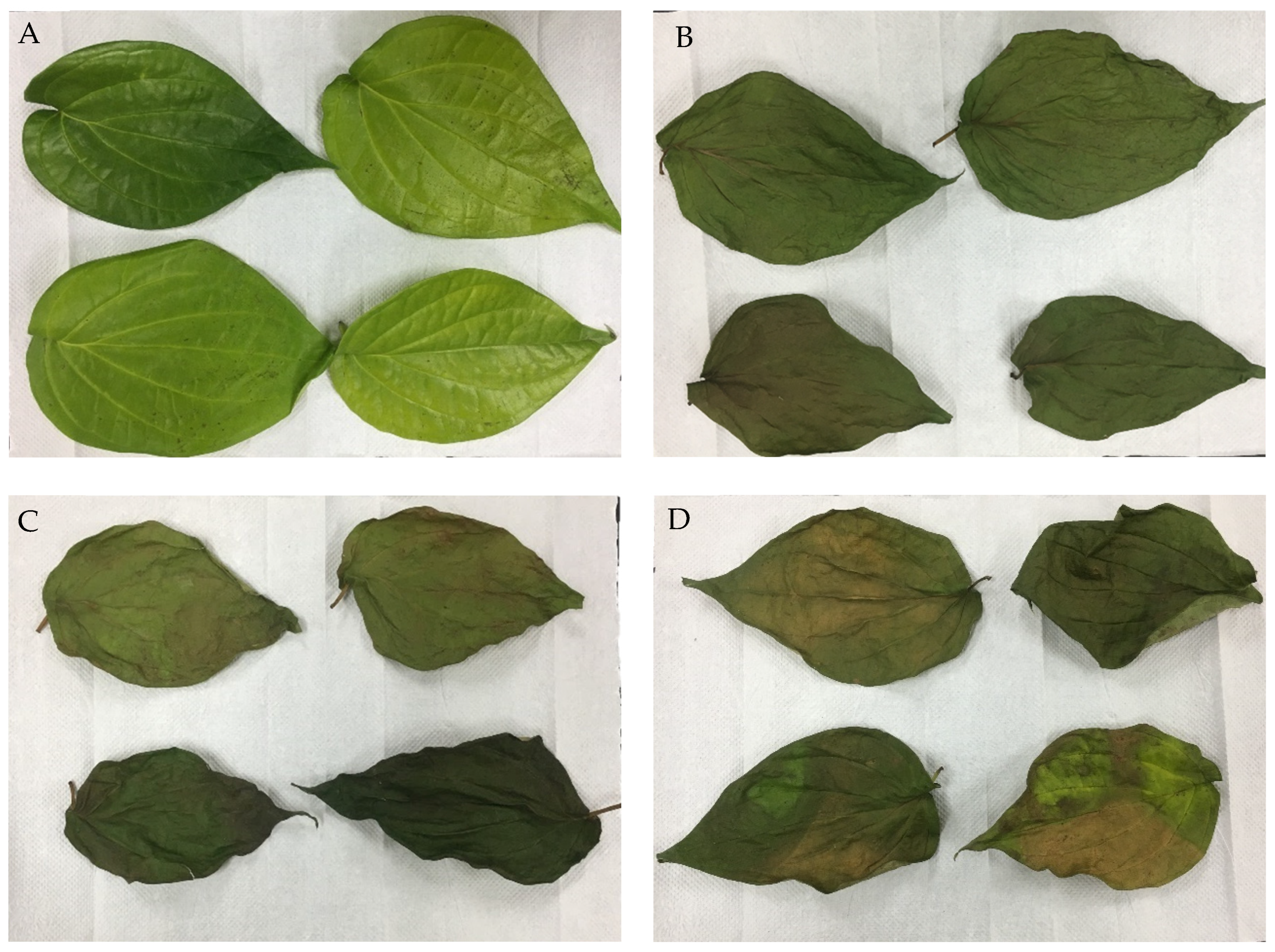
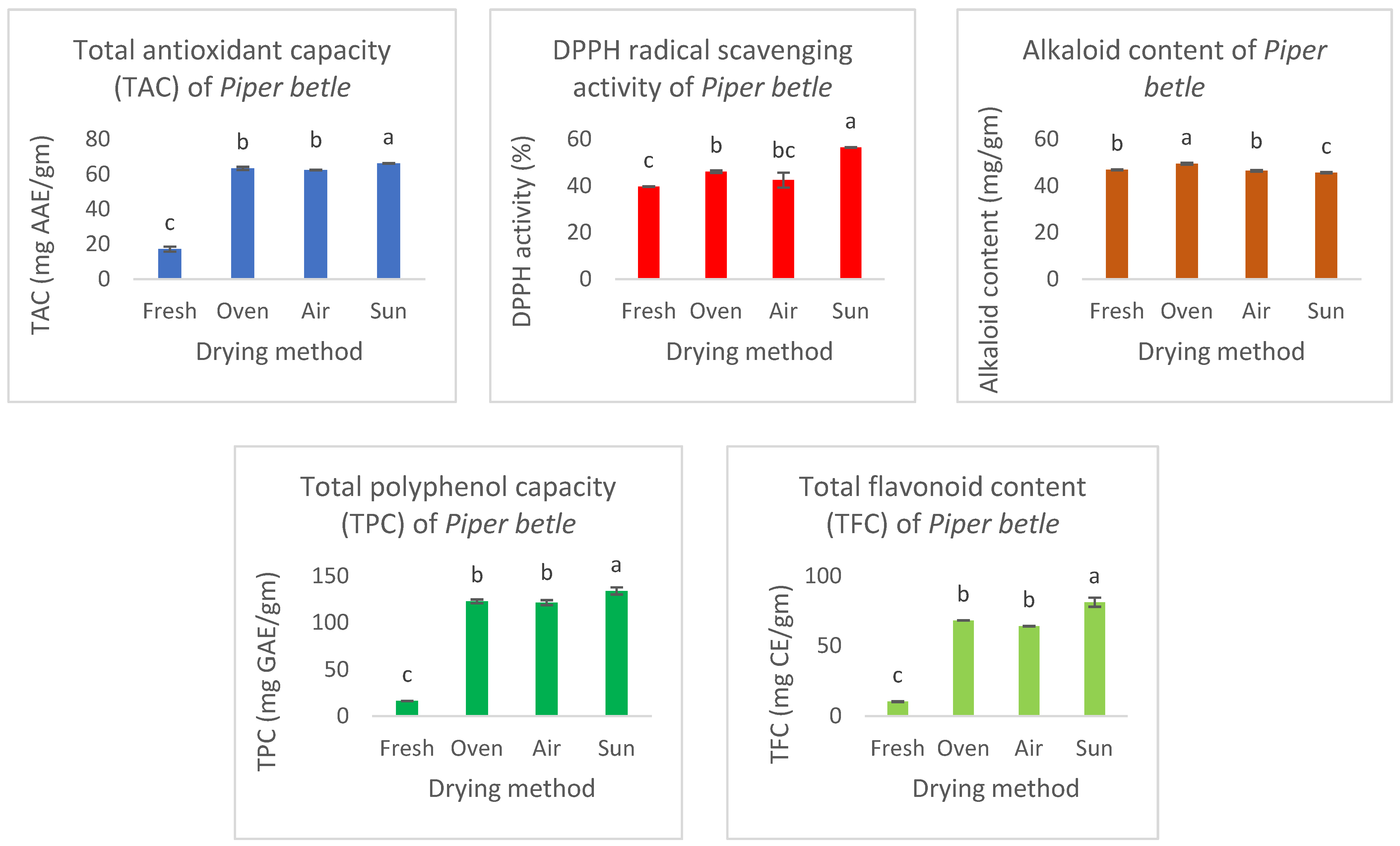
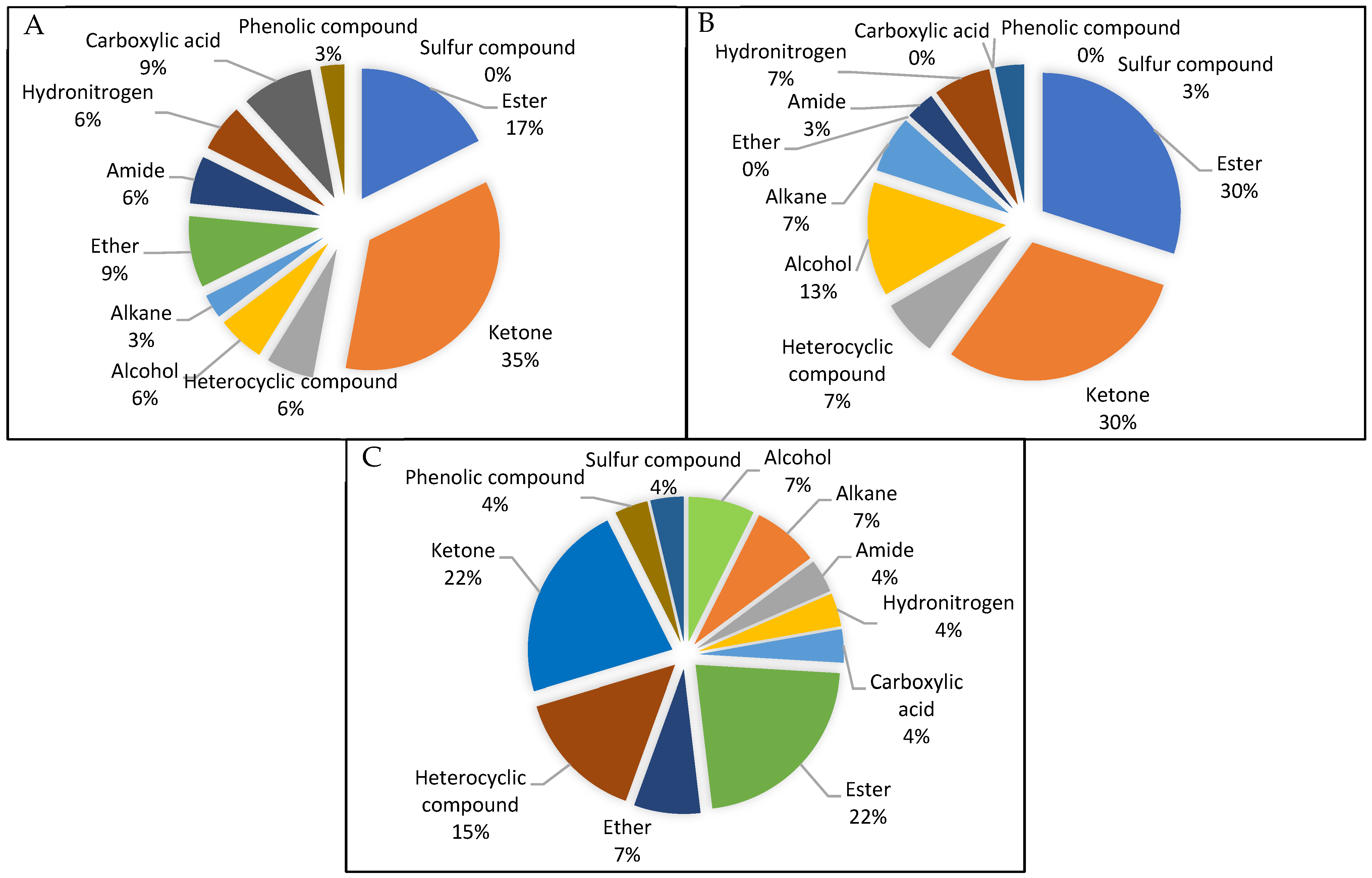
2. Results
3. Discussion
4. Materials and Methods
4.1. Sample Preparation
4.2. Drying Methods
4.3. Colour Measurement
4.4. Antioxidant Analysis
4.4.1. Preparation of Extract
4.4.2. Total Polyphenol Content
4.4.3. Total Antioxidant Capacity
4.4.4. DPPH Radical Scavenging Assay
4.4.5. Total Flavonoid Content
4.5. Gas Chromatography-Mass Spectroscopy (GCMS) Analysis
4.5.1. Sample Preparation
4.5.2. Screening of Compounds
4.6. Statistical Analysis
5. Conclusions
Author Contributions
Funding
Institutional Review Board Statement
Informed Consent Statement
Data Availability Statement
Acknowledgments
Conflicts of Interest
References
- Sarma, C.; Rasane, P.; Kaur, S.; Singh, J.; Singh, J.; Gat, Y.; Dhawan, K. Antioxidant and antimicrobial potential of selected varieties of Piper betle L. (Betel leaf). An. Acad. Bras. Cienc. 2018, 90, 3871–3878. [Google Scholar] [CrossRef]
- Umar, R.A.; Zahary, M.N.; Rohin, M.A.K.; Ismail, S. Chemical Composition and The Potential Biological Activities of Piper Betel–A Review. Malays. J. Appl. Sci. 2018, 3, 1–8. [Google Scholar]
- Madhumita, M.; Guha, P.; Nag, A. Extraction of betel leaves (Piper betle L.) essential oil and its bio-actives identification: Process optimization, GC-MS analysis and antimicrobial activity. Ind. Crops Prod. 2019, 138, 111578. [Google Scholar] [CrossRef]
- Pin, K.Y.; Chuah, T.G.; Rashih, A.A.; Law, C.L.; Rasadah, M.A.; Choong, T.S.Y. Drying of Betel Leaves (Piper betle L.): Quality and Drying Kinetics. Drying Technol. 2009, 27, 149–155. [Google Scholar] [CrossRef]
- Das, S.; Parida, R.; Sandeep, I.S.; Nayak, S.; Mohanty, S. Biotechnological intervention in betelvine (Piper betle L.): A review on recent advances and future prospects. Asian Pacific J. Trop. Med. 2016, 9, 938–946. [Google Scholar] [CrossRef]
- Biswas, P.; Anand, U.; Saha, S.C.; Kant, N.; Mishra, T.; Masih, H.; Dey, A. Betelvine (Piper betle L.): A comprehensive insight into its ethnopharmacology, phytochemistry, and pharmacological, biomedical and therapeutic attributes. J. Cell Mol. Med. 2022, 26, 3083–3119. [Google Scholar] [CrossRef]
- Rathee, J.S.; Patro, B.S.; Mula, S.; Gamre, S.; Chattopadhyay, S. Antioxidant activity of Piper betel leaf extract and its constituents. J. Agric. Food Chem. 2006, 54, 9046–9054. [Google Scholar] [CrossRef] [PubMed]
- Dwijayanti, D.R.; Puspitarini, S.; Widodo, N. Piper betle L. Leaves Extract Potentially Reduce the Nitric Oxide Production on LPS-Induced RAW 264.7 Cell Lines. J. Exp. Life Sci. 2023, 13, 78–83. [Google Scholar] [CrossRef]
- Aulia, H.R.; Wienaldi, W.; Fioni, F. Effectiveness of green betel leaf extract cream in healing cut wounds. J. Prima Medika Sains 2023, 5, 187–195. [Google Scholar] [CrossRef]
- Abrahim, N.N.; Kanthimathi, M.S.; Abdul-Aziz, A. Piper betle shows antioxidant activities, inhibits MCF-7 cell proliferation and increases activities of catalase and superoxide dismutase. BMC Complement. Altern. Med. 2012, 12, 220. [Google Scholar] [CrossRef] [PubMed]
- Nur Sazwi, N.; Nalina, T.; Rahim, Z.H.A. Antioxidant and cytoprotective activities of Piper betle, Areca catechu, Uncaria gambir and betel quid with and without calcium hydroxide. BMC Complement. Altern. Med. 2013, 13, 351. [Google Scholar] [CrossRef]
- Sahoo, B.C.; Singh, S.; Sahoo, S.; Kar, B. Comparative Metabolomics of High and Low Essential Oil Yielding Landraces of Betelvine (Piper betel L.) by Two-Dimensional Gas Chromatography Time-of-Flight Mass Spectrometry (GC× GC TOFMS). Anal. Lett. 2023, 57, 1–17. [Google Scholar]
- Babu, A.K.; Kumaresan, G.; Raj, V.A.A.; Velraj, R. Review of leaf drying: Mechanism and influencing parameters, drying methods, nutrient preservation, and mathematical models. Renew. Sustain. Energy Rev. 2018, 90, 536–556. [Google Scholar] [CrossRef]
- Thi, P.V.; Thi, T.N.; Nguyen, M.T.; Thi, D.N.P.; Trong, N.K.T.; Thi, C.H.C. The Comparison of Antioxidative and Cytotoxic Activities of Fresh and Dried Piper betle L. leave Extracts on MCF-7, HELA and SK-LU-1. Asian J. Res. Bot. 2023, 6, 339–351. [Google Scholar]
- Vernekar, A.A.; Vijayalaxmi, K.G. Nutritional composition of fresh and dehydrated betel leaves. J. Pharm. Innov. 2019, 8, 602–605. [Google Scholar]
- Sahu, C.K.; Balan, A.; Bayineni, V.K.; Banerjee, S. Study of physicochemical and antioxidant synergy efficacy of betel leaf dried paste powder. Coatian J. Food Sci. Technol. 2021, 13, 155–159. [Google Scholar]
- Seo, J.; Lee, U.; Seo, S.; Wibowo, A.E.; Pongtuluran, O.B.; Lee, K.; Cho, S. Anti-inflammatory and antioxidant activities of methanol extract of Piper betle Linn. (Piper betle L.) leaves and stems by inhibiting NF-κB/MAPK/Nrf2 signaling pathways in RAW 264.7 macrophages. Biomed. Pharmacother. 2022, 155, 113734. [Google Scholar] [CrossRef] [PubMed]
- Vikrama Chakravarthi, P.; Murugesan, S.; Arivuchelvan, A.; Sukumar, K.; Arulmozhi, A.; Jagadeeswaran, A. GC-MS profiling of methanolic extract of Piper betle (Karpoori Variety) leaf. J. Pharmacogn. Phytochem. 2018, 7, 2449–2452. [Google Scholar]
- Therdthai, N.; Zhou, W. Characterisation of microwave vacuum drying and hot air drying of mint leaves (Mentha cordifolia Opiz ex Fresen). J. Food Eng. 2009, 91, 482–489. [Google Scholar] [CrossRef]
- Rudra, S.G.; Singh, H.; Basu, S.; Shivhare, U. Enthalpy entropy compensation during thermal degradation of chlorophyll in mint and coriander puree. J. Food Eng. 2008, 86, 379–387. [Google Scholar] [CrossRef]
- Doymaz, İ.; Tugrul, N.; Pala, M. Drying characteristics of dill and parsley leaves. J. Food Eng. 2006, 77, 559–565. [Google Scholar] [CrossRef]
- Ali, A.; Chua, B.L.; Ashok, G.A. Effective Extraction of Natural Antioxidants from Piper betle with The Aid of Ultrasound: Drying And Extraction Kinetics. J. Eng. Sci. Technol. 2018, 13, 1–16. [Google Scholar]
- Alara, O.R.; Ukaegbu, C.I.; Abdurahman, N.H.; Alara, J.A.; Ali, H.A. Plant-sourced Antioxidants in Human Health: A State-of-Art Review. Curr. Nutr. Food Sci. 2023, 19, 817–830. [Google Scholar] [CrossRef]
- Stiller, A.; Garrison, K.; Gurdyumov, K.; Kenner, J.; Yasmin, F.; Yates, P.; Song, B.H. From fighting critters to saving lives: Polyphenols in plant defense and human health. Int. J. Mol. Sci. 2021, 22, 8995. [Google Scholar] [CrossRef] [PubMed]
- Roshanak, S.; Rahimmalek, M.; Goli, S.A.H. Evaluation of seven different drying treatments in respect to total flavonoid, phenolic, vitamin C content, chlorophyll, antioxidant activity and color of green tea (Camellia sinensis or C. assamica) leaves. J. Food Sci. Technol. 2016, 53, 721–729. [Google Scholar] [CrossRef]
- Salamatullah, A.M.; Ahmed, M.A.; Hayat, K.; Husain, F.M.; Arzoo, S.; Alzahrani, A.; Ahmad, S.R. Different drying techniques effect on the bioactive properties of rose petals. J. King Saud. Univ. Sci. 2024, 36, 103025. [Google Scholar] [CrossRef]
- Martínez-Las Heras, R.; Heredia, A.; Castelló, M.; Andrés, A. Influence of drying method and extraction variables on the antioxidant properties of persimmon leaves. Food Biosci. 2014, 6, 1–8. [Google Scholar] [CrossRef]
- Zhang, X.; Wang, X.; Wang, M.; Cao, J.; Xiao, J.; Wang, Q. Effects of different pretreatments on flavonoids and antioxidant activity of Dryopteris erythrosora leaves. PLoS ONE 2019, 14, e0200174. [Google Scholar] [CrossRef]
- Wanigasekera, W.; Joganathan, A.; Pethiyagoda, R.; Yatiwella, L.; Attanayake, H. Comparison of antioxidant activity, Phenolic and Flavonoid contents of selected medicinal plants in Sri Lanka. Ceylon J. Sci. 2019, 48, 155–162. [Google Scholar] [CrossRef]
- Lou, S.-N.; Lai, Y.-C.; Huang, J.-D.; Ho, C.-T.; Ferng, L.-H.A.; Chang, Y.-C. Drying effect on flavonoid composition and antioxidant activity of immature kumquat. Food Chem. 2015, 171, 356–363. [Google Scholar] [CrossRef]
- Bahloul, N.; Boudhrioua, N.; Kouhila, M.; Kechaou, N. Effect of convective solar drying on colour, total phenols and radical scavenging activity of olive leaves (Olea europaea L.). Int. J. Food Sci. Technol. 2009, 44, 2561–2567. [Google Scholar] [CrossRef]
- Romero-Márquez, J.M.; Navarro-Hortal, M.D.; Forbes-Hernández, T.Y.; Varela-López, A.; Puentes, J.G.; Pino-García, R.D.; Quiles, J.L. Exploring the Antioxidant, Neuroprotective, and Anti-Inflammatory Potential of Olive Leaf Extracts from Spain, Portugal, Greece, and Italy. Antioxidants 2023, 12, 1538. [Google Scholar] [CrossRef]
- Ramarao, K.D.R.; Somasundram, C.; Razali, Z.; Kunasekaran, W.; Jin, T.L. The antioxidant properties and microbial load of Moringa oleifera leaves dried using a prototype convective air-dryer. Saudi J. Biol. Sci. 2022, 29, 103290. [Google Scholar] [CrossRef] [PubMed]
- Djas, M.; Henczka, M. Reactive extraction of carboxylic acids using organic solvents and supercritical fluids: A review. Sep. Purif. Technol. 2018, 201, 106–119. [Google Scholar] [CrossRef]
- ElGamal, R.; Song, C.; Rayan, A.M.; Liu, C.; Al-Rejaie, S.; ElMasry, G. Thermal Degradation of Bioactive Compounds during Drying Process of Horticultural and Agronomic Products: A Comprehensive Overview. Agronomy 2023, 13, 1580. [Google Scholar] [CrossRef]
- Zhang, Y.R.; Lü, C.W.; Liu, T.; Zhen, J.M. Effect of different drying methods on volatile flavor components in Agaricus blazei. Food Sci. 2016, 37, 116–121. [Google Scholar]
- George, M.; Nagaraja, K.S.; Balasubramanian, N. Spectrophotometric determination of hydrazine. Anal. Lett. 2007, 40, 2597–2605. [Google Scholar] [CrossRef]
- Haque, A.M.J.; Kumar, S.; del Río, J.S.; Cho, Y.K. Highly sensitive detection of hydrazine by a disposable, Poly (Tannic Acid)-Coated carbon electrode. Biosens. Bioelectron. 2020, 150, 111927. [Google Scholar] [CrossRef]
- Matsumoto, M.; Kano, H.; Suzuki, M.; Katagiri, T.; Umeda, Y.; Fukushima, S. Carcinogenicity and chronic toxicity of hydrazine monohydrate in rats and mice by two-year drinking water treatment. Regul. Toxicol. Pharmacol. 2016, 76, 63–73. [Google Scholar] [CrossRef] [PubMed]
- Newsome, W.H. Determination of daminozide residues on foods and its degradation to 1, 1-dimethylhydrazine by cooking. J. Agric. Food. Chem. 1980, 28, 319–321. [Google Scholar] [CrossRef]
- Prodhan, M.D.H.; Afroze, M.; Begum, A.; Ahmed, M.S.; Sarker, D. Optimization of a QuEChERS based analytical method for the determination of organophosphorus and synthetic pyrethroid pesticide residues in betel Leaf. Int. J. Environ. Anal. Chem. 2023, 103, 1292–1303. [Google Scholar] [CrossRef]
- Konwar, L.J.; Mikkola, J.-P.; Bordoloi, N.; Saikia, R.; Chutia, R.S.; Kataki, R. Chapter 3—Sidestreams from Bioenergy and Biorefinery Complexes as a Resource for Circular Bioeconomy. In Waste Biorefinery; Bhaskar, T., Pandey, A., Mohan, S.V., Lee, D.J., Khanal, S.K., Eds.; Elsevier: Amsterdam, The Netherlands, 2018; pp. 85–125. [Google Scholar]
- Araújo, R.S.; Sousa, K.R.S.; Sousa, F.C.B.; Oliveira, A.C.; Dourado, L.R.B.; Guimarães, S.E.F.; Silva, W.; Biagiotti, D.; Bayão, G.F.V.; Araujo, A.C. Effect of glycerin supplementation on the expression of antioxidant and mitochondrial genes in broilers. Anim. Prod. Sci. 2018, 59, 408–415. [Google Scholar] [CrossRef]
- Jananie, R.K.; Priya, V.; Vijayalakshmi, K. Determination of bioactive components of Cynodon dactylon by GC-MS analysis. N. Y. Sci. J. 2011, 4, 1–5. [Google Scholar]
- Chung, J.H.; Choi, S.Y.; Kim, J.Y.; Kim, D.H.; Lee, J.W.; Choi, J.S.; Chung, H.Y. 3-Methyl-1, 2-cyclopentanedione down-regulates age-related NF-κB signaling cascade. J. Agric. Food Chem. 2007, 55, 6787–6792. [Google Scholar] [CrossRef]
- Kim, A.R.; Zou, Y.; Kim, H.S.; Choi, J.S.; Chang, G.Y.; Kim, Y.J.; Chung, H.Y. Selective peroxynitrite scavenging activity of 3-methyl-1, 2-cyclopentanedione from coffee extract. J. Pharm. Pharmacol. 2002, 54, 1385–1392. [Google Scholar] [CrossRef]
- Rao, K.M.; Rao, K.S.V.K.; Ha, C.S. Stimuli responsive poly (vinyl caprolactam) gels for biomedical applications. Gels 2016, 2, 6. [Google Scholar] [CrossRef]
- Alexakis, A.E.; Ayyachi, T.; Mousa, M.; Olsén, P.; Malmström, E. 2-Methoxy-4-Vinylphenol as a Biobased Monomer Precursor for Thermoplastics and Thermoset Polymers. Polymers 2023, 15, 2168. [Google Scholar] [CrossRef]
- Yue, G.G.; Lee, J.K.; Kwok, H.F.; Cheng, L.; Wong, E.C.; Jiang, L.; Yu, H.; Leung, H.W.; Wong, Y.L.; Leung, P.C.; et al. Novel PI3K/AKT targeting anti-angiogenic activities of 4-vinylphenol, a new therapeutic potential of a well-known styrene metabolite. Sci. Rep. 2015, 8, 11149. [Google Scholar] [CrossRef]
- Jeong, J.B.; Hong, S.C.; Jeong, H.J.; Koo, J.S. Anti-inflammatory effect of 2-methoxy-4-vinylphenol via the suppression of NF-κB and MAPK activation, and acetylation of histone H3. Arch. Pharm. Res. 2011, 34, 2109–2116. [Google Scholar] [CrossRef]
- Fragnière, C.; Aebischer, J.N.; Dudler, V.; Sager, F. A short study on the formation of styrene in cinnamon. Mitteilungen Leb. Hyg. 2003, 94, 609–620. [Google Scholar]
- Ramarao, K.D.R.; Razali, Z.; Somasundram, C. Mathematical models to describe the drying process of Moringa oleifera leaves in a convective-air dryer. Czech J. Food Sci. 2021, 39, 444–451. [Google Scholar] [CrossRef]
- Ali, M.; Yusof, Y.; Chin, N.; Ibrahim, M.; Basra, S. Drying kinetics and colour analysis of Moringa oleifera leaves. Agric. Agric. Sci. Procedia 2014, 2, 394–400. [Google Scholar] [CrossRef]
- Uribe, E.; Delgadillo, A.; Giovagnoli-Vicuña, C.; Quispe-Fuentes, I.; Zura-Bravo, L. Extraction Techniques for Bioactive Compounds and Antioxidant Capacity Determination of Chilean Papaya (Vasconcellea pubescens) Fruit. J. Chem. 2015, 8, 347532. [Google Scholar] [CrossRef]
- Jaiswal, S.G.; Patel, M.; Saxena, D.K.; Naik, S.N. Antioxidant properties of Piper betel (L) leaf extracts from six different geographical domain of India. J. Bioresour. Eng. Technol. 2014, 1, 18–26. [Google Scholar]
- Bae, S.H.; Suh, H.J. Antioxidant activities of five different mulberry cultivars in Korea. LWT-Food Sci. Technol. 2007, 40, 955–962. [Google Scholar] [CrossRef]
- Prieto, P.; Pineda, M.; Aguilar, M. Spectrophotometric quantitation of antioxidant capacity through the formation of a phosphomolybdenum complex: Specific application to the determination of vitamin E. Anal. Biochem. 1999, 269, 337–341. [Google Scholar] [CrossRef] [PubMed]
- Sakanaka, S.; Tachibana, Y.; Okada, Y. Preparation and antioxidant properties of extracts of Japanese persimmon leaf tea (kakinoha-cha). Food Chem. 2005, 89, 569–575. [Google Scholar] [CrossRef]
- Madi, N.; Dany, M.; Abdoun, S.; Usta, J. Moringa oleifera’s nutritious aqueous leaf extract has anticancerous effects by compromising mitochondrial viability in an ROS-dependent manner. J. Am. Coll. Nutr. 2016, 35, 604–613. [Google Scholar] [CrossRef]


| Level | Classification |
|---|---|
| Kingdom | Plantae |
| Division | Magnoliophyta |
| Class | Magnoliopsida |
| Order | Piperales |
| Family | Piperaceae |
| Genus | Piper |
| Species | betle |
| Drying Method | Fresh | Oven | Convective | Sun | |
|---|---|---|---|---|---|
| Colour Parameter | |||||
| L* | 41.8 ± 4.53 a | 33.17 ± 2.96 a | 33.13 ± 2.91 a | 33.47 ± 2.29 a | |
| a* | −7.53 ± 0.54 a | −5.13 ± 0.86 b | −3.20 ± 0.67 bc | −2.70 ± 0.67 c | |
| b* | 28.07 ± 6.76 a | 16.70 ± 2.07 ab | 15.97 ± 2.38 ab | 15.23 ± 1.53 b | |
| a*/b* | −0.29 ± 0.10 a | −0.31 ± 0.03 a | −0.21 ± 0.07 a | −0.18 ± 0.05 a | |
| Drying Method: Sun-Dried Extraction Method: First | ||||
| Peak | R Time | Area % | Name | Nature of Compound |
| 1 | 3.144 | 43.56 | Hydrazine, 1,2-dimethyl- | Hydronitrogen |
| 2 | 3.262 | 4.04 | Ethyl aminomethylformimidate | Ester |
| 3 | 3.598 | 1.55 | Acetic acid, hydroxy-, methyl ester | Ester |
| 4 | 3.701 | 1.49 | Acetoin | Ketone |
| 5 | 3.974 | 10.35 | Glycerin | Alcohol |
| 6 | 4.119 | 2.30 | Propanoic acid, 2-hydroxy-, methyl ester, (+/−)- | Ester |
| 7 | 6.297 | 3.34 | 2-Cyclopenten-1-one | Ketone |
| 8 | 8.110 | 0.87 | 1,3,5,7-Cyclooctatetraene | Heterocyclic compound |
| 9 | 9.832 | 15.50 | 1,2-Cyclopentanedione | Ketone |
| 10 | 12.656 | 2.21 | Oxirane, [(2-propenyloxy)methyl]- | Ether |
| 11 | 13.926 | 1.21 | 1,2-Cyclopentanedione, 3-methyl- | Ketone |
| 12 | 14.086 | 2.20 | 1,2-Cyclopentanedione, 3-methyl- | Ketone |
| 13 | 17.801 | 0.88 | 2-Cyclopenten-1-one, 3-ethyl-2-hydroxy- | Ketone |
| 14 | 19.695 | 1.15 | Silane, dimethyldi(but-3-enyloxy)- | Ether |
| 15 | 21.990 | 0.86 | Benzofuran, 2,3-dihydro- | Heterocyclic compound |
| 16 | 23.041 | 1.88 | Caprolactam | Amide |
| 17 | 30.767 | 6.62 | Benzoic acid, 2,5-dimethyl- | Carboxylic acid |
| Drying Method: Sun-Dried Extraction Method: Second | ||||
| Peak | R time | Area % | Name | Nature of compound |
| 1 | 3.133 | 45.09 | Hydrazine, 1,2-dimethyl- | Hydronitrogen |
| 2 | 3.258 | 3.78 | Ethyl aminomethylformimidate | Ester |
| 3 | 3.599 | 1.46 | Acetic acid, hydroxy-, methyl ester | Ester |
| 4 | 3.699 | 1.56 | Acetoin | Ketone |
| 5 | 3.972 | 9.95 | Glycerin | Alcohol |
| 6 | 4.121 | 1.38 | Propanoic acid, 2-hydroxy-, methyl ester, (+/−)- | Ester |
| 7 | 6.297 | 2.77 | 2-Cyclopenten-1-one | Ketone |
| 8 | 9.819 | 14.12 | 1,2-Cyclopentanedione | Ketone |
| 9 | 12.651 | 1.88 | Octane, 4-ethyl- | Alkane |
| 10 | 13.921 | 1.19 | 1,2-Cyclopentanedione, 3-methyl- | Ketone |
| 11 | 14.083 | 2.03 | 1,2-Cyclopentanedione, 3-methyl- | Ketone |
| 12 | 17.799 | 1.09 | 2-Cyclopenten-1-one, 3-ethyl-2-hydroxy- | Ketone |
| 13 | 19.693 | 1.04 | Silane, dimethyldi(but-3-enyloxy)- | Ether |
| 14 | 23.036 | 2.04 | Caprolactam | Amide |
| 15 | 25.080 | 0.54 | 2-Methoxy-4-vinylphenol | Phenolic compound |
| 16 | 30.760 | 10.09 | Benzoic acid, 2,5-dimethyl- | Carboxylic acid |
| Drying Method: None (Fresh Leaves) Extraction Method: First | ||||
| Peak | R time | Area % | Name | Nature of compound |
| 1 | 3.031 | 16.59 | Hydrazine, 1,2-dimethyl- | Hydronitrogen |
| 2 | 3.253 | 9.74 | Ethyl aminomethylformimidate | Ester |
| 3 | 3.344 | 2.41 | Trimethylsilyl ethaneperoxoate | Ester |
| 4 | 3.985 | 29.41 | Glycerin | Alcohol |
| 5 | 4.133 | 3.87 | Propanoic acid, 2-hydroxy-, methyl ester, (+/−)- | Ester |
| 6 | 4.366 | 2.45 | 2-Propenoic acid, methyl ester | Ester |
| 7 | 4.653 | 3.56 | Alpha-monopropionin | Alcohol |
| 8 | 6.341 | 4.63 | 2-Cyclopenten-1-one | Ketone |
| 9 | 9.708 | 10.65 | 1,2-Cyclopentanedione | Ketone |
| 10 | 9.804 | 5.77 | 1,2-Cyclopentanedione | Ketone |
| 11 | 12.666 | 2.42 | Decane | Alkane |
| 12 | 14.070 | 6.26 | 2-Cyclopenten-1-one, 2-hydroxy-3-methyl- | Ketone |
| 13 | 22.891 | 2.23 | Caprolactam | Amide |
| Drying Method: None (Fresh Leaves) Extraction Method: Second | ||||
| Peak | R time | Area % | Name | Nature of compound |
| 1 | 3.041 | 19.88 | Hydrazine, 1,2-dimethyl- | Hydronitrogen |
| 2 | 3.200 | 1.15 | Ethyl aminomethylformimidate | Ester |
| 3 | 3.261 | 9.83 | Allyl mercaptan | Sulfur compound |
| 4 | 3.342 | 2.20 | Trimethylsilyl ethaneperoxoate | Ester |
| 5 | 3.606 | 1.54 | Acetic acid, hydroxy-, methyl ester | Ester |
| 6 | 3.982 | 27.88 | Glycerin | Alcohol |
| 7 | 4.129 | 3.60 | Propanoic acid, 2-hydroxy-, methyl ester, (+/−)- | Ester |
| 8 | 4.427 | 2.08 | Pyrrole | Heterocyclic compound |
| 9 | 4.554 | 1.40 | Glycerin | Alcohol |
| 10 | 4.646 | 1.82 | Propanoic acid, 1-methylpropyl ester | Ester |
| 11 | 6.311 | 4.64 | 2-Cyclopenten-1-one | Ketone |
| 12 | 8.115 | 0.97 | Styrene | Heterocyclic compound |
| 13 | 9.714 | 11.16 | 1,2-Cyclopentanedione | Ketone |
| 14 | 9.792 | 4.65 | 1,2-Cyclopentanedione | Ketone |
| 15 | 12.655 | 3.48 | Decane | Alkane |
| 16 | 13.922 | 1.70 | 1,2-Cyclopentanedione, 3-methyl- | Ketone |
| 17 | 14.061 | 2.03 | 1,2-Cyclopentanedione, 3-methyl- | Ketone |
Disclaimer/Publisher’s Note: The statements, opinions and data contained in all publications are solely those of the individual author(s) and contributor(s) and not of MDPI and/or the editor(s). MDPI and/or the editor(s) disclaim responsibility for any injury to people or property resulting from any ideas, methods, instructions or products referred to in the content. |
© 2024 by the authors. Licensee MDPI, Basel, Switzerland. This article is an open access article distributed under the terms and conditions of the Creative Commons Attribution (CC BY) license (https://creativecommons.org/licenses/by/4.0/).
Share and Cite
Ramarao, K.D.R.; Razali, Z.; Somasundram, C.; Kunasekaran, W.; Jin, T.L. Effects of Drying Methods on the Antioxidant Properties of Piper betle Leaves. Molecules 2024, 29, 1762. https://doi.org/10.3390/molecules29081762
Ramarao KDR, Razali Z, Somasundram C, Kunasekaran W, Jin TL. Effects of Drying Methods on the Antioxidant Properties of Piper betle Leaves. Molecules. 2024; 29(8):1762. https://doi.org/10.3390/molecules29081762
Chicago/Turabian StyleRamarao, Kivaandra Dayaa Rao, Zuliana Razali, Chandran Somasundram, Wijenthiran Kunasekaran, and Tan Li Jin. 2024. "Effects of Drying Methods on the Antioxidant Properties of Piper betle Leaves" Molecules 29, no. 8: 1762. https://doi.org/10.3390/molecules29081762
APA StyleRamarao, K. D. R., Razali, Z., Somasundram, C., Kunasekaran, W., & Jin, T. L. (2024). Effects of Drying Methods on the Antioxidant Properties of Piper betle Leaves. Molecules, 29(8), 1762. https://doi.org/10.3390/molecules29081762