Article

The Antiproliferative Activity and NO Inhibition of Neo-Clerodane Diterpenoids from Salvia guevarae in RAW 264.7 Macrophages

by
Juan Pablo Torres-Médicis
1,
Celia Bustos-Brito
1,
Leovigildo Quijano
1,
Brenda Y. Bedolla-García
2,
Sergio Zamudio
3,
Teresa Ramírez-Apan
1,
Diego Martínez-Otero
1 and
Baldomero Esquivel
1,*
1
Instituto de Química, Universidad Nacional Autónoma de México, Circuito Exterior, Ciudad Universitaria, Ciudad de México 04510, Mexico
2
Instituto de Ecología, A.C., Centro Regional del Bajío, Apartado Postal 386, Pátzcuaro 61600, Mexico
3
Independent Researcher, Apartado Postal 392, Pátzcuaro 61600, Mexico
*
Author to whom correspondence should be addressed.
Molecules 2025, 30(7), 1628; https://doi.org/10.3390/molecules30071628
Submission received: 12 March 2025 / Revised: 1 April 2025 / Accepted: 2 April 2025 / Published: 5 April 2025
(This article belongs to the Special Issue Natural Products with Pharmaceutical Activities)

Abstract

:
In this study, nine neo-clerodane-type diterpenoids (19) were isolated from the dichloromethane extract of Salvia guevarae Bedolla & Zamudio leaves. Compounds 16 were new natural products, and 79 were acetone artifacts. In addition, four neo-clerodanes diterpenoids (1013) previously described from different sources and six triterpenoids—identified as 3β,20,25-trihydroxylupane, oleanolic acid, 3β-O-acetyl-oleanolic acid, ursolic acid, 3β-O-acetyl-betulinic acid, and 3β,28-O-diacetyl-betulin—were isolated. Additionally, five flavonoids were also isolated from the methanol extract: quercetin-3-O-β-xylopyranosyl-(1 → 2)-β-galactopyranoside, taxifolin-7-O-β-glucopyranoside, naringenin-7-O-β-glucopyranoside, a mixture of 2R and 2S eriodictyol-7-O-β-glucopyranoside, caffeic acid, the methyl ester of rosmarinic acid, and rosmarinic acid. The structure of the isolated compounds was established by spectroscopic means, mainly 1H and 13C NMR, including 1D and 2D homo- and heteronuclear experiments. The absolute configuration of 1 and 10 was ascertained via an X-ray analysis, and that of the other compounds via ECD. The antiproliferative activity of some diterpenoids was determined using the sulforhodamine B method, where guevarain B (2) and 6α-hydroxy-patagonol acetonide (7) showed moderate activity against the K562 line, with IC50 (μM) = 33.1 ± 1.3 and 39.8 ± 1.5, respectively. The NO inhibition in RAW 264.7 macrophage activity was also determined for some compounds, where 2-oxo-patagonal (6), 6α-hydroxy-patagonol acetonide (7), and 7α-acetoxy-ent-clerodan-3,13-dien-18,19:16,15-diolide (10) were proven to be active, with IC50 (μM) of 26.4 ± 0.4, 17.3 ± 0.5, and 13.7 ± 2.0, respectively. The chemotaxonomy of Salvia guevarae is also discussed.

Graphical Abstract

1. Introduction

Salvia L. (Lamiaceae) is one of the most species-rich genera of angiosperm plants, with about 1000 species with a sub-cosmopolitan distribution [1]. Recent taxonomic and phylogenetic analyses have led to the organization of the genus into 11 subgenera [2], with the subgenera Audibertia and Calosphace being endemic to the American continents. With a biogeographic distribution ranging from North America to Central America, the Caribbean, and South America, the subgenus Calosphace, with an estimated 550 species, is the most diverse, comprising 55% of the whole genus. The Salvia species growing in Mexico mainly belong to this subgenus. With an estimated 312 species, with 57% of species from the subgenus Calosphace and 31% from the subgenus Mundial, Salvia is the most species-rich genus in Mexico, which is considered, due to this diversity and an estimated endemism of 82%, the center of the origin and diversification of this subgenus [3]. Several Salvia species are widely cultured in gardens around the world and in Mexico and profusely used as medicinal plants for different ailments including gastrointestinal, nervous system, gynecological, and folk illnesses [4].
From systematic phytochemical studies on Mexican Salvia species, starting in 1984, diterpenoids have been found to be the most diversified secondary metabolites. Diterpenoids with neo-clerodane-type (sometimes also referred to as ent-clerodanes) [5] and abietane-type skeletons are the most frequently isolated, although pimarane-, totarane-, and recently labdane-type diterpenoids [6] have also been described from some members of the genus. Several rearranged scaffolds derived mainly from the neo-clerodane type and, to a lesser extent, the abietane- and pimarane-type skeletons, have also frequently been described, highlighting the chemical features of Mexican Salvia species. The distribution of these types of compounds, in some cases, parallels that of the botanical classification proposed by Epling, as in the case of sections Erythrostachys, Fulgentes, Angulatae, Tomentellae, and Scorodonia. Another interesting result is that the distribution of diterpenoids in species of the subgenus Calosphace differs markedly from that found in species from Europe and Asia, from which mainly abietane and abietane-derived diterpenoids have been isolated. The presence of diterpenoids in the genus Salvia is, therefore, of chemo-systematic relevance [7].
As a result of intensive botanical field work during the past three decades all over the country, the number of Salvia spp. has constantly grown in Mexico, with several new species being described, all belonging to the subgenus Calosphace. Since 2007, 62 new species have been described, some of which have been classified up to the taxonomic section rank [5]. Salvia guevarae Bedolla & Zamudio was recently described and determined to be a new botanical species [8]. The morphology of this species, with its red markedly ventricose corollas with short lips, allowed it to be classified in section Holwaya (Ramamoorthy) [9] and suggests its close relation to S. involucrata and S. puberula. Recently, we described the diterpenoid content of the newly described S. carranzae Bedolla & Zamudio, which led us to propose that this species is closely related to those classified in section Erythrostachys (Epling) [7]. In this study, we report on the phytochemical analysis of S. guevarae, supporting its inclusion in section Holwaya, and evaluate the biological activity of some isolated secondary metabolites. The dichloromethane and methanol extracts of this species were thoroughly analyzed using chromatographic techniques to afford nine not previously described neo-clerodane-type diterpenoids, with three of them being acetone artifacts, four neo-clerodane diterpenoids, six triterpenoids, and eight polyphenolic compounds, already known. Their antiproliferative activities against human cancer cell cultures and capacity for the inhibition of nitric oxide (NO) production in RAW 264.7 macrophages were also evaluated.

2. Results and Discussion

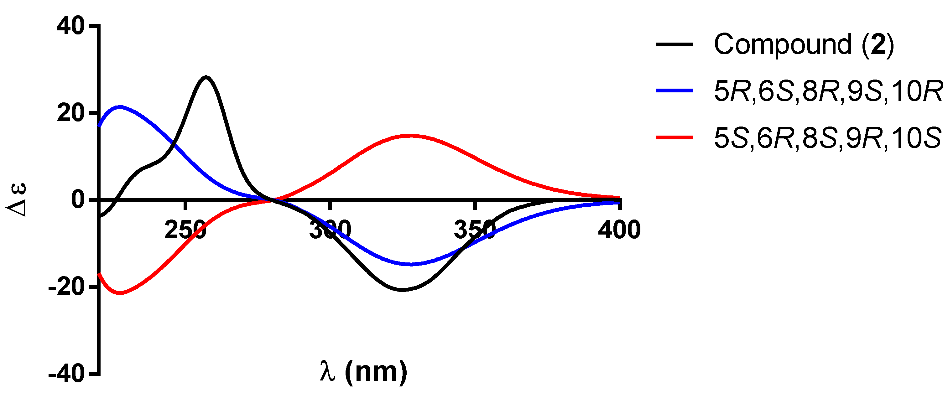
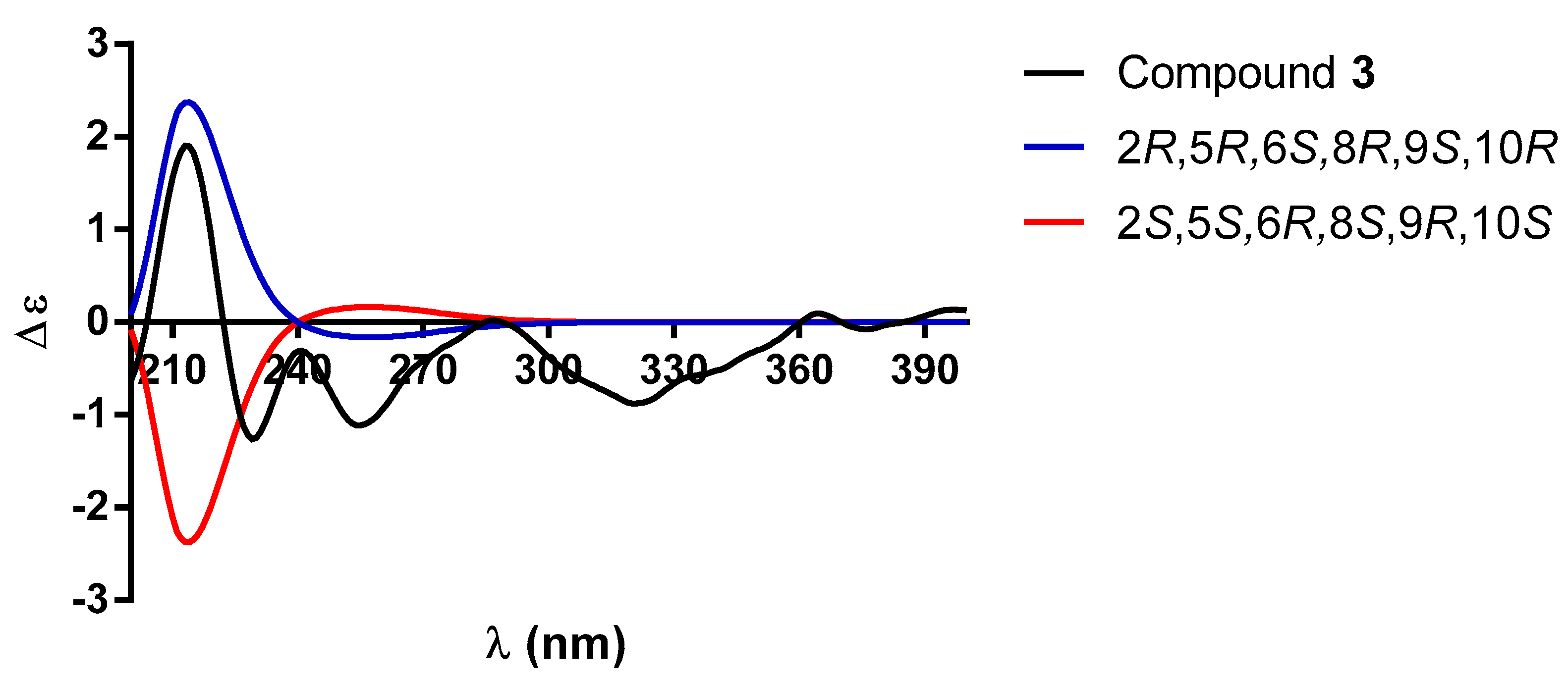
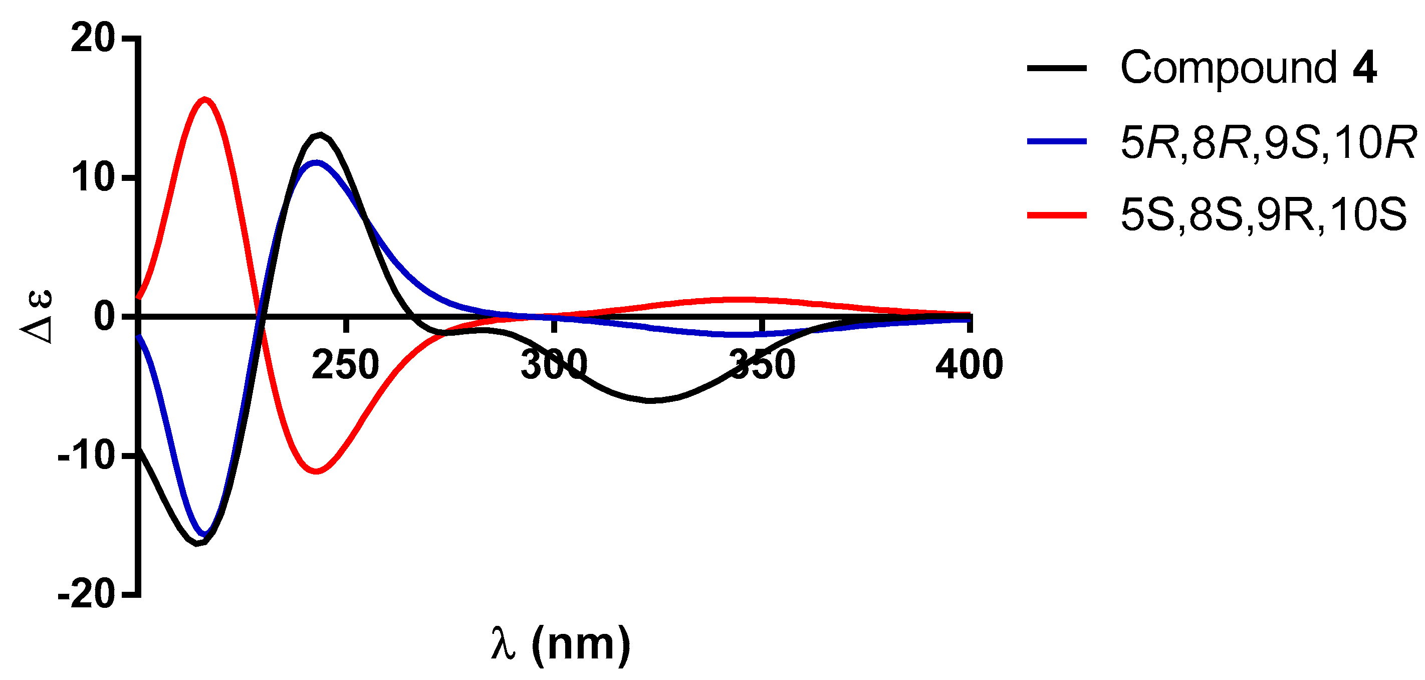
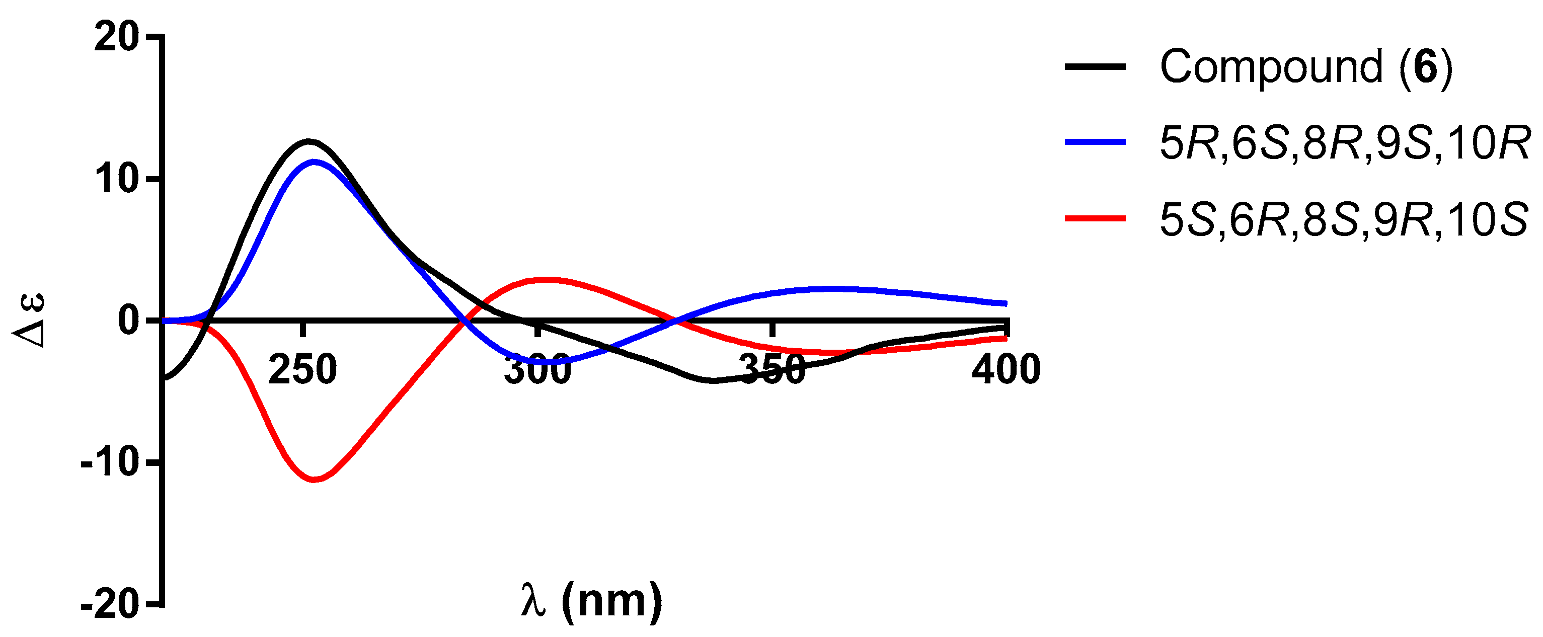
2.1. Characterization

The leaves and flowers of S. guevarae afforded, after dichloromethane extraction and thorough chromatographic purification, 13 neo-clerodane derivatives (113) (Figure 1). Compounds 16 are undescribed neo-clerodane diterpenoids with structures related to those of patagonol (14) and patagonal (15), two neo-clerodane diterpenoids previously isolated from a liana collected from the Surinam rainforest, tentatively identified as a Casimirella species [10]. Diterpenoids 79 are artifacts obtained, most likely, via a reaction with acetone, used in the purification process, and products 1013 are previously known compounds isolated from several sources. The structural elucidation of all the isolated compounds was carried out via spectroscopic and chemical methods and a comparison with literature data.
Compound 1 was the major component and was isolated as a crystalline solid at mp 136–138 °C. The HR-DART-MS showed a pseudo-molecular ion [M + H − H2O]+ at m/z 317.2103 (calculated for C20H30O4 + H − H2O, 317.2117) consistent with the molecular formula C20H30O4 minus the loss of an H2O molecule from the molecular ion. Its IR spectrum exhibited characteristic absorptions for α,β-unsaturated-γ-lactone function (1755 cm−1), hydroxyl groups (3606 cm−1), and double bounds (1654, 1602 cm−1). The UV spectrum [MeOH, λmax (log ε) 207 (6.32) nm] was consistent with these assignments.
In the 1H NMR spectrum of 1 (Table 1), signals of an α-substituted butenolide were observed at C-12. A quintet at δH 7.41 (J = 1.7 Hz) was assigned to the vinylic H-14 coupled with two methylene groups with signals at δH 4.79 and 2.14/2.05 due to CH2-15 and CH2-12, respectively, and with similar 3J and 4J coupling constant values to those of H-14 and H-12, according to their COSY spectra (Table 1, Figure 2). The 13C NMR spectrum of 1 (Table 1) supports the above assumption, since the corresponding signals of C-13, C-14, C-15, and C-16 were observed at δC 134.3, 146.2, 71.0, and 174.7, respectively, which is in accordance with the results obtained from the HSQC and HMBC experiments (Figure 2). The presence of an α-substituted butenolide group is a frequent feature in neo-clerodane-type diterpenoids isolated from Salvia species [9] and is present in all diterpenoids (113) isolated from S. guevarae.
The other relevant signals observed in the 1H NMR spectrum of 1 (Table 1) included a broad singlet at δH 5.19, which was ascribed to the vinylic H-3, and multiple signals at δH 4.13, assigned to the allylic proton H-2, geminal to the hydroxyl group at the C-2 position. In the COSY spectrum of 1 (Figure 2), cross peaks of correlation were observed between the H-2 signal (δH 4.13) with H-3 (δH 5.19); a three-proton signal at δH 1.84; and the hydrogens of a methylene group at δH 1.94 (ddd, J = 12.6, 7.0, 1.4 Hz) and 1.49 (td, J = 12.6, 10.1 Hz), which were ascribed to the vinylic CH3-18 and to H-1 equatorial and H-1 axial, respectively. All other relevant COSY interactions are depicted in Figure 2.
The 1H NMR spectrum of 1 (Table 1) also exhibited the signal for a second hydrogen atom geminal to a hydroxyl group, which was observed at δH 3.48 (dt, J = 9.5, 5.6 Hz). The multiplicity and the coupling constant values led us to propose that the hydroxyl group is located at the C-6 position with an α equatorial orientation. Signals due to characteristic C-17, C-19, and C-20 methyl groups of a neo-clerodane-type diterpenoid were also observed at δH 0.84 (d, J = 6.8 Hz), 1.06 (s), and 0.75 (s), respectively. Finally, a doublet of doublets at δH 1.35 (J = 12.6, 1.4 Hz), ascribed to the β-axially oriented H-10, coupled with the methylene signals of -CH2-1 at δH 1.94 (ddd, J = 12.6, 7.0, 1.4 Hz)/1.49, td (J = 12.6, 10.1 Hz), was also observed in the 1H NMR spectrum of 1 (Table 1). These facts, in combination with the chemical shifts observed for C-17 and C-20 in the 13C NMR spectrum of 1 (Table 1), indicated a trans fusion of A/B rings, which is the most frequently observed for the neo-clerodane diterpenoids hitherto isolated from Salvia spp. [5]. All other signals in the 13C NMR spectrum of 1 (Table 1) agreed with the proposed structure, and all connectivities were confirmed with the aid of the HMBC spectrum (Figure 2).
The NOESY spectrum supports relative stereochemistry since it showed NOE correlations between the β-axially oriented H-10 and H-2 (Figure 2), indicating that the latter must also be β-axially oriented; therefore, the C-2 hydroxyl group must be α-equatorially oriented. On the other hand, the cross peaks of correlation between H-10 and H-8 established that the C-17 methyl group was also α-equatorially oriented. The NOE effect observed between the C-19 and C-20 methyl groups indicated that both were cofacial [10], as shown in Figure 1. Therefore, compound 1 corresponds to a new neo-clerodane compound that we named guevarain A.
The absolute configuration of guevarain A was established via an X-ray diffraction analysis. Guevarain A was crystallized as a monohydrate in the monoclinic P2(1) space group, and the absolute configuration was determined to have the following parameters: Flack(x), −0.02(3); Hooft(y), −0.02(2); and Parsons(z), −0.019(16). This configuration confirms the assignment of the crystal model as well as the configuration of the chiral carbon atoms as being 2R, 5R, 6S, 8R, 9S, and 10R. The molecular structure was shown in Figure 3, and the crystallographic details are presented in the Supplementary Materials (Table S1). Figure 4 shows the crystalline packing, where three discrete hydrogen bonds described as D 1 1 (2) were found, labeled as a, c, and d, where one water molecule participate as donor (c, d) and one (a) as an acceptor. An eight-membered one-dimensional chain, described as C 1 1 (8) in terms of graph set descriptors, labeled as b, formed through the O-H fragments of compound 1. The hydrogen bond distances and angles corresponding to guevarain A (1) are shown in the Supplementary Materials (Table S2).
Diterpenoid 2, C20H28O4, (HR-DART-MS), showed a similar IR spectrum to that of guevarain A (1), except for the presence of an intense band at 1644 cm−1 due to an α,β-unsaturated carbonyl group, also supported by the UV spectrum, since a λmax at 239.8 nm (log ε = 4.07) was observed. The 1H and 13C NMR spectra of 2 (Table 1) confirmed the presence of a C2-C3-C4 α,β-unsaturated ketone, given that a broad singlet at δH 5.72, ascribed to the vinylic H-3, was found to be coupled to a three-proton doublet at δH 2.14 (J = 1.3 Hz), assigned to C-18 vinylic methyl, in the 1H NMR spectrum. Signals for C-2 carbonyl, C-3:C-4 double bond, and C-18 methyl were observed at δC 199.7, 126.5, 172.8, and 23.0, respectively, in the 13C NMR spectrum. All these assignments were supported by COSY and HMBC spectra (Figure 5).
The signal for the hydrogen geminal to the hydroxyl group at C-6 in compound 2 was observed at δH 3.70 (J = 11.1, 4.8 Hz), similarly to that of 1. The main difference in the 1H NMR spectrum was in the signals associated with the presence of a carbonyl group at C-2. Thus, in the COSY spectrum of compound 2 (Figure 5), the CH2-1 signals at δH 2.48 (J = 17.8, 13.9 Hz) and 2.32 (J = 17.8, 3.5 Hz) showed couplings only with the doublet of doublets at δH 1.88 (J = 13.9, 3.6 Hz) ascribed to the β-axially oriented H-10. The characteristic signals of the methyl groups of a neo-clerodane skeleton were observed at δH 0.89 (d, J = 6.8 Hz), 1.12 (s), and 0.84 (s) and ascribed to CH3-17, CH3-19, and CH3-20, respectively. The 13C NMR data (Table 1) agree with the proposed structure for compound 2. All NMR assignments for diterpenoid 2 were confirmed with the aid of HMBC (Figure 5) and HSQC spectra.
The relative configuration depicted in 2 was established with the aid of a NOESY spectrum (Figure 5), since cross peaks of correlation were observed between the β-axially oriented H-10 and H-6, indicating cofacial orientations for these protons. On the other hand, the signal for H-6 showed NOE with multiple signals at δH 1.73, ascribed to H-8, also β-axially oriented.
Based on the previous discussion, the structure of compound 2 was established as the oxidation product of the secondary OH group at C-2 of guevarain A (1) and named guevarain B (2). The absolute configuration of guevarain B was established via a comparison of the experimental ECD spectrum and the enantiomeric calculated spectra [11]. Figure 6 indicates that the absolute configuration of this diterpenoid corresponds to the 5R, 6S, 8R, 9S, 10R enantiomer.
The treatment of guevarain A (1) with pyridinium chlorochromate (PCC) in CH2Cl2 afforded a mixture of two reaction products. The main product (44.7% yield) was identical in all aspects to guevarain B (2), thus confirming the proposed structure for compound 2. Structure 1a was assigned to the minor product (21.8% yield) based on its spectroscopic properties. The 1H NMR spectrum of 1a (Table 2) lacked the signals for the geminal protons of the hydroxyl groups at C-2 and C-6 observed in guevarain A (1), while the 13C NMR spectrum (Table 2) displayed two carbonyls due to the C-2 and C-6 ketone groups at δC 197.8 and 210.7, respectively, thus confirming oxidation at the C-2 and C-6 positions.
The structure of compound 3 was deduced mainly from the 1H NMR spectrum and MS. The molecular formula C20H30O5 was established via HR-DART-MS, indicating an index of hydrogen deficiency Ω = 6. The three degrees of unsaturation can be readily explained by the presence of the butenolide moiety at C-12.
The 1H NMR of compound 3 (Table 2) was like that of compound 1, with the main differences being the lack of a vinylic methyl signal and the presence of two AB doublets at δH 4.08 and 4.23 (J = 13.5 Hz), coupled with the vinylic hydrogen broad singlet at δH 5.56 (H-3), which in turn was found to be coupled with overlapping multiple signals at δH 4.23 (H-2), in accordance with the COSY spectrum (Figure 7). These facts, in addition to their chemical shifts, led us to ascribe these signals to the C-18 methylene protons and the geminal hydrogen to the hydroxyl group at C-2. The H-2 signal, in turn, showed cross peaks of correlation with two methylene protons at δH 2.02 (ddt, J = 12.5, 6.8, 1.3 Hz) and 1.57 (td, J = 12.5, 10.3 Hz) due to H-1 equatorial and H-1 axial, respectively. A β-axially oriented H-10 appeared at δH 1.40 (brd, J = 10.3 Hz) and the hydrogen atom geminal to the hydroxyl group at C-6 appeared at δH 3.58 as multiple signals similarly to that in guevarain A (1).
The relative configuration of 3 was established based on NOE interactions (Figure 7). In the NOESY spectrum, the β-axially oriented H-10 signal showed cross peaks of correlation with H-2 and H-6, while H-6 showed correlation with H-8, indicating that H-10, H-2, H-6, and H-8 were cofacial. On the other hand, the C-19 methyl hydrogens exhibited NOE with the C-20 methyl group, thus indicating that they were on the same face of the molecule. Based on all the above data, the structure of compound 3 was established as the 18-hydroxy derivative of compound 1, a structure closely related to that of patagonol (14); thus, it was named 2α,6α-dihydroxy-patagonol. The absolute configuration of 3 was established as 2R, 5R, 6S, 8R, 9S, and 10R based on the comparison between the theoretical (blue and red lines) and experimental (black line, Figure 8) ECD curves.
The structure of diterpenoid 4 was closely related to that of compound 3, mainly deduced from its NMR data. The 1H NMR spectrum (Table 3) displayed a broad singlet at δH 6.10 due to the vinylic H-3, which showed cross peaks of correlation in the COSY spectrum (Figure 9) with the C-18 allylic methylene signals at δH 4.40 (dd, J = 17.6, 1.8 Hz) and 4.35 (dd, J = 17.6, 1.8 Hz). An ABC system was observed at δH 2.44 (dd, J = 17.8, 13.5 Hz), 2.37 (dd, J = 17.8, 4.3 Hz), and 1.98 (dd, J = 13.5, 4.3 Hz), while the AB part of this system corresponds to the hydrogen atoms of the CH2-1, and the C part is ascribed to the β-axial H-10. The COSY spectrum of 4 (Figure 9) supports these assignments since the expected cross peaks of correlation were observed. In the 1H NMR spectrum of 4, despite being similar to that of compound 3, the lack of an additional hydrogen atom geminal to a hydroxyl function indicated that 4 was devoid of the secondary hydroxyl group at C-6 present in 3.
On the other hand, the 13C NMR spectrum (Table 3) indicated the presence of three methyl, seven methylene (including two bonded to oxygen), and four methine (two of them of olefinic nature) groups and six fully substituted carbons (including two carbonyls, two sp2 olefins, and two fully substituted sp3 carbons). The UV spectrum exhibited two absorption maxima at λmax 218 nm (log ε = 3.65) and 235.4 nm (log ε = 3.52), indicating that in addition to the α,β-unsaturated-γ-lactone, an α,β-unsaturated ketone must be present in compound 4, as in the case of 2. The carbon signals at δC 200.0, 121.4, and 173.6 were assigned to C-2, C-3, and C-4 of this chromophore. These facts led us to establish the structure of compound 4 as 2-oxo-patagonol, as depicted in Figure 1. The absolute configuration of 4 was established as 5R, 8R, 9S, and 10R via a comparison between the theoretical and experimental ECD data (Figure 10).
Diterpenoid 5, C20H28O5 (HR-DART-MS), had similar IR and NMR spectral data to those of 4, except for the presence of multiple signals at δH 3.83 in the 1H NMR spectrum of 5 (Table 3), ascribed to the hydrogen atom geminal to the hydroxyl group attached to C-6. The 13C NMR spectrum of 5 supports the presence of this oxygenated function since a signal was observed at δC 73.8, ascribed to C-6. In agreement with the presence of a ketone carbonyl at C-2, the signals of an ABC system were observed in the 1H NMR spectrum at δH 2.54 (dd, J = 17.8, 13.9 Hz), 2.37 (dd, J = 17.8, 3.4 Hz), and 1.91 (dd, J = 13.9, 3.6 Hz) (Table 3). The AB part was ascribed to the hydrogen atoms of the C-1 methylene and the C part to the β-axial H-10. The rest of the spectrum was similar to that obtained for 2-oxo-patagonol (4). As in the case of the previously described diterpenoids 24, the absolute configuration of 5 was established via ECD spectroscopy. Figure 11 shows a comparison between the experimental and theoretical ECD spectra that led us to establish that the absolute configuration of 5 was 5R, 6S, 8R, 9S, and 10R, as depicted. Based on all the above data, compound 5 was identified as a patagonol (14) derivative named 2-oxo-6α-hydroxy-patagonol.
Compound 6 was isolated as a white powder at mp 48−50 °C. The UV spectrum showed two absorption maxima at λmax 217 (log ε = 3.93) and 233 nm (log ε 3.86), thus indicating the presence of chromophores similar to those present in compounds 4 and 5. The IR spectrum of compound 6 was similar to those of 2-oxo-patagonol (4) and 2-oxo-6α-hydroxy-patagonol (5), although the intensity of the band at 1676 cm−1 ascribed to the α,β-unsaturated ketone was higher, indicating the presence of an additional conjugated carbonyl group. This carbonyl group was part of an aldehyde moiety, since in the 13C NMR spectrum of 6 (Table 4), a signal at δC 197.5 was observed, which correlated in the HSQC spectrum with a signal at δH 9.75 (Table 4). These facts indicated that the diterpenoid 6 was closely related to patagonal (15), a diterpenoid isolated together with patagonol (14) from the Casimirella species [10]. Another relevant signal observed in the 1H NMR spectrum of 6 was a doublet of doublets at δH 3.75 (J = 11.1, 4.7 Hz), which was ascribed to the hydrogen atom geminal to a secondary hydroxyl group located at C-6. The coupling constants, J = 11.1, 4.7 Hz, indicated that the hydroxyl was equatorially oriented. Based on the previous discussion, the diterpenoid 6 was identified as 6α-hydroxy-patagonal. The absolute configuration of 6α-hydroxy-patagonal (6) was also established via ECD. Figure 12 shows a comparison between the experimental and calculated ECD spectra that led us to the conclusion that compound 6 possessed the same absolute configuration as diterpenoids 15.
The treatment of 2α,6α-dihydroxy-patagonol (3) with PCC afforded, after 24 h of stirring at room temperature, a mixture of compounds identical in all aspects to 5 (10.9% yield) and 6 (31.4% yield), thus confirming the proposed structure for these diterpenoids.
Compounds 79 exhibited similar 1H and 13C NMR spectra to those of 3 and 5 (see the experimental section). In addition to the signals that indicated their neo-clerodane nature, the 13C NMR spectra of 79 displayed signals of quaternary carbons at δC 101.1, 101.3, and 101.6, respectively. These signals, in addition to the signals of two additional methyl groups, in the range of 23.9–24.9, indicated the presence of an acetonide group in compounds 79. Their 1H NMR spectra support the presence of the acetonide group since, besides the characteristic methyl groups of a neo-clerodane-type diterpenoid, the signals of two additional methyl groups at δH 1.34–1.37 were observed. The HMBC spectra of 79 showed correlation cross peaks between the C-18 methylene hydrogens (δH 3.65–4.59) and H-6 (δH 3.58–3.75), with the quaternary carbons (δC 100.1–100.6) indicating that the acetonide was formed between the OH groups at the C-18 and C-6 positions. Acetonides 79 must have formed during the chromatographic purification since acetone was used as an eluent. An inspection of the Dreiding models of compounds 79 and the precursor diols indicated that the formation of the acetonide is highly favored. That the plausible precursor diols for acetonides 8 and 9 are 2α,6α-dihydroxy-patagonol (3) and 2-oxo-6α-hydroxy-patagonol (5) was proven via the treatment of 2α,6α-dihydroxy-patagonol (3) with anhydrous acetone in the presence of anhydrous CuSO4, yielding 8, and further oxidation of 8 with PCC in dichloromethane, affording compound 9. While the diols 3 and 5 were obtained from the DCM extract of S. guevarae, the precursor of 7 (6α-hydroxy-patagonol) was not isolated; thus, we considered that it was present in the crude extract in small quantities. Based on the previous discussion, acetonides 79 should be considered acetone artifacts, instead of natural products.
Compound 10 was isolated as colorless crystals at mp 182-184 °C and identified as 7α-acetoxy-ent-clerodan-3,13-dien-18,19:16,15-diolide based mainly on its NMR data. Diterpenoid 10 was previously isolated from a population of Salvia melissodora Lag. [12] collected in the State of San Luis Potosí, Mexico. The undescribed high-resolution NMR and DART-MS data are recorded in the experimental section. On the other hand, in this study, we report the X-ray analysis of 10 since the absolute configuration of this diterpenoid was not established in its original description. The molecular structure of 10 is shown in Figure 13, and the crystallographic details are presented in the Supplementary Materials (Table S1). Compound 10 was crystallized in the orthorhombic P2(1)2(1)2(1) space group, and its absolute structure parameters—Flack(x), 0.01(5); Hooft(y), 0.00(5); and Parsons(z), 0.00(3)—confirm the assignment of the crystal model as well as the configuration of the chiral carbon atoms as being 5S, 7R, 8S, 9R, and 10R.
While products 11 and 12 have previously been obtained from S. melissodora and identified as 7-keto-ent-clerodan-3,13-dien-18,19:16,15-diolide and 7α-hydroxy-ent-clerodan-3,13-dien-18,19:16,15-diolide, respectively [12], compound 13 was identical in all aspect to mkapwanin, previously isolated from Dodonea angustifolia (Sapindaceae) [13]. The absolute configuration of these diterpenoids, as depicted in 1113, was established via a comparison of their experimental ECD spectra with theoretically calculated curves (see Supplemental Figure S61 and Figure S62, and S63). The undescribed high-resolution NMR and DART-MS data are recorded in the experimental section.
In addition to the previously described neo-clerodane diterpenoids, six already known triterpenoids were isolated from the DCM extract of S. guevarae, identified as 3β,20,25-trihydroxylupane [14], oleanolic acid [15], 3β-O-acetyl-oleanolic acid [16], ursolic acid [17], 3β-O-acetyl-betulinic acid [18], and 3β,28-O-diacetyl-betulin [19].
Chromatographic purification of the methanol extract of S. guevarae afforded several previously described phenolic compounds identified via spectroscopic and spectrometric methods and a comparison with literature data: quercetin-3-O-β-xylopyranosil-(1 → 2)-β-galactopyranoside, taxifolin-7-O-β-glucopyranoside, naringenin 7-O-β-glucopyranoside, a mixture of 2R and 2S eriodictyol-7-O-β-glucopyranoside, caffeic acid, the methyl ester of rosmarinic acid, and rosmarinic acid. Quercetin-3-O-β-xylopyranosil-(1 → 2)-β-galactopyranoside was originally isolated from the horseradish Amoracia rusticana (Brassicaceae) [20]; white clover Trifolium repens (Fabaceae) [21]; Asclepias syriaca (Asclepidaceae) [22]; Prunus padus (Rosaceae) [23]; and recently, Elaeagnus umbellata (Elaeagnaceae) [24]. Taxifolin-7-O-β-glucopyranoside was isolated for the first time from Podocarpus nivalis (Podocarpaceae) [25] and from species belonging to different plant families including Pinaceae [26,27,28], Rosaceae, and Lamiaceae [29]. While the herbal drugs Crataegus pentagyna (Rosaceae) (Crataegi folium cum flore) [30] and Lysimachia patungensis (Primulaceae) are natural sources of naringenin 7-O-β-glucopyranoside, a mixture of diastereomers (2R) and (2S)-eriodictyol 7-O-β-D-glucopyranoside was isolated from the parasitic plant Balanophora involucrata (Balanophoraceae) [31]. Caffeic acid is a widely distributed compound and has been reported in several Salvia spp. [32,33,34]. Rosmarinic acid is an ester of caffeic acid and 3,4-dihydroxyphenyllactic acid, commonly found in members of the Lamiaceae family, and exhibits a wide range of biological activities, such as antioxidant, antibacterial, antiviral, and anti-inflammatory activities [35]. The methyl ester of rosmarinic acid is also widely distributed in genera of the Lamiaceae family such as Salvia and Melissa [36].

2.2. Biological Activity

2.2.1. Antiproliferative Activity

The Lamiaceae and Euphorbiaceae families are a rich source of diterpenoids with promising anticancer activity. In the Lamiaceae family, although several abietane- and icetexane-type diterpenoids exhibited interesting antiproliferative activity in different cancer cell lines, some trans-clerodane diterpenoids also have demonstrated interesting cytotoxic activity [37]. Therefore, we evaluated the antiproliferative activity of some of the diterpenoids isolated from S. guevarae against a panel of six human cancer cell lines: U251 (glioblastoma), PC-3 (prostate cancer), K562 (chronic myelogenous leukemia), HCT-15 (colon cancer), MCF-7 (breast cancer), and SKLU-1 (lung adenocarcinoma). The cell line COS-7 (monkey kidney) was used for an evaluation of the cytotoxicity in a normal cell line. The results of the primary screening of the crude DCM extract at 25 ppm and diterpenoids 13,1a, 3, and 511 at 25 µM are included in Table S3. Significant inhibition of the K562 cell line was observed for the crude DCM extract, with 96.6%, and no significant cytotoxicity was observed in the other cancer cell lines of the panel. Guevarain B (2) and the acetonide of 6α-hydroxy-patagonol (7) showed only moderate, although selective, activity against K562, with 43.3 and 46.9% inhibition, respectively, being non-cytotoxic to normal cell line COS-7 and the rest of the panel. Weak cytotoxic activity against K562 was observed for 6α-hydroxy-patagonal (6) (34.7%) and compound 10 (31.7%). Compound 1a and 6α-hydroxy-patagonal (6) also exhibited weak activity against U251 (35.2%) and PC-3 (38.1%), respectively. Only diterpenoid 10 proved to be weakly cytotoxic against COS-7 (30.6%). Table 5 shows the results of the IC50 of guevarain B (2) and the acetonide of 6α-hydroxy-patagonol (7) against the chronic myelogenous leukemia K562 cell line compared with that of Adriamycin.

2.2.2. Anti-Inflammatory Activity Due to the Inhibition of NO Production in RAW 264.7 Macrophage Cells

Terpenoids have been widely studied due to their great structural diversity and the wide range of biological activities they have exhibited, among which an anti-inflammatory effect can be listed. Diterpenes belonging to different molecular arrangements, including labdane-, kaurane-, pimarane-, and clerodane-type diterpenoids, have been shown to have interesting anti-inflammatory effects in different experimental models, among them the inhibition of NO release in lipopolysaccharide-stimulated RAW 264.7 macrophage cells [38]. Based on these backgrounds, the evaluation of the anti-inflammatory effect of some of the diterpenes obtained from S. guevarae was carried out through the quantification of the release of NO via nitrite formation, through the Griess reaction, in stimulated RAW 264.7 macrophages. The induction of NO was promoted using E. coli lipopolysaccharide at 1 µg/mL (E. coli, serotype 055 B5, Sigma). Table S4 shows the results obtained for diterpenoids 13, 57, and 910. Aminoguanidine and celecoxib, at a concentration of 100 µM, were used as standards. Cell viability was evaluated via the MTT method. It is important to mention that none of the evaluated diterpenes generated nitrites in the supernatant, which indicates that they do not induce the formation of NO. Compound 5 showed moderate inhibition; however, a higher percentage of activity was observed for compounds 6, 7, and 10. Table 6 shows the IC50 value for the most active compounds compared with that of aminoguanidine. 6α-Hydroxy-patagonal (6) showed an activity comparable with that of the standard; however, a 15% decrease in the viability of macrophages was observed (data shown in Table S4). On the other hand, compounds 7 and 10 showed an IC50 lower than that observed for aminoguanidine, with high viability in the RAW 264.7 cells. These results indicate that products 6, 7, and 10 have promising anti-inflammatory activity.

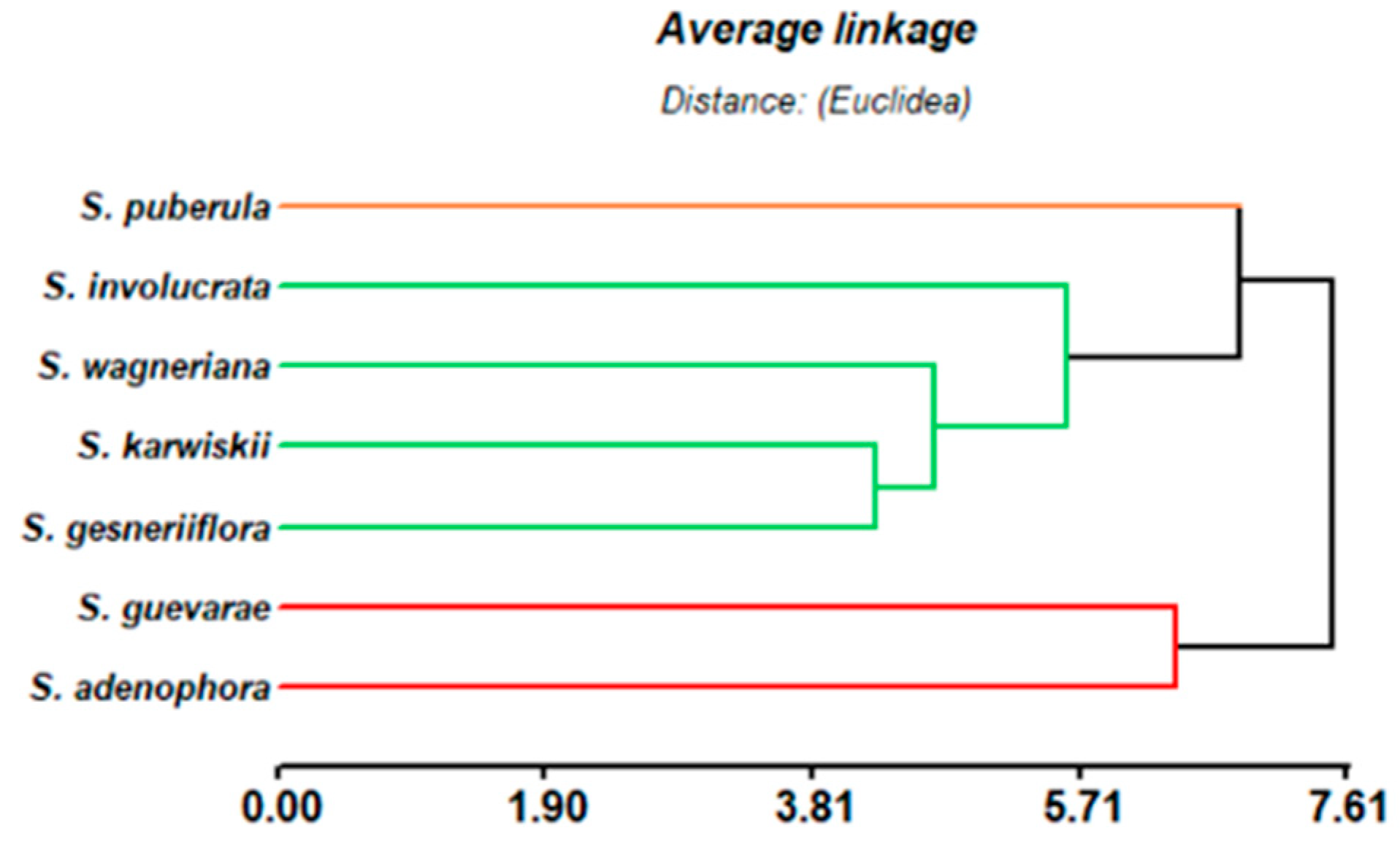
2.3. Taxonomic Considerations

In its original description, S. guevarae was classified in section Holwaya Ramamoorthy [9], together with the Mexican species S. holwayi Blake, S. karwinskii Benth., S. involucrata Cav., S. puberula Fernald, S. stolonifera Benth., and S. wagneriana Polak [8]. On the other hand, in 1991, during a taxonomic revision of section Nobiles Epling, S. adenophora Fernald, S. disjuncta Fernald, and S. gesneriflora Lindl & Paxton were transferred to section Holwaya [39]. Although from the taxonomic point of view, S. guevarae is closely related to S. karwinskii, S. wagneriana, S. involucrata, and S. holwayii, a hierarchical cluster analysis of the diterpenoid content of the members of section Holwaya phytochemically studied thus far, such as S. adenophora [40], S. gesneriflora [41,42], S. involucrata [43], S. puberula [44], S. karwinskii [45], and S. wagneriana [46], including the results obtained in this work from S. guevarae, indicated the presence of three groups that can be distinguished (Figure 14). S. puberula is, from the chemosystematic point of view, different from the rest of the species in section Holwaya, since S. puberula contents only rearranged isosalvipuberulane and salvipuberulane diterpenoids. On the other hand, S. wagneriana, S. karwinskii, S. involucrata, and S. gesneriflora constitute a second group with similar diterpenoid constituents. The third group indicates a close similarity between the diterpenoids obtained from S. adenophora and S. guevarae. Although no phylogenetical analysis has been conducted on S. guevarae, the oxidation and substitution pattern of the neo-clerodane described in this work support the inclusion of S. guevarae in section Holwaya.

3. Materials and Methods

3.1. General Experimental Procedures

The melting points (uncorrected) were determined on a Fisher Johns apparatus (Fisher Scientific Company, Pittsburgh, PA, USA). Optical rotations were measured on a PerkinElmer 341 polarimeter (PerkinElmer Inc., London, UK). The UV spectra were recorded on a Shimadzu UV 160U spectrophotometer (Shimadzu, Kyoto, Japan). The ECD spectra were measured using a JASCO J-1500 Circular Dichroism Spectrophotometer (JASCO Inc., Easton, MD, USA). The IR spectra were obtained on a Bruker Tensor 27 spectrometer (Bruker Corporation, Billerica, MA, USA); 1D and 2D NMR experiments were performed on a Bruker Advance III HD spectrometer (Bruker Corporation, Billerica, MA, USA) at 700 MHz for 1H and 175 MHz for 13C, a Bruker AVANCE III HD spectrometer (Bruker Corporation, Billerica, MA, USA) at 500 MHz for 1H and 125 MHz for 13C, or a Bruker Avance III (Bruker Corporation, Billerica, MA, USA) at 400 MHz for 1H and 100 MHz for 13C. CDCl3, (CD3)2CO, and CD3OD were used as solvents as indicated, and the chemical shifts were attributed to the residual solvents CHCl3 (δH = 7.26, δC = 77.16), CH3OH (δH = 3.31, δC = 49), and (CH3)2CO (δH = 2.05, δC = 29.84). The HR-DART-MS data were obtained on The AccuTOF JMS-T100LC mass spectrometer created by Jeol (Jeol Ltd., Tokyo, Japan) or the HPLC-ESI-QTOF-MS 1260-G6530 model (Agilent Tech. Santa Clara, CA, USA) using a column Zorbax Extend-C18 (Agilent, 50 × 3.0 mm × 3.5 μm), with the mobile phase as follows: A, H2O with CH3COOH 0.1% and B, CH3CN (in a gradient (0–12 min (85% A, 15% B): 100% B), 0.400 mL/min 600.00 bar). The X-ray data were collected on a Bruker APEX II DUO diffractometer (Bruker Corporation, Billerica, MA, USA). Silica gel 230–400 mesh (Macherey-Nagel; Düren, Germany) and Sephadex LH-20 (Pharmacia Biotech AB, Uppsala, Sweden) were used for Column Chromatography (CC). Precoated silica gel TLC plates (Merck, St. Louis, MO, USA) were used for thin layer chromatography (TLC).

3.2. Plant Materials

Salvia guevarae was collected at Xilitla, San Luis Potosi, Mexico, 21°24′34″ N, −99°5′9″ W, and 2100 m altitude, in October 2018. The plant material was identified by Dr. Brenda Y. Bedolla-Garcia and Dr. Sergio Zamudio and deposited in the Herbarium of the Instituto de Ecología, A. C., Centro Regional del Bajío (voucher IEB-266884).

3.3. Extraction and Isolation

The dried and powdered leaves of S. guevarae (361 g) were extracted via percolation with CH2Cl2 (2 L). The CH2Cl2 extract was concentrated at reduced pressures to yield 10.5 g of residue, a portion (9.5 g) of which was subjected to column chromatography (CC, 21 cm in length by 5 cm in internal diameter) on silica gel using a gradient elution with C6H14/CH3COOCH2CH3 (100:0-0:100); (CH3)2CO (100%); and finally, CH3OH as the mobile phase to obtain 80 eluates, 150 mL each, which were combined into 22 major fractions (A−U) after thin-layer chromatography (TLC) evaluation. The acetylation of fraction H (50 mg) and subsequent purification using preparative TLC on silica gel, eluting with C6H14/CH3COOCH2CH3 (80:20) as the mobile phase, gave 3β,28-O-diacetyl betulin (13.2 mg) and a mixture of 3β-O-acetyl oleanolic acid and 3β-O-acetyl betulinic acid (18.9 mg). Oleanolic and ursolic acids were identified as the constituents of fraction I (1296 mg). Fraction M (277 mg) was subjected to CC (35 × 2.5 cm) on silica gel, eluting with C6H14/CH3COOCH2CH3 (40:60) to obtain 25 subfractions, 50 mL each, which were combined into 8 major subfractions (MA−MH) based on a TLC evaluation. Subfraction MC (48 mg) was purified via preparative TLC on silica gel, eluting with CH2Cl2/CH3COOCH2CH3 (70:30) to give 13 (1.7 mg). Compound 10 (13.7 mg) was obtained as a solid from fraction N, and the mother liquors (369 mg) of this fraction were subjected to CC (20 × 35cm) on silica gel using C6H14/(CH3)2CO (80:20) as the eluent to obtain 14 subfractions (NA−NN) based on a TLC profile. Compound 7 (11 mg) was identified from fraction NC. Compound 10 (116 mg) was crystallized from C6H14/CH3COOCH2CH3 in fraction NJ. Subfraction NK (28.5 mg) was subjected to preparative TLC on silica gel, eluting with CH2Cl2/CH3COOCH2CH3 (8:2) to give 12 (2 mg). Crystals of compound 1 (711 mg) were obtained from fraction O, and the mother liquor’s residue (1079 mg) was subjected to CC on silica gel using CH2Cl2/(CH3)2CO (90:10) to obtain 18 subfractions (OA−OQ) based on a TLC evaluation. Subfraction OF (156 mg) was subjected to CC (32.5 × 2 cm) on Sephadex LH-20 using CH3OH as an eluent to obtain four subfractions (OFA−OFD) based on a TLC evaluation. Subfraction OFC (29 mg) was subjected to preparative TLC on silica gel, eluting with CH2Cl2/CH3COOCH2CH3 (70:30) to give 11 (8 mg). Subfraction OI (55 mg) was purified via TLC on silica gel in the C8 reverse phase (RP-8) using CH3OH/H2O (70:30) as the mobile phase to give 2 (30 mg). Compound 3β,20,25-trihydroxylupane (18 mg) was identified as the constituent of fraction ON. Fraction P (398 mg) was subjected to CC (15 × 1.8 cm) on Sephadex LH-20, eluting with methanol to obtain six subfractions (PA−PF) based on a TLC evaluation. Subfraction PC (261 mg) was subjected to CC (12 × 2 cm) on silica gel using C6H14/(CH3)2CO (50:50) to obtain 12 subfractions (PCA−PCL) based on a TLC evaluation. Compound 4 (3 mg) was purified from subfraction PCF via TLC on silica gel, eluting with C6H14/CH3COOCH2CH3/CH3OH/H2O (100:100:8:3.5). Compound 3 (410 mg) was obtained as a powder from fraction Q. Subfraction R (717 mg) was subjected to CC (22 × 2.5 cm) on silica gel using CH2Cl2/(CH3)2CO (60:40) to obtain 13 subfractions (RA−RM) after TLC evaluation. Subfraction RC (48 mg) was purified via preparative TLC using C6H14/C7H8/CH3COOCH2CH3 (30:30:40) as the mobile phase to obtain compound 6 (1.8 mg). Subfraction RD (146 mg) was purified via CC (11 × 1.8 cm) on silica gel using C6H14/CH3COOCH2CH3 (70:30) as the mobile phase to obtain 9 subfractions (RDA−RDI) based on a TLC evaluation. Compound 8 (61.8 mg) was obtained from subfraction RDE. Subfraction RF (100 mg) was purified via CC (11 × 2.5 cm) on silica gel using C6H14/CH3COOCH2CH3 (30:70) as an eluent to obtain 7 subfractions (RFA−RFG) based on a TLC profile. Compound 5 (4 mg) was obtained from subfraction RFD. Subfraction RFE (54 mg) was treated with anhydrous acetone (2 mL) and copper sulfate (120 mg) to obtain a mix of compounds. Compound 9 (2 mg) was obtained after purification via TLC on silica gel, eluting with CH2Cl2/(CH3)2CO (70:30) as the mobile phase.
The material previously extracted with dichloromethane was subjected to percolation with methanol to obtain 12.8 g of an extract. The methanolic extract was subjected to a liquid–liquid extraction process with a mixture of (C6H14:C6H6)/(CH3OH:H2O) (25:25:45:5), and the CH3OH:H2O fraction (11.4 g) was fractionated on a Sephadex LH 20 column using methanol as the mobile phase to obtain 7 fractions (A–G). Subfraction C (600 mg) was subjected to flash chromatography (14 × 5 cm) in the C18 reverse phase (RP-18), eluting with a mixture of CH3CN:H2O (0.1% formic acid) (5:100-100:0) to obtain 20 subfractions (CA-CT) based on an analytical TLC evaluation. Taxifolin-7-O-β-glucopyranoside (45 mg) was obtained from subfraction CF. Subfraction CI (35 mg) was purified via TLC on RP-18 using CH3COOCH2CH3/CH3OH (70:30) to give quercetin-3-O-β-xylopyranosyl-(1 → 2)-β-galactopyranoside (12 mg) and caffeic acid (2.5 mg). A mixture of 2R and 2S eriodictyol-7-O-β-glucopyranoside (1.8 mg) was obtained from subfraction CJ. Subfraction CN (20 mg) was subjected to TLC on RP-18 using CH3OH/H2O (40:60) as the mobile phase to give naranginin-7-O-β-glucopyranoside (6 mg) and rosmarinic acid (10 mg). 3-O-Methylrosmarinic acid was identified from subfraction CR.
Guevarain A (1): colorless crystals; mp 136−138 °C; [α] 25D +23.3 (c 0.0045, MeOH); UV (MeOH) λmax (log ε) 207 (3.16) nm; IR (ATR) νmax 3606, 2964, 2932, 2876, 1755, 1654, 1602, 1451, 1349, 1203, 1076, 998 cm−1; 1H and 13C NMR ((CD3)2CO): see Table 1; HR-DART-MS m/z 317.2103 [M + H − H2O]+; (calcd for C20H29O3, 317.2117).
Guevarain B (2): colorless crystals; mp 112−114 °C; [α]25D −5.6 (c 0.0006, MeOH); UV (MeOH) λmax (log ε) 214.4 (4.08), 239.8 (4.07), 227.8 (4.01) nm; ECD (c 3.01 mM, MeOH) [θ]257 +92349.6, [θ]278 0, [θ]325 −68602.6; IR (ATR) νmax 3417, 2957, 2930, 2869, 1747, 1644, 1607, 826 cm−1; 1H and 13C NMR (CDCl3): see Table 1; HR-DART-MS m/z 333.2063 [M+H]+; (calcd for C20H29O4, 333.2066).
2α,6α-Dihydroxy-patagonol (3): white powder; mp 98−100 °C; [α]25D +23.1 (c 0.0016, MeOH); UV (MeOH) λmax (log ε) 207.5 (6.45) nm; ECD (c 3.14 mM, MeOH) [θ]213 +6266.6, [θ]229 −4287.7, [θ]255 −5936.8, [θ]321 −2968.4; IR (ATR) νmax 3266, 1754, 1649, 1642 cm−1; 1H and 13C NMR (CD3OD): see Table 2; HR-DART-MS m/z 350.2090 [M-H]+; (calcd for C20H30O5, 349.2093).
2-Oxo-patagonol (4): white powder; mp 103−105 °C; [α]25D −25.6 (c 0.0018, MeOH); UV (MeOH) λmax (log ε) 218.0 (3.65) nm; ECD (c 5.41 mM, MeOH) [θ]215 −53430.8, [θ]244 +42876.6, [θ]322 −20448.8; IR (ATR) νmax 3411, 2927, 2872, 1747, 1651, 1050, 773 cm−1; 1H and 13C NMR (CDCl3): see Table 3; HR-DART-MS m/z 333.2066 [M+H]+; (calcd for C20H29O4, 333.2066).
6α-Hydroxy-2-oxo-patagonol (5): white powder; mp 92–94 °C; [α]25D +16 (c 0.001, MeOH); UV (MeOH) λmax (log ε) 241.2 (3.85) nm; ECD (c 2.87 mM, MeOH) [θ]245 +40897.7, [θ]284 0, [θ]326 21108.5; IR (ATR) νmax 3400, 2956, 2929, 2874, 1741, 1650, 1449, 1266, 1209, 1006, 733 cm−1; 1H and 13C NMR (CDCl3): see Table 3; HR-DART-MS m/z 349.2025 [M+H]+; (calcd for C20H29O5, 349.2015).
6α-Hydroxy-2-oxo-patagonal (6): white powder; mp 48−50 °C; [α]25D −12.7 (c 0.0011, MeOH); UV (MeOH) λmax (log ε) 241.4 (4.08) nm; ECD (c 3.17 mM, MeOH) [θ]251 +41557.3, [θ]295 0, [θ]338 −14512.1; IR (ATR) νmax 3439, 2958, 2931, 2876, 1744, 1676, 1449, 1348, 1261, 1206, 1072, 1048, 832 cm−1; 1H and 13C NMR (CDCl3): see Table 4; HR-DART-MS m/z 347.1859 [M + H]+; (calcd for C20H29O4, 347.1859).
2,6-Dioxo-guevarain (1a): Treatment of 1 (50 mg) with pyridinium chlorochromate (20.1 mg) in DCM gave a complex mixture, which was subjected to CC in silica gel using EtOAc as the mobile phase to afford guevarain B (2) (14.4 mg) and 1a (7 mg) as a white powder; mp 148−150 °C; [α]25D −36 (c 0.003, MeOH); UV (MeOH) λmax (log ε) 241.4 (4.08) nm; IR (ATR) νmax 3416, 1746, 1643, 1607 cm−1; 1H and 13C NMR (CDCl3): see Table 2; HR-DART-MS m/z 331.1898 [M + H]+; (calcd for C20H27O4, 331.1909).
6α-Hydroxy-2-oxo-patagonal (6): Treatment of 3 (24.8 mg) with pyridinium chlorochromate (15.25 mg) gave a complex mixture, which was subjected to CC in silica gel using EtOAc as the mobile phase to afford 5 (2.7 mg) and 6 (7.7 mg) as white powders.
Acetonide 7. Compound 3 (51.4 mg) was dissolved in freshly distilled acetone and treated with anhydrous CuSO4 (35 mg) and stirred for 1 h. A work-up of the reaction afforded 34.4 mg of a compound, identical in all aspects to 7.
Acetonide 9. The treatment of synthetic diterpenoid 7 (34 mg) was oxidized with 57 mg of PCC in DCM. After stirring at room temperature for 24 h, 26 mg (76%) of a compound identical to acetonide 9 was obtained.

3.4. Electronic Circular Dichroism Calculations

The compounds 26 and 1013 were constructed, and their geometry was optimized using a semiempirical method (PM3), as implemented in Spartan’10. A conformational analysis was performed using the same software and force field. All conformers were filtered and checked for redundancy. Subsequently, the conformers with relativity energies ≤ 2.0 or 2.5 Kcal/mol were minimized and optimized with Gaussian 09 using a DFT force field at the B3YLP/DGZVP level of theory for optimization and frequency. The ECD calculations for compounds 27 and 1214 in a MeOH solution were carried out by employing a TD-SCF force field at the B3LYP/6-31G(d) theory level, with the default solvent model. The calculated excitation energy (nm) and rotatory strength (R) in dipole velocity (Rvel) form were simulated into an ECD curve using Equation (1), as implemented in SpecDis software (Version 1.71), where E0k and R0k are the transition energy and rotatory strength of kth electronic transition, respectively, and σ is the exponential half width.
Δ ε = 1 2.297 × 10 39   ×   1 σ π k E 0 k R 0 k e x p [ { E E 0 k σ } 2 ]
All calculations were performed on the HP Cluster Platform 3000SL Miztli, a parallel supercomputer with a Linux operating system, containing 25,312 cores and a total of 45,000 GB of RAM.

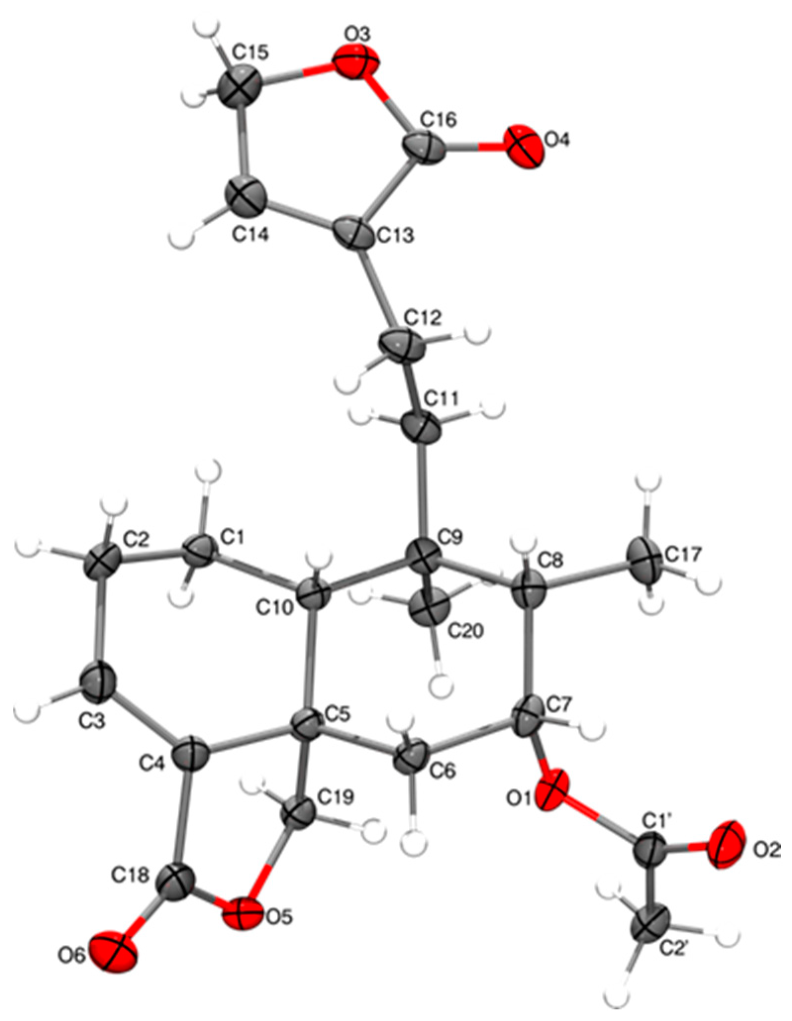
3.5. Single-Crystal X-Ray Diffraction Analysis of 1 and 10

The relevant details of the crystals, data collection, and structure refinement for compounds 1 and 10 can be found in Table S1. The data for guevarain A (1) and diterpenoid 10 were collected on a Bruker APEX II CCD Diffractometer at 100 K, using Cu-Ka radiation (k = 1.54178 Å) from an Incoatec ImuS source and Helios optic monochromator. Suitable crystals were coated with hydrocarbon oil, picked up with a nylon loop, and mounted in the cold nitrogen stream of the diffractometer. Frames were collected using ω scans and integrated with SAINT multi-scan absorption correction (SADABS). The structures were solved using direct methods and refined via full-matrix least-squares on F2 with SHELXL-2018 [47] using the SHELXLE GUI [48]. The hydrogen atoms of the C–H bonds were placed in idealized positions, the hydrogens of the O–H moiety and the hydrogens of the water molecules were found on a map of residual density, and their position was refined with Uiso = aUeq (where a is 1.5 for –CH3 and –OH moieties and 1.2 for others). The molecular graphics were prepared using the POV-RAY and GIMP programs. The crystallographic data for the crystal’s structures have been deposited at the Cambridge Crystallographic Data Centre under reference numbers CCDC 2,430,636 and CCDC 2430637 for compounds 1 and 10, respectively.
The crystal data of 1 were collected from a colorless prism (0.433 × 0.405 × 0.290 mm3) at 100(2) K: C20H32O5, MW = 352.45, monoclinic, space group P21, unit cell dimensions a = 8.78990(10) Ǻ, b = 10.45200(10) Ǻ, c = 10.27570(10) Ǻ, α = γ = 90°, β = 90.2546(4)°, V = 944.040(17) Ǻ3, Z = 2, Dc = 1.240 Mg/m3, F(000) = 384. A total of 33,896 reflections were collected in the range 4.302° < θ < 71.003°, with 3601 independent reflections [R(int) = 0.0304]; completeness to θmax was 99.8%. The structure was solved using direct methods and refined via full-matrix least squares on F2, with anisotropic temperature factors for non-hydrogen atoms converging at R final indices [I > 2σ(I)], R1 = 0.0266, wR2 = 0.0725; R indices (all data), R1 = 0.0266, wR2 = 0.0725. The absolute Flack, Hooft, and Parsons structure parameters were −0.02(3), −0.02(2), and −0.019(16), respectively, thus indicating that the absolute configuration of compound 1 is that depicted in Figure 3. The configuration of the chiral carbon atoms is 2R, 5R, 6S, 8R, 9S, and 10R.
Crystallographic data for compound 10 were collected from a colorless prism (0.401 × 0.319 × 0.278 mm3) at 100(2) K, orthorhombic, space group P212121 (Table S1). A total of 32,163 reflections were collected, and the structure was solved using direct methods. The absolute Flack, Hooft, and Parsons structure parameters were 0.01(5), 0.00(5), and 0.00(3), thus indicating that the absolute configuration of compound 10 is that depicted in Figure 13.

3.6. Chemometric Analysis

A cluster analysis was carried out using a complete chaining method. The data consisted of structural characteristics of 47 diterpenoids reported from the Salvia species classified in section Holwaya, including S. guevarae. The type of diterpenoid scaffolds, the unsaturation index, the oxidation pattern, and the presence of lactones or furan groups in the structure were analyzed. The analysis was carried out using InfoStat software (version 2017, released in 2020).

3.7. Antiproliferative Activity

The crude DCM extracts at 25 ppm and diterpenoids 13, 1a, 3, and 511 at 25 µM were evaluated in vitro against a panel of six human cancer cell lines: U251 (glioblastoma), PC-3 (prostate cancer), K562 (chronic myelogenous leukemia), HCT-15 (colon cancer), MCF-7 (breast cancer), and SKLU-1 (lung adenocarcinoma). The cell line COS-7 (monkey kidney) was used for the evaluation of the cytotoxicity in a normal cell line. The cell lines were supplied by the National Cancer Institute (USA) and American Type Culture Cells (ATCC). The cytotoxicity was evaluated using the protein-binding dye sulforhodamine B (SRB) in a microculture assay as previously reported by Monks et al. [7,49].

3.8. NO Inhibition in RAW 264.7 Macrophages

RAW 264.7 cells were obtained from the American Type Culture Collection (ATCC TIB-71), and cultured as previously described. Diterpenoids 13, 57, and 910 were dissolved in DMSO at 25 µM. The nitrite concentration in the medium was quantified as an indirect indicator of NO production following the Griess reaction and the experimental protocol previously reported [50]. Cell viability was evaluated via the MTT method, according to the protocol described by Mosmann and coworkers [51]. Table S4 shows the results obtained for the inhibition in nitrite production, and Table 6 shows the IC50 value for the most active compounds compared with those of aminoguanidine.

4. Conclusions

From the dichloromethane and methanolic extracts of the leaves of the new species Salvia guevarae, 13 neo-clerodanes diterpenoids (113) were isolated, of which 16 have yet to be described in the literature. The three acetonides 79 were artifacts produced via the reaction of diol derivatives with the acetone used in some parts of the separation process. The neo-clerodane diterpenoids 1013 were known diterpenoids previously described from different sources. Six triterpenoids were also isolated and identified: 3β,20,25-trihydroxylupane, oleanolic acid, 3β-O-acetyl-oleanolic acid, ursolic acid, 3β-O-acetyl-betulinic acid, and 3β,28-O-diacetyl-betulin. Additionally, from the methanol extract of S. guevarae, several phenolic derivatives were isolated and identified: quercetin-3-O-β-xylopyranosyl-(1 → 2)-β-galactopyranoside, taxifolin-7-O-β-glucopyranoside, naringenin-7-O-β-glucopyranoside, a mixture of 2R and 2S eriodictyol-7-O-β-glucopyranoside, caffeic acid, the methyl ester of rosmarinic acid, and rosmarinic acid. The structure of the isolated compounds was established by spectroscopic means, mainly 1H and 13C NMR, including 1D and 2D homo- and heteronuclear experiments and some chemical reactions. The antiproliferative activity of some diterpenoids was determined via the sulforhodamine B method, where guevarain B (2) and 6α-hydroxy patagonol acetonide (7) showed moderate activity against the K562 line, with IC50 (μM) = 33.0 ± 1.3 and 39.8 ± 1.5, respectively. The NO inhibition in the RAW 264.7 macrophage activity via nitrite formation was also determined for some compounds, where 2-oxo-patagonal (6), 6α-hydroxy patagonol acetonide (7), and 7α-acetoxy-ent-clerodan-3,13-dien-18,19:16,15-diolide (10) were proven to be active, with IC50 (μM) = 26.4 ± 0.4, 17.3 ± 0.5, and 13.7 ± 2.0, respectively.
The hierarchical cluster analysis of the diterpenoid content of the members of section Holwaya, including those obtained in this work, indicated a close phytochemical similarity between S. adenophora and S. guevarae, thus supporting the classification of the latter in section Holwaya.

Supplementary Materials

The following supporting information can be downloaded at https://www.mdpi.com/article/10.3390/molecules30071628/s1, Figures S1–S60: Copies of the 1D and 2D NMR spectra of compounds 19; Figures S61–S63: Comparison between the experimental and calculated ECD for the enantiomers of 11, 12, and 13; Tables S1 and S2: Crystallographic data for compounds 1 and 10; Table S3: Screening for the antiproliferative activity of the isolated compounds from Salvia guevarae (25 μM) and the dichloromethane extract of S. guevarae (25 ppm); Table S4: Screening for the inhibition of nitrite (NO) production for compounds 13 and 510 in the RAW 264.7 macrophages (25 μM); Spectroscopic data of compounds 713.

Author Contributions

Conceptualization, B.E.; methodology, J.P.T.-M., C.B.-B. and B.E.; data curation, J.P.T.-M., D.M.-O., B.Y.B.-G. and S.Z.; validation, J.P.T.-M., C.B.-B., L.Q. and B.E.; formal analysis, J.P.T.-M., T.R.-A. and D.M.-O.; investigation, J.P.T.-M., C.B.-B., L.Q. and B.E.; resources, L.Q. and B.E.; writing—original draft preparation, J.P.T.-M., C.B.-B., D.M.-O., L.Q. and B.E.; writing—review and editing, L.Q., B.E., B.Y.B.-G. and S.Z.; visualization, J.P.T.-M., C.B.-B., B.E., B.Y.B.-G. and D.M.-O.; supervision, C.B.-B., L.Q. and B.E.; project administration, J.P.T.-M., C.B.-B., L.Q. and B.E.; funding acquisition, L.Q. and B.E. All authors have read and agreed to the published version of the manuscript.

Funding

This research received no external funding.

Institutional Review Board Statement

Not applicable.

Informed Consent Statement

Not applicable.

Data Availability Statement

The crystallographic data for compounds 1 and 10 have been deposited at the Cambridge Crystallographic Data Centre under the reference numbers CCDC 2430636 and CCDC 2430637, respectively. Copies of the data can be obtained free of charge via application to the CCDC, 2 Union Road, Cambridge CB2 IEZ, UK. Fax: +44-(0)1223-336033 or email: deposit@ccdc.cam.ac.uk.

Acknowledgments

The authors acknowledge B. Quiroz, E. Huerta, A. Peña, R. A. Romo, C. Garcia, J. Pérez, and E. Tapia for collecting the NMR, UV, IR, and MS data. This study made use of UNAM’s NMR lab, LURMN at IQ-UNAM, which was funded under CONACYT-Mexico (project 0224747). J.P.T.-M. is grateful to CONACyT/CONAHCyT for the fellowship provided to pursue graduate studies (CONACyT CVU 968805).

Conflicts of Interest

The authors declare no conflicts of interest.

References

  1. Lara-Cabrera, S.I.; de la Luz Perez-Garcia, M.; Maya-Lastra, C.A.; Montero-Castro, J.C.; Godden, G.T.; Cibrian-Jaramillo, A.; Fisher, A.E.; Porter, J.M. Phylogenomics of Salvia L. Subgenus Calosphace (Lamiaceae). Front. Plant Sci. 2021, 12, 725900. [Google Scholar] [CrossRef]
  2. Hu, G.X.; Takano, A.; Drew, B.T.; Liu, E.; De Soltis, D.E.; Soltis, P.S.; Peng, H.; Xiang, C.L. Phylogeny and Staminal Evolution of Salvia (Lamiaceae, Nepetoideae) in East Asia. Ann. Bot. 2018, 122, 649–668. [Google Scholar] [CrossRef] [PubMed]
  3. González-Gallegos, J.G.; Bedolla-García, B.Y.; Cornejo-Tenorio, G.; Fernández-Alonso, J.L.; Fragoso-Martínez, I.; García-Peña, M.D.R.; Harley, R.M.; Klitgaard, B.; Martínez-Gordillo, M.J.; Wood, J.R.I.; et al. Richness and Distribution of Salvia Subg. Calosphace (Lamiaceae). Int. J. Plant Sci. 2020, 181, 831–856. [Google Scholar] [CrossRef]
  4. Jenks, A.A.; Kim, S.C. Medicinal Plant Complexes of Salvia Subgenus Calosphace: An Ethnobotanical Study of New World Sages. J. Ethnopharmacol. 2013, 146, 214–224. [Google Scholar] [CrossRef]
  5. Li, R.; Morris-Natschke, S.L.; Lee, K.H. Clerodane Diterpenes: Sources, Structures, and Biological Activities. Nat. Prod. Rep. 2016, 33, 1166–1226. [Google Scholar] [CrossRef] [PubMed]
  6. Bustos-Brito, C.; Nieto-Camacho, A.; Hernandez-Ortega, S.; Rivera-Chávez, J.; Quijano, L.; Esquivel, B. Structural Elucidation of Malonylcommunol and 6β-Hydroxy-Trans-Communic Acid, Two Undescribed Diterpenes from Salvia cinnabarina. First Examples of Labdane Diterpenoids from a Mexican Salvia Species. Molecules 2020, 25, 1808. [Google Scholar] [CrossRef]
  7. Bustos-Brito, C.; Torres-Medicis, J.P.; Bedolla-García, B.Y.; Zamudio, S.; Ramírez-Apan, T.O.; Macías-Rubalcava, M.L.; Quijano, L.; Esquivel, B. Structure, Absolute Configuration, Antiproliferative and Phytotoxic Activities of Icetexane and Abietane Diterpenoids from Salvia carranzae and Chemotaxonomic Implications. Molecules 2024, 29, 1226. [Google Scholar] [CrossRef]
  8. Bedolla-García, B.Y.; Zamudio, S. Nueva Especie de Salvia (Lamiaceae) del Centro de México. Phytoneuron 2017, 66, 1–12. [Google Scholar]
  9. Ramamoorthy, T.P. Typifications in Salvia (Lamiaceae). Taxon 1984, 33, 322–324. [Google Scholar]
  10. Adou, E.; Williams, R.B.; Schilling, J.K.; Malone, S.; Meyer, J.; Wisse, J.H.; Frederik, D.; Koese, D.; Werkhoven, M.C.M.; Snipes, C.E.; et al. Cytotoxic Diterpenoids from Two Lianas from the Suriname Rainforest. Bioorganic Med. Chem. 2005, 13, 6009–6014. [Google Scholar] [CrossRef]
  11. Bruhn, T.; Schaumlöffel, A.; Hemberger, Y.; Bringmann, G. SpecDis: Quantifying the Comparison of Calculated and Experimental Electronic Circular Dichroism Spectra. Chirality 2013, 25, 243–249. [Google Scholar] [CrossRef] [PubMed]
  12. Esquivel, B.; Hernandez, M.; Cardenas, J.; Ramamoorthy, T.P.; Rodríguez-Hahn, L. Further ent-Clerodane Diterpenoids from Salvia melissodora. Phytochemistry 1989, 28, 566. [Google Scholar] [CrossRef]
  13. Omosa, L.K.; Midiwo, J.O.; Derese, S.; Yenesew, A.; Peter, M.G.; Heydenreich, M. Neo-Clerodane Diterpenoids from the Leaf Exudate of Dodonaea angustifolia. Phytochem. Lett. 2010, 3, 217–220. [Google Scholar] [CrossRef]
  14. Yürüker, A.; Orjala, J.; Sticher, O.; Rali, T. Triterpenes from Rhus taitensis. Phytochemistry 1988, 48, 863–866. [Google Scholar]
  15. Ikuta, A.; Itokawa, H. The Triterpenes from Stauntonia hexaphylla Callus Tissues and Their Biosynthetic Significance. J. Nat. Prod. 1989, 52, 37. [Google Scholar] [CrossRef]
  16. Kolak, U.; Topçu, G.; Birteksöz, S.; Ötük, G.; Ulubelen, A. Terpenoids and Steroids from the Roots of Salvia blepharochlaena. Turk. J. Chem. 2005, 29, 177–186. [Google Scholar]
  17. Migas, P.; Cisowski, W.; Dembinska-Migas, W. Isoprene Derivatives from the Leaves and Callus Cultures of Vaccinium corymbosum Var. Bluecrop. Acta Pol. Pharmacéutica-Drug Res. 2025, 62, 45–51. [Google Scholar]
  18. Sun, I.-C.; Wang, H.-K.; Kashiwada, Y.; Shen, J.-K.; Cosentino, L.M.; Chen, C.-H.; Yang, L.-M.; Lee, K.-H. Anti-AIDS Agents. 34. Synthesis and Structure−Activity Relationships of Betulin Derivatives as Anti-HIV Agents. J. Med. Chem. 1998, 41, 4648–4657. [Google Scholar] [CrossRef]
  19. Symon, A.V.; Veselova, N.N.; Kaplun, A.P.; Vlasenkova, N.K.; Fedorova, G.A.; Lyutik, A.I.; Gerasimova, G.K.; Shvets, V.I. Synthesis and Antitumor Activity of Cyclopropane Derivatives of Betulinic and Betulonic Acids. Russ. J. Bioorganic Chem. 2005, 31, 286–291. [Google Scholar] [CrossRef]
  20. Melchior Larsen, L.; Kvist Nielsen, J. Identification of 3-O-[2-O-(β-D-Xylopyranosyl)-β-D-Glactopyranosil] Flavonoids in Horseradish Leaves Acting as Feeding Stimulants for a Flea Beetle. Phytochemistry 1982, 21, 1029–1033. [Google Scholar]
  21. Hofmann, R.W.; Swinny, E.E.; Bloor, S.J.; Markham, K.R.; Ryan, K.G.; Campbell, B.D.; Jordan, B.R.; Fountain, D.W. Responses of Nine Trifolium repens L. Populations to Ultraviolet-B Radiation: Differential Flavonol Glycoside Accumulation and Biomass Production. Ann. Bot. 2000, 86, 527–537. [Google Scholar] [CrossRef]
  22. Sikorska, M.; Matławska, I. Quercetin and Its Glycosides in the Flowers of Asclepias syriaca L. Acta Pol. Pharm. 2000, 57, 321–324. [Google Scholar]
  23. Olszewska, M.A.; Kwapisz, A. Metabolite Profiling and Antioxidant Activity of Prunus padus L. Flowers and Leaves. Nat. Prod. Res. 2011, 25, 1115–1131. [Google Scholar] [CrossRef]
  24. Iannuzzi, A.M.; Giacomelli, C.; De Leo, M.; Pietrobono, D.; Camangi, F.; De Tommasi, N.; Martini, C.; Trincavelli, M.L.; Braca, A. Antioxidant Activity of Compounds Isolated from Elaeagnus umbellata Promotes Human Gingival Fibroblast Well-Being. J. Nat. Prod. 2020, 83, 626–637. [Google Scholar] [CrossRef] [PubMed]
  25. Markham, K.R.; Webby, R.F.; Vilain, C. 7-O-Methyl-(2R: 3R)-Dihidroquercetin 5-O-B-D-Glucoside and Other Flavonooids from Podocarpus nivalis. Phytochemistry 1984, 23, 2049–2052. [Google Scholar] [CrossRef]
  26. Yeap Foo, L.; Karchesy, J.J. Polyphenolic Glycosides from Douglas Fir Inner Bark. Phytochemistry 1989, 28, 1237–1240. [Google Scholar] [CrossRef]
  27. Slimestad, R.; Andersen, Ø.M.; Francis, G.W. Ampelopsin 7-Glucoside and Other Dihydroflavonol 7-Glucosides from Needles of Picea abies. Phytochemistry 1994, 35, 550–552. [Google Scholar] [CrossRef]
  28. Álvarez-Fernández, M.A.; Hornedo-Ortega, R.; Cerezo, A.B.; Troncoso, A.M.; García-Parrilla, M.C. Determination of Nonanthocyanin Phenolic Compounds Using High-Resolution Mass Spectrometry (UHPLC-Orbitrap-MS/MS) and Impact of Storage Conditions in a Beverage Made from Strawberry by Fermentation. J. Agric. Food Chem. 2016, 64, 1367–1376. [Google Scholar] [CrossRef]
  29. Xu, J.; Yu, Y.; Shi, R.; Xie, G.; Zhu, Y.; Wu, G.; Qin, M. Organ-Specific Metabolic Shifts of Flavonoids in Scutellaria baicalensis at Different Growth and Development Stages. Molecules 2018, 23, 428. [Google Scholar] [CrossRef]
  30. Rayyan, S.; Fossen, T.; Solheim Nateland, H.; Andersen, M. Isolation and Identification of Flavonoids, Including Flavone Rotamers, from the Herbal Drug “Crateagi Folium Cum Flore” (Hawthorn). Phytochem. Anal. 2005, 16, 334–341. [Google Scholar] [CrossRef]
  31. Pan, J.; Zhang, S.; Yan, L.; Tai, J.; Xiao, Q.; Zou, K.; Zhou, Y.; Wu, J. Separation of Flavanone Enantiomers and Flavanone Glucoside Diastereomers from Balanophora involucrata Hook. f. by Capillary Electrophoresis and Reversed-Phase High-Performance Liquid Chromatography on a C18 Column. J. Chromatogr. A 2008, 1185, 117–129. [Google Scholar] [CrossRef]
  32. da Rosa, E.; do Amaral, Q.D.F.; Duarte, J.A.; Limberger, J.T.; Chaves, P.E.E.; Zuravski, L.; de Oliveira, L.F.S.; Machado, M.M. Antigenotoxic, Antimutagenic and Cytoprotective Potential of Salvia hispanica L. Seed Extract on Human Leukocytes Exposed to Oxidative Damage. J. Funct. Foods 2017, 38, 505–509. [Google Scholar] [CrossRef]
  33. Kim, M.H.; Jung, K.; Nam, K.H.; Jang, H.J.; Lee, S.W.; Kim, Y.; Park, C.S.; Lee, T.H.; Park, J.H.; Choi, J.H.; et al. Salvia Plebeia R.Br. Inhibits Signal Transduction of IL-6 and Prevents Ovariectomy-Induced Bone Loss by Suppressing Osteoclastogenesis. Arch. Pharm. Res. 2016, 39, 1671–1681. [Google Scholar] [CrossRef]
  34. Bahadori, M.B.; Asghari, B.; Dinparast, L.; Zengin, G.; Sarikurkcu, C.; Abbas-Mohammadi, M.; Bahadori, S. Salvia nemorosa L.: A Novel Source of Bioactive Agents with Functional Connections. LWT-Food Sci. Technol. 2017, 75, 42–50. [Google Scholar] [CrossRef]
  35. Petersen, M.; Simmonds, M.S.J. Molecules of Interest Rosmarinic Acid. Phytochemistry 2003, 62, 121–125. [Google Scholar] [CrossRef] [PubMed]
  36. Sharma, Y.; Velamuri, R.; Fagan, J.; Schaefer, J. Full-Spectrum Analysis of Bioactive Compounds in Rosemary (Rosmarinus officinalis L.) as Influenced by Different Extraction Methods. Molecules 2020, 25, 4599. [Google Scholar] [CrossRef] [PubMed]
  37. Forzato, C.; Nitti, P. New Diterpenes with Potential Antitumoral Activity Isolated from Plants in the Years 2017–2022. Plants 2022, 11, 2240. [Google Scholar] [CrossRef]
  38. Prakash, V. Terpenoids as Source of Anti-Inflamatory Compounds. Asian J. Pharm. Clin. Res. 2017, 10, 68. [Google Scholar] [CrossRef]
  39. Santos, E.P.; Harley, R.M. Notes on Salvia Section Nobiles (Lamiaceae) and Two New Species from Brazil. Kew Bull. 2004, 59, 103–109. [Google Scholar]
  40. Bisio, A.; Schito, A.M.; Ebrahimi, S.N.; Hamburger, M.; Mele, G.; Piatti, G.; Romussi, G.; Dal Piaz, F.; De Tommasi, N. Antibacterial Compounds from Salvia adenophora Fernald (Lamiaceae). Phytochemistry 2015, 110, 120–132. [Google Scholar] [CrossRef]
  41. Jimenez, M.; Moreno, E.D.; Díaz, E. Diterpenos de La Salvia gensneraefolia I. Estructuras de La Gensnerofolinas A y B. Rev. Latinoam. Quim. 1978, 10, 166–171. [Google Scholar]
  42. Esquivel, B.; Flores, E.A. A New neo-Clerodane Diterpenoid from Salvia gensneraeflora (Labiatae). Heterocycles 2001, 55, 505–509. [Google Scholar]
  43. Bustos-Brito, C.; Pérez-Juanchi, D.; Rivera-Chávez, J.A.; Hernández-Herrera, A.D.; Bedolla-García, B.Y.; Zamudio, S.; Ramírez-Apan, T.O.; Quijano, L.; Esquivel, B. Clerodane and 5,10-seco-Clerodane-Type Diterpenoids from Salvia involucrata. J. Mol. Struct. 2021, 1237, 130367. [Google Scholar] [CrossRef]
  44. Rodriguez-Hahn, L.; Esquivel, B.; Sánchez, A.-A.; Cárdenas, J.; Tovar, O.G.; Soriano-Garcia, M.; Toscano, A. Puberulin and Isopuberulin, Benzonorcaradiene and Benzocycloheptatriene Diterpenoids of Clerodanic Origin from Salvia puberula. J. Org. Chem. 1988, 53, 3933–3936. [Google Scholar]
  45. Esquivel, B.; Calderon, J.S.; Sánchez, L. Neo-Clerodane and Languidulane Diterpenoids from Salvia sousae and S. Karwinskii. Phytochemistry 1997, 45, 781–783. [Google Scholar] [CrossRef]
  46. Bisio, A.; De Tommasi, N.; Romussi, G. Diterpenoids from Salvia wagneriana. Planta Med. 2004, 70, 452–457. [Google Scholar] [CrossRef] [PubMed]
  47. Sheldrick, G.M. A Short History of SHELX. Acta Crystallogr. A 2008, 64, 112–122. [Google Scholar] [CrossRef] [PubMed]
  48. Hübschle, C.B.; Sheldrick, G.M.; Dittrich, B. ShelXle: A Qt Graphical User Interface for SHELXL. J. Appl. Crystallogr. 2011, 44, 1281–1284. [Google Scholar] [CrossRef]
  49. Monks, A.; Scudiero, D.; Skehan, P.; Shoemaker, R.; Paull, K.; Vistica, D.; Hose, C.; Langley, J.; Cronise, P.; Vaigro-Wolff, A.; et al. Feasibility of a High-Flux Anticancer Drug Screen Using a Diverse Panel of Cultured Human Tumor Cell Lines. J. Natl. Cancer Inst. 1991, 83, 757–766. [Google Scholar] [CrossRef]
  50. Del-Ángel, M.; Nieto, A.; Ramírez-Apan, T.; Delgado, G. Anti-Inflammatory Effect of Natural and Semi-Synthetic Phthalides. Eur. J. Pharmacol. 2015, 752, 40–48. [Google Scholar] [CrossRef]
  51. Mosmann, T. Rapid Colorimetric Assay for Cellular Growth and Survival: Application to Proliferation and Cytotoxicity Assays. J. Lmmunol. Methods 1983, 65, 55–63. [Google Scholar] [CrossRef] [PubMed]
Figure 1. Compounds isolated from S. guevarae.
Molecules 30 01628 g001
Figure 2. Selected COSY, HMBC, and NOESY correlations for 1.
Molecules 30 01628 g002
Figure 3. The molecular structure of compound 1. The thermal ellipsoids are set at 50% of the probability level.
Molecules 30 01628 g003
Figure 4. Hydrogen bonds in the packing structure found in compound 1.
Molecules 30 01628 g004
Figure 5. Selected COSY, HMBC, and NOESY correlations for 2.
Molecules 30 01628 g005
Figure 6. Comparison between the experimental (black line) and calculated ECD spectra for enantiomers (5R,6S,8R,9S,10R)-2 (blue line) and (5S,6R,8S,9R,10S)-2 (red line).
Molecules 30 01628 g006
Figure 7. Selected COSY, HMBC, and NOESY correlations for 3.
Molecules 30 01628 g007
Figure 8. Comparison between the experimental (black line) and calculated ECD spectra for enantiomers (2R, 5R, 6S, 8R, 9S, 10R)-3 (blue line) and (2S, 5S, 6R, 8S, 9R, 10S)-3 (red line).
Molecules 30 01628 g008
Figure 9. Selected COSY, HMBC, and NOESY correlations for 4.
Molecules 30 01628 g009
Figure 10. Comparison between the experimental (black line) and calculated ECD spectra for enantiomers (5R, 8R, 9S, 10R)-4 (blue line) and (5S, 8S, 9R, 10S)-4 (red line).
Molecules 30 01628 g010
Figure 11. Comparison between the experimental (black line) and calculated ECD spectra for enantiomers (5R, 6S, 8R, 9S, 10R)-5 (blue line) and (5S, 6R, 8S, 9R, 10S)-5 (red line).
Molecules 30 01628 g011
Figure 12. Comparison between the experimental (black line) and calculated ECD spectra for enantiomers (5R, 6S, 8R, 9S, 10R)−6 (blue line) and (5S, 6R, 8S, 9R, 10S)−6 (red line).
Molecules 30 01628 g012
Figure 13. The molecular structure of compound 10. The thermal ellipsoids are set at 50% of the probability level.
Molecules 30 01628 g013
Figure 14. Hierarchical cluster analysis of the Salvia species from section Holwaya with phytochemicals studies, including S. guevarae.
Molecules 30 01628 g014
Table 1. NMR data (700 MHz) of 1 and 2.
1 a2 b
PositionδCTypeδH, Multiplicity (J in Hz)δCTypeδH, Multiplicity (J in Hz)
129.2CH21.94, ddd (12.6, 7.0, 1.4)34.4CH22.48, dd (17.8, 13.9)
1.49, td (12.6, 10.1)2.32, dd (17.8, 3.6)
269.1CH4.13, m199.7C
3127.9CH5.19, brs126.5CH5.72, brs
4146.7C 172.8C
545.2C 45.5C
675.3CH3.48, dt (9.5, 5.6)73.5CH3.70, dd (11.1, 4.8)
738.7CH21.57, m37.9CH21.69 ddd(13.0, 4.7, 3.7)
1.62, ddd (13.0, 12.4, 11.1)
835.2CH1.70, m34.4CH1.73, m
938.8C 38.7C
1045.4CH1.35, dd (12.6, 1.4)44.9CH1.88, dd (13.9, 3.6)
1136.8CH21.62, ddd (14.6, 12.7, 5.1)35.0CH21.58, m
1.55, m1.47, ddd (14.6, 13.0, 4.9)
1219.4CH22.14, m18.7CH22.16, m
2.05, m2.00, m
13134.3C 134.3C
14146.2CH7.41, p (1.7) *144.0CH7.09, p (1.7) *
1571.0CH24.79, q (1.8)70.4CH24.76, p (1.7)
16174.7C 174.3C
1716.0CH30.84, d (6.8)15.5CH30.89, d (6.8)
1822.6CH31.84, dd (1.6, 1.7)23.0CH32.14, d (1.3)
1915.6CH31.06, s13.6CH31.12, s
2018.3CH30.75, s17.4CH30.84, s
2 OH 3.6, d (6.1)
6 OH 3.39, d (5.6)
a (CD3)2CO; b CDCl3; * 4J.
Table 2. NMR data of 1a and 3.
1a a3 b
PositionδCTypeδH, Multiplicity (J in Hz)δCTypeδH, Multiplicity (J in Hz)
134.2CH22.53, dd (17.0, 14.3)28.9CH22.02, ddt (12.5, 6.8, 1.3)
2.39, dd (17.0, 3.1)1.57, td (12.3, 10.3)
2197.8C 69.5CH4.23, m
3127.9CH5.82, brs129.6CH5.56, brs
4167.2C 149.4C
555.4C 45.6C
6210.7C 75.9CH3.58, m
743.7CH22.85, dd (12.7, 12.6)37.20CH21.64, m
2.19, dd (12.7, 4.0)
840.5CH2.13, m35.9CH1.76, m
939.3C 39.2C
1048.2CH2.25, dd (14.2, 3.2)45.6CH1.40, brd (12.3)
1135.4CH21.68, ddd (14.2, 12.7, 4.2)37.2CH21.68, ddd (13.7, 12.3, 4.9)
1.53, ddd (14.6, 12.7, 4.9)1.61, m
1219.1CH22.14, m19.7CH22.21, m
2.00, m2.18, m
13133.8C 134.7C
14144.5CH7.10, p (1.6) *147.4CH7.36, p (1.5) *
1570.3CH24.76, q (1.8)72.1CH24.82, q (1.7)
16174.1C 176.8C
1716.2CH31.01, d (6.6)16.0CH30.89, d (6.8)
1821.4CH32.17, d (1.3)65.5CH24.08, d (13.5)
4.23, brd (13.5)
1918.4CH31.51, s16.5CH31.13, s
2017.9CH31.06, s18.6CH30.79, s
a 500 MHz, b CDCl3, 700 MHz; CD3OD; * 4J.
Table 3. NMR data (500 MHz, CDCl3) of 4 and 5.
45
PositionδCTypeδH, Multiplicity (J in Hz)δCTypeδH, Multiplicity (J in Hz)
135.3CH22.44, dd (17.8, 13.5)34.7CH22.54, dd (17.8, 13.9)
2.37, dd (17.8, 4.3)2.37, dd (17.8, 3.4)
2200.1C 200.2C
3121.4CH6.10, br s125.4CH5.97, br s
4173.6C 170.6C
539.2,C 35.0C
634.6CH21.57, m73.8CH3.83, dd (8.7, 6.9)
1.45, m
726.7CH21.53, m37.0CH21.63, m
836.2CH1.58, m 1.55, m
938.9C 34.5CH1.70, m
1045.8CH1.98, dd (13.5, 4.3)45.1CH1.91, dd (13.9, 3.6)
1135.1CH21.54, m35.0CH22.51, ddd (13.7, 12.3, 4.9)
1.49, m
1218.9CH22.21, m18.8CH22.13, m
2.04, m1.96, m
13134.4C 134.2C
14143.9CH7.09, q (1.7)144.2CH7.10, p (1.4)
1570.4CH24.77, m70.4CH2477, q (1.7)
16173.6C 170.6C
1715.8CH30.87, d (6.4)14.8CH30.91, d (6.4)
1860.6CH24.40, dd (17.6, 1.8)65.1CH24.51, d (15.0)
4.35, dd (17.6, 1.8)4.37, br d (15.0)
1919.6CH31.20, s17.6CH31.22, s
2017.9CH30.85, s15.5CH30.85, s
Table 4. NMR data (500 MHz, CDCl3) of 6.
66
PositionδCTypeδH (J in Hz)PositionδCTypeδH (J in Hz)
135.3CH22.65 dd (18.1, 14.1)1135CH21.62, m
2.56, dd (18.1, 3.6)1.51, ddd (14.7, 12.8, 4.8)
2199.9C 1218.7CH22.18, tdd (12.9, 4.3, 2.0)
3141.1CH6.43, br s1.97, m
4163.8C 13134.1C
545.8C 14144.1CH7.10, br s
672.1CH3.75, dd (11.1, 4.7)1570.4CH24.77, q (2.1)
735.5CH2177, ddd (13.3, 4.8, 3.0)16174.2C
1.65, m1715.6CH30.84, d (6.6)
834.2CH1.71, m18197.5CH9.75, s
938.6C 1915.2CH31.13, s
1044.8CH1.95, dd (14.1, 3.5)2017.1CH30.78, s
6-OH 4.63, s
Table 5. IC50 (µM) values of the antiproliferative activity for compounds 2 and 7 against K562.
CompoundsIC50
233.1 ± 1.3
739.8 ± 1.5
Adriamycin0.2 ± 0.0
Table 6. IC50 (µM) values for the inhibition of nitrite production for compounds 7, 8, and 11.
CompoundIC50 (µM)
626.4 ± 0.4
717.3 ± 0.5
1013.7 ± 2.0
Aminoguanidine26.2 ± 0.4
The results are expressed as the mean ± S.E.M. of three determinations.
Disclaimer/Publisher’s Note: The statements, opinions and data contained in all publications are solely those of the individual author(s) and contributor(s) and not of MDPI and/or the editor(s). MDPI and/or the editor(s) disclaim responsibility for any injury to people or property resulting from any ideas, methods, instructions or products referred to in the content.

