Review

Noncovalent Labeling of Biomolecules with Red and Near- Infrared Dyes

Department of Chemistry, Georgia State University, University Plaza, Atlanta, Georgia 30303, USA
*
Author to whom correspondence should be addressed.
Molecules 2004, 9(3), 40-49; https://doi.org/10.3390/90300040
Submission received: 25 February 2004 / Accepted: 27 February 2004 / Published: 28 February 2004
(This article belongs to the Special Issue Biologically Relevant Heterocyclic Compounds)

Abstract

:
Biopolymers such as proteins and nucleic acids can be labeled with a fluorescent marker to allow for their detection. Covalent labeling is achieved by the reaction of an appropriately functionalized dye marker with a reactive group on a biomolecule. The recent trend, however, is the use of noncovalent labeling that results from strong hydrophobic and/or ionic interactions between the marker and biomolecule of interest. The main advantage of noncovalent labeling is that it affects the functional activity of the biomolecule to a lesser extent. The applications of luminescent cyanine and squarylium dyes are reviewed.

Introduction

Optical probes for labeling of biomolecules have been widely used in science and clinical analysis. The label molecule may contain a reactive functional group for a covalent attachment to the biomolecule of interest. Alternatively, a non-functionalized marker molecule may exhibit a strong noncovalent interaction with the biomolecule that allows for the formation of a stable complex. The noncovalent labeling can involve ionic, electrostatic, hydrophobic, and hydrogen bonding interactions. Although the interactions resulting from noncovalent labeling are substantially less stable than the covalent linkages, they occur at a faster rate and at a physiological pH range. Purification steps are usually unnecessary when the stoichiometry of the complex is known or if noncovalent labeling is used in conjunction with separation techniques, e.g. capillary electrophoresis. Another disadvantage of covalent labeling is that analyte molecules often have varying numbers of dyes molecules attached to them, leading to band broadening [1]. In both methodologies the trend is to use dye labels that absorb and fluoresce in the red (600-700 nm) or near-infrared (>700 nm) regions of the electromagnetic spectrum. The far-wavelength detection takes advantage of low interference from solvents and the biological matrix in this region.
Patonay and co-workers [2] were among the first researchers to study the noncovalent method of labeling human serum albumin (HSA) with near-infrared dyes by using high-performance liquid chromatography (HPLC) and absorption detection. While high detection sensitivity has been achieved, this method has been found damaging to size-exclusion columns because of the presence of free unassociated dye. It has been found that the use of a nonionic detergent, such as Triton-X, minimizes the unwanted effects of free dyes on HPLC columns.
Fluorescence detection offers a much greater sensitivity than the measurements based on absorption, especially when coupled with a novel analytical technique of capillary electrophoresis (CE). The CE technique involves the application of high voltage across buffer-filled capillaries to achieve separation. In particular, laser induced fluorescence (LIF) is a very sensitive detection method for CE. The LIF method involves the measurements of the optical emission from molecules that have been excited to higher energy levels by absorption of electromagnetic radiation from an appropriate laser source. As already noted, the main advantage of fluorescence detection compared to absorption measurements is much greater sensitivity. This is due to the fact that the fluorescence signal encounters very low background fluorescence and noise, especially in the NIR region. For molecules that can be excited at the output wavelength of the laser, LIF may provide highly selective excitation of the analyte to reduce interferences.
Ideally, the fluorescent probe used to label biomolecules should have a good quantum yield and a large Stokes' shift (a difference between the fluorescence and absorption maxima), and not be significantly affected by the presence of fluorescence quenching molecules. Quenchers may be present in complex biological mixtures. Noncovalent labels frequently are shielded by the biomolecule from those quenching effects, hence adding additional advantage. Practical fluorescent markers developed recently for noncovalent labeling of biomolecules are squarylium and cyanine dyes.

Squarylium Dyes

Zwitterionic non-symmetrical squarylium dyes are synthesized from readily available starting materials, as exemplified in Scheme 1 by the preparation of compounds 1-4 [3,4]. A similar methodology has been used in the synthesis of 5 and 6. On the other hand, symmetrical squaraines are easily accessible by condensation of squaric acid (3,4-dihydroxycyclobut-3-ene-1,2-dione) with N,N-disubstituted anilines [5] or heterocyclic azonium salts containing an activated methyl group at the position adajacent to the quarternized ring nitrogen atom [6]. For example, condensation of 3-ethyl-1,1,2-trimethyl-1H-benz[e]indolium iodide with squaric acid yields indolium derivative 7.
Scheme 1.
Scheme 1.
Molecules 09 00040 g004
In general, squarylium dyes absorb in the red region of the visible spectrum and are weakly fluorescent in solution. They show a strong noncovalent binding with proteins with a concomitant bathochromic shift in absorption (up to 90 nm) in the complex. Importantly, due to immobilization, they become highly fluorescent in the complexes [7,8,9,10,11]. The highest increases in fluorescence quantum yields have been reported for binding of 3 and 6 to human serum albumin (HSA), from 0.03 and 0.02 in solution to 0.92 and 0.97 in the complex with HSA, respectively. An extremely large value of association constant of 107 M-1 has been determined for noncovalent binding of 7 to HSA and greatly increased fluorescence efficiency upon binding has been observed by using the CE-LIF technique. Following binding studies of 7 with additional proteins, it has been concluded that dye 7 is an excellent reagent capable of fluorescently labeling analyte proteins for analysis by CE-LIF without itself being significantly fluorescent in solution in the absence of proteins [4].

Cyanine Dyes

As shown by the general structure 8, cyanine dyes are cationic molecules in which two heterocyclic units are joined by a polyene chain [12,13,14].
Molecules 09 00040 i001
Their common names depend on the number of methine groups in the polyene chain. For example, compounds 8 with n = 0 and n = 3 are referred to as monomethine and heptamethine cyanines, respectively. Particular examples of monomethine cyanine dyes are 9-11 (Scheme 2), and heptamethine analogs 12-14 are shown in Scheme 3. While there is a plethora of well developed synthetic routes to monomethine and trimethine cyanines [14], the pentamethine, heptamethine and longer analogs are preferably synthesized by condensation of methyl-substituted quaternized heterocyclic compounds with an α,ω-dialdehyde or equivalent (Scheme 3) [14,15,16,17,18]. The monomethine cyanines show absorption in the visible region, and each extension of the chromophore by one vinylene moiety (CH=CH) causes a bathochromic shift of about 100 nm [18].
Scheme 2.
Scheme 2.
Molecules 09 00040 g005
Substituents at the chromophore cause additional shifts in absorption. Of particular interest are facile displacement reactions of the chlorine atom in heptamethine cyanines, such as 13, in the presence of nucleophiles that are good single-electron donors. These nucleophilic displacement reactions proceed by an SRN1 mechanism, and a reduction product, such as 14, can also be obtained efficiently from an intermediate cation radical [17]. Depending on the nucleophile, compound 13 and similar chloro-substituted heptamethine cyanine dyes can be transformed into derivatives with an absorption range spanning from 615 nm for a methylamino-substituted derivative to 800 nm for diphenylphosphino substitution [16,17]. An unusual property of these dyes derived from 13 and analogs is an extremely high extinction coefficient of up to 260,000 M-1cm-1.
Scheme 3.
Scheme 3.
Molecules 09 00040 g006
As with squarylium dyes, the fluorescence efficiency of the conformationally flexible cyanine counterparts is greatly enhanced upon immobilization in complexes with biomolecules. Polycationic cyanine derivatives including 9-11 have been synthesized for a strong binding to nucleic acids [19,20,21,22,23,24,25]. On the other hand, dyes 12-14 with a net charge of –1 show strong interaction with proteins [26,27,28,29,30]. The binding studies are discussed in more detail as follows.
The dicationic cyanine 9 shows absorption at 515 nm and is not fluorescent in aqueous solution. However, a strong fluorescence is observed in the presence of double-stranded (ds) DNA [19]. Its tetracationic analog 10 and similar homodimers with different linkages between the terminal cyanine moieties show even stronger noncovalent binding with ds DNA. In the presence of ds DNA the Stokes' shifts of about 30 nm and the fluorescence quantum yields in the range 0.48-0.96 have been observed. These homodimers are bisintercalators with selectivity toward the 5'-CTGAG-3' binding site of oligonucleotides [21,24,25]. Interestingly, compound 10 and analogs also bind strongly with single-stranded (ss) DNA. In the complexes with ss DNA they also fluoresce strongly with quantum yields in the range 0.3-0.8 [21]. The isoxazolium homodimer 11 is less valuable for noncovalent labeling of DNA due to hypsochromically shifted absorption relative to that of 10 [22].
The use of indocyanine green (12 in Scheme 3) for determination of human serum albumin (HSA) [26,27] and a number of other proteins [27] by capillary electrophoresis with diode laser-induced fluorescence detection (CE-LIF) has been investigated recently. This negatively charged dye shows a strong absorption at 780 nm and a weak fluorescence at 820 nm in aqueous solution. Its binding with proteins results in a bathochromic shift of absorption and a substantial enhancement of fluorescence. All studied proteins have been successfully resolved in a short period of time without any interference from excess, unbound dye.
In general, the capillary electrophoresis based methods for binding constant studies require the use of multiple runs at varying concentrations of either drug or protein. Therefore, the methods are time consuming and demanding, requiring large amounts of reagents. A novel dye-displacement capillary electrophoresis based method for determining binding constants of drugs to subdomain IIIA of human serum albumin has been developed in our laboratories. The method uses a competitive-type interaction between the drug of interest and a dye probe, as a noncovalent label for HSA, to arrive at a binding constant. A heptamethine cyanine dye 13 has been used as a probe [28,29].
The first step in the development of the CE-based dye displacement assay was to analyze the stoichiometry of the dye:protein complex and the change in quantum yield of the dye upon complexation. Since a predominant stoichiometry of NIR dye-protein complex could not be determined due to a peak broadening, this result suggests that there may be multiple dye-protein complexes present. The change in quantum yield of the dye upon complexation with HSA was found to be increased by an order of magnitude.
The next step of the experiment involved determination of binding constants. The noncovalent labeling of HSA was achieved by mixing the appropriate volumes of the dye and protein solutions. The sample was vortex-mixed for two minutes prior to injection (Figure 1). Various dye-protein ratios and concentrations were used. Dye displacement experiments were achieved by addition of an appropriate drug solution to an equilibrated dye-protein solution (Figure 2). The electrophoretic separation conditions were optimized in such a way that they would permit complete resolution of free dye from the dye complexed with HSA. Such separations have the potential to be problematic due to adsorption effects between the untreated silica capillary and the protein [30]. Adjusting the pH of run buffers to pH values greater than the pI of the protein has been shown to reduce protein adsorption to the capillary wall. It was found that a pH 8.5 borate buffer provided the best results.
Figure 1. Illustration of noncovalent labeling of HSA with a dye.
Molecules 09 00040 g001
When a drug is introduced into an equilibrated mixture of the dye and protein, there are two possible pathways by which the dye and protein can interact. The example of noncompetitive interactions is shown in Figure 2. They occur when the dye and drug bind at different sites on the protein. While noncompetitive interactions do not allow for the quantification of binding constants, they do serve some purpose concerning the binding site of the dye. It was found that the dye and warfarin, which is known to bind at site I on HSA, do not make any competitive interactions. From that, it is obvious that the dye 13 does not bind at site I.
Figure 2. Illustration of noncompetitive binding of the drug and dye with HSA.
Molecules 09 00040 g002
Figure 3 shows the idea behind competitive interactions between the drug and dye. The drug and dye bind at the same site on HSA and, due to a deficit of binding areas relative to drug, compete for binding. Therefore, drug binding may be monitored and measured through changes in either the free dye concentration or the amount of dye bound to protein. It was found that the degree to which the drugs displace the dye is related to the drug’s binding constants with HSA.
Figure 3. Illustration of competitive binding of the drug and dye with HSA.
Molecules 09 00040 g003
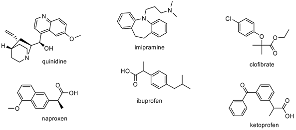
Dye displacement was calculated by using the difference of the peak areas of the dye-protein peaks in the presence and absence of drug. Four drugs known to bind to subdomain IIIA of HSA, clofibrate, imipramine, quinidine, and ketoprofen were chosen for their wide range of binding constants [29]. Determined binding constants are shown in Table 1. The binding constants of ibuprofen and naproxen were estimated using the dye displacement assay. The determined constants are in line with results found in the literature [31,32].
Table 1. Binding constants of drugs to HSA Molecules 09 00040 i002
DrugBinding Constant [M-1]DrugBinding Constant [M-1]
CE MetdodaOtder metdodsbCE MetdodaOtder metdodsb
Quinidine7.9x1041.6x103 Naproxen8x1051.2x106
Imipramine1.9x1052.5x104 Ibuprofen1x1062.7x106
Clofibrate7.6x1057.6x105Ketoprofen3.8x106__
a Ref. [28], b Refs. [31,32]

Conclusions

Over the past decade, the applicability of capillary electrophoresis based dye labeling techniques for detection and separation of biomolecules has been shown. The noncovalent labeling occurs fast at a physiological pH range. Advantages of the near-infrared dye displacement method for the determination of drug binding constants to human serum albumin were presented. The dye displacement technique utilizes both competitive and noncompetitive binding between drug and dye to HSA. The method is fast and sensitive, requires small amounts of reagents, and is amenable to automation.

References

  1. Craig, D. B.; Dovichi, N. J. Multiple Labeling of Proteins. Anal. Chem. 1998, 70, 2493–2494. [Google Scholar]
  2. Williams, R. J.; Lipowska, M.; Patonay, G.; Strekowski, L. Comparison of covalent and noncovalent labeling with near-infrared dyes for the high-performance liquid chromatographic determination of human serum albumin. Anal. Chem. 1993, 65, 601–605. [Google Scholar]
  3. Yagi, S.; Hyodo, Y.; Matsumoto, S.; Takahashi, N.; Kono, H.; Nakazumi, H. Synthesis of novel unsymmetrical squarylium dyes absorbing in the near-infrared region. J. Chem. Soc., Perkin Trans. 1 2000, 599–603. [Google Scholar]
  4. Welder, F.; Beverly, P.; Nakazumi, H.; Yagi, S.; Colyer, Ch. L. Symmetric and asymmetric squarylium dyes as noncovalent protein labels: a study by fluorimetry and capillary electrophoresis. J. Chromatogr. B. 2003, 793, 93–105. [Google Scholar]
  5. Bello, K. A.; Ajayi, J. O. Near-Infrared absorbing squarylium dyes. Dyes and Pigments 1996, 31, 79–87. [Google Scholar]
  6. Tong, L.; Bixian, P. The influence of N-alkyl groups on the properties of squarylium cyanine dyes. Dyes and Pigments 1998, 391, 201–209. [Google Scholar]
  7. Terpetsching, E.; Szmacinski, H.; Lakowicz, J. R. Synthesis, spectral properties and photostabilities of symmetrical and unsymmetrical squarines; a new class of flourophores with long-wavelength exiciation and emmision. Anal. Chim. Acta 1993, 282, 633–641. [Google Scholar]
  8. Meadows, F.; Narayanan, N.; Patonay, G. Determination of protein–dye association by near infrared fluorescence-detected circular dichroism. Talanta 2000, 50, 1149–1155. [Google Scholar]
  9. Sophianopoulos, A. J.; Lipowski, J.; Narayanan, N.; Patonay, G. Association of near-infrared dyes with bovine serum albumin. Appl. Spectrosc. 1997, 51, 1511–1515. [Google Scholar]
  10. Nakazumi, H.; Colyer, Ch. L.; Kaihara, K.; Yagi, S.; Hyodo, Y. Red luminescent squarylium dyes for noncovalent HSA labeling. Chem. Lett. 2003, 32, 804–805. [Google Scholar]
  11. Swamy, A. R.; Mason, J. C.; Lee, H.; Meadows, F.; Baars, M.; Strekowski, L.; Patonay, G. Encyclopedia of Analytical Chemistry; Wiley: London, 2000; p. 11. [Google Scholar]
  12. Tyutyulkov, N.; Fabian, J.; Mehlhorn, A.; Dietz, F.; Tadjer, A. Polymethine Dyes: Structure and Properties; St. Kliment Ohridski University Press: Sofia, Bulgaria, 1991. [Google Scholar]
  13. Fabian, J.; Nakazumi, H.; Matsuoka, M. Near-infrared absorbing dyes. Chem. Rev. 1992, 92, 1197–1226. [Google Scholar]
  14. Hammer, F. M. The Cyanine Dyes and Related Compounds; Wiley: New York, 1964. [Google Scholar]
  15. Narayanan, N.; Strekowski, L.; Lipowska, M.; Patonay, G. A New method for the synthesis of heptamethine cyanine dyes: Synthesis of new near-infrared fluorescent labels. J. Org. Chem. 1997, 62, 9387. [Google Scholar]
  16. Strekowski, L.; Lipowska, M.; Patonay, G. Facile derivatizations of heptamethine cyanine dyes. Synth. Commun. 1992, 22, 2593–2598. [Google Scholar]
  17. Strekowski, L.; Lipowska, M.; Patonay, G. Substitution reactions of a nucleofugal group in heptamethine cyanine dyes. Synthesis of an isothiocyanato derivative for labeling of proteins with a near-infrared chromophore. J. Org. Chem. 1992, 57, 4578–4580. [Google Scholar]
  18. Mishra, A.; Behera, R. K.; Behera, P. K.; Mishra, B. K.; Behera, G. B. Cyanines during the 1990s: a review. Chem. Rev. 2000, 100, 1973–2011. [Google Scholar]
  19. Deligeorgiev, T. G.; Zaneva, D. A.; Kim, S. H.; Sabnis, R. W. Preparation of monomethine cyanine dyes for nucleic acid detection. Dyes and Pigments 1998, 37, 205–211. [Google Scholar]
  20. Gadjev, N. I.; Deligeorgiev, T. G.; Kim, S. H. Preparation of monomethine cyanine dyes as noncovalent labels for nucleic acids. Dyes and Pigments 1999, 40, 181–186. [Google Scholar]
  21. Deligeorgiev, T. G.; Gadjev, N. I.; Timtcheva, I. I.; Maximova, V. A.; Katerinopoulos, H. E.; Foukaraki, E. Synthesis of homodimeric monomethine cyanine dyes as noncovalent nucleic acid labels and their absorption and fluorescence spectral characteristics. Dyes and Pigments 1999, 44, 131–136. [Google Scholar]
  22. Gadjev, N. I.; Deligeorgiev, T. G.; Timcheva, I.; Maximova, V. Synthesis and properties of YOYO-1-type homodimeric monomethine cyanine dyes as noncovalent nucleic acid labels. Dyes and Pigments 2003, 57, 161–164. [Google Scholar]
  23. Rye, H.S.; Yue, S.; Wemmer, D.E.; Quesada, M.A.; Haugland, R.P.; Maties, R.A.; Glazer, A.N. Stable fluorescent complexes of double-stranded DNA with bis-intercalating asymmetric cyanine dyes: properties and applications. Nucleic Acids Res. 1992, 20, 2803–2812. [Google Scholar]
  24. Stark, D.; Hamed, A. A.; Pedersen, E. B.; Jacobsen, J. P. Bisintercalation of homodimeric thiazole orange dyes in DNA: effect of modifying the linker. Bioconjugate Chem. 1997, 8, 869–877. [Google Scholar]
  25. Jacobsen, J. P.; Pedersen, J. B.; Hansen, L. F.; Wemmer, D. E. Site selective bis-intercalation of a homodimeric thiazole orange dye in DNA oligonucleotides. Nucleic Acids Res. 1995, 23, 753–760. [Google Scholar]
  26. Moody, E. D.; Viskari, P. J.; Colyer, Ch. L. Non-covalent labeling of human serum albumin with indocyanine green: a study by capillary electrophoresis with diode laser-induced fluorescence detection. J. Chromatogr. B. 1999, 729, 55–64. [Google Scholar]
  27. McCorquodale, E. M.; Colyer, Ch. L. Indocyanine green as a noncovalent, pseudofluorogenic label for protein determination by capillary electrophoresis. Electrophoresis 2001, 22, 2403–2408. [Google Scholar]
  28. Sowell, J. D. The use of covalent and noncovalent labeling schemes for the study of bioaffinity interactions via capillary electrophoresis with near-infrared laser induced fluorescence detection. PhD Dissertation, Georgia State University, Atlanta, 2003. [Google Scholar]
  29. Andrews-Wilberforce, D.; Patonay, G. Investigation of near-infrared laser dye albumin complexes. Spectrochim. Acta 1990, 46A, 1153–62. [Google Scholar]
  30. Legendre, B. L., Jr.; Soper, S. A. Binding properties of near-IR dyes to proteins and separation of the dye/protein complexes using capillary electrophoresis with laser-induced fluorescence detection. Appl. Spectrosc. 1996, 50, 1196–1202. [Google Scholar]
  31. Peters, T. All About Albumin: Biochemistry, Genetics, and Medical Applications; Academic Press: San Diego, 1996. [Google Scholar]
  32. Borga, O.; Borga, B. Serum protein binding of nonsteroidal antiinflammatory drugs: a comparative study. J. Pharmacokinet. Biopharm. 1997, 25, 63–77. [Google Scholar]