Review

Urinary Bladder Cancer Susceptibility Markers. What Do We Know about Functional Mechanisms?

by
Aleksandra M. Dudek
1,2,3,*,
Anne J. Grotenhuis
2,4,
Sita H. Vermeulen
2,4,
Lambertus A. L. M. Kiemeney
1,2,4 and
Gerald W. Verhaegh
1,3
1
Department of Urology, Radboud University Medical Centre, Geert Grooteplein 16, Nijmegen 6525 GA, The Netherlands
2
Department for Health Evidence, Radboud University Medical Centre, Geert Grooteplein 21, Nijmegen 6525 EZ, The Netherlands
3
Nijmegen Centre for Molecular Life Sciences, Geert Grooteplein 28, Nijmegen 6525 GA, The Netherlands
4
Nijmegen Centre for Evidence Based Practice, Geert Grooteplein 21, Nijmegen 6525 GA, The Netherlands
*
Author to whom correspondence should be addressed.
Int. J. Mol. Sci. 2013, 14(6), 12346-12366; https://doi.org/10.3390/ijms140612346
Submission received: 6 May 2013 / Revised: 23 May 2013 / Accepted: 30 May 2013 / Published: 10 June 2013
(This article belongs to the Special Issue Molecular Research in Urology)

Abstract

:
Genome-wide association studies (GWAS) have been successful in the identification of the several urinary bladder cancer (UBC) susceptibility loci, pointing towards novel genes involved in tumor development. Despite that, functional characterization of the identified variants remains challenging, as they mostly map to poorly understood, non-coding regions. Recently, two of the UBC risk variants (PSCA and UGT1A) were confirmed to have functional consequences. They were shown to modify bladder cancer risk by influencing gene expression in an allele-specific manner. Although the role of the other UBC risk variants is unknown, it can be hypothesized—based on studies from different cancer types—that they influence cancer susceptibility by alterations in regulatory networks. The insight into UBC heritability gained through GWAS and further functional studies can impact on cancer prevention and screening, as well as on the development of new biomarkers and future personalized therapies.

1. Introduction

For many years, the identification of disease-susceptibility genes was based on linkage studies in high-risk families. Although rare, high-penetrance mutations have been identified, most of the genetic susceptibility in sporadic cases remained unexplained. Recently, however, genome-wide association studies (GWAS) have been performed for many complex diseases, including cancer. Cancer is a multifactorial disease, arising from the complex interplay between genetic and environmental factors. The GWAS approach allows a search for novel susceptibility loci throughout the genome in a hypothesis-free manner [1]. Most of the identified variants are common and confer small risk (odds ratios between 1.1 and 1.3). However, the involvement of many low-penetrance loci can explain differences in cancer susceptibility between individuals [2]. Despite the large number of identified risk variants, only a modest number of functional studies have so far been published. The majority of identified genetic variants map to non-coding regions in the genome and require fine-mapping, and therefore finding the causative variants, as well as understanding their functionality and role in cancer development, remains challenging [3].
Urinary bladder cancer (UBC) is a common malignancy of the urinary tract, with 180,500 estimated new cases each year and 38,200 deaths in the European Union [4], and with 73,510 estimated new cases and 14,880 deaths in United States [5]. Despite many years of research and the identification of several genes involved in bladder cancer pathogenesis, the large part of its heritability remains unknown [6]. In the past, many hypothesis-driven candidate gene studies were performed. However the majority of results were not reproduced, with the exception of GSTM1 and NAT2 [1]. GWAS has successfully identified several bladder cancer risk variants. Understanding the functional consequences of these single nucleotide polymorphisms (SNPs) will help in understanding the role of the identified variants in bladder carcinogenesis. This can lead to the development of new biomarkers or therapies for selected patients, making personalized medicine for bladder cancer reality [7].
The aim of this review is to give a comprehensive overview of the bladder cancer risk of SNPs and the functional consequences of these genetic variants. We will include some functional studies performed for the risk SNPs that are located in UBC susceptibility regions, but were performed in relation to other cancer types. Finally, we will suggest some future research lines that will improve our understanding of the heritability underlying UBC.

2. Bladder Cancer

Urinary bladder cancer occurs frequently, with the highest incidence rates in developed countries. It is typically diagnosed in older patients (55 years of age or older) [8]. Up to 95% of the patients develop urothelial cell carcinoma. The remaining types include adenocarcinoma, squamous cell carcinoma and other rare histological types [9]. Urothelial cell carcinoma also can include such histological variants, which can influence prognosis [10]. About 75% of patients are diagnosed with non-muscle-invasive bladder cancer (NMIBC), containing the clinical stages Ta, T1 and CIS (carcinoma in situ). The Ta stage is characterized by the presence of noninvasive papillary lesions of low and high grade, of which the latter tends to recur most frequently. T1 tumors invade into the subepithelial connective tissue but not into the muscle layer. T1 tumors harbor a higher risk of progression to muscle-invasive disease, and even come into consideration for cystectomy. CIS is a flat high-grade lesion and has a higher progression rate to muscle invasive bladder cancer (MIBC) [11]. The treatment of NMIBC involves the removal of the tumor by transurethral resection (TUR). Surgical removal is usually followed by one, or a course of intravesical installations of chemotherapy or immunotherapy, which reduce the risk of recurrence. However, the impact on progression and cancer-specific survival remains uncertain [12].
The remaining 25% of patients are diagnosed with MIBC (T2 stage or higher). Due to the high rate of progression to metastatic disease, MIBC, if still confined to the bladder, is mostly treated by radical cystectomy [13], but systemic chemotherapy, and radical radiotherapy are also widely used in some countries [14].
The main risk factor for developing bladder cancer is tobacco smoking, accounting for an estimated 50% of cases among men and 35% of cases among women [15]. Smoking cessation reduces the risk of developing bladder cancer immediately. However, due to the fact that many carcinogens are present in tobacco smoke, which alter gene expression and damage the DNA, the increased risk is still present even after 25 years [16]. Bladder cancer risk is also correlated with various occupational (mainly aromatic amines) and environmental exposures (e.g., arsene in drinking water). Other risk factors include exposure to ionizing radiation, chronic inflammation or schistosomiasis [17].

3. Genome-Wide Association Studies

The environmental risk factors for developing bladder cancer, such as smoking, are common, although only a fraction of people exposed to them will eventually suffer from this disease. This suggests the contribution of genetic variation in determining bladder cancer risk [18]. In recent years, GWAS have emerged as a powerful approach in the discovery of genetics underlying complex traits like cancer.

3.1. Principle of GWAS

Until now, over 1500 genome-wide association studies have been performed [19], allowing to test, in an unbiased way, the most common genetic variation (SNPs) for the association with particular, complex traits. GWAS is usually based on the case-control design. Both controls and cases affected by the particular trait of interest are genotyped for a large set of SNPs (tagSNPs), spaced across the genome, using commercial chips. These proxy markers represent the regions of strong linkage disequilibrium (LD), characterized by a high correlation (r2 > 0.8) between SNPs. Therefore, genotyping of particular tagSNPs can give information about other SNPs in high LD with the measured one. Because of the large number of analyzed loci there will be many false positive findings in the discovery set. Particular standards have been adopted in the field. For example, all identified variants must reach the genome-wide significance threshold (p < 5 × 10⁻⁸) and the association of identified variants needs to be confirmed in independent replication sets of cases and controls. Additionally, data from several GWAS studies can be pooled in meta-analysis leading to the discovery of variants with even smaller effect sizes. Typically, GWAS examine hundreds of thousands of SNPs in at least a thousand cases and a thousand controls, and replicate in series that are even larger [20].

3.2. GWAS for Bladder Cancer Risk

So far, three GWAS for UBC risk have been conducted. Firstly, a European GWAS was led by the Radboud University Nijmegen Medical Center (The Netherlands) and deCODE Genetics (Iceland), resulting in the identification of 3 UBC risk loci. The three identified loci are located in TP63 (rs710521), MYC (rs9642880) [21] and FGFR3/TACC3 (rs798766) [22]. The second GWAS was conducted by the MD Anderson Cancer Center (USA) leading to the discovery of a SNP in the PSCA locus (rs2294008) [23]. The US National Cancer Institute performed a third GWAS, in which three susceptibility loci were revealed: CBX6/APOBEC3A (rs1014971), CCNE1 (rs8102137), and UGT1A (rs11892031). The association of NAT2 variants and the GSTM1 deletion, discovered through candidate gene studies (see above), were confirmed [24]. The association of the SLC14A1 locus was independently reported by two groups: Rafnar et al. [25] and Garcia-Closas et al. [26]. Finally, 2 SNPs in the TERT-CLPTM1L region (rs2736098; rs401681), previously identified by a GWAS for basal carcinoma of the skin, were also found to be associated with the risk of UBC by targeted genotyping of selected variants [27]. A summary of all identified UBC risk loci is shown in Table 1.

4. Functional Studies of Risk SNPs in Bladder Cancer

Although GWAS have led to discovery of several UBC risk variants, evaluating the role of these SNPs remains challenging. Firstly, the identified variants are unlikely causative by themselves, therefore further fine-mapping, in search of stronger associated variants in the identified region, needs to be performed. Secondly, the SNPs are located mostly in the intronic and intergenic regions, and their role is still poorly understood [3]. Until now, only two of the UBC risk loci have been functionally characterized.

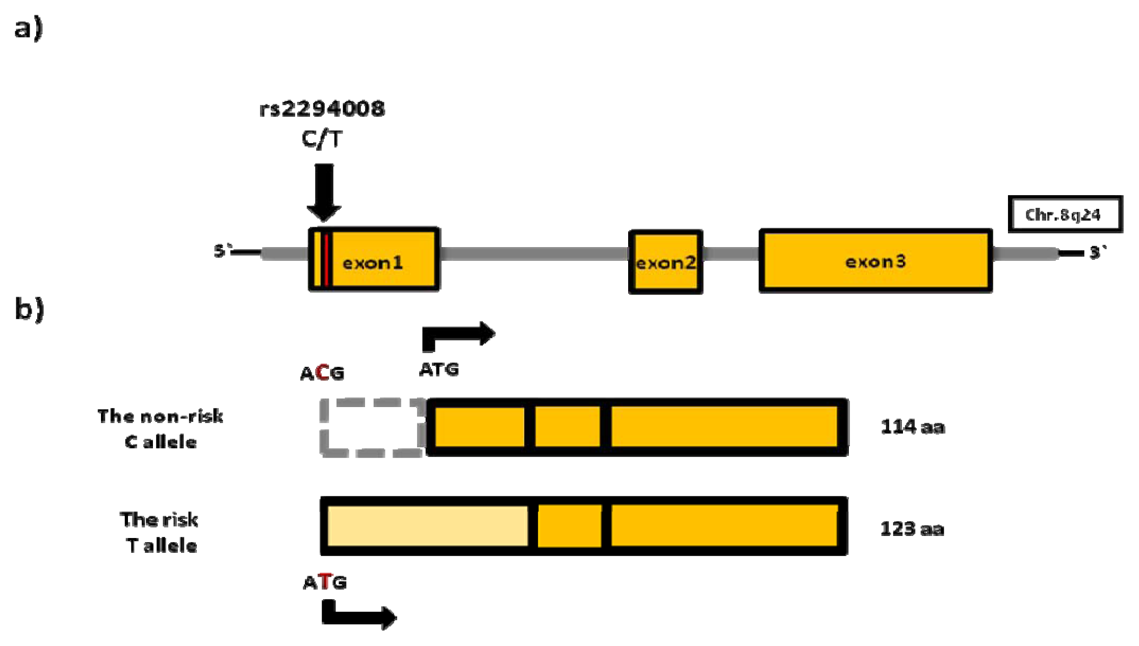
4.1. rs2294008 (PSCA)

The prostate stem cell antigen (PSCA) gene encodes the glycosylphosphatidylinositol (GPI)-anchored cell surface protein belonging to the Thy-1/Ly-6 family. PSCA is expressed in a variety of normal tissues including prostate, kidney, skin, esophagus, stomach and placenta. Alterations in PSCA expression were described in prostate cancer, renal cell carcinoma, pancreatic cancer, ovarian cancer, non-small-cell lung carcinoma, esophageal and gastric cancer [29]. In normal urothelium, PSCA is expressed at low levels and the gene is highly over-expressed in the majority of NMIBC and MIBC [30]. Although PSCA function is unknown, other members of the Thy-1/Ly-6 family, including PSCA, have been implicated in tumorigenesis. They are involved in cell adhesion, signal transduction, apoptosis, or T cell activation. It is hypothesized that PSCA may regulate self-renewal and proliferation of progenitor/stem cells [31]. Because of frequent PSCA over-expression in both primary and metastatic prostatic cancer, PSCA was chosen as a target for immunotherapy. It was shown that anti-PSCA antibodies are able to inhibit tumor development by reducing tumor growth and metastasis. However, the precise role of PSCA in this process remains to be determined [32].
The rs2294008 (C/T) SNP, located in the 8q24.3 region is, until now, the most well-studied bladder cancer susceptibility variant. This missense variant is located in exon 1 of PSCA and causes a change of a nucleotide in the initiation codon. As a result, a novel ATG is used for translation initiation, leading to expression of a protein 9 amino acids (aa) longer (Figure 1).
Rs2294008 has been identified previously to be associated with diffuse-type gastric cancer in the Japanese population. It was shown that the risk T allele reduced PSCA transcriptional activity, however no difference in the protein localization between haplotypes containing either the C or T allele was observed [33]. By contrast, a study by Tanikawa et al. showed that rs2294008 altered cellular localization and stability of PSCA in an allele-specific manner [34]. In bladder cancer cell lines, all haplotypes containing the risk T allele were shown to have reduced PSCA promoter activity, in comparison to the non-risk C allele, confirming the aforementioned results. These data suggest that the SNP in exon 1 of PSCA is also important for the transcription initiation of PSCA by, for example, affecting the binding of transcription factors (TFs) in an allele-specific manner. Unfortunately, no correlation of rs2294008 with PSCA mRNA expression was detected in bladder cancer cell lines [23]. To study tissue-specific gene regulatory effects, Fu et al. evaluated the PSCA mRNA expression in normal bladder and bladder cancer tissue specimens. Surprisingly, the risk T allele was found to be correlated with increased PSCA mRNA expression in both tissue types. Moreover, PSCA mRNA expression was also found to be strongly up-regulated in bladder cancer samples in comparison to adjacent normal tissue. Additionally, the authors fine-mapped the 8q24 region by imputation, and found a second variant (rs2978974) located in the alternative exon 1 of PSCA. This variant was found to be associated with UBC, although this still has to be confirmed in an independent replication series [35]. Rs2294008 was shown to be associated with allelic expression imbalance in normal and tumor samples, which are heterozygous for rs2294008. In normal and tumor tissue, transcripts containing the T allele were far more abundant than transcripts containing the C allele. On the protein level, cells transfected with allele-specific PSCA constructs showed higher PSCA cell surface expression when the risk T allele was present. Importantly, rs2294008 was also found to be strongly associated with PSCA protein expression levels in a large set of bladder tumors.
Taken together, the functional studies of PSCA variants indicate that PSCA is subjected to allele-specific regulation by rs2294008. The results suggest the possibility of a genotype-based patient selection for PSCA immunotherapy [36]. However, the role of PSCA in bladder cancer as well as the mechanism by which rs2294008 affects PSCA mRNA expression is unknown. Recently, the concept of the androgen-sensitive bladder cancer including PSCA and rs2294008 was proposed. Firstly, UBC is diagnosed 3–4 times more frequently in men, but female gender is associated with a worse prognosis [37]. Secondly, studies in mice indicated that gender-specific differences in bladder cancer susceptibility may be regulated by androgen signaling. It was shown that, when exposed to aromatic amines, male mice were more susceptible for bladder cancer than females and castrated males. None of androgen receptor (AR) knockout (ARKO) mice developed bladder tumors [38]. Although reports about AR expression in bladder cancer are conflicting [39] and rs2294008 is located outside of the androgen responsive element (ARE) mapped within the PSCA promoter, it can be hypothesized that other variants linked to rs2294008 can influence bladder cancer risk by lowering affinity of the AR to ARE in the PSCA promoter region. This event can possibly predispose cancer cells to escape from androgen regulation, leading to more aggressive and metastatic disease [40]. This hypothesis remains to be proven, but the possible influence of androgens on bladder cancer development and progression may lead to the future development of new therapies targeting hormonal signaling [41].

4.2. rs17863783 (UGT1A)

The UDP-glucuronosyltransferase (UGT) 1A locus is located on chromosome 2q37. This complex gene is subjected to extensive alternative splicing, leading to the presence of at least nine functional isoforms. Each isoform has a unique alternative exon 1, whilst exons 2–5 are shared among all isoforms [42]. The UGT1A isoforms are expressed in the majority of tissues, including the liver, kidney, gastrointestinal tract, and the urinary bladder [43]. The UGT1A family is involved in the detoxification of many endogenous (steroids, bilirubin) and xenobiotic (drugs, carcinogens) substrates, mainly in the liver but also in extrahepatic tissues [44]. They are involved in inactivation of aromatic amines through glucoronidation, which increases the solubility and excretability of these compounds. However, the N-glucoronidation facilitated by UGT1A is reversible in the acidic urine, leading to the accumulation of aromatic amines in the urothelium and formation of DNA mutagenic adducts which contribute to carcinogenesis [45]. Moreover, exposure to aromatic amines (e.g., by smoking and certain occupations) is a known risk factor for developing bladder cancer [46]. Genetic variants in the aromatic amines detoxification enzymes have been identified to be associated with bladder cancer risk, including the GSTM1 deletion (null genotype) and the NAT2 slow acetylator genotypes [28]. Additionally, genetic variants in UGT1A have been implicated in the modulation of toxicity of the chemotherapeutic agent irinotecan, influencing the clinical outcome of treatment [47].
One of bladder cancer susceptibility loci maps to UGT1A. Rs11892031 (A/C), located in intron 1 of UGT1A8 and UGT1A10, has been discovered by Rothman et al. [24]. In follow-up studies, fine-mapping of the region was performed by re-sequencing, leading to the identification of a causative genetic variant rs17863783 (G/T), which explained the association in this region found by GWAS. This synonymous variant is located in the long isoforms of UGT1A6 (UGT1A6.1) (Figure 2).
Although this variant does not change the amino acid sequence, Tang et al. hypothesized that it can modify the UGT1A6.1 expression level by influencing the exonic splicing enhancer (ESE) in an allele-specific manner. In an in vitro experiment, the presence of the protective T allele caused a significant increase in UGT1A6.1 expression, compared to the G allele. Due to the fact that no carriers of the uncommon T allele (MAF = 0.025) were detected in bladder samples, rs17863783 was correlated with UGT1A6.1 expression in normal liver samples. The protective T allele was found to be correlated with increased expression of UGT1A6.1. The increased expression of UGT1A6 in bladder tissue can lead to enhanced removal of carcinogens dissociated from glucoronids from bladder tissue into the urine, thereby reducing bladder cancer risk [48].
Several studies have shown the importance of enzymes involved in the detoxification of aromatic amines and other carcinogens in bladder cancer susceptibility [49]. Tang et al. have provided compelling evidence that a variant in the UGT1A6.1 gene is functional and affects RNA splicing and gene expression in an allele-specific manner. This effect of a genetic variant on UGT1A6.1 gene expression may lead to enhanced detoxification of the bladder tissue and therefore to a reduced bladder cancer risk.

4.3. Functional Studies of Risk SNPs in UBC Susceptibility Regions

4.3.1. MYC Locus on 8q24

The v-myc myelocytomatosis viral oncogene homolog (MYC) gene is one of the most well-studied proto-oncogenes. This transcription factor regulates hundreds to thousands of genes involved in a wide variety of biological processes, both in normal and malignant cells [50]. MYC is known to influence proliferation, apoptosis, differentiation, self-renewal/senescence and angiogenesis [51]. It is also one of the most frequently up-regulated genes in cancer, suggesting its importance in tumorigenesis [52]. Increased MYC expression in tumors was found to be caused by different mechanisms, including MYC gene amplification, chromosomal translocations, changes in the MYC regulatory network, and mutations targeting the upstream signaling pathways or MYC protein stability [53].
The MYC gene is located in the 8q24 region, in which several susceptibility loci were identified by GWAS for many epithelial cancers including colorectal cancer [54], prostate cancer [55], breast cancer [56] and bladder cancer. The bladder cancer susceptibility variant rs9642880 (G/T) is located 30 kb upstream of the MYC gene and is in weak LD with the other risk variants on the 8q24. No correlation of rs9642880 with MYC expression was detected in whole blood or adipose tissue. It is, however, possible that the MYC regulatory mechanism, which may be modified by rs9642880, is tissue-specific and therefore detectable only in urothelium [21]. The association of rs9642880 was replicated in a Chinese population and it was shown that the risk T allele was correlated with increased MYC mRNA expression in normal bladder tissue and protein expression in tumors [57]. These data suggest that this variant or a variant in high LD is indeed functional. Until now, no functional studies of rs9642880 have been published. On the other hand, several functional studies have been performed on SNPs that are scattered over a 500 kb region upstream of the MYC gene, and that are associated with several cancer types. No transcriptional (i.e., promoter) activity of the 8q24 risk-alleles was detected in colorectal, prostate or breast cancer. However, it was shown that the risk loci, located far upstream of MYC, were shown to physically interact with the MYC promoter in a tissue-specific manner, possibly influencing MYC expression levels [58]. Furthermore, the risk variants lie within regions containing chromatin marks for regulatory elements and were shown to act as enhancers [59,60]. The most extensively studied genetic variant in the 8q24 gene desert upstream of MYC gene is the colorectal and prostate cancer risk SNP rs6983267. It was shown that rs6983267 maps to a highly conserved TCF4 transcription factor binding site within a functional enhancer region (MYC-335). TCF4 is known to play a role in Wnt signaling (frequently deregulated in colorectal cancer) and MYC is one of the target genes of the Wnt signaling pathway [61]. Indeed, TCF4 binding at rs6983267 was found to be allele-specific, with increased affinity to the risk G allele. The rs6983267 region was found to be able to interact with the MYC promoter via chromatin looping, but no difference in efficiency of chromatin looping between alleles was observed. Conversely, rs69833267 was found to regulate enhancer activity in an allele-specific manner in vitro [62,63] and in vivo in transgenic mice [64]. Despite the reported enhancer activity of the rs6983267 locus, no correlation with MYC expression has been found so far, neither in colorectal nor in prostate cancer tissue samples [65]. Interestingly, Sur et al. showed that the presence of the MYC-355 enhancer, containing rs6983267, is not crucial for normal intestinal development and function. Surprisingly, MYC-355 only seems to be involved in tumorigenesis, as lack of the enhancer region reduced the formation of polyps in an Apcmin mouse strain that spontaneously develops colorectal tumors [66].
Studies in the other cancer types show that the risk variants at the 8q24 are able to affect MYC promoter activity in an allele-specific manner, by influencing transcription factors binding affinity. It is therefore reasonable to hypothesize that rs9642880 lies within such a regulatory region and that altered TFs binding may modify MYC expression in a similar way. Given that frequent MYC over-expression or amplification has been described in bladder cancer [67], MYC seems to play an important role in bladder cancer development. Many therapies targeting MYC expression, function, or interaction with other proteins, are currently being developed [68]. Thus, future studies elucidating the role of MYC and rs9642880 in bladder cancer may have an impact on the selection of patients who may benefit from MYC-targeted therapies.

4.3.2. TERT/CLPTM1L Locus on 5p15

Multiple studies linked numerous variants at the 5p15 region to cancer risk, including breast cancer, pancreatic cancer, and glioma [69]. Furthermore, two bladder cancer risk variants in the 5p15 were discovered including rs2736098, a synonymous variant located in the exon of the TERT gene and rs401681 in intron 4 of CLPTM1L. Additionally, both variants were found to be associated with increased risk of basal cell carcinoma, lung cancer and cervical cancer, which are known to be strongly connected to environmental exposures. Interestingly, rs401681 was identified as a protective variant for melanoma [27].
Telomerase is a ribonucleoprotein, which consists of the integral telomerase RNA (TER) and telomerase reverse transcriptase (TERT). Telomerase prevents loss of the chromosomal ends, which occurs during DNA replication. This, in turn, ensures genome stability and integrity. In normal cells telomerase is expressed at low levels and it is tightly controlled through transcriptional and epigenetic regulation. The expression of telomerase decreases with subsequent cell divisions, leading to an increase in telomeres shortening. Telomere shortening eventually leads to senescence and apoptosis [70,71]. The balance between telomere shortening and maintenance is known to influence cancer risk [72]. Remarkably, telomerase (TERT) expression is deregulated in approximately 85% of cancers, through TERT amplification or 5p chromosomal gains. The up-regulation of TERT expression leads to maintenance of telomeres length, resulting in unlimited proliferation and immortalization of cancer cells, one of the hallmarks of carcinogenesis [73]. A second gene in the 5p15 locus, upstream of TERT is, the less-studied cleft lip and palate transmembrane protein 1-like protein (CLPTM1L). CLPTM1L was shown to influence the sensitivity of ovarian cancer cells to cisplatin-induced apoptosis [74]. It was also found to be over-expressed in doxorubicin-resistant breast cancer, although the mechanism by which CLPTM1L affects drug resistance is unknown [75].
Based on the importance of TERT in carcinogenesis, several studies aimed to evaluate the influence of discovered variants. In prostate cancer, fine-mapping of the original GWAS signal was performed, leading to the identification of four distinct TERT regions independently associated with prostate cancer risk. Interestingly, the identified regions were found to be located within the active regulatory elements predicted by The Encyclopedia of DNA Elements (ENCODE). Additionally, SNPs in one of the identified risk regions influenced TERT mRNA expression in benign prostate tissue [76]. In the previously mentioned study by Rafnar et al. the risk alleles of the two identified SNPs were found to be correlated with a shorter length of telomeres in blood of older, but not younger, women. It was therefore hypothesized that risk variants may affect telomere length gradually over time, leading to increased genomic instability of proliferating cells [27]. The telomere length is known to modify bladder cancer risk and is affected by cigarette smoking [77,78]. By contrast, studies in lung cancer showed that risk variants in TERT are associated with longer telomeres, possibly promoting tumorigenesis by increasing the number of cell divisions, leading to acquisition of more somatic changes in DNA [79]. Recent studies in breast and ovarian cancer indicated, however, that regulation of TERT expression and the role of the identified risk variants could be much more complex. The risk SNPs in the TERT region were shown to differentially influence TERT promoter activity and affect telomere lengths. For example, it was shown that some variants did not affect telomere length, but silenced TERT promoter activity and led to expression of an alternatively spliced, truncated TERT product [80]. The impact of genetic variation on CLPTM1L expression is less studied. Recently, however, it was shown that the risk allele of the intronic rs402710 was associated with increased levels of DNA adduct accumulation in lung tissue. This suggests that variants identified in CLPTM1L are involved in metabolism of carcinogens present in, for example, tobacco smoke [81]. A study by James et al. showed that CLPTM1L is frequently over-expressed in lung cancer. In addition, CLPTM1L was shown to play an anti-apoptotic role by protecting cancer cells from genotoxic-stress induced DNA damage [82].
In conclusion, the 5p15 locus was frequently identified to be associated with increased risk for different cancer types, suggesting an important role in cancer etiology. Firstly, it can be hypothesised that rs2736098 and/or other variants in LD with rs2736098 can influence TERT transcription levels or telomerase activity, leading to increased UBC susceptibility. As new therapies targeting TERT signaling are being developed [83], elucidating TERT regulation in UBC is of particular interest. Secondly, considering that smoking is a strong risk factor for developing lung and bladder cancer, it is possible that the risk variants identified at the 5p15 locus affect the CLPTM1L gene, thereby influencing the metabolism of carcinogens, and hence modify cancer risk. So far, functional studies on the TERT and CLPTM1L variants in other cancer types have given conflicting data. Therefore, detailed studies are needed to evaluate the role of the 5p15 risk SNPs in UBC development and to unravel their possible interaction with environmental risk factors.

5. Future of Functional Studies

5.1. Possible Role of Variants in Non-Coding Regions

Until now, the majority of UBC risk variants have not been functionally characterized. However, an increasing amount of data is published about the role of the risk variants in modification of cancer susceptibility. Variations in the human genome can influence cellular processes on different levels. Exonic genetic variants may affect protein folding, cellular localization, stability, or expression, and may disrupt splicing. Intronic and intergenic variants may influence, amongst others, gene expression regulation by alterations in transcription factors binding sites, microRNAs sequence or target sites, non-coding RNAs or RNA splicing.
Frequent mutations in the protein coding regions, involved in tumor development, have been described in many cancer types [84], including bladder cancer [85]. The majority of GWAS signals map to the still poorly understood non-coding regions of the genome. Initiatives like ENCODE proved that large parts of the genome, including non-coding regions, are transcribed and probably functional [86]. Over 75% of the GWAS risk variants were shown to map to regulatory elements or to be in strong LD with SNPs in active regions [87]. Genetic variants regulate gene expression in a cis- (local) and trans- (distal) manner, resulting in heritable differences in gene expression levels between individuals, which may be found through expression quantitative trait loci (eQTLs) analysis [88]. Several studies in different cancer types confirmed that, indeed, the intergenic risk variants map to novel, tissue-specific regulatory elements. Furthermore, these SNPs were shown to alter the affinity of transcription factors binding, leading to an allelic imbalance in the target gene expression [8991]. Secondly, it is known that long non-coding RNAs (lncRNAs) regulate a large variety of biological processes and play important roles in cancer development and, as such, these lncRNAs can act as oncogenes or tumor-suppressors [92]. Some risk variants identified by GWAS were found to influence the expression of tissue-specific lncRNAs in an allele-specific way [93]. For example, Jendrzejewski et al. showed that the non-coding papillary thyroid carcinoma risk variant maps to the novel, thyroid-specific, long intergenic non-coding RNA, PTCSC3. The expression of this tumor-suppressor RNA was found to be strongly down-regulated in cancer in an allele-specific manner, leading to an increase in cell growth [94]. Thirdly, alternative splicing is known to be frequently deregulated in cancer and will lead to exon skipping or aberrantly excised intronic sequences [95]. Consequently, altered protein expression may influence proliferation, survival and metastatic potential of cancer cells [96]. Several SNPs in the exonic and intronic regions were shown to influence the patterns of splicing by altering the splicing regulatory sequences, including the exonic and intronic splicing enhancers or silencers. In prostate cancer the intronic risk SNP was shown to lie within a splicing enhancer region of the KLF6 tumor-suppressor gene. This variant was found to increase the transcription of alternatively spliced KLF6 isoforms, which act in a dominant-negative way on wild-type KLF6, resulting in increased transcriptional activation of growth stimulating genes [97]. Fourthly, microRNAs (miRNAs) are post-transcriptional regulators of gene expression and are known to be involved in regulation of a wide range of genes and biological processes [98]. Kim et al. showed that SNPs located in miR-1206 at the 8q24 and miR-612 at the 11q13.3 regions influence the biogenesis of the corresponding mature miRNAs in an allele-specific manner [99]. Furthermore, it was shown that in esophageal squamous cell carcinoma, a risk SNP in the 3′UTR of the RAP1A gene, affects the binding site of miRNA-196a. This led to RAP1A over-expression resulting in more aggressive tumor behavior [100].
Taken together, the GWAS results suggest that cancer susceptibility is in general modified by the alterations or variations in gene expression regulatory mechanisms, rather than by amino acid sequence changes. Several diseases have already been linked to mutations in non-coding regions including Hirschsprung disease [101], colorectal cancer [102], or melanoma [103,104], pointing towards a crucial role of cis-regulatory regions in health and disease [105]. Considering the rapid development of the techniques suitable for analysis of the non-coding regions, as well as the more accurate and more comprehensive annotation of tissue-specific regulatory elements, functional studies of the intronic and intergenic variants will become more straightforward in the future [106].

5.2. Rapid Development of New Technologies

Despite the success of GWAS in the identification of novel risk loci, only a small part of an individual’s cancer susceptibility can be explained by genetic variants. The majority of studies so far focused on common variants following the “common disease–common variant” hypothesis. With improvements in technologies the “missing heritability” problem is expected to be (at least partly) solved by the discovery of multiple low-effect size, rare and/or structural variants associated with a particular disease [107].
Initiatives like The International 1000 Genomes Project will improve the characterization of human genome variation by sequencing individuals from different populations, leading to the annotation not only common of variants (MAF > 5%), but also low frequency (MAF < 1%) and rare variants (MAF < 0.5%) [108,109]. In the past years, genotyping chips allowed for the detection of common variants. Recently, new chips have been designed that allow the detection of less common variants. However, next generation sequencing, including whole-genome and whole-exome sequencing, are emerging as new, powerful tools in the detection of rare variants [110]. It is possible that multiple low penetrance variants associated with disease remain to be discovered. Larger GWAS initiatives and meta-analyses of the existing data are needed in order to discover the common variants with even smaller effects contributing to overall disease susceptibility [111]. Despite improvements in technologies that detect copy number variants (CNVs) through SNP arrays, calling of CNVs pose a particular challenge. With further developments of methodology and annotation of CNVs our understanding of the full spectrum of this variation will become more complete [112].

5.3. The Impact of Context

Until now, the bladder cancer risk loci confer only a small increased risk, suggesting that pathways involved in cancer development can be much more complex than expected and that the risk variants need to be studied in particular cancer subtypes or environmental context.
The number of identified risk loci differs significantly between cancer types. This highlights the polygenic nature of cancer and the differences in etiology between different tumors. Alternatively, some of the tumor types can be more heterogeneous than others, which complicates the discovery of more general variants [113]. Secondly, GWAS risk signals show a remarkable subtype specificity. In breast cancer, several risk variants were shown to be associated only with the more aggressive ER-negative tumors but not with ER-positive cancer, confirming observed differences in clinical behavior of both tumor types [114]. Interestingly, several bladder cancer risk variants including MYC [21] and FGFR3/TACC3 [22] show stronger association with low risk tumors, confirming the heterogeneous nature of bladder cancer [115]. In this regard, identification of pathways affected by subtype-specific variants and involved in less or more aggressive disease development will be important for cancer prevention. The collection of larger sample sizes corresponding to disease subtypes can result in the identification of more loci which are subtype-specific [116]. Finally, interactions between identified SNPs and environmental and lifestyle factors can modify individual cancer susceptibility, but this has not yet been extensively studied [117]. Garcia-Closas et al. showed that 6 of 12 UBC susceptibility variants showed significant additive gene–environment interactions, most notably true for NAT2 and UGT1A6. They also demonstrated how GWAS data can be used for predicting the absolute risk of bladder cancer, taking into account smoking [118]. It would be of particular interest to study the additive effects of the identified UBC susceptibility variants in other environmental and lifestyle contexts, although it requires large sample sizes and above all valid phenotypic and exposure information [119].

6. Conclusions

The GWAS approach has been successful in the identification of UBC risk genetic variants. Until now, only two of the identified loci (PSCA and UGT1A) have been functionally characterized. These studies, however, confirmed that variants identified by GWAS have functional consequences and influence gene expression in an allele-specific manner. Furthermore, the GWAS approach pointed towards new genes involved in bladder cancer development, possibly leading to the development of new therapies in the future. In this regard, the heritable differences in gene expression related to risk SNPs have potential to be used in the selection of patients who will benefit from a particular treatment. As our understanding of the non-coding regions is still far from complete, the role of other bladder cancer risk variants remains unknown. An increasing amount of data is being published about the influence of intergenic and intronic SNPs in modification of various biological processes. It can, therefore, be hypothesized that UBC risk variants affect gene expression by alterations in urothelium-specific regulatory networks through one of the aforementioned mechanisms. A large part of UBC susceptibility remains to be uncovered. Bladder cancer occurs frequently and is associated with high economic costs, due to extensive follow-up [120]. Screening programs for bladder cancer are currently not thought to reduce disease specific outcomes. Tailoring of such programs according to genetic risks and patterns of exposure to carcinogens may enable the identification or selection of people at risk [121]. Until now, individual genetic risk assessment using the risk variants has not been clinically relevant, however, it cannot be excluded that, with increasing numbers of identified variants, genetic counseling of high risk groups, based on GWAS results, will become possible [122]. In conclusion, GWAS results are very promising in bringing us closer to the era of personalized medicine. However, they still pose a particular challenge in the interpretation of their functional significance.

Conflict of Interest

The authors declare no conflict of interest.

References

  1. Chung, C.C.; Magalhaes, W.C.; Gonzalez-Bosquet, J.; Chanock, S.J. Genome-wide association studies in cancer—Current and future directions. Carcinogenesis 2010, 31, 111–120. [Google Scholar]
  2. Wu, X.; Hildebrandt, M.A.; Chang, D.W. Genome-wide association studies of bladder cancer risk: A field synopsis of progress and potential applications. Cancer Metastasis Rev 2009, 28, 269–280. [Google Scholar]
  3. Freedman, M.L.; Monteiro, A.N.; Gayther, S.A.; Coetzee, G.A.; Risch, A.; Plass, C.; Casey, G.; de Biasi, M.; Carlson, C.; Duggan, D.; et al. Principles for the post-GWAS functional characterization of cancer risk loci. Nat. Genet 2011, 43, 513–518. [Google Scholar]
  4. Burger, M.; Catto, J.W.; Dalbagni, G.; Grossman, H.B.; Herr, H.; Karakiewicz, P.; Kassouf, W.; Kiemeney, L.A.; la Vecchia, C.; Shariat, S.; et al. Epidemiology and risk factors of urothelial bladder cancer. Eur. Urol 2013, 63, 234–241. [Google Scholar]
  5. Siegel, R.; Naishadham, D.; Jemal, A. Cancer statistics, 2012. CA Cancer J. Clin 2012, 62, 10–29. [Google Scholar]
  6. Kiemeney, L.A.; Grotenhuis, A.J.; Vermeulen, S.H.; Wu, X. Genome-wide association studies in bladder cancer: First results and potential relevance. Curr. Opin. Urol 2009, 19, 540–546. [Google Scholar]
  7. Kiltie, A.E. Common predisposition alleles for moderately common cancers: Bladder cancer. Curr. Opin. Genet. Dev 2010, 20, 218–224. [Google Scholar]
  8. Ploeg, M.; Aben, K.K.; Kiemeney, L.A. The present and future burden of urinary bladder cancer in the world. World J. Urol 2009, 27, 289–293. [Google Scholar]
  9. Ploeg, M.; Aben, K.K.; Hulsbergen-van de Kaa, C.A.; Schoenberg, M.P.; Witjes, J.A.; Kiemeney, L.A. Clinical epidemiology of nonurothelial bladder cancer: Analysis of the Netherlands cancer registry. J. Urol 2010, 183, 915–920. [Google Scholar]
  10. Amin, M.B. Histological variants of urothelial carcinoma: Diagnostic, therapeutic and prognostic implications. Mod. Pathol 2009, 22, S96–S118. [Google Scholar]
  11. Babjuk, M.; Oosterlinck, W.; Sylvester, R.; Kaasinen, E.; Bohle, A.; Palou-Redorta, J.; Roupret, M. European Association of, U. EAU guidelines on non-muscle-invasive urothelial carcinoma of the bladder, the 2011 update. Eur. Urol 2011, 59, 997–1008. [Google Scholar]
  12. Van den Bosch, S.; Alfred Witjes, J. Long-term cancer-specific survival in patients with high-risk, non-muscle-invasive bladder cancer and tumour progression: A systematic review. Eur. Urol 2011, 60, 493–500. [Google Scholar]
  13. Sawhney, R.; Bourgeois, D.; Chaudhary, U.B. Neo-adjuvant chemotherapy for muscle-invasive bladder cancer: A look ahead. Ann. Oncol 2006, 17, 1360–1369. [Google Scholar]
  14. Merseburger, A.S.; Kuczyk, M.A. The value of bladder-conserving strategies in muscle-invasive bladder carcinoma compared with radical surgery. Curr. Opin. Urol 2007, 17, 358–362. [Google Scholar]
  15. Murta-Nascimento, C.; Schmitz-Drager, B.J.; Zeegers, M.P.; Steineck, G.; Kogevinas, M.; Real, F.X.; Malats, N. Epidemiology of urinary bladder cancer: From tumor development to patient’s death. World J. Urol 2007, 25, 285–295. [Google Scholar]
  16. Volanis, D.; Kadiyska, T.; Galanis, A.; Delakas, D.; Logotheti, S.; Zoumpourlis, V. Environmental factors and genetic susceptibility promote urinary bladder cancer. Toxicol. Lett 2010, 193, 131–137. [Google Scholar]
  17. Kiriluk, K.J.; Prasad, S.M.; Patel, A.R.; Steinberg, G.D.; Smith, N.D. Bladder cancer risk from occupational and environmental exposures. Urol. Oncol 2012, 30, 199–211. [Google Scholar]
  18. Chang, D.W.; Gu, J.; Wu, X. Germline prognostic markers for urinary bladder cancer: Obstacles and opportunities. Urol. Oncol 2012, 30, 524–532. [Google Scholar]
  19. Division of Genomic Medicine. A Catalog of Published Genome-Wide Association Studies. Available online: http://www.genome.gov/gwastudies (accessed on 11 April 2013).
  20. Chung, C. C.; Chanock, S. J. Current status of genome-wide association studies in cancer. Hum. Genet 2011, 130, 59–78. [Google Scholar]
  21. Kiemeney, L.A.; Thorlacius, S.; Sulem, P.; Geller, F.; Aben, K.K.; Stacey, S.N.; Gudmundsson, J.; Jakobsdottir, M.; Bergthorsson, J.T.; Sigurdsson, A.; et al. Sequence variant on 8q24 confers susceptibility to urinary bladder cancer. Nat. Genet 2008, 40, 1307–1312. [Google Scholar]
  22. Kiemeney, L.A.; Sulem, P.; Besenbacher, S.; Vermeulen, S.H.; Sigurdsson, A.; Thorleifsson, G.; Gudbjartsson, D.F.; Stacey, S.N.; Gudmundsson, J.; Zanon, C.; et al. A sequence variant at 4p16.3 confers susceptibility to urinary bladder cancer. Nat. Genet 2010, 42, 415–419. [Google Scholar] [Green Version]
  23. Wu, X.; Ye, Y.; Kiemeney, L.A.; Sulem, P.; Rafnar, T.; Matullo, G.; Seminara, D.; Yoshida, T.; Saeki, N.; Andrew, A.S.; et al. Genetic variation in the prostate stem cell antigen gene PSCA confers susceptibility to urinary bladder cancer. Nat. Genet 2009, 41, 991–995. [Google Scholar]
  24. Rothman, N.; Garcia-Closas, M.; Chatterjee, N.; Malats, N.; Wu, X.; Figueroa, J.D.; Real, F.X.; van Den Berg, D.; Matullo, G.; et al. A multi-stage genome-wide association study of bladder cancer identifies multiple susceptibility loci. Nat. Genet 2010, 42, 978–984. [Google Scholar]
  25. Rafnar, T.; Vermeulen, S.H.; Sulem, P.; Thorleifsson, G.; Aben, K.K.; Witjes, J.A.; Grotenhuis, A.J.; Verhaegh, G.W.; Hulsbergen-van de Kaa, C.A.; Besenbacher, S.; et al. European genome-wide association study identifies SLC14A1 as a new urinary bladder cancer susceptibility gene. Hum. Mol. Genet 2011, 20, 4268–4281. [Google Scholar]
  26. Garcia-Closas, M.; Ye, Y.; Rothman, N.; Figueroa, J.D.; Malats, N.; Dinney, C.P.; Chatterjee, N.; Prokunina-Olsson, L.; Wang, Z.; Lin, J.; et al. A genome-wide association study of bladder cancer identifies a new susceptibility locus within SLC14A1, a urea transporter gene on chromosome 18q12.3. Hum. Mol. Genet 2011, 20, 4282–4289. [Google Scholar]
  27. Rafnar, T.; Sulem, P.; Stacey, S.N.; Geller, F.; Gudmundsson, J.; Sigurdsson, A.; Jakobsdottir, M.; Helgadottir, H.; Thorlacius, S.; Aben, K.K.; et al. Sequence variants at the TERT-CLPTM1L locus associate with many cancer types. Nat. Genet 2009, 41, 221–227. [Google Scholar]
  28. Garcia-Closas, M.; Malats, N.; Silverman, D.; Dosemeci, M.; Kogevinas, M.; Hein, D.W.; Tardon, A.; Serra, C.; Carrato, A.; Garcia-Closas, R.; et al. NAT2 slow acetylation, GSTM1 null genotype, and risk of bladder cancer: Results from the Spanish bladder cancer study and meta-analyses. Lancet 2005, 366, 649–659. [Google Scholar]
  29. Ono, H.; Hiraoka, N.; Lee, Y.S.; Woo, S.M.; Lee, W.J.; Choi, I.J.; Saito, A.; Yanagihara, K.; Kanai, Y.; Ohnami, S.; et al. Prostate stem cell antigen, a presumable organ-dependent tumor suppressor gene, is down-regulated in gallbladder carcinogenesis. Genes Chromos. Cancer 2012, 51, 30–41. [Google Scholar]
  30. Amara, N.; Palapattu, G.S.; Schrage, M.; Gu, Z.; Thomas, G.V.; Dorey, F.; Said, J.; Reiter, R.E. Prostate stem cell antigen is overexpressed in human transitional cell carcinoma. Cancer Res 2001, 61, 4660–4665. [Google Scholar]
  31. Zhigang, Z.; Wenlv, S. Prostate stem cell antigen (PSCA) expression in human prostate cancer tissues and its potential role in prostate carcinogenesis and progression of prostate cancer. World J. Surg. Oncol 2004, 2, 13. [Google Scholar]
  32. Saffran, D.C.; Raitano, A.B.; Hubert, R.S.; Witte, O.N.; Reiter, R.E.; Jakobovits, A. Anti-PSCA mAbs inhibit tumor growth and metastasis formation and prolong the survival of mice bearing human prostate cancer xenografts. Proc. Natl. Acad. Sci. USA 2001, 98, 2658–2663. [Google Scholar]
  33. Sakamoto, H.; Yoshimura, K.; Saeki, N.; Katai, H.; Shimoda, T.; Matsuno, Y.; Saito, D.; Sugimura, H.; Tanioka, F.; et al. Study Group of Millennium Genome Project for Cancer. Genetic variation in PSCA is associated with susceptibility to diffuse-type gastric cancer. Nat. Genet. 2008, 40, 730–740. [Google Scholar]
  34. Tanikawa, C.; Urabe, Y.; Matsuo, K.; Kubo, M.; Takahashi, A.; Ito, H.; Tajima, K.; Kamatani, N.; Nakamura, Y.; Matsuda, K. A genome-wide association study identifies two susceptibility loci for duodenal ulcer in the Japanese population. Nat. Genet 2012, 44, 430–434, , S1–S2.. [Google Scholar]
  35. Fu, Y.P.; Kohaar, I.; Rothman, N.; Earl, J.; Figueroa, J.D.; Ye, Y.; Malats, N.; Tang, W.; Liu, L.; Garcia-Closas, M.; et al. Common genetic variants in the PSCA gene influence gene expression and bladder cancer risk. Proc. Natl. Acad. Sci. USA 2012, 109, 4974–4979. [Google Scholar]
  36. Kohaar, I.; Porter-Gill, P.; Lenz, P.; Fu, Y.P.; Mumy, A.; Tang, W.; Apolo, A.B.; Rothman, N.; Baris, D.; Schned, A.R.; et al. Genetic variant as a selection marker for anti-prostate stem cell antigen immunotherapy of bladder cancer. J. Natl. Cancer Instit 2013, 105, 69–73. [Google Scholar]
  37. Shariat, S.F.; Sfakianos, J.P.; Droller, M.J.; Karakiewicz, P.I.; Meryn, S.; Bochner, B.H. The effect of age and gender on bladder cancer: A critical review of the literature. BJU Int 2010, 105, 300–308. [Google Scholar]
  38. Miyamoto, H.; Yang, Z.; Chen, Y.T.; Ishiguro, H.; Uemura, H.; Kubota, Y.; Nagashima, Y.; Chang, Y.J.; Hu, Y.C.; Tsai, M.Y.; et al. Promotion of bladder cancer development and progression by androgen receptor signals. J. Natl. Cancer Instit 2007, 99, 558–568. [Google Scholar]
  39. Miyamoto, H.; Yao, J.L.; Chaux, A.; Zheng, Y.; Hsu, I.; Izumi, K.; Chang, C.; Messing, E.M.; Netto, G.J.; Yeh, S. Expression of androgen and oestrogen receptors and its prognostic significance in urothelial neoplasm of the urinary bladder. BJU Int 2012, 109, 1716–1726. [Google Scholar]
  40. Gakis, G.; Stenzl, A. Gender-specific differences in muscle-invasive bladder cancer: The concept of sex steroid sensitivity. World J. Urol. 2013. [Google Scholar] [CrossRef]
  41. Miyamoto, H.; Zheng, Y.; Izumi, K. Nuclear hormone receptor signals as new therapeutic targets for urothelial carcinoma. Curr. Cancer Drug Targets 2012, 12, 14–22. [Google Scholar]
  42. Bellemare, J.; Rouleau, M.; Harvey, M.; Guillemette, C. Modulation of the human glucuronosyltransferase UGT1A pathway by splice isoform polypeptides is mediated through protein-protein interactions. J. Biol. Chem 2010, 285, 3600–3607. [Google Scholar]
  43. Nakamura, A.; Nakajima, M.; Yamanaka, H.; Fujiwara, R.; Yokoi, T. Expression of UGT1A and UGT2B mRNA in human normal tissues and various cell lines. Drug Metab. Dispos. Biol. Fate Chem 2008, 36, 1461–1464. [Google Scholar]
  44. Tukey, R.H.; Strassburg, C.P. Human UDP-glucuronosyltransferases: Metabolism, expression, and disease. Ann. Rev. Pharmacol. Toxicol 2000, 40, 581–616. [Google Scholar]
  45. Ciotti, M.; Lakshmi, V.M.; Basu, N.; Davis, B.B.; Owens, I.S.; Zenser, T.V. Glucuronidation of benzidine and its metabolites by cDNA-expressed human UDP-glucuronosyltransferases and pH stability of glucuronides. Carcinogenesis 1999, 20, 1963–1969. [Google Scholar]
  46. Zenser, T.V.; Lakshmi, V.M.; Hsu, F.F.; Davis, B.B. Metabolism of N-acetylbenzidine and initiation of bladder cancer. Mutat. Res. 2002, 506–507, 29–40. [Google Scholar]
  47. Levesque, E.; Belanger, A.S.; Harvey, M.; Couture, F.; Jonker, D.; Innocenti, F.; Cecchin, E.; Toffoli, G.; Guillemette, C. Refining the UGT1A haplotype associated with irinotecan-induced hematological toxicity in metastatic colorectal cancer patients treated with 5-fluorouracil/irinotecan-based regimens. J. Pharmacol. Exp. Ther 2013, 345, 95–101. [Google Scholar]
  48. Tang, W.; Fu, Y.P.; Figueroa, J.D.; Malats, N.; Garcia-Closas, M.; Chatterjee, N.; Kogevinas, M.; Baris, D.; Thun, M.; Hall, J.L.; et al. Mapping of the UGT1A locus identifies an uncommon coding variant that affects mRNA expression and protects from bladder cancer. Hum. Mol. Genet 2012, 21, 1918–1930. [Google Scholar]
  49. Wu, X.; Ros, M.M.; Gu, J.; Kiemeney, L. Epidemiology and genetic susceptibility to bladder cancer. BJU Int 2008, 102, 1207–1215. [Google Scholar]
  50. Eilers, M.; Eisenman, R.N. Myc’s broad reach. Genes Dev 2008, 22, 2755–2766. [Google Scholar]
  51. Felsher, D.W. MYC inactivation elicits oncogene addiction through both tumor cell-intrinsic and host-dependent mechanisms. Genes Cancer 2010, 1, 597–604. [Google Scholar]
  52. Nilsson, J.A.; Cleveland, J.L. Myc pathways provoking cell suicide and cancer. Oncogene 2003, 22, 9007–9021. [Google Scholar]
  53. Lin, C.Y.; Loven, J.; Rahl, P.B.; Paranal, R.M.; Burge, C.B.; Bradner, J.E.; Lee, T.I.; Young, R.A. Transcriptional amplification in tumor cells with elevated c-Myc. Cell 2012, 151, 56–67. [Google Scholar]
  54. Theodoratou, E.; Montazeri, Z.; Hawken, S.; Allum, G.C.; Gong, J.; Tait, V.; Kirac, I.; Tazari, M.; Farrington, S.M.; Demarsh, A.; et al. Systematic meta-analyses and field synopsis of genetic association studies in colorectal cancer. J. Natl. Cancer Instit 2012, 104, 1433–1457. [Google Scholar]
  55. Al Olama, A.A.; Kote-Jarai, Z.; Giles, G.G.; Guy, M.; Morrison, J.; Severi, G.; Leongamornlert, D.A.; Tymrakiewicz, M.; Jhavar, S.; Saunders, E.; et al. Multiple loci on 8q24 associated with prostate cancer susceptibility. Nat. Genet 2009, 41, 1058–1060. [Google Scholar]
  56. Easton, D.F.; Pooley, K.A.; Dunning, A.M.; Pharoah, P.D.; Thompson, D.; Ballinger, D.G.; Struewing, J.P.; Morrison, J.; Field, H.; Luben, R.; et al. Genome-wide association study identifies novel breast cancer susceptibility loci. Nature 2007, 447, 1087–1093. [Google Scholar]
  57. Wang, M.; Wang, M.; Zhang, W.; Yuan, L.; Fu, G.; Wei, Q.; Zhang, Z. Common genetic variants on 8q24 contribute to susceptibility to bladder cancer in a Chinese population. Carcinogenesis 2009, 30, 991–996. [Google Scholar]
  58. Ahmadiyeh, N.; Pomerantz, M.M.; Grisanzio, C.; Herman, P.; Jia, L.; Almendro, V.; He, H.H.; Brown, M.; Liu, X.S.; Davis, M.; et al. 8q24 prostate, breast, and colon cancer risk loci show tissue-specific long-range interaction with MYC. Proc. Natl. Acad. Sci. USA 2010, 107, 9742–9746. [Google Scholar]
  59. Jia, L.; Landan, G.; Pomerantz, M.; Jaschek, R.; Herman, P.; Reich, D.; Yan, C.; Khalid, O.; Kantoff, P.; Oh, W.; et al. Functional enhancers at the gene-poor 8q24 cancer-linked locus. PLoS Genet 2009, 5, e1000597. [Google Scholar]
  60. Ting, M.C.; Liao, C.P.; Yan, C.; Jia, L.; Groshen, S.; Frenkel, B.; Roy-Burman, P.; Coetzee, G.A.; Maxson, R. An enhancer from the 8q24 prostate cancer risk region is sufficient to direct reporter gene expression to a subset of prostate stem-like epithelial cells in transgenic mice. Dis. Models Mech 2012, 5, 366–374. [Google Scholar]
  61. Tuupanen, S.; Turunen, M.; Lehtonen, R.; Hallikas, O.; Vanharanta, S.; Kivioja, T.; Bjorklund, M.; Wei, G.; Yan, J.; Niittymaki, I.; et al. The common colorectal cancer predisposition SNP rs6983267 at chromosome 8q24 confers potential to enhanced Wnt signaling. Nat. Genet 2009, 41, 885–890. [Google Scholar]
  62. Pomerantz, M.M.; Ahmadiyeh, N.; Jia, L.; Herman, P.; Verzi, M.P.; Doddapaneni, H.; Beckwith, C.A.; Chan, J.A.; Hills, A.; Davis, M.; et al. The 8q24 cancer risk variant rs6983267 shows long-range interaction with MYC in colorectal cancer. Nat. Genet 2009, 41, 882–884. [Google Scholar]
  63. Wright, J.B.; Brown, S.J.; Cole, M.D. Upregulation of c-MYC in cis through a large chromatin loop linked to a cancer risk-associated single-nucleotide polymorphism in colorectal cancer cells. Mol. Cell. Biol 2010, 30, 1411–1420. [Google Scholar]
  64. Wasserman, N.F.; Aneas, I.; Nobrega, M.A. An 8q24 gene desert variant associated with prostate cancer risk confers differential in vivo activity to a MYC enhancer. Genome Res 2010, 20, 1191–1197. [Google Scholar]
  65. Pomerantz, M.M.; Beckwith, C.A.; Regan, M.M.; Wyman, S.K.; Petrovics, G.; Chen, Y.; Hawksworth, D.J.; Schumacher, F.R.; Mucci, L.; Penney, K.L.; et al. Evaluation of the 8q24 prostate cancer risk locus and MYC expression. Cancer Res 2009, 69, 5568–5574. [Google Scholar]
  66. Sur, I.K.; Hallikas, O.; Vaharautio, A.; Yan, J.; Turunen, M.; Enge, M.; Taipale, M.; Karhu, A.; Aaltonen, L.A.; Taipale, J. Mice lacking a Myc enhancer that includes human SNP rs6983267 are resistant to intestinal tumors. Science 2012, 338, 1360–1363. [Google Scholar]
  67. Mhawech-Fauceglia, P.; Cheney, R.T.; Schwaller, J. Genetic alterations in urothelial bladder carcinoma: An updated review. Cancer 2006, 106, 1205–1216. [Google Scholar]
  68. Ponzielli, R.; Katz, S.; Barsyte-Lovejoy, D.; Penn, L.Z. Cancer therapeutics: Targeting the dark side of Myc. Eur. J. Cancer 2005, 41, 2485–2501. [Google Scholar]
  69. Baird, D.M. Variation at the TERT locus and predisposition for cancer. Expert Rev. Mol. Med 2010, 12, e16. [Google Scholar]
  70. Daniel, M.; Peek, G.W.; Tollefsbol, T.O. Regulation of the human catalytic subunit of telomerase (hTERT). Gene 2012, 498, 135–146. [Google Scholar]
  71. Cifuentes-Rojas, C.; Shippen, D.E. Telomerase regulation. Mutat. Res 2012, 730, 20–27. [Google Scholar]
  72. Hou, L.; Zhang, X.; Gawron, A.J.; Liu, J. Surrogate tissue telomere length and cancer risk: Shorter or longer? Cancer Lett 2012, 319, 130–135. [Google Scholar]
  73. Cao, Y.; Bryan, T.M.; Reddel, R.R. Increased copy number of the TERT and TERC telomerase subunit genes in cancer cells. Cancer Sci 2008, 99, 1092–1099. [Google Scholar]
  74. Yamamoto, K.; Okamoto, A.; Isonishi, S.; Ochiai, K.; Ohtake, Y. A novel gene, CRR9, which was up-regulated in CDDP-resistant ovarian tumor cell line, was associated with apoptosis. Biochem. and Biophys. Res. Commun 2001, 280, 1148–1154. [Google Scholar]
  75. Folgueira, M.A.A.K.; Carraro, D.M.; Brentani, H.; Patra, D.F.D.; Barbosa, E.M.; Netto, M.M.; Caldeira, J.R.F.; Katayama, M.L.H.; Soares, F.A.; Oliveira, C.T.; et al. Gene expression profile associated with response to doxorubicin-based therapy in breast cancer. Clin. Cancer Res 2005, 11, 7434–7443. [Google Scholar]
  76. Kote-Jarai, Z.; Saunders, E.J.; Leongamornlert, D.A.; Tymrakiewicz, M.; Dadaev, T.; Jugurnauth-Little, S.; Ross-Adams, H.; al Olama, A.A.; Benlloch, S.; Halim, S.; et al. Fine-mapping identifies multiple prostate cancer risk loci at 5p15, one of which associates with TERT expression. Hum. Mol. Genet 2013, 22, 2520–2528. [Google Scholar]
  77. McGrath, M.; Wong, J.Y.; Michaud, D.; Hunter, D.J.; de Vivo, I. Telomere length, cigarette smoking, and bladder cancer risk in men and women. Cancer Epidemiol. Biomark. Prev 2007, 16, 815–819. [Google Scholar]
  78. Gu, J.; Chen, M.; Shete, S.; Amos, C.I.; Kamat, A.; Ye, Y.; Lin, J.; Dinney, C.P.; Wu, X. A genome-wide association study identifies a locus on chromosome 14q21 as a predictor of leukocyte telomere length and as a marker of susceptibility for bladder cancer. Cancer Prev. Res 2011, 4, 514–521. [Google Scholar]
  79. Lan, Q.; Cawthon, R.; Gao, Y.; Hu, W.; Hosgood, H.D., III; Barone-Adesi, F.; Ji, B.T.; Bassig, B.; Chow, W.H.; Shu, X.; et al. Longer telomere length in peripheral white blood cells is associated with risk of lung cancer and the rs2736100 (CLPTM1L-TERT) polymorphism in a prospective cohort study among women in China. PLoS One 2013, 8, e59230. [Google Scholar]
  80. Bojesen, S.E.; Pooley, K.A.; Johnatty, S.E.; Beesley, J.; Michailidou, K.; Tyrer, J.P.; Edwards, S.L.; Pickett, H.A.; Shen, H.C.; Smart, C.E.; et al. Multiple independent variants at the TERT locus are associated with telomere length and risks of breast and ovarian cancer. Nat. Genet 2013, 45, 371–384. [Google Scholar]
  81. Zienolddiny, S.; Skaug, V.; Landvik, N.E.; Ryberg, D.; Phillips, D.H.; Houlston, R.; Haugen, A. The TERT-CLPTM1L lung cancer susceptibility variant associates with higher DNA adduct formation in the lung. Carcinogenesis 2009, 30, 1368–1371. [Google Scholar]
  82. James, M.A.; Wen, W.D.; Wang, Y.A.; Byers, L.A.; Heymach, J.V.; Coombes, K.R.; Girard, L.; Minna, J.; You, M. Functional characterization of CLPTM1L as a lung cancer risk candidate gene in the 5p15.33 Locus. PLoS One 2012, 7, e36116. [Google Scholar]
  83. Rahman, R.; Grundy, R. Histone deacetylase inhibition as an anticancer telomerase-targeting strategy. Int. J. Cancer 2011, 129, 2765–2774. [Google Scholar]
  84. Vogelstein, B.; Papadopoulos, N.; Velculescu, V.E.; Zhou, S.; Diaz, L.A., Jr; Kinzler, K.W. Cancer genome landscapes. Science 2013, 339, 1546–1558. [Google Scholar]
  85. Kompier, L.C.; Lurkin, I.; van der Aa, M.N.; van Rhijn, B.W.; van der Kwast, T.H.; Zwarthoff, E.C. FGFR3, HRAS, KRAS, NRAS and PIK3CA mutations in bladder cancer and their potential as biomarkers for surveillance and therapy. PLoS One 2010, 5, e13821. [Google Scholar]
  86. Consortium, E.P.; Dunham, I.; Kundaje, A.; Aldred, S.F.; Collins, P.J.; Davis, C.A.; Doyle, F.; Epstein, C.B.; Frietze, S.; Harrow, J.; et al. An integrated encyclopedia of DNA elements in the human genome. Nature 2012, 489, 57–74. [Google Scholar]
  87. Maurano, M.T.; Humbert, R.; Rynes, E.; Thurman, R.E.; Haugen, E.; Wang, H.; Reynolds, A.P.; Sandstrom, R.; Qu, H.; Brody, J.; et al. Systematic localization of common disease-associated variation in regulatory DNA. Science 2012, 337, 1190–1195. [Google Scholar]
  88. Cheung, V.G.; Spielman, R.S. Genetics of human gene expression: Mapping DNA variants that influence gene expression. Nat. Rev. Genet 2009, 10, 595–604. [Google Scholar]
  89. Pittman, A.M.; Naranjo, S.; Jalava, S.E.; Twiss, P.; Ma, Y.; Olver, B.; Lloyd, A.; Vijayakrishnan, J.; Qureshi, M.; Broderick, P.; et al. Allelic variation at the 8q23.3 colorectal cancer risk locus functions as a cis-acting regulator of EIF3H. PLoS Genet 2010, 6, e1001126. [Google Scholar]
  90. Akamatsu, S.; Takata, R.; Ashikawa, K.; Hosono, N.; Kamatani, N.; Fujioka, T.; Ogawa, O.; Kubo, M.; Nakamura, Y.; Nakagawa, H. A functional variant in NKX3.1 associated with prostate cancer susceptibility down-regulates NKX3.1 expression. Hum. Mol. Genet 2010, 19, 4265–4272. [Google Scholar]
  91. Schodel, J.; Bardella, C.; Sciesielski, L.K.; Brown, J.M.; Pugh, C.W.; Buckle, V.; Tomlinson, I.P.; Ratcliffe, P.J.; Mole, D.R. Common genetic variants at the 11q13.3 renal cancer susceptibility locus influence binding of HIF to an enhancer of cyclin D1 expression. Nat. Genet 2012, 44, 420–425, , S1–S2.. [Google Scholar]
  92. Qi, P.; Du, X. The long non-coding RNAs, a new cancer diagnostic and therapeutic gold mine. Mod. Pathol 2013, 26, 155–165. [Google Scholar]
  93. Kumar, V.; Westra, H.J.; Karjalainen, J.; Zhernakova, D.V.; Esko, T.; Hrdlickova, B.; Almeida, R.; Zhernakova, A.; Reinmaa, E.; Vosa, U.; et al. Human disease-associated genetic variation impacts large intergenic non-coding RNA expression. PLoS Genet 2013, 9, e1003201. [Google Scholar]
  94. Jendrzejewski, J.; He, H.; Radomska, H.S.; Li, W.; Tomsic, J.; Liyanarachchi, S.; Davuluri, R.V.; Nagy, R.; de la Chapelle, A. The polymorphism rs944289 predisposes to papillary thyroid carcinoma through a large intergenic noncoding RNA gene of tumor suppressor type. Proc. Natl. Acad. Sci. USA 2012, 109, 8646–8651. [Google Scholar]
  95. Lee, Y.; Gamazon, E.R.; Rebman, E.; Lee, Y.; Lee, S.; Dolan, M.E.; Cox, N.J.; Lussier, Y.A. Variants affecting exon skipping contribute to complex traits. PLoS Genet 2012, 8, e1002998. [Google Scholar]
  96. Kim, Y.J.; Kim, H.S. Alternative splicing and its impact as a cancer diagnostic marker. Genomics Inf 2012, 10, 74–80. [Google Scholar]
  97. Narla, G.; Difeo, A.; Reeves, H.L.; Schaid, D.J.; Hirshfeld, J.; Hod, E.; Katz, A.; Isaacs, W.B.; Hebbring, S.; Komiya, A.; et al. A germline DNA polymorphism enhances alternative splicing of the KLF6 tumor suppressor gene and is associated with increased prostate cancer risk. Cancer Res 2005, 65, 1213–1222. [Google Scholar]
  98. Gong, J.; Tong, Y.; Zhang, H.M.; Wang, K.; Hu, T.; Shan, G.; Sun, J.; Guo, A.Y. Genome-wide identification of SNPs in microRNA genes and the SNP effects on microRNA target binding and biogenesis. Hum. Mutat 2012, 33, 254–263. [Google Scholar]
  99. Kim, H.K.; Prokunina-Olsson, L.; Chanock, S.J. Common genetic variants in miR-1206 (8q24.2) and miR-612 (11q13.3) affect biogenesis of mature miRNA forms. PloS One 2012, 7, e47454. [Google Scholar]
  100. Wang, K.; Li, J.; Guo, H.; Xu, X.; Xiong, G.; Guan, X.; Liu, B.; Li, J.; Chen, X.; Yang, K.; et al. MiR-196a binding-site SNP regulates RAP1A expression contributing to esophageal squamous cell carcinoma risk and metastasis. Carcinogenesis 2012, 33, 2147–2154. [Google Scholar]
  101. Fitze, G.; Schierz, M.; Kuhlisch, E.; Schreiber, M.; Ziegler, A.; Roesner, D.; Schackert, H.K. Novel intronic polymorphisms in the RET proto-oncogene and their association with Hirschsprung disease. Hum. Mutat 2003, 22, 177. [Google Scholar]
  102. Hienonen, T.; Sammalkorpi, H.; Enholm, S.; Alhopuro, P.; Barber, T.D.; Lehtonen, R.; Nupponen, N.N.; Lehtonen, H.; Salovaara, R.; Mecklin, J.P.; et al. Mutations in two short noncoding mononucleotide repeats in most microsatellite-unstable colorectal cancers. Cancer Res 2005, 65, 4607–4613. [Google Scholar]
  103. Horn, S.; Figl, A.; Rachakonda, P.S.; Fischer, C.; Sucker, A.; Gast, A.; Kadel, S.; Moll, I.; Nagore, E.; Hemminki, K.; et al. TERT promoter mutations in familial and sporadic melanoma. Science 2013, 339, 959–961. [Google Scholar]
  104. Huang, F.W.; Hodis, E.; Xu, M.J.; Kryukov, G.V.; Chin, L.; Garraway, L.A. Highly recurrent TERT promoter mutations in human melanoma. Science 2013, 339, 957–959. [Google Scholar]
  105. Epstein, D.J. Cis-regulatory mutations in human disease. Brief. Funct. Genomics Proteomics 2009, 8, 310–316. [Google Scholar]
  106. Ward, L.D.; Kellis, M. Interpreting noncoding genetic variation in complex traits and human disease. Nat. Biotechnol 2012, 30, 1095–1106. [Google Scholar]
  107. Manolio, T.A.; Collins, F.S.; Cox, N.J.; Goldstein, D.B.; Hindorff, L.A.; Hunter, D.J.; McCarthy, M.I.; Ramos, E.M.; Cardon, L.R.; Chakravarti, A.; et al. Finding the missing heritability of complex diseases. Nature 2009, 461, 747–753. [Google Scholar]
  108. Genomes Project, C.; Abecasis, G.R.; Auton, A.; Brooks, L.D.; DePristo, M.A.; Durbin, R.M.; Handsaker, R.E.; Kang, H.M.; Marth, G.T.; McVean, G.A. An integrated map of genetic variation from 1092 human genomes. Nature 2012, 491, 56–65. [Google Scholar]
  109. Abecasis, G.R.; Altshuler, D.; Auton, A.; Brooks, L.D.; Durbin, R.M.; Gibbs, R.A.; Hurles, M.E.; McVean, G.A. 1000 Genomes Project Consortium. A map of human genome variation from population-scale sequencing. Nature 2010, 467, 1061–1073. [Google Scholar] [Green Version]
  110. Cirulli, E.T.; Goldstein, D.B. Uncovering the roles of rare variants in common disease through whole-genome sequencing. Nat. Rev. Genet 2010, 11, 415–425. [Google Scholar]
  111. Park, J.H.; Wacholder, S.; Gail, M.H.; Peters, U.; Jacobs, K.B.; Chanock, S.J.; Chatterjee, N. Estimation of effect size distribution from genome-wide association studies and implications for future discoveries. Nat. Genet 2010, 42, 570–575. [Google Scholar]
  112. McCarroll, S.A. Extending genome-wide association studies to copy-number variation. Hum. Mol. Genet 2008, 17, R135–R142. [Google Scholar]
  113. Easton, D.F.; Eeles, R.A. Genome-wide association studies in cancer. Hum. Mol. Genet 2008, 17, R109–R115. [Google Scholar]
  114. Garcia-Closas, M.; Couch, F.J.; Lindstrom, S.; Michailidou, K.; Schmidt, M.K.; Brook, M.N.; Orr, N.; Rhie, S.K.; Riboli, E.; Feigelson, H.S.; et al. Genome-wide association studies identify four ER negative-specific breast cancer risk loci. Nat. Genet 2013, 45, 392–398. [Google Scholar]
  115. Goebell, P.J.; Knowles, M.A. Bladder cancer or bladder cancers? Genetically distinct malignant conditions of the urothelium. Urol. Oncol 2010, 28, 409–428. [Google Scholar]
  116. Kraft, P.; Haiman, C.A. GWAS identifies a common breast cancer risk allele among BRCA1 carriers. Nat. Genet 2010, 42, 819–820. [Google Scholar]
  117. Le Marchand, L.; Wilkens, L.R. Design considerations for genomic association studies: Importance of gene-environment interactions. Cancer Epidemiol. Biomark. Prev 2008, 17, 263–267. [Google Scholar]
  118. Garcia-Closas, M.; Rothman, N.; Figueroa, J.D.; Prokunina-Olsson, L.; Han, S.S.; Baris, D.; Jacobs, E.J.; Malats, N.; de Vivo, I.; Albanes, D.; et al. Common genetic polymorphisms modify the effect of smoking on absolute risk of bladder cancer. Cancer Res 2013, 73, 2211–2220. [Google Scholar]
  119. Hindorff, L.A.; Gillanders, E.M.; Manolio, T.A. Genetic architecture of cancer and other complex diseases: Lessons learned and future directions. Carcinogenesis 2011, 32, 945–954. [Google Scholar]
  120. Stenzl, A.; Hennenlotter, J.; Schilling, D. Can we still afford bladder cancer? Curr. Opin. Urol 2008, 18, 488–492. [Google Scholar]
  121. Larre, S.; Catto, J.W.; Cookson, M.S.; Messing, E.M.; Shariat, S.F.; Soloway, M.S.; Svatek, R.S.; Lotan, Y.; Zlotta, A.R.; Grossman, H.B. Screening for bladder cancer: Rationale, limitations, whom to target, and perspectives. Eur. Urol 2013, 63, 1049–1058. [Google Scholar]
  122. Gibson, G. Hints of hidden heritability in GWAS. Nat. Genet 2010, 42, 558–560. [Google Scholar]
Figure 1. (a) PSCA locus and location of rs2294008; (b) Influence of rs2294008 on PSCA protein length.
Ijms 14 12346f1
Figure 2. UGT1A locus and location of the functional UBC variant.
Ijms 14 12346f2
Table 1. Urinary bladder cancer risk variants.
Chrom.GeneSNP (identified allele)LocationMAFOR (95% CI)Study typeRef.
8q24MYCrs9642880 [T]Intergenic0.451.22 (1.15–1.29)GWAS[21]
3q28TP63rs710521 [A]Intergenic0.731.19 (1.12–1.27)GWAS[21]
5p15CLPTM1Lrs401681[C]Intronic0.531.12 (1.06–1.18)Follow-up GWAS[27]
5p15TERTrs2736098 [A]Exonic0.251.16 (1.08–1.23)Follow-up GWAS[27]
8q24PSCArs2294008 [T]Exonic0.461.15 (1.10–1.20)GWAS[23]
4p16FGFR3/TACC3rs798766 [T]Intronic0.191.24 (1.17–1.32)GWAS[22]
22q13CBX6/APOBEC3Ars1014971 [C]Intergenic0.380.88 (0.85–0.91)GWAS[24]
19q12CCNE1rs8102137 [C]Intergenic0.331.13 (1.09–1.17)GWAS[24]
2q37UGT1Ars11892031 [C]Intronic0.080.84 (0.79–0.89)GWAS[24]
18q12SLC14A1rs17674580 [T]Intronic0.331.17 (1.11–1.22)GWAS[25]
18q12SLC14A1rs7238033 [T]Intronic0.431.20 (1.13–1.28)Meta-analysis[26]
1p13GSTM1Null *0.511.5 (1.3–1.6)Candidate gene, meta-analysis[28]
8p22NAT2Slow acetylator *0.561.4 (1.2–1.6)Candidate gene, meta-analysis[28]
*NAT2 slow acetylator and GSTM1 null refer to the risk genotypes not the risk SNPs; MAF: Minor allele frequency; OR: Odds ratio; CI: Confidence intervals.

