Glutathionylation of the L-type Ca2+ Channel in Oxidative Stress-Induced Pathology of the Heart


Abstract
:1. Introduction
2. The Role of Calcium and Reactive Oxygen Species (ROS) in Oxidative Stress Responses
2.1. Calcium Homeostasis and Myocyte Contraction
2.2. Ca2+ and ROS-Induced ROS-Release

2.3. Post-Ischemic Persistent Elevation of ROS and Ca2+ Is Pathological

2.4. Mitochondrial Complex III Is the Locus for Superoxide Generation
3. Glutathionylation of the L-type Ca2+ Channel during Oxidative Stress
3.1. Overview of Protein Glutathionylation
3.2. Glutathionylation of the L-type Ca2+ Channel
3.3. Functional and Clinical Consequences of L-type Ca2+ Channel Glutathionylation

3.4. S-Nitrosylation of the L-type Ca2+ Channel as an Alternate Means of Channel Modification during Oxidative Stress
4. Novel Interventions to Alleviate Ischemic Injury
4.1. Overview of Ischemia-Reperfusion Injury
4.2. Reduced Ischemia-Reperfusion Injury Using Targeted Peptide Delivery

4.3. Targeted Peptide Delivery Decreases Infarct Size and Restores Contractility in Vivo
5. Concluding Remarks
Acknowledgments
Author Contributions
Conflicts of Interest
References
- Dhalla, N.S.; Temsah, R.F.; Netticadan, T. Role of oxidative stress in cardiovascular diseases. J. Hyperten. 2000, 18, 655–673. [Google Scholar]
- Turrens, J.F. Mitochondrial formation of reactive oxygen species. J. Physiol. 2003, 552, 335–344. [Google Scholar] [PubMed]
- Schafer, F.Q.; Buettner, G.R. Redox environment of the cell as viewed through the redox state of the glutathione disulfide/glutathione couple. Free Radic. Biol. Med. 2001, 30, 1191–1212. [Google Scholar] [CrossRef] [PubMed]
- Sauer, H.; Wartenberg, M.; Hescheler, J. Reactive oxygen species as intracellular messengers during cell growth and differentiation. Cell. Physiol. Biochem. 2001, 11, 173–186. [Google Scholar] [CrossRef] [PubMed]
- Thannickal, V.J.; Fanburg, B.L. Reactive oxygen species in cell signaling. Am. J. Physiol. Lung Cell. Mol. Physiol. 2000, 279, L1005–L1028. [Google Scholar] [PubMed]
- Valko, M.; Leibfritz, D.; Moncol, J.; Cronin, M.T.; Mazur, M.; Telser, J. Free radicals and antioxidants in normal physiological functions and human disease. Int. J. Biochem. Cell. Biol. 2007, 39, 44–84. [Google Scholar] [CrossRef] [PubMed]
- Droge, W. Free radicals in the physiological control of cell function. Physiol. Rev. 2002, 82, 47–95. [Google Scholar] [PubMed]
- Turpaev, K.T. Reactive oxygen species and regulation of gene expression. Biochemistry 2002, 67, 281–292. [Google Scholar] [PubMed]
- Finkel, T. Signal transduction by reactive oxygen species. J. Cell. Biol. 2011, 194, 7–15. [Google Scholar] [CrossRef] [PubMed]
- Yan, Y.; Wei, C.L.; Zhang, W.R.; Cheng, H.P.; Liu, J. Cross-talk between calcium and reactive oxygen species signaling. Acta Pharmacol. Sin. 2006, 27, 821–826. [Google Scholar] [CrossRef] [PubMed]
- Feissner, R.F.; Skalska, J.; Gaum, W.E.; Sheu, S.S. Crosstalk signaling between mitochondrial Ca2+ and ROS. Front. Biosci. 2009, 14, 1197–1218. [Google Scholar] [CrossRef]
- Bers, D.M. Calcium cycling and signaling in cardiac myocytes. Annu. Rev. Physiol. 2008, 70, 23–49. [Google Scholar] [CrossRef] [PubMed]
- Winslow, R.L.; Greenstein, J.L. Cardiac myocytes and local signaling in nano-domains. Prog. Biophys. Mol. Biol. 2011, 107, 48–59. [Google Scholar] [CrossRef] [PubMed]
- Kirichok, Y.; Krapivinsky, G.; Clapham, D.E. The mitochondrial calcium uniporter is a highly selective ion channel. Nature 2004, 427, 360–364. [Google Scholar] [PubMed]
- Bodi, I.; Mikala, G.; Koch, S.E.; Akhter, S.A.; Schwartz, A. The L-type calcium channel in the heart: The beat goes on. J. Clin. Investig. 2005, 115, 3306–3317. [Google Scholar] [CrossRef] [PubMed]
- Mildaziene, V.; Baniene, R.; Nauciene, Z.; Bakker, B.M.; Brown, G.C.; Westerhoff, H.V.; Kholodenko, B.N. Calcium indirectly increases the control exerted by the adenine nucleotide translocator over 2-oxoglutarate oxidation in rat heart mitochondria. Arch. Biochem. Biophys. 1995, 324, 130–134. [Google Scholar] [CrossRef] [PubMed]
- Hansford, R.G.; Zorov, D. Role of mitochondrial calcium transport in the control of substrate oxidation. Mol. Cell. Biochem. 1998, 184, 359–369. [Google Scholar] [CrossRef] [PubMed]
- Das, A.M.; Harris, D.A. Control of mitochondrial ATP synthase in heart cells: Inactive to active transitions caused by beating or positive inotropic agents. Cardiovasc. Res. 1990, 24, 411–417. [Google Scholar] [CrossRef] [PubMed]
- Traaseth, N.; Elfering, S.; Solien, J.; Haynes, V.; Giulivi, C. Role of calcium signaling in the activation of mitochondrial nitric oxide synthase and citric acid cycle. Biochim. Biophys. Acta 2004, 1658, 64–71. [Google Scholar] [CrossRef] [PubMed]
- McCormack, J.G.; Denton, R.M. Mitochondrial Ca2+ transport and the role of intramitochondrial Ca2+ in the regulation of energy metabolism. Dev. Neurosci. 1993, 15, 165–173. [Google Scholar] [CrossRef] [PubMed]
- Chen, L.B. Mitochondrial membrane potential in living cells. Annu. Rev. Cell. Biol. 1988, 4, 155–181. [Google Scholar] [CrossRef] [PubMed]
- Hatefi, Y. The mitochondrial electron transport and oxidative phosphorylation system. Annu. Rev. Biochem. 1985, 54, 1015–1069. [Google Scholar] [CrossRef] [PubMed]
- Chaban, Y.; Boekema, E.J.; Dudkina, N.V. Structures of mitochondrial oxidative phosphorylation supercomplexes and mechanisms for their stabilisation. Biochim. Biophys. Acta 2014, 1837, 418–426. [Google Scholar] [CrossRef] [PubMed]
- Murphy, M.P. How mitochondria produce reactive oxygen species. Biochem. J. 2009, 417, 1–13. [Google Scholar] [CrossRef] [PubMed]
- Sheu, S.S.; Nauduri, D.; Anders, M.W. Targeting antioxidants to mitochondria: A new therapeutic direction. Biochim. Biophys. Acta 2006, 1762, 256–265. [Google Scholar] [CrossRef] [PubMed]
- Gunter, T.E.; Sheu, S.S. Characteristics and possible functions of mitochondrial Ca2+ transport mechanisms. Biochim. Biophys. Acta 2009, 1787, 1291–1308. [Google Scholar] [CrossRef] [PubMed]
- Balaban, R.S.; Nemoto, S.; Finkel, T. Mitochondria, oxidants, and aging. Cell. 2005, 120, 483–495. [Google Scholar] [CrossRef] [PubMed]
- Chance, B.; Sies, H.; Boveris, A. Hydroperoxide metabolism in mammalian organs. Physiol. Rev. 1979, 59, 527–605. [Google Scholar] [PubMed]
- Rasmussen, H.H.; Hamilton, E.J.; Liu, C.C.; Figtree, G.A. Reversible oxidative modification: Implications for cardiovascular physiology and pathophysiology. Trends Cardiovasc. Med. 2010, 20, 85–90. [Google Scholar] [CrossRef] [PubMed]
- Zorov, D.B.; Filburn, C.R.; Klotz, L.O.; Zweier, J.L.; Sollott, S.J. Reactive oxygen species (ROS)-induced ros release: A new phenomenon accompanying induction of the mitochondrial permeability transition in cardiac myocytes. J. Exp. Med. 2000, 192, 1001–1014. [Google Scholar] [CrossRef] [PubMed]
- Aon, M.A.; Cortassa, S.; Marban, E.; O’Rourke, B. Synchronized whole cell oscillations in mitochondrial metabolism triggered by a local release of reactive oxygen species in cardiac myocytes. J. Biol. Chem. 2003, 278, 44735–44744. [Google Scholar] [CrossRef] [PubMed]
- Zorov, D.B.; Juhaszova, M.; Sollott, S.J. Mitochondrial ROS-induced ROS release: An update and review. Biochim. Biophys. Acta 2006, 1757, 509–517. [Google Scholar] [CrossRef] [PubMed]
- Zinkevich, N.S.; Gutterman, D.D. ROS-induced ROS release in vascular biology: Redox-redox signaling. Am. J. Physiol. Heart Circ. Physiol. 2011, 301, H647–H653. [Google Scholar] [CrossRef] [PubMed]
- Brady, N.R.; Hamacher-Brady, A.; Westerhoff, H.V.; Gottlieb, R.A. A wave of reactive oxygen species (ROS)-induced ROS release in a sea of excitable mitochondria. Antioxid. Redox Signal. 2006, 8, 1651–1665. [Google Scholar] [CrossRef] [PubMed]
- Hool, L.C. Hypoxia increases the sensitivity of the L-type Ca2+ current to β-adrenergic receptor stimulation via a C2 region-containing protein kinase C isoform. Circ. Res. 2000, 87, 1164–1171. [Google Scholar] [CrossRef] [PubMed]
- Hool, L.C. Hypoxia alters the sensitivity of the L-type Ca2+ channel to α-adrenergic receptor stimulation in the presence of β-adrenergic receptor stimulation. Circ. Res. 2001, 88, 1036–1043. [Google Scholar] [CrossRef] [PubMed]
- Sims, C.; Harvey, R.D. Redox modulation of basal and β-adrenergically stimulated cardiac L-type Ca2+ channel activity by phenylarsine oxide. Br. J. Pharmacol. 2004, 142, 797–807. [Google Scholar] [CrossRef] [PubMed]
- Akaishi, T.; Nakazawa, K.; Sato, K.; Saito, H.; Ohno, Y.; Ito, Y. Hydrogen peroxide modulates whole cell Ca2+ currents through L-type channels in cultured rat dentate granule cells. Neurosci. Lett. 2004, 356, 25–28. [Google Scholar] [CrossRef] [PubMed]
- Fearon, I.M.; Palmer, A.C.; Balmforth, A.J.; Ball, S.G.; Varadi, G.; Peers, C. Hypoxic and redox inhibition of the human cardiac L-type Ca2+ channel. Adv. Exp. Med. Biol. 2000, 475, 209–218. [Google Scholar] [PubMed]
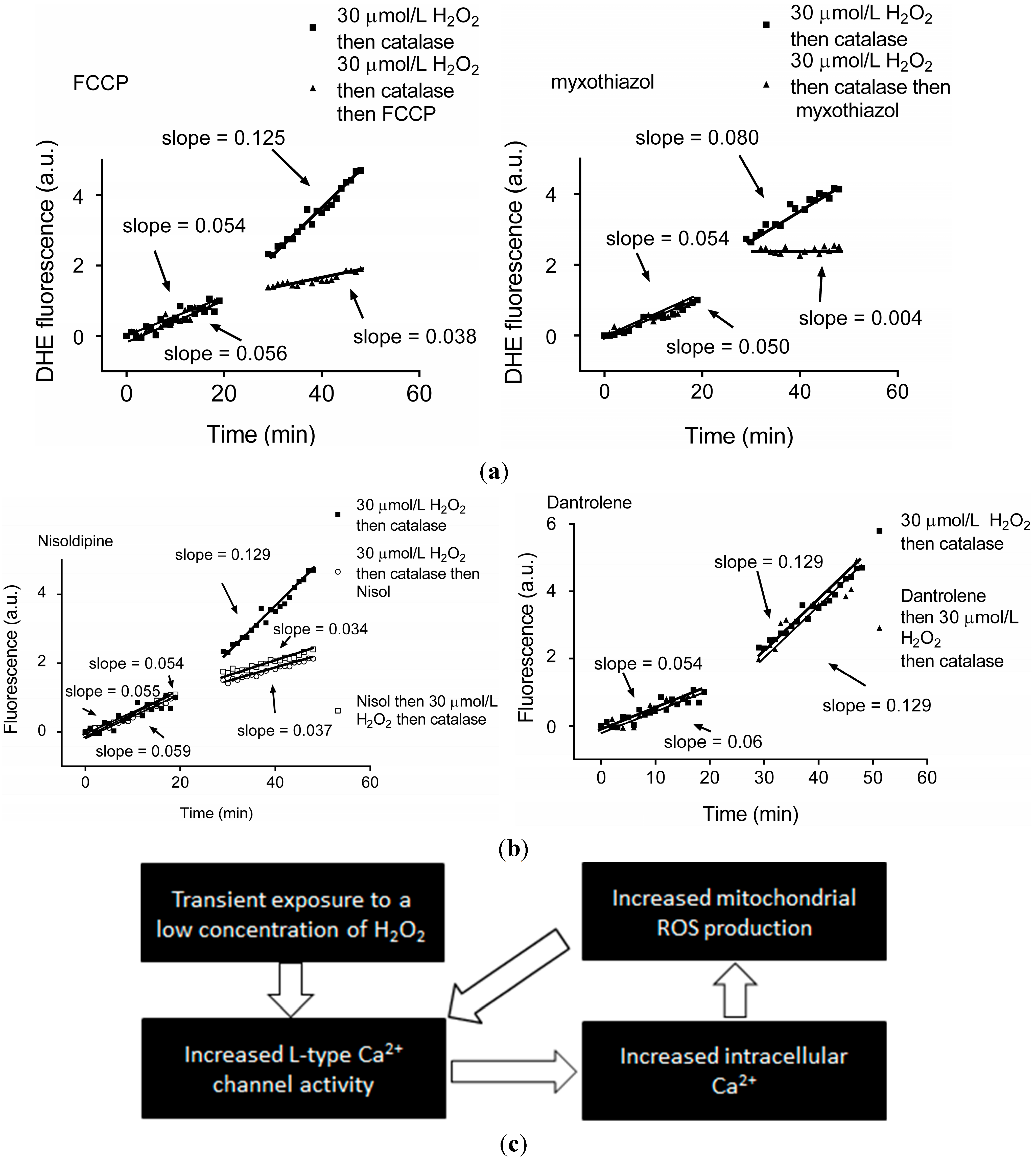
- Viola, H.M.; Arthur, P.G.; Hool, L.C. Transient exposure to hydrogen peroxide causes an increase in mitochondria-derived superoxide as a result of sustained alteration in L-type Ca2+ channel function in the absence of apoptosis in ventricular myocytes. Circ. Res. 2007, 100, 1036–1044. [Google Scholar] [CrossRef] [PubMed]
- Li, A.; Segui, J.; Heinemann, S.H.; Hoshi, T. Oxidation regulates cloned neuronal voltage-dependent Ca2+ channels expressed in xenopus oocytes. J. Neurosci. 1998, 18, 6740–6747. [Google Scholar] [PubMed]
- Chiamvimonvat, N.; O’Rourke, B.; Kamp, T.J.; Kallen, R.G.; Hofmann, F.; Flockerzi, V.; Marban, E. Functional consequences of sulfhydryl modification in the pore-forming subunits of cardiovascular Ca2+ and Na+ channels. Circ. Res. 1995, 76, 325–334. [Google Scholar] [CrossRef] [PubMed]
- Frey, N.; Olson, E.N. Cardiac hypertrophy: The good, the bad, and the ugly. Annu. Rev. Physiol. 2003, 65, 45–79. [Google Scholar] [CrossRef] [PubMed]
- Heineke, J.; Molkentin, J.D. Regulation of cardiac hypertrophy by intracellular signalling pathways. Nat. Rev. Mol. Cell. Biol. 2006, 7, 589–600. [Google Scholar] [CrossRef] [PubMed]
- Wilkins, B.J.; Molkentin, J.D. Calcium-calcineurin signaling in the regulation of cardiac hypertrophy. Biochem. Biophys. Res. Commun. 2004, 322, 1178–1191. [Google Scholar] [CrossRef] [PubMed]
- Takimoto, E.; Kass, D.A. Role of oxidative stress in cardiac hypertrophy and remodeling. Hypertension 2007, 49, 241–248. [Google Scholar] [CrossRef] [PubMed]
- Sag, C.M.; Santos, C.X.; Shah, A.M. Redox regulation of cardiac hypertrophy. J. Mol. Cell. Cardiol 2014, 73, 103–111. [Google Scholar] [PubMed]
- Hafstad, A.D.; Nabeebaccus, A.A.; Shah, A.M. Novel aspects of ROS signalling in heart failure. Basic Res. Cardiol. 2013, 108, 359. [Google Scholar] [CrossRef] [PubMed]
- Becker, L.B. New concepts in reactive oxygen species and cardiovascular reperfusion physiology. Cardiovasc. Res. 2004, 61, 461–470. [Google Scholar] [CrossRef] [PubMed]
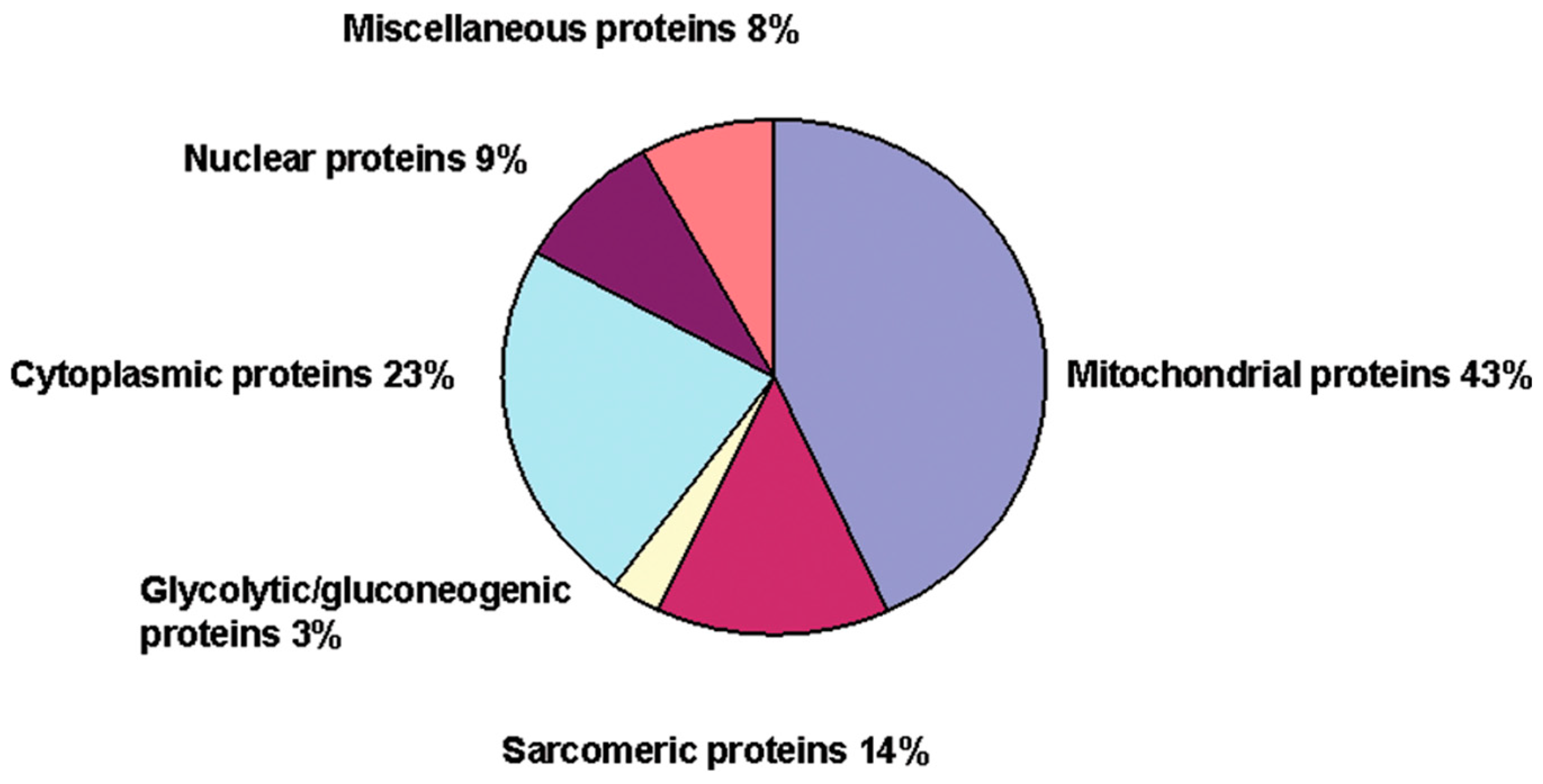
- Seenarain, V.; Viola, H.M.; Ravenscroft, G.; Casey, T.M.; Lipscombe, R.J.; Ingley, E.; Laing, N.G.; Bringans, S.D.; Hool, L.C. Evidence of altered guinea pig ventricular cardiomyocyte protein expression and growth in response to a 5 min in vitro exposure to H2O2. J. Proteome Res. 2010, 9, 1985–1994. [Google Scholar] [CrossRef] [PubMed]
- Anderson, M.E.; Brown, J.H.; Bers, D.M. Camkii in myocardial hypertrophy and heart failure. J. Mol. Cell. Cardiol. 2011, 51, 468–473. [Google Scholar] [CrossRef] [PubMed]
- Pokharel, S.; Sharma, U.C.; Pinto, Y.M. Left ventricular hypertrophy: Virtuous intentions, malign consequences. Int. J. Biochem. Cell. Biol. 2003, 35, 802–806. [Google Scholar] [CrossRef] [PubMed]
- St-Pierre, J.; Buckingham, J.A.; Roebuck, S.J.; Brand, M.D. Topology of superoxide production from different sites in the mitochondrial electron transport chain. J. Biol. Chem. 2002, 277, 44784–44790. [Google Scholar] [CrossRef] [PubMed]
- Jezek, P.; Hlavata, L. Mitochondria in homeostasis of reactive oxygen species in cell, tissues, and organism. Int. J. Biochem. Cell. Biol. 2005, 37, 2478–2503. [Google Scholar] [CrossRef] [PubMed]
- Viola, H.M.; Hool, L.C. Qo site of mitochondrial Complex III is the source of increased superoxide after transient exposure to hydrogen peroxide. J. Mol. Cell. Cardiol. 2010, 49, 875–885. [Google Scholar] [CrossRef] [PubMed]
- Vanden Hoek, T.L.; Becker, L.B.; Shao, Z.; Li, C.; Schumacker, P.T. Reactive oxygen species released from mitochondria during brief hypoxia induce preconditioning in cardiomyocytes. J. Biol. Chem. 1998, 273, 18092–18098. [Google Scholar]
- Collins, Y.; Chouchani, E.T.; James, A.M.; Menger, K.E.; Cocheme, H.M.; Murphy, M.P. Mitochondrial redox signalling at a glance. J. Cell. Sci. 2012, 125, 801–806. [Google Scholar] [CrossRef]
- Groitl, B.; Jakob, U. Thiol-based redox switches. Biochim. Biophys. Acta 2014, 1844, 1335–1343. [Google Scholar] [CrossRef] [PubMed]
- Ghezzi, P. Oxidoreduction of protein thiols in redox regulation. Biochem. Soc. Trans. 2005, 33, 1378–1381. [Google Scholar] [CrossRef]
- Ghezzi, P. Protein glutathionylation in health and disease. Biochim. Biophys. Acta 2013, 1830, 3165–3172. [Google Scholar] [CrossRef] [PubMed]
- Cooper, A.J.; Pinto, J.T.; Callery, P.S. Reversible and irreversible protein glutathionylation: Biological and clinical aspects. Expert Opin. Drug Metab. Toxicol. 2011, 7, 891–910. [Google Scholar] [CrossRef] [PubMed]
- Meister, A.; Anderson, M.E. Glutathione. Annu. Rev. Biochem. 1983, 52, 711–760. [Google Scholar] [CrossRef] [PubMed]
- Hurd, T.R.; Costa, N.J.; Dahm, C.C.; Beer, S.M.; Brown, S.E.; Filipovska, A.; Murphy, M.P. Glutathionylation of mitochondrial proteins. Antioxid. Redox Signal. 2005, 7, 999–1010. [Google Scholar] [CrossRef] [PubMed]
- Hurd, T.R.; Filipovska, A.; Costa, N.J.; Dahm, C.C.; Murphy, M.P. Disulphide formation on mitochondrial protein thiols. Biochem. Soc. Trans. 2005, 33, 1390–1393. [Google Scholar] [CrossRef] [PubMed]
- Ghezzi, P. Regulation of protein function by glutathionylation. Free Radic. Res. 2005, 39, 573–580. [Google Scholar] [CrossRef] [PubMed]
- Petrushanko, I.Y.; Yakushev, S.; Mitkevich, V.A.; Kamanina, Y.V.; Ziganshin, R.H.; Meng, X.; Anashkina, A.A.; Makhro, A.; Lopina, O.D.; Gassmann, M.; et al. S-Glutathionylation of the Na, k-atpase catalytic α subunit is a determinant of the enzyme redox sensitivity. J. Biol. Chem. 2012, 287, 32195–32205. [Google Scholar] [CrossRef] [PubMed]
- Fratelli, M.; Demol, H.; Puype, M.; Casagrande, S.; Eberini, I.; Salmona, M.; Bonetto, V.; Mengozzi, M.; Duffieux, F.; Miclet, E.; et al. Identification by redox proteomics of glutathionylated proteins in oxidatively stressed human t lymphocytes. Proc. Natl. Acad. Sci. USA 2002, 99, 3505–3510. [Google Scholar] [CrossRef] [PubMed]
- Cabiscol, E.; Levine, R.L. The phosphatase activity of carbonic anhydrase III is reversibly regulated by glutathiolation. Proc. Natl. Acad. Sci. USA 1996, 93, 4170–4174. [Google Scholar] [CrossRef] [PubMed]
- Klatt, P.; Molina, E.P.; Lamas, S. Nitric oxide inhibits c-jun DNA binding by specifically targeted S-glutathionylation. J. Biol. Chem. 1999, 274, 15857–15864. [Google Scholar] [CrossRef] [PubMed]
- Pineda-Molina, E.; Klatt, P.; Vazquez, J.; Marina, A.; Garcia de Lacoba, M.; Perez-Sala, D.; Lamas, S. Glutathionylation of the p50 subunit of NF-κB: A mechanism for redox-induced inhibition of DNA binding. Biochemistry 2001, 40, 14134–14142. [Google Scholar] [CrossRef] [PubMed]
- Davis, D.A.; Newcomb, F.M.; Starke, D.W.; Ott, D.E.; Mieyal, J.J.; Yarchoan, R. Thioltransferase (glutaredoxin) is detected within HIV-1 and can regulate the activity of glutathionylated HIV-1 protease in vitro. J. Biol. Chem. 1997, 272, 25935–25940. [Google Scholar] [CrossRef] [PubMed]
- Liang, J.N.; Pelletier, M.R. Destabilization of lens protein conformation by glutathione mixed disulfide. Exp. Eye Res. 1988, 47, 17–25. [Google Scholar] [CrossRef] [PubMed]
- Pastore, A.; Piemonte, F. Protein glutathionylation in cardiovascular diseases. Int. J. Mol. Sci. 2013, 14, 20845–20876. [Google Scholar] [CrossRef] [PubMed]
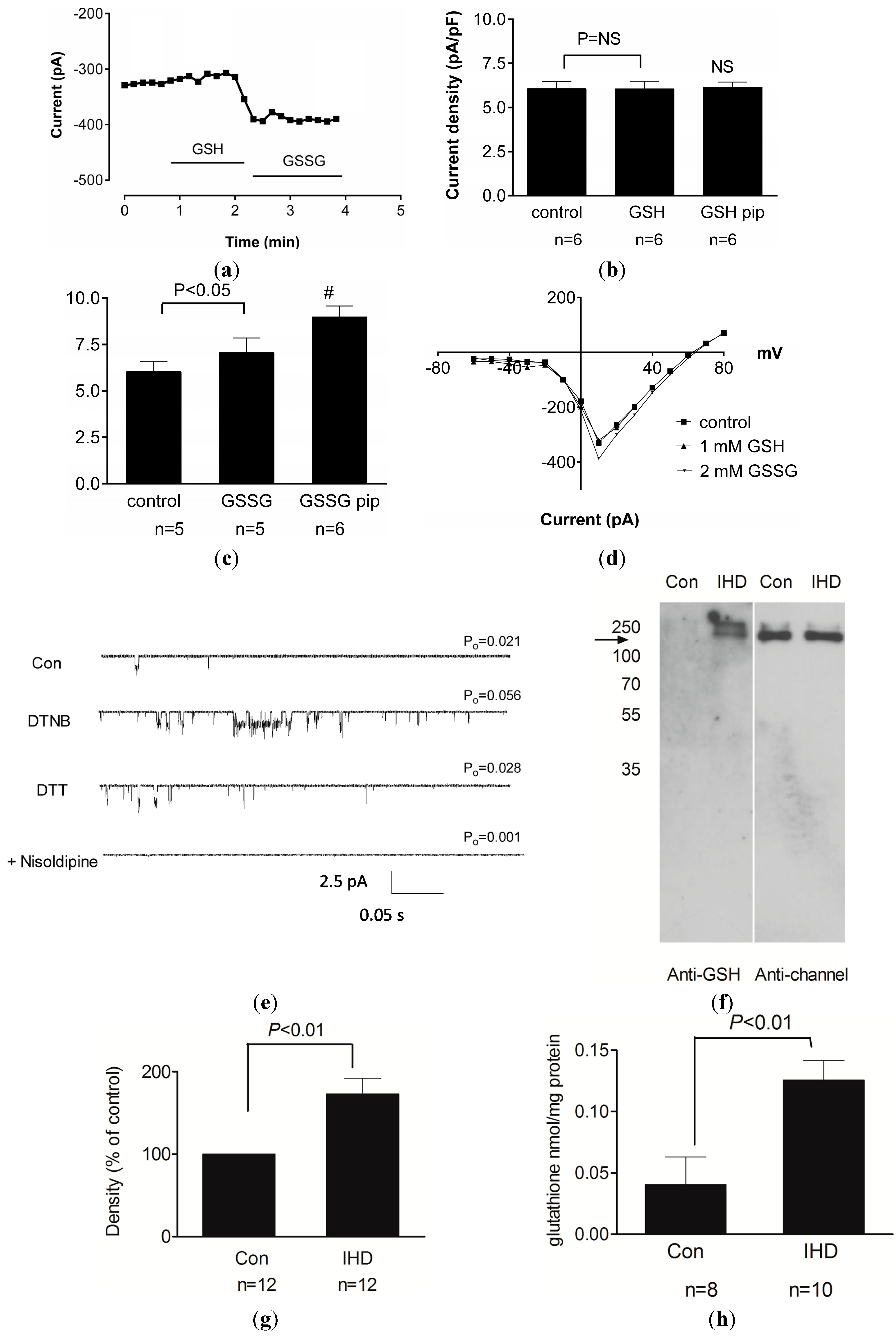
- Tang, H.; Viola, H.M.; Filipovska, A.; Hool, L.C. Cav1.2 calcium channel is glutathionylated during oxidative stress in guinea pig and ischemic human heart. Free Radic. Biol. Med. 2011, 51, 1501–1511. [Google Scholar] [CrossRef] [PubMed]
- Hudasek, K.; Brown, S.T.; Fearon, I.M. H2O2 regulates recombinant Ca2+ channel α1c subunits but does not mediate their sensitivity to acute hypoxia. Biochem. Biophys. Res. Commun. 2004, 318, 135–141. [Google Scholar] [CrossRef] [PubMed]
- Yang, L.; Xu, J.; Minobe, E.; Yu, L.; Feng, R.; Kameyama, A.; Yazawa, K.; Kameyama, M. Mechanisms underlying the modulation of L-type Ca2+ channel by hydrogen peroxide in guinea pig ventricular myocytes. J. Physiol. Sci. 2013, 63, 419–426. [Google Scholar] [CrossRef] [PubMed]
- Fernandes, A.P.; Holmgren, A. Glutaredoxins: Glutathione-dependent redox enzymes with functions far beyond a simple thioredoxin backup system. Antioxid. Redox Signal. 2004, 6, 63–74. [Google Scholar] [CrossRef] [PubMed]
- Jerng, H.H.; Pfaffinger, P.J. S-Glutathionylation of an auxiliary subunit confers redox sensitivity to Kv4 channel inactivation. PLoS One 2014, 9, e93315. [Google Scholar] [CrossRef] [PubMed]
- Kemp, M.; Donovan, J.; Higham, H.; Hooper, J. Biochemical markers of myocardial injury. Br. J. Anaesth. 2004, 93, 63–73. [Google Scholar] [CrossRef] [PubMed]
- Dalle-Donne, I.; Rossi, R.; Colombo, G.; Giustarini, D.; Milzani, A. Protein S-glutathionylation: A regulatory device from bacteria to humans. Trends Biochem. Sci. 2009, 34, 85–96. [Google Scholar] [CrossRef] [PubMed]
- Yang, Y.; Shi, W.; Chen, X.; Cui, N.; Konduru, A.S.; Shi, Y.; Trower, T.C.; Zhang, S.; Jiang, C. Molecular basis and structural insight of vascular KATP channel gating by S-glutathionylation. J. Biol. Chem. 2011, 286, 9298–9307. [Google Scholar] [CrossRef] [PubMed]
- Bredt, D.S.; Snyder, S.H. Nitric oxide: A physiologic messenger molecule. Annu. Rev. Biochem. 1994, 63, 175–195. [Google Scholar] [CrossRef] [PubMed]
- Broniowska, K.A.; Hogg, N. The chemical biology of S-nitrosothiols. Antioxid. Redox Signal. 2012, 17, 969–980. [Google Scholar] [CrossRef] [PubMed]
- Campbell, D.L.; Stamler, J.S.; Strauss, H.C. Redox modulation of L-type calcium channels in ferret ventricular myocytes. Dual mechanism regulation by nitric oxide and S-nitrosothiols. J. Gen. Physiol. 1996, 108, 277–293. [Google Scholar] [CrossRef] [PubMed]
- Hu, H.; Chiamvimonvat, N.; Yamagishi, T.; Marban, E. Direct inhibition of expressed cardiac L-type Ca2+ channels by S-nitrosothiol nitric oxide donors. Circ. Res. 1997, 81, 742–752. [Google Scholar] [CrossRef] [PubMed]
- Grech, E.D. Pathophysiology and investigation of coronary artery disease. BMJ 2003, 326, 1027–1030. [Google Scholar] [CrossRef] [PubMed]
- Carden, D.L.; Granger, D.N. Pathophysiology of ischaemia-reperfusion injury. J. Pathol. 2000, 190, 255–266. [Google Scholar] [CrossRef] [PubMed]
- Eltzschig, H.K.; Collard, C.D. Vascular ischaemia and reperfusion injury. Br. Med. Bull. 2004, 70, 71–86. [Google Scholar] [CrossRef] [PubMed]
- Solares, J.; Garcia-Dorado, D.; Oliveras, J.; Gonzalez, M.A.; Ruiz-Meana, M.; Barrabes, J.A.; Gonzalez-Bravo, C.; Soler-Soler, J. Contraction band necrosis at the lateral borders of the area at risk in reperfused infarcts. Observations in a pig model of in situ coronary occlusion. Virchows Arch. 1995, 426, 393–399. [Google Scholar] [CrossRef] [PubMed]
- Whalen, D.A.; Hamilton, D.G.; Ganote, C.E.; Jennings, R.B. Effect of a transient period of ischemia on myocardial cells. I. Effects on cell volume regulation. Am. J. Pathol. 1974, 74, 381–398. [Google Scholar] [PubMed]
- Kloner, R.A.; Ganote, C.E.; Whalen, D.A., Jr.; Jennings, R.B. Effect of a transient period of ischemia on myocardial cells. II. Fine structure during the first few minutes of reflow. Am. J. Pathol. 1974, 74, 399–422. [Google Scholar] [PubMed]
- Gross, G.J.; Kersten, J.R.; Warltier, D.C. Mechanisms of postischemic contractile dysfunction. Ann. Thorac. Surg. 1999, 68, 1898–1904. [Google Scholar] [CrossRef] [PubMed]
- Piper, H.M.; Garcia-Dorado, D. Prime causes of rapid cardiomyocyte death during reperfusion. Ann. Thorac. Surg. 1999, 68, 1913–1919. [Google Scholar] [CrossRef] [PubMed]
- Ambrosio, G.; Weisfeldt, M.L.; Jacobus, W.E.; Flaherty, J.T. Evidence for a reversible oxygen radical-mediated component of reperfusion injury: Reduction by recombinant human superoxide dismutase administered at the time of reflow. Circulation 1987, 75, 282–291. [Google Scholar] [CrossRef] [PubMed]
- VanBenthuysen, K.M.; McMurtry, I.F.; Horwitz, L.D. Reperfusion after acute coronary occlusion in dogs impairs endothelium-dependent relaxation to acetylcholine and augments contractile reactivity in vitro. J. Clin. Investig. 1987, 79, 265–274. [Google Scholar] [CrossRef] [PubMed]
- Pearson, P.J.; Schaff, H.V.; Vanhoutte, P.M. Long-term impairment of endothelium-dependent relaxations to aggregating platelets after reperfusion injury in canine coronary arteries. Circulation 1990, 81, 1921–1927. [Google Scholar] [CrossRef] [PubMed]
- Bolli, R.; Marban, E. Molecular and cellular mechanisms of myocardial stunning. Physiol. Rev. 1999, 79, 609–634. [Google Scholar] [PubMed]
- Wong, D.T.; Puri, R.; Richardson, J.D.; Worthley, M.I.; Worthley, S.G. Myocardial “no-reflow”—Diagnosis, pathophysiology and treatment. Int. J. Cardiol. 2013, 167, 1798–1806. [Google Scholar] [CrossRef] [PubMed]
- Galasso, G.; Schiekofer, S.; D’Anna, C.; Gioia, G.D.; Piccolo, R.; Niglio, T.; Rosa, R.D.; Strisciuglio, T.; Cirillo, P.; Piscione, F.; et al. No-reflow phenomenon: Pathophysiology, diagnosis, prevention, and treatment. A review of the current literature and future perspectives. Angiology 2014, 65, 180–189. [Google Scholar] [CrossRef] [PubMed]
- Lefer, A.M.; Tsao, P.S.; Lefer, D.J.; Ma, X.L. Role of endothelial dysfunction in the pathogenesis of reperfusion injury after myocardial ischemia. FASEB J. 1991, 5, 2029–2034. [Google Scholar] [PubMed]
- Szocs, K. Endothelial dysfunction and reactive oxygen species production in ischemia/reperfusion and nitrate tolerance. Gen. Physiol. Biophys. 2004, 23, 265–295. [Google Scholar] [PubMed]
- Zweier, J.L.; Talukder, M.A. The role of oxidants and free radicals in reperfusion injury. Cardiovasc. Res. 2006, 70, 181–190. [Google Scholar] [CrossRef] [PubMed]
- Bolli, R.; Jeroudi, M.O.; Patel, B.S.; DuBose, C.M.; Lai, E.K.; Roberts, R.; McCay, P.B. Direct evidence that oxygen-derived free radicals contribute to postischemic myocardial dysfunction in the intact dog. Proc. Natl. Acad. Sci. USA 1989, 86, 4695–4699. [Google Scholar] [CrossRef] [PubMed]
- Viola, H.M.; Arthur, P.G.; Hool, L.C. Evidence for regulation of mitochondrial function by the L-type Ca2+ channel in ventricular myocytes. J. Mol. Cell. Cardiol. 2009, 46, 1016–1026. [Google Scholar] [CrossRef] [PubMed]
- Hool, L.C. Evidence for the regulation of L-type Ca2+ channels in the heart by reactive oxygen species: Mechanism for mediating pathology. Clin. Exp. Pharmacol. Physiol. 2008, 35, 229–234. [Google Scholar] [PubMed]
- Catterall, W.A. Structure and regulation of voltage-gated Ca2+ channels. Annu. Rev. Cell. Dev. Biol. 2000, 16, 521–555. [Google Scholar] [CrossRef] [PubMed]
- Takahashi, M.; Catterall, W.A. Dihydropyridine-sensitive calcium channels in cardiac and skeletal muscle membranes: Studies with antibodies against the α subunits. Biochemistry 1987, 26, 5518–5526. [Google Scholar] [CrossRef] [PubMed]
- Davies, A.; Hendrich, J.; van Minh, A.T.; Wratten, J.; Douglas, L.; Dolphin, A.C. Functional biology of the α2δ subunits of voltage-gated calcium channels. Trends Pharmacol. Sci. 2007, 28, 220–228. [Google Scholar] [CrossRef] [PubMed]
- Dolphin, A.C. β Subunits of voltage-gated calcium channels. J. Bioenerg. Biomembr. 2003, 35, 599–620. [Google Scholar] [CrossRef] [PubMed]
- Hohaus, A.; Person, V.; Behlke, J.; Schaper, J.; Morano, I.; Haase, H. The carboxyl-terminal region of ahnak provides a link between cardiac L-type Ca2+ channels and the actin-based cytoskeleton. FASEB J. 2002, 16, 1205–1216. [Google Scholar] [CrossRef] [PubMed]
- Haase, H.; Alvarez, J.; Petzhold, D.; Doller, A.; Behlke, J.; Erdmann, J.; Hetzer, R.; Regitz-Zagrosek, V.; Vassort, G.; Morano, I. Ahnak is critical for cardiac Cav1.2 calcium channel function and its β-adrenergic regulation. FASEB J. 2005, 19, 1969–1977. [Google Scholar] [CrossRef] [PubMed]
- Alvarez, J.; Hamplova, J.; Hohaus, A.; Morano, I.; Haase, H.; Vassort, G. Calcium current in rat cardiomyocytes is modulated by the carboxyl-terminal ahnak domain. J. Biol. Chem. 2004, 279, 12456–12461. [Google Scholar] [CrossRef] [PubMed]
- Hohaus, A.; Poteser, M.; Romanin, C.; Klugbauer, N.; Hofmann, F.; Morano, I.; Haase, H.; Groschner, K. Modulation of the smooth-muscle L-type Ca2+ channel α1 subunit (α1c-b) by the β2a subunit: A peptide which inhibits binding of β to the I–II linker of α1 induces functional uncoupling. Biochem. J. 2000, 348, 657–665. [Google Scholar] [CrossRef] [PubMed]
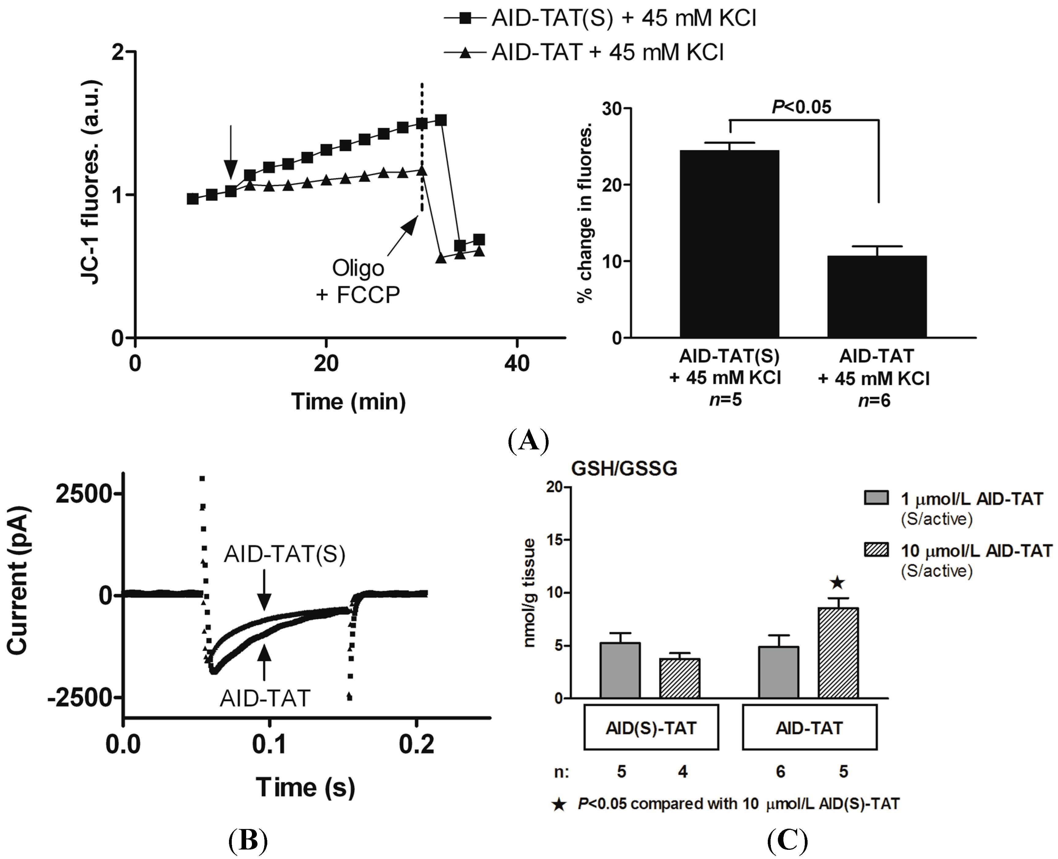
- Viola, H.M.; Jordan, M.C.; Roos, K.R.; Hool, L.C. Decreased myocardial injury and improved contractility after administration of a peptide derived against the α-interacting domain of the L-type calcium channel. J. Am. Heart Assoc. 2014, 3, e000961. [Google Scholar] [CrossRef] [PubMed]
- Viola, H.M.; Adams, A.M.; Davies, S.M.; Fletcher, S.; Filipovska, A.; Hool, L.C. Impaired functional communication between the L-type calcium channel and mitochondria contributes to metabolic inhibition in the mdx heart. Proc. Natl. Acad. Sci. USA 2014, 111, E2905–E2914. [Google Scholar] [CrossRef] [PubMed]
- Hammerman, H.; Moscovitz, M.; Hir, J. Beneficial effect of nisoldipine in repeated coronary reperfusion. Coron. Artery Dis. 1997, 8, 97–100. [Google Scholar] [CrossRef] [PubMed]
- Sheiban, I.; Tonni, S.; Chizzoni, A.; Marini, A.; Trevi, G. Recovery of left ventricular function following early reperfusion in acute myocardial infarction: A potential role for the calcium antagonist nisoldipine. Cardiovasc. Drugs Ther. 1997, 11, 5–16. [Google Scholar] [CrossRef] [PubMed]
- Liu, X.; Engelman, R.M.; Wei, Z.; Bagchi, D.; Rousou, J.A.; Nath, D.; Das, D.K. Attenuation of myocardial reperfusion injury by reducing intracellular calcium overloading with dihydropyridines. Biochem. Pharmacol. 1993, 45, 1333–1341. [Google Scholar] [CrossRef] [PubMed]
- Akita, T.; Abe, T.; Kato, S.; Kodama, I.; Toyama, J. Protective effects of diltiazem and ryanodine against ischemia-reperfusion injury in neonatal rabbit hearts. J. Thorac. Cardiovasc. Surg. 1993, 106, 55–66. [Google Scholar] [PubMed]
- Herzog, W.R.; Vogel, R.A.; Schlossberg, M.L.; Edenbaum, L.R.; Scott, H.J.; Serebruany, V.L. Short-term low dose intracoronary diltiazem administered at the onset of reperfusion reduces myocardial infarct size. Int. J. Cardiol. 1997, 59, 21–27. [Google Scholar] [CrossRef] [PubMed]
- Borchgrevink, P.C.; Bergan, A.S.; Bakoy, O.E.; Jynge, P. Magnesium and reperfusion of ischemic rat heart as assessed by 31p-NMR. Am. J. Physiol. 1989, 256, H195–H204. [Google Scholar] [PubMed]
- Hara, A.; Matsumura, H.; Abiko, Y. Beneficial effect of magnesium on the isolated perfused rat heart during reperfusion after ischaemia: Comparison between pre-ischaemic and post-ischaemic administration of magnesium. N-S Arch. Pharmacol. 1990, 342, 100–106. [Google Scholar] [CrossRef]
- Bolli, R.; Becker, L.; Gross, G.; Mentzer, R., Jr.; Balshaw, D.; Lathrop, D.A. Myocardial protection at a crossroads: The need for translation into clinical therapy. Circ. Res. 2004, 95, 125–134. [Google Scholar] [CrossRef] [PubMed]
- Cannon, R.O., 3rd. Mechanisms, management and future directions for reperfusion injury after acute myocardial infarction. Nat. Clin. Pract. Cardiovasc. Med. 2005, 2, 88–94. [Google Scholar] [CrossRef] [PubMed]
- Dirksen, M.T.; Laarman, G.J.; Simoons, M.L.; Duncker, D.J. Reperfusion injury in humans: A review of clinical trials on reperfusion injury inhibitory strategies. Cardiovasc. Res. 2007, 74, 343–355. [Google Scholar] [CrossRef] [PubMed]
- Clozel, J.P.; Veniant, M.; Osterrieder, W. The structurally novel Ca2+ channel blocker Ro 40–5967, which binds to the [3h] desmethoxyverapamil receptor, is devoid of the negative inotropic effects of verapamil in normal and failing rat hearts. Cardiovasc. Drugs Ther. 1990, 4, 731–736. [Google Scholar] [CrossRef] [PubMed]
- Billman, G.E. Ro 40–5967, a novel calcium channel antagonist, protects against ventricular fibrillation. Eur. J. Pharmacol. 1992, 229, 179–187. [Google Scholar] [CrossRef] [PubMed]
- Reimer, K.A.; Califf, R.M. Good news for experimental concept but bad news for clinically effective therapy. Circulation 1999, 99, 198–200. [Google Scholar] [CrossRef] [PubMed]
© 2014 by the authors; licensee MDPI, Basel, Switzerland. This article is an open access article distributed under the terms and conditions of the Creative Commons Attribution license (http://creativecommons.org/licenses/by/4.0/).
Share and Cite
Johnstone, V.P.A.; Hool, L.C. Glutathionylation of the L-type Ca2+ Channel in Oxidative Stress-Induced Pathology of the Heart. Int. J. Mol. Sci. 2014, 15, 19203-19225. https://doi.org/10.3390/ijms151019203
Johnstone VPA, Hool LC. Glutathionylation of the L-type Ca2+ Channel in Oxidative Stress-Induced Pathology of the Heart. International Journal of Molecular Sciences. 2014; 15(10):19203-19225. https://doi.org/10.3390/ijms151019203
Chicago/Turabian StyleJohnstone, Victoria P. A., and Livia C. Hool. 2014. "Glutathionylation of the L-type Ca2+ Channel in Oxidative Stress-Induced Pathology of the Heart" International Journal of Molecular Sciences 15, no. 10: 19203-19225. https://doi.org/10.3390/ijms151019203
APA StyleJohnstone, V. P. A., & Hool, L. C. (2014). Glutathionylation of the L-type Ca2+ Channel in Oxidative Stress-Induced Pathology of the Heart. International Journal of Molecular Sciences, 15(10), 19203-19225. https://doi.org/10.3390/ijms151019203