Article

Dynamic TMT-Based Quantitative Proteomics Analysis of Critical Initiation Process of Totipotency during Cotton Somatic Embryogenesis Transdifferentiation

State Key Laboratory of Crop Biology, College of Agronomy, Shandong Agricultural University, Tai’an 271018, China
*
Author to whom correspondence should be addressed.
These authors contributed equally to this work.
Int. J. Mol. Sci. 2019, 20(7), 1691; https://doi.org/10.3390/ijms20071691
Submission received: 21 February 2019 / Revised: 27 March 2019 / Accepted: 2 April 2019 / Published: 4 April 2019

Abstract

:
The somatic embryogenesis (SE) process of plants, as one of the typical responses to abiotic stresses with hormone, occurs through the dynamic expression of different proteins that constitute a complex regulatory network in biological activities and promotes plant totipotency. Plant SE includes two critical stages: primary embryogenic calli redifferentiation and somatic embryos development initiation, which leads to totipotency. The isobaric labels tandem mass tags (TMT) large-scale and quantitative proteomics technique was used to identify the dynamic protein expression changes in nonembryogenic calli (NEC), primary embryogenic calli (PEC) and globular embryos (GEs) of cotton. A total of 9369 proteins (6730 quantified) were identified; 805, 295 and 1242 differentially accumulated proteins (DAPs) were identified in PEC versus NEC, GEs versus PEC and GEs versus NEC, respectively. Eight hundred and five differentially abundant proteins were identified, 309 of which were upregulated and 496 down regulated in PEC compared with NEC. Of the 295 DAPs identified between GEs and PEC, 174 and 121 proteins were up- and down regulated, respectively. Of 1242 differentially abundant proteins, 584 and 658 proteins were up- and down regulated, respectively, in GEs versus NEC. We have also complemented the authenticity and accuracy of the proteomic analysis. Systematic analysis indicated that peroxidase, photosynthesis, environment stresses response processes, nitrogen metabolism, phytohormone response/signal transduction, transcription/posttranscription and modification were involved in somatic embryogenesis. The results generated in this study demonstrate a proteomic molecular basis and provide a valuable foundation for further investigation of the roles of DAPs in the process of SE transdifferentiation during cotton totipotency.

Graphical Abstract

1. Introduction

Somatic embryogenesis (SE) is a notable illustration of cell totipotency as one of the typical responses to abiotic stresses with hormone, which processes the developmental reprogramming of somatic cells toward the embryogenesis pathway. Cotton (Gossypium hirsutum L.), as the foremost natural fiber source [1] and one of the most important economic crops worldwide, has a global socioeconomic impact worth approximately $56 billion [2]. However, plant regeneration in SE is still a limiting method for transgenic development in cotton [1,3]. Somatic embryogenesis represents a unique phenomenon in the plant kingdom [4]. This developmental pathway is one of the most striking examples of plant cell developmental plasticity [5,6]. It includes a series of characteristic events, including somatic dedifferentiation, cell division activation, metabolism alterations and gene expression pattern reprogramming [4]. During SE, the development of somatic cells is reprogrammed to the embryogenic pathway, and SE forms the basis of cellular totipotency in higher plants [7]. Each transformed cell has the potential to produce a plant from the callus [8]. Somatic embryogenesis and subsequent plant regeneration have been reported in most major crop varieties [9]. Soybeans and cotton have proven to be the most difficult to regenerate [10].
In cotton, only a few percent of somatic embryos are able to mature and regenerate into plantlets. Most embryos develop abnormally, redifferentiate in to calli, or become necrotic and die [11]. Sakhanokho and Rajasekaran [12] obtained a variety of factors affecting cotton during in vitro regeneration, including plant growth regulators, explants, compositions of media and environmental conditions. The transition of somatic cells into embryogenic cells is the most intriguing and the least understood part of somatic embryogenesis [13,14,15]. Now, it is generally accepted that stress and hormones play a crucial role in collectively inducing cell dedifferentiation and initiation of the embryogenic program in plants with responsive genotypes [16,17,18].
Since the first observations of somatic embryo formation in suspension cultures of carrot cells by Stewards [7] and Reinert [19], the potential for SE has been demonstrated to be characteristic of extensive tissue culture systems from both dicotyledonous and monocotyledonous plants [20,21]. Considerable efforts have been expended in identifying the various factors that control SE [22,23]. An important gene that marks embryonic cells is the transcription factor gene WUSCHEL (WUS) [24]. Using a genetic gain-of-function screening approach, Zuo et al. [25] found that overexpression of WUS in roots, leaf petioles, stems, or leaves of Arabidopsis can induce the formation of somatic embryos. These results indicate that WUS participates in the promotion and/or maintenance of totipotent embryogenic stem cells. However, the wus mutants are still able to produce somatic embryos, suggesting that multiple alternative pathways can lead to the expression of totipotent potential. WUS is the only transcription factor that has been found to be involved in regulating meristematic stem cells (pluripotent) and embryogenic stem cells (totipotent) [15]. In addition, when auxin biosynthesis rates were manipulated in Arabidopsis embryos, polar auxin transport activity apparently buffered the normal distribution of auxin, suggesting a compensatory mechanism for buffering auxin gradients in the embryo, with PIN1 and PIN4 being the most important genes [26]. The results by Su [27] suggested that the establishment of auxin gradients and the polar distribution of PIN1 are critical for the regulation of WUS expression during somatic embryogenesis. ERF plays an important role in hormone signal transduction and interconnecting different hormone pathways [28]. Inhibition of gibberellin (GA) biosynthesis increases the fraction of lec1-tnp seedlings displaying the mutant phenotype, suggesting that reduced GA levels enhance maturation processes induced by LEAFY COTYLEDON 1 (LEC1) [29]. The plant hormone abscisic acid (ABA) regulates many important plant developmental processes and induces epigenetic reprogramming against tolerance to different stresses, including drought, salinity, low temperature and some pathogens [30,31]. ABA serves as a critical chemical messenger for stress responses. The roles of several genes in somatic embryogenesis masses (SEM) have been well-characterized, including Arabinogalactan protein 1 (AGP1) [32], Glutathione-S-transferase (GST) [33], SOMATIC EMBRYO RELATED FACTOR1 (MtSERF1) [34], BABY BOOM (BBM) [35], Agamous-like 15 (AGL15) [36,37] and SOMATIC EMBRYOGENESIS RECEPTOR-LIKE KINASE (SERK) from Daucus carota, which was the first identified marker gene with a crucial role in SEM [38].
At present, a great number of SE-related genes and transcription factors have been identified at the transcription level. For example, Zeng [39], with the suppression subtractive hybridization (SSH) technique, identified 671 cDNAs in the initial period of SE in cotton. Nonetheless, reports on the identification of cotton SE at high-throughput proteins levels are still insufficient, especially during the initial stage of SE transdifferentiation. Proteomics is a powerful approach aimed at systematic studies of protein structure, function, interaction, and dynamics [4]. To further investigate the molecular regulatory mechanisms of somatic embryogenesis, the protein dynamics of NEC, PEC and GE were identified by TMT quantitative proteomics techniques. The highly sensitive proteomic platform based on the isobaric labels tandem mass tags was recently developed as one of the most robust proteomics techniques [40,41]. Through identification and annotation of DAPs, we uncovered the key genes/proteins and pathways involved in cotton SE transdifferentiation. The results generated in this study provide a valuable foundation for further investigation of the roles of DAPs in cotton SE.

2. Results

2.1. Somatic Embryogenesis in Cotton

PEC were formed from NEC after approximately 3 months of culture. The initial development period of GEs from embryogenic callus was the most restrictive step during cotton SEM (Figure 1). To identify proteins related to SE and morphogenesis in cotton, we sampled the critical representative periods of NEC, PEC and GEs for protein preparation and TMT-based quantitative proteomics analyses.

2.2. TMT-Based Quantitative Proteomic Basis Data Analysis and Overall Protein Identification

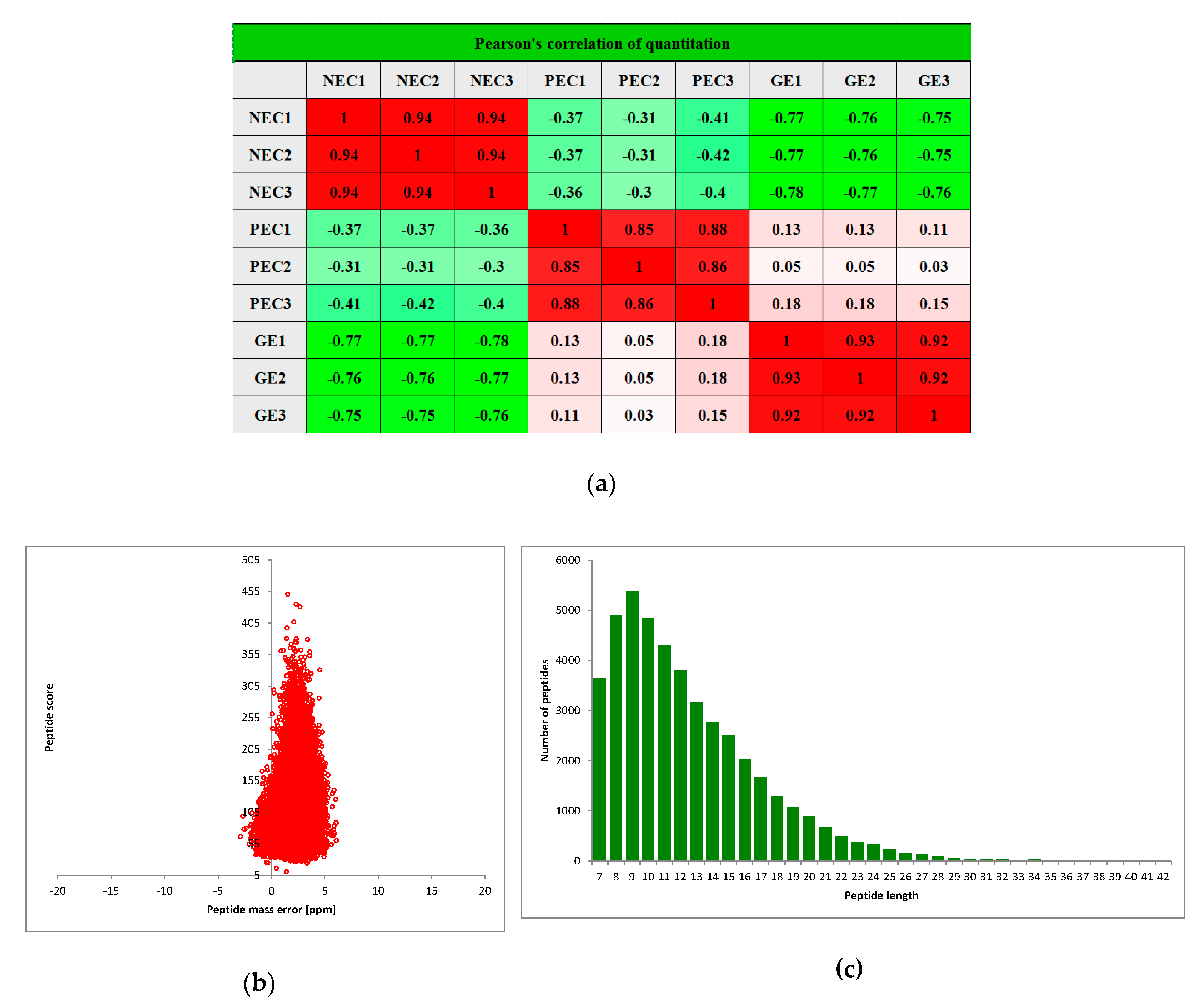
TMT-based quantitative proteomics was conducted to assess protein changes among NEC, PEC and GEs in cotton. Pair wise Pearson’s correlation coefficients displayed sufficient reproducibility of this experiment (Figure 2a). After quality validation, a total of 360,720 (74,579 matched) spectra were obtained. Of these spectra, 45062 identified peptides (27,673 unique peptides) and 9369 identified proteins (6730 quantified proteins) were detected (Table 1), and the average peptides mass error was <10 ppm, indicating a high mass accuracy of the MS data (Figure 2b). The lengths of most identified peptides were 8 to 20 amino acid residues (Figure 2c), suggesting that our sampling met the required standard. The detail information of identified proteins, including protein accession, protein description, gene name, peptide number, matching scores, carried charges and delta mass, is shown in Supplementary Table S1.
To further understand their functions, all identified proteins were annotated according to different categories, including subcellular localizations, Gene Ontology (GO) terms, Kyoto Encyclopedia of Genes and Genomes (KEGG) pathways, predicted functional domains and other data. The detailed information of all identified proteins is listed in Supplementary Table S1.

2.3. Enrichment of the Chloroplast Subcellular Location and GO Functional Classification of All Identified Proteins

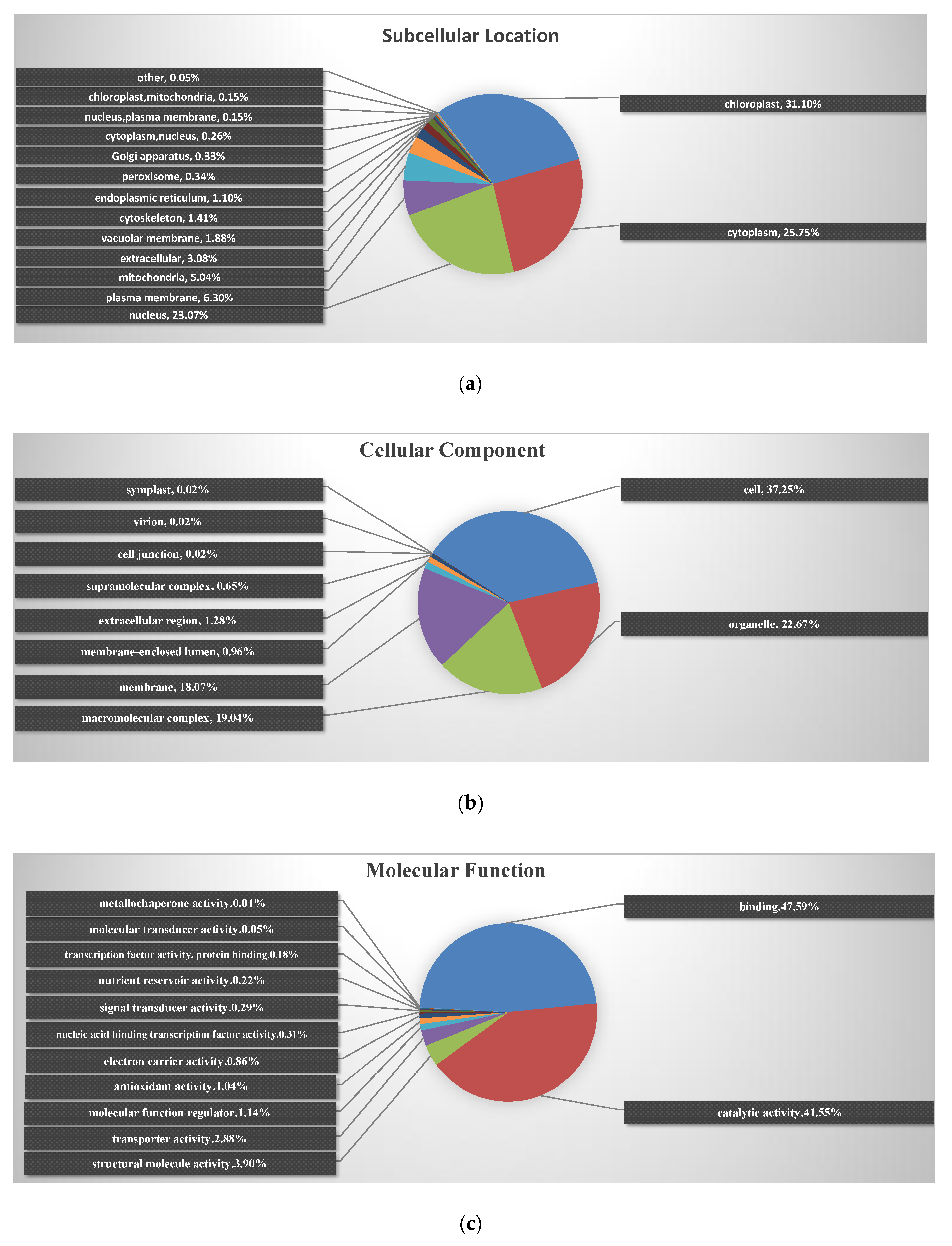
To characterize the subcellular locations and functions of the identified differential proteins among NEC, PEC and GEs in cotton, subcellular locations and GO functional classification were performed (Figure 3; Supplementary Table S1). Subcellular distribution predictions (Figure 3a) showed that the identified proteins were distributed predominantly in chloroplast (31.10%), cytoplasm (25.75%) and nucleus (23.07%) during the transformation periods of somatic embryogenesis. Significantly, the highest proportion of differential proteins was enriched in chloroplasts, highlighting that this organelle plays an important role in cotton SE.
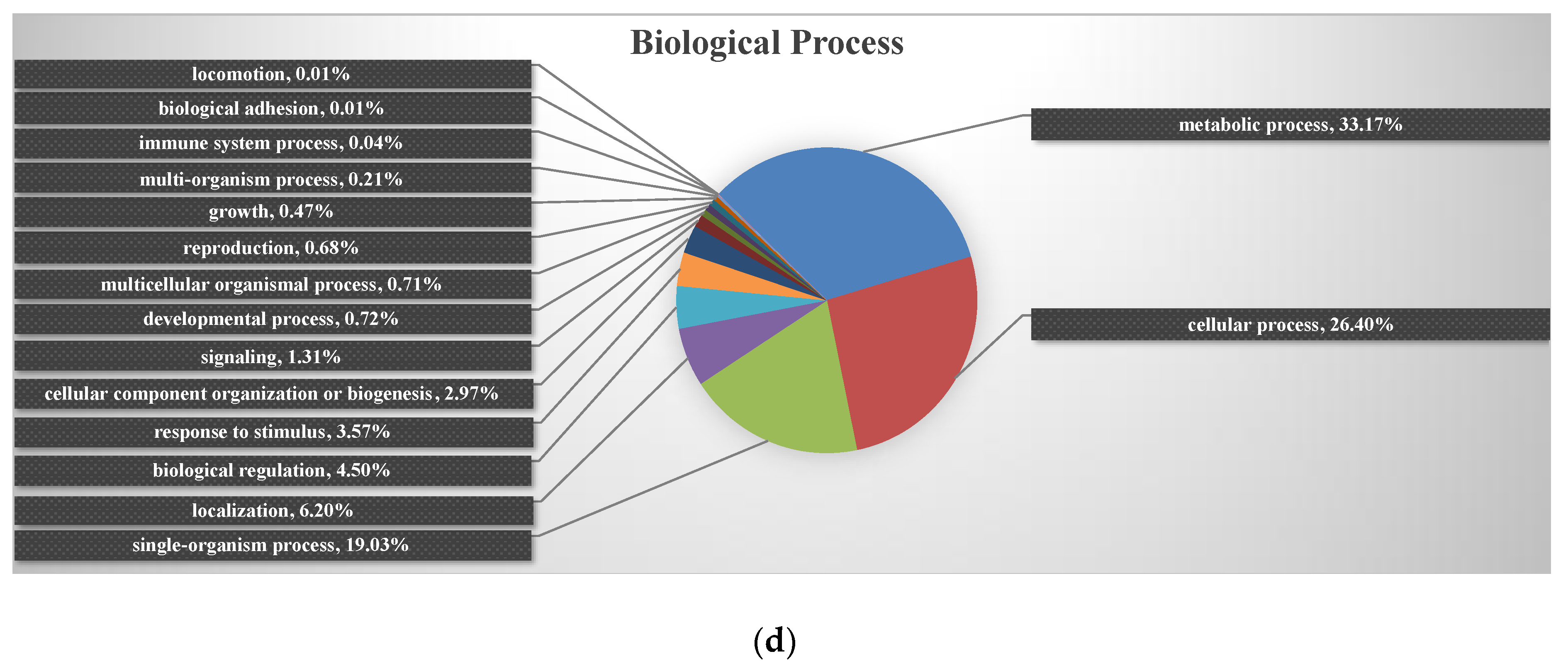
The results of cellular component analysis further revealed that 22.67% of the identified proteins were catalogued in organelles, 19.04% with macromolecular complexes and 18.07% with the membrane (Figure 3b). Regarding molecular function, the largest two GO categories, binding and catalytic activity, accounted for 47.59 and 41.55% of the identified proteins, respectively (Figure 3c). At the biological process level, proteins involved in the metabolic process, cellular process and single-organism process accounted for 33.17, 26.40 and 19.03% of identified proteins, respectively (Figure 3d). These results demonstrated that the identified proteins are found in multiple cellular components, have diversified molecular functions, and are involved in a variety of biological processes.

2.4. Identification of Differentially Abundant Proteins

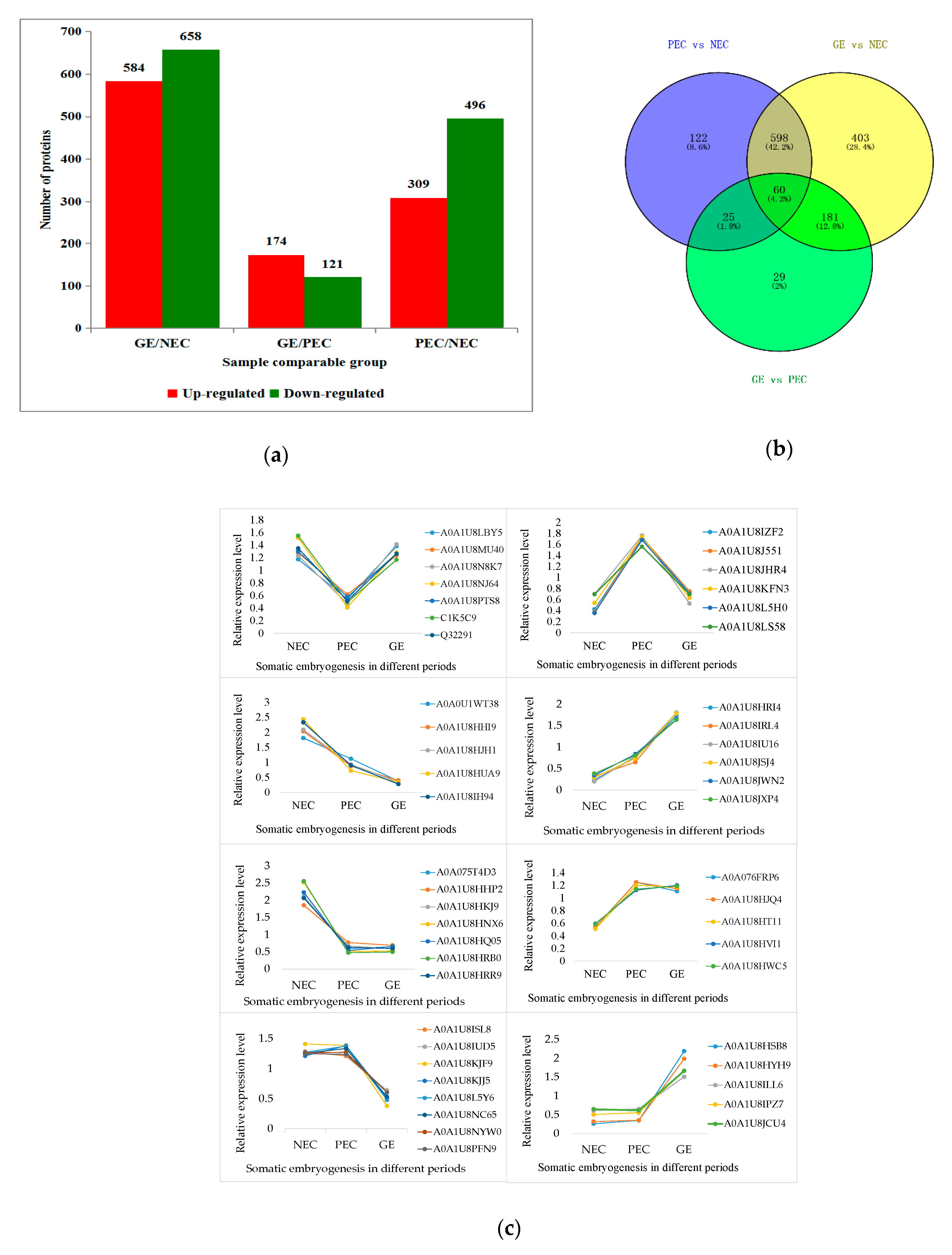
Differentially abundant proteins were defined as those with a ≥2-fold or ≤0.5-fold change in relative abundance (p < 0.05) between PEC and NEC, GEs and PEC, and GEs and NEC. In total, 805, 295 and 1242 DAPs were identified in comparing PEC versus NEC, GEs versus PEC and GEs versus NEC, respectively. In PEC compared with NEC, 805 proteins differentially accumulated were identified, 309 of which were up regulated and 496 of which were down-regulated. Of the 295 DAPs identified between GEs and PEC, 174 and 121 proteins were up- and down regulated in GEs, respectively. Of 1242 proteins differentially accumulated in GEs compared to NEC, 584 and 658 proteins were up- and down regulated, respectively (Figure 4a; Supplementary Table S2).
To identify the commonly and specifically changed proteins between PEC and NEC, GEs and PEC or between GEs and NEC, a Venn diagram was generated (Figure 4b). It clearly showed that 122 and 29 proteins were specifically expressed in PEC and GE processes, respectively, and 85 common proteins (25 and 60) were involved in both PEC and GE.
To investigate the overall dynamics of proteome changes in SEM, we performed eight types of protein expression pattern analyses for the DAPs identified in NEC, PEC and GEs (Figure 4c). These analyses suggested protein expression patterns including down- to up regulation, up- to down regulation, down- to down regulation, up- to up regulation, down- to constant-regulation, up- to constant-regulation, constant- to down regulation and constant- to up regulation.

2.5. Enrichment Analysis of DAPs in GO, KEGG and Protein Domain

In total, 1418 proteins (nonrepetitive DAPs) were differentially accumulated and significantly regulated by the NEC, PEC and GEs under the given culture conditions (Supplementary Table S2/Total DAPs). The biological functions of the DAPs could also be identified by their GO terms, KEGG pathways and protein domain enrichment, as summarized in Figure 5, Figure 6 and Figure 7.

2.5.1. Enrichment Cluster Analysis of DAPs between the Groups in GO Terms

In the different GO functional classifications, we carried out comparative cluster analysis between the sample groups, indicating the change of the co-expression trends of different proteins between the groups.

The Enzyme Metabolism Activity of Molecular Function Category in Cotton SE

For up-regulated DAPs, ‘protein dimerization activity’ and ‘protein heterodimerization activity’ showed a certain degree of enrichment in PEC versus NEC, GE versus PEC and GE versus NEC, especially in GE versus NEC. ‘DNA helicase activity’ and ‘helicase activity’ were functional categories which lower in GE versus NEC compared to PEC versus NEC. The up regulation of ‘peroxiredoxin activity’ and ‘nutrient reservoir activity’ in GE versus PEC was greater than in GE versus NEC, indicating that the enzyme and nutritional protein activities were higher in GE (Figure 5a). Most of the differential proteins are clearly clustered among the down regulated proteins. Four DAPs of peptidase-related protein activity, proteins from ‘hydrolase activity’ to ‘hydrolase activity, hydrolyzing O-glycosyl compounds’ and other enzyme activity were significantly down regulated in PEC versus NEC, but the enrichment of these DAPs was not significant in GE versus PEC; there were various degree of enrichment in GE versus NEC. Additionally, ‘glutamate dehydrogenase (NAD+) activity’, two proteins of ‘oxidoreductase activity’ and ‘phosphoenolpyruvate carboxykinase (ATP) activity’ accumulated to a certain extent in GE versus PEC (Figure 5a).
The results above indicated that enzyme metabolism activity affected the SE of cotton, with dynamic features in NEC, PEC, and GE.

The Photosynthesis-Related Proteins of the Cellular Component Category in Cotton SE

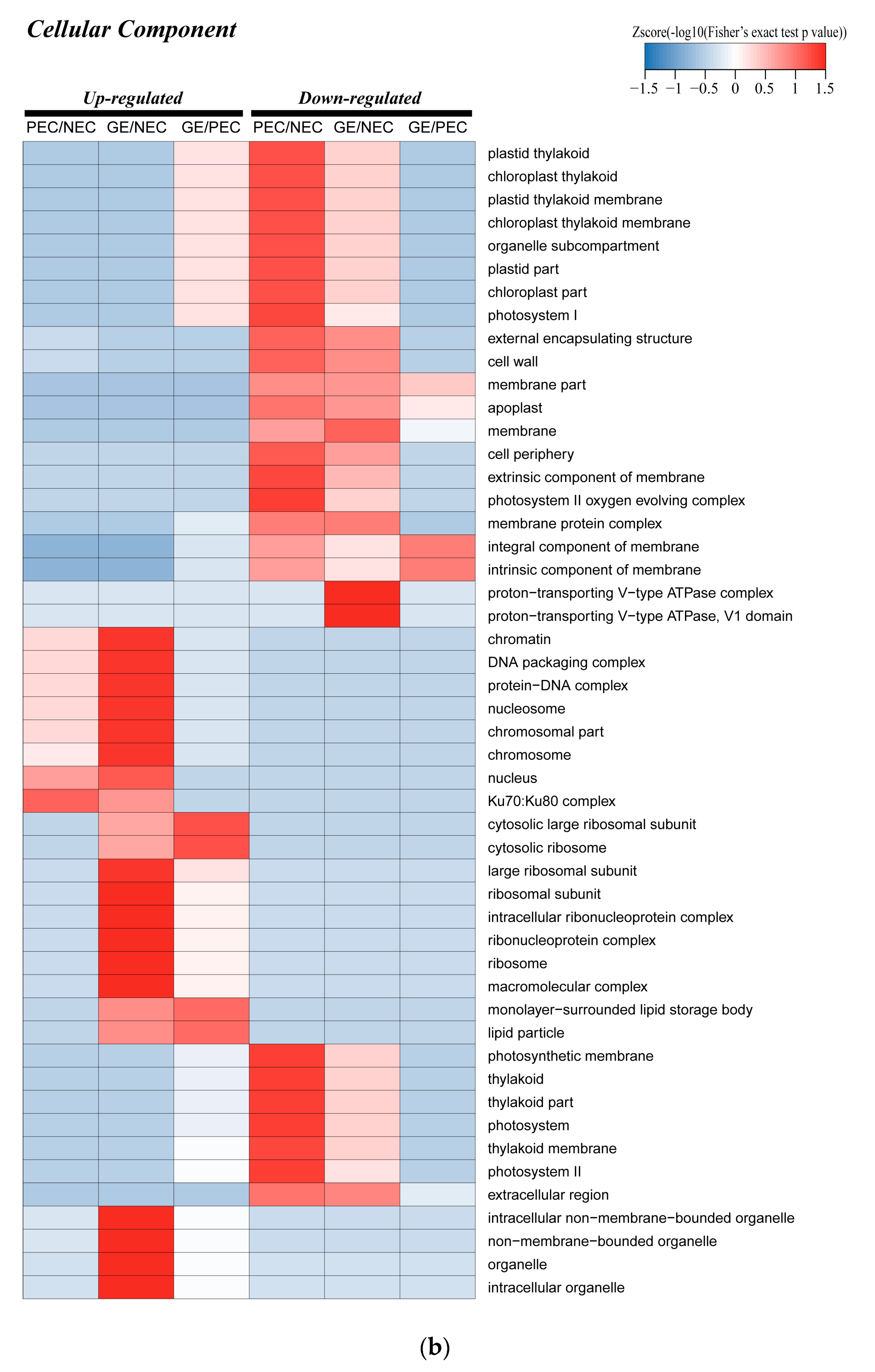
In the cellular component category of PEC versus NEC and GE versus PEC, a large number of DAPs were clustered in photosynthesis-related cellular components and proteins from ‘plastid thylakoid’ to ‘photosystem I’, indicating a significant decrease of photosynthesis in PEC versus NEC. Furthermore, in GE versus PEC, the corresponding photosynthetic cell components showed slightly up regulated enrichment. However, the photosynthetic effect of GE was not higher than NEC; this result showed that the photosynthesis-related DAPs of the cell component classification in GE versus NEC were concentrated in the down regulated expression region (Figure 5b). Additionally, the DAPs’ of expression pattern from ‘photosynthetic membrane’ to ‘photosystem II’, photosynthesis-related proteins’ was similar to the above results in that there was significant down regulation in PEC versus NEC (Figure 5b).
The analysis above showed that photosynthesis is a critical process involved in SE of cotton, which is consistent with our subcellular localization results.

The Regulation, Response and Metabolism-related Proteins of the Biological Process Category in Cotton SE

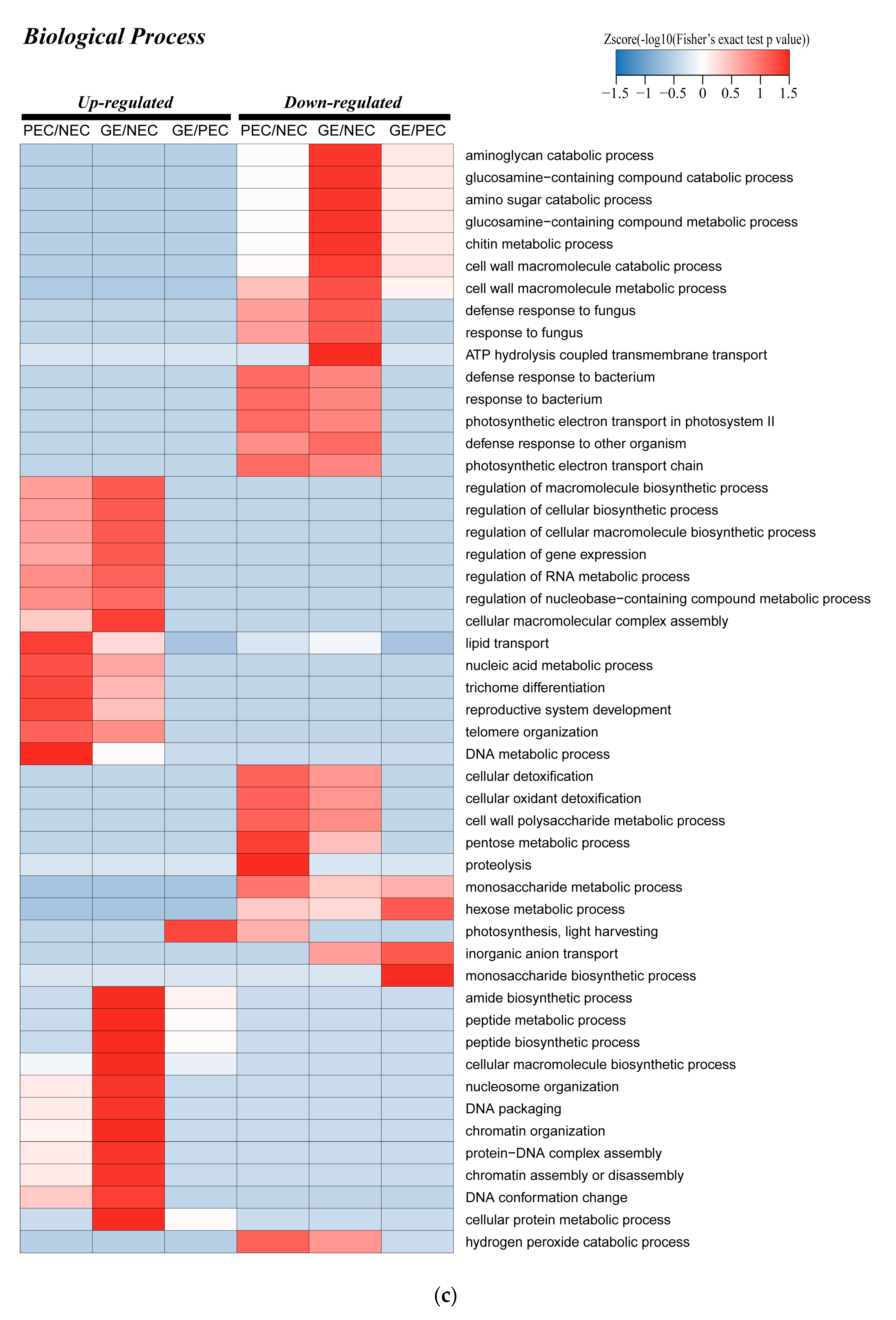
Proteins related to ‘lipid transport’, ‘reproductive system development’, ‘DNA metabolic process’ and ‘regulation’ were up regulated with different degrees of enrichment in PEC versus NEC and GE versus NEC. In addition, we also found that from ‘amide biosynthetic process’ to ‘cellular protein metabolic process’ proteins in GE versus NEC were uniformly enriched in up regulation (Figure 5c). Furthermore, ‘monosaccharide metabolic process’, ‘hexose metabolic process’, glycometabolism related proteins and the DAPs from ‘aminoglycan catabolic process’ to ‘cell wall macromolecule metabolic process’ were down regulated to different degrees in the three sample groups. Other proteins involved in response regulation were also enriched in the down regulated region in PEC versus NEC and GE versus NEC (Figure 5c).
Interestingly and consistently, the photosynthesis-related proteins in ‘photosynthesis, light harvesting’ were down regulated in PEC versus NEC and up regulated in GE versus PEC (Figure 5c). The above results demonstrated that SE of cotton might frequently involve proteins associated with environmental stress response, biological regulation, central metabolic processes, and photosynthetic metabolism.

2.5.2. Enrichment Analysis in KEGG of the DAPs Involved in Phenylpropanoid Biosynthesis, Nitrogen Metabolism, Photosynthesis and Other Related Biological Processes

Enrichment Analysis in KEGG Clusters of Related Biological Processes among Groups

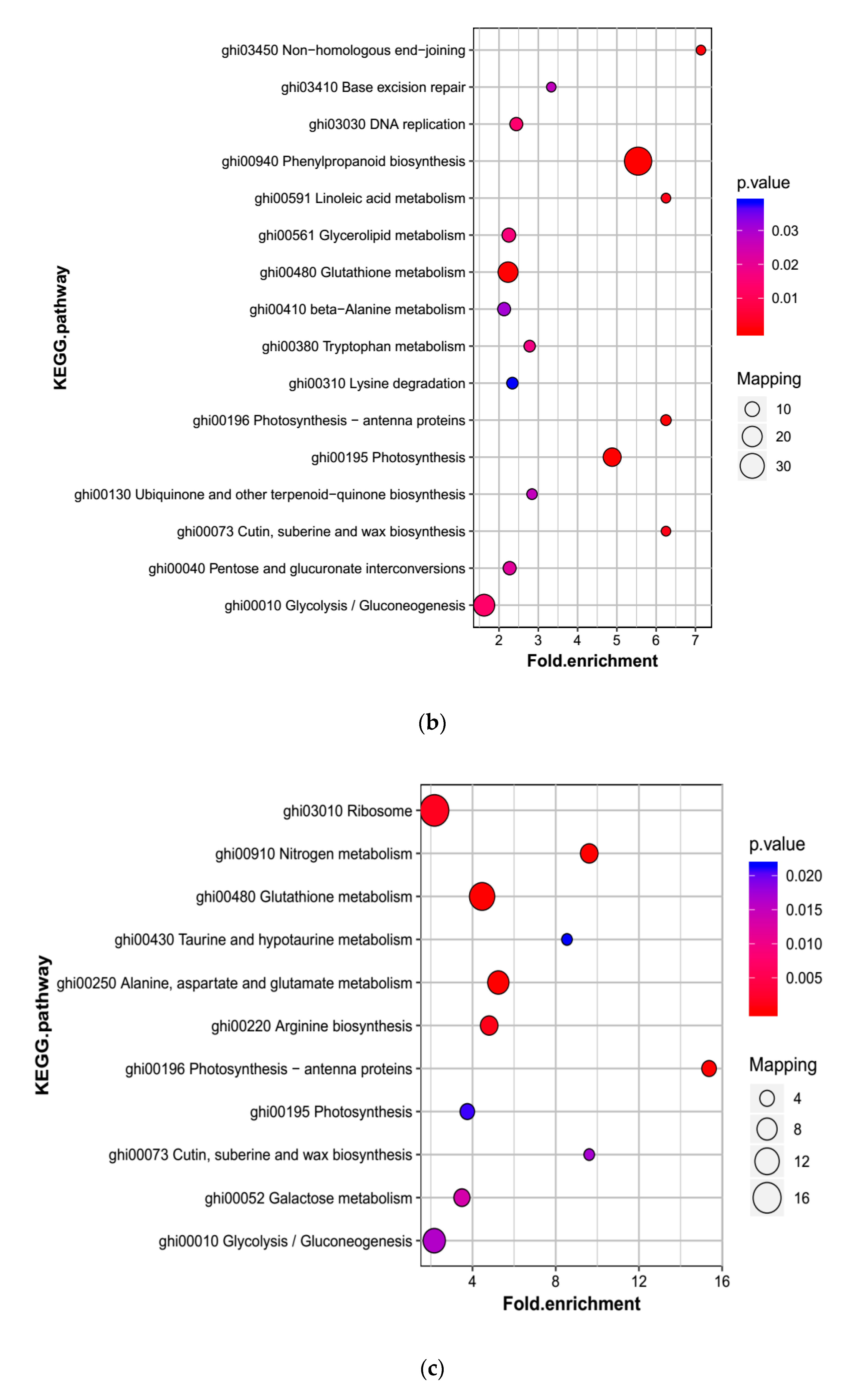
To further understand the function of SE-related proteins, we analyzed the differences and dynamic changes among groups of rich clustering classes in KEGG pathways. Cluster analysis of up regulated expression pathways showed that protein from ‘ribosome biogenesis in eukaryotes’ and ‘DNA replication’ were slightly enriched to varying degrees in PEC versus NEC and GE versus NEC (Figure 6a). In the down regulated enrichment region, multiple types of proteins were enriched in different levels in different sample groups, of which ‘phenylpropanoid biosynthesis’ and ‘nitrogen metabolism’ were enriched significantly in PEC versus NEC and GE versus PEC, respectively (Figure 6a). The categories ‘photosynthesis−antenna proteins’, ‘glycolysis/gluconeogenesis’ and ‘carbon fixation in photosynthetic organisms’ were down regulated in PEC versus NEC and up regulated in GE versus PEC (Figure 6a).
Phenylpropanoid biosynthesis and nitrogen metabolism were significantly enriched and photosynthesis was re-enriched. The study suggesting the above biological processes possible involvement in cotton SE transformation.

KEGG Pathway Enrichment Analysis of Related Biological Processes within the Sample Groups

In PEC versus NEC, KEGG pathway enrichment analysis demonstrated that the ‘phenylpropanoid biosynthesis’, ‘photosynthesis’, ‘glutathione metabolism’ and ‘glycolysis/gluconeogenesis’ were the most significantly affected pathways. A great deal of peroxidase proteins of the phenylpropanoid biosynthesis pathway were down regulated in PEC and NEC. Furthermore, a variety of proteins related to ‘photosynthesis’ and a slight number of DAPs of the ‘glycolysis/gluconeogenesis’ pathway were also down-regulated in PEC and NEC. Additionally, the ‘glutathione metabolism’ pathway was down regulated in PEC and NEC (Figure 6b).
KEGG analysis of GE versus PEC indicated that the enriched pathways of the DAPs were most remarkably associated with ‘glutathione metabolism’ and ‘nitrogen metabolism’ pathways, which were both down regulated. Once again, ‘photosynthesis-antenna proteins’ and ‘photosynthesis’ were significantly enriched up regulated pathways (Figure 6c).
In addition, DAPs of GE versus NEC were classified into various KEGG pathways, of which 6 metabolic pathways were significantly enriched. Interestingly, the largest number of DAPs was once more enriched in ‘phenylpropanoid biosynthesis’ and ‘photosynthesis’ were down-regulated. Moreover, ‘ribosome’ was up regulated pathways that were also enriched (Figure 6d).
The results of the KEGG pathway analysis further indicated that the three metabolic pathways, including phenylpropanoid biosynthesis, nitrogen metabolism and photosynthesis, are essential for the process of cotton SE transformation.

2.5.3. Enrichment Cluster Analysis of Differential Proteins Functional Domain

Domain enrichment analysis of up regulated proteins revealed that ‘histone’-related domain, ‘ribosomal’-related domain, ‘seed maturation protein’, ‘translation protein SH3−like domain’, ‘RmlC−like jelly roll fold’, ‘Cupin 1’ and ‘RmlC−like cupin domain’ were enriched in the three sample groups with different dynamic expression patterns. The degree of enrichment of ’rmlC-like jelly roll fold’ and ‘cupin 1’, seed storage protein-related domain, was extremely high in GE versus PEC (Figure 7). This result indicates that the development of GE requires storage proteins to provide nutrients for the regeneration of somatic embryos. For the down regulated expression region, domains including the ‘aspartic peptidase domain’ and the ‘START−like domain’ were abundant in three or two samples groups, including glutathione S−transferase domain. Furthermore, domains related to ‘haem peroxidase, plant/fungal/bacterial’, ‘secretory peroxidase’, ‘aquaporin−like’ and ‘glycoside hydrolase’ were equally enriched in PEC versus NEC and GE versus NEC (Figure 7).
The cluster analysis of dynamic enrichment changes through different functional domains showed that the ‘rmlC−like cupin domain’, seed maturation protein, glutathione S−transferase and peroxidase-related domain were driving diverse tasks in different development processes of cotton SE.

2.6. Enrichment Analysis of the Major Biological Process between Different Comparison Groups

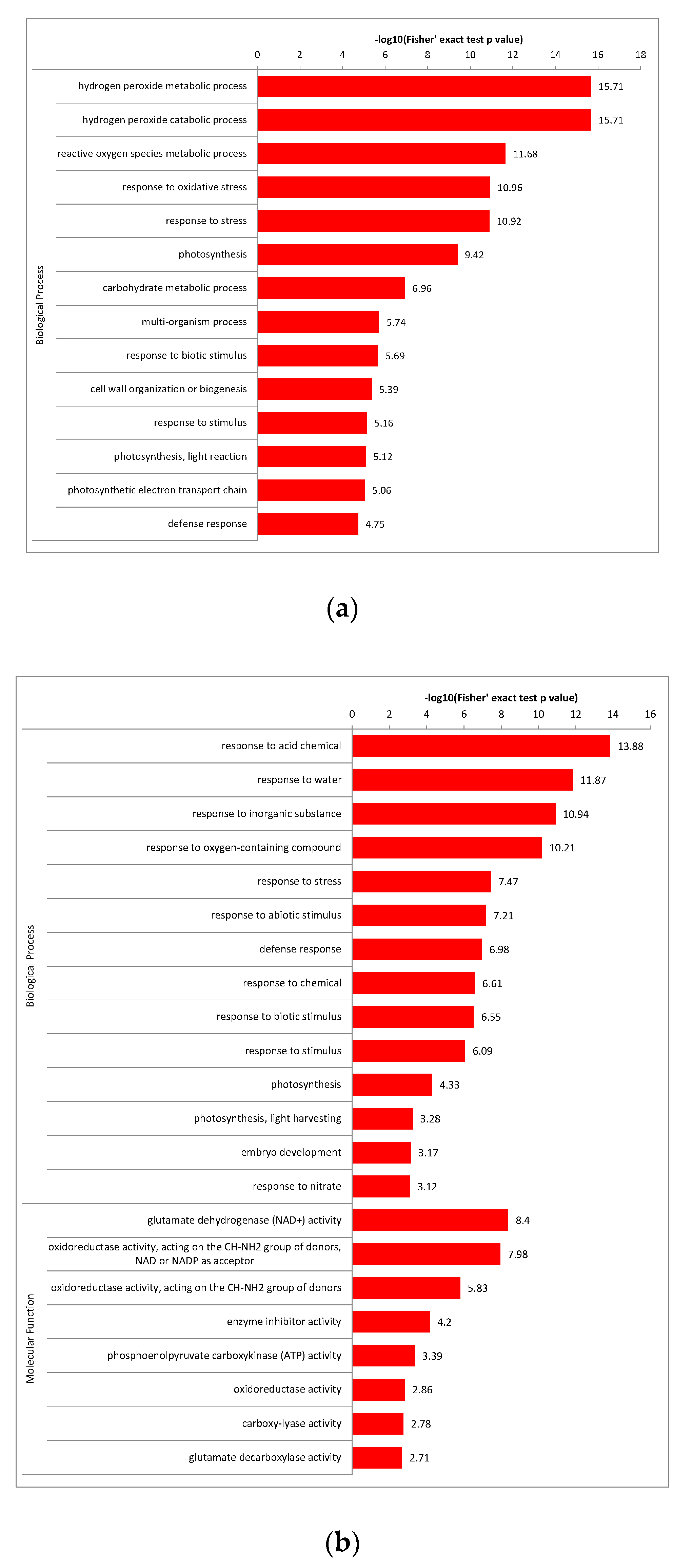
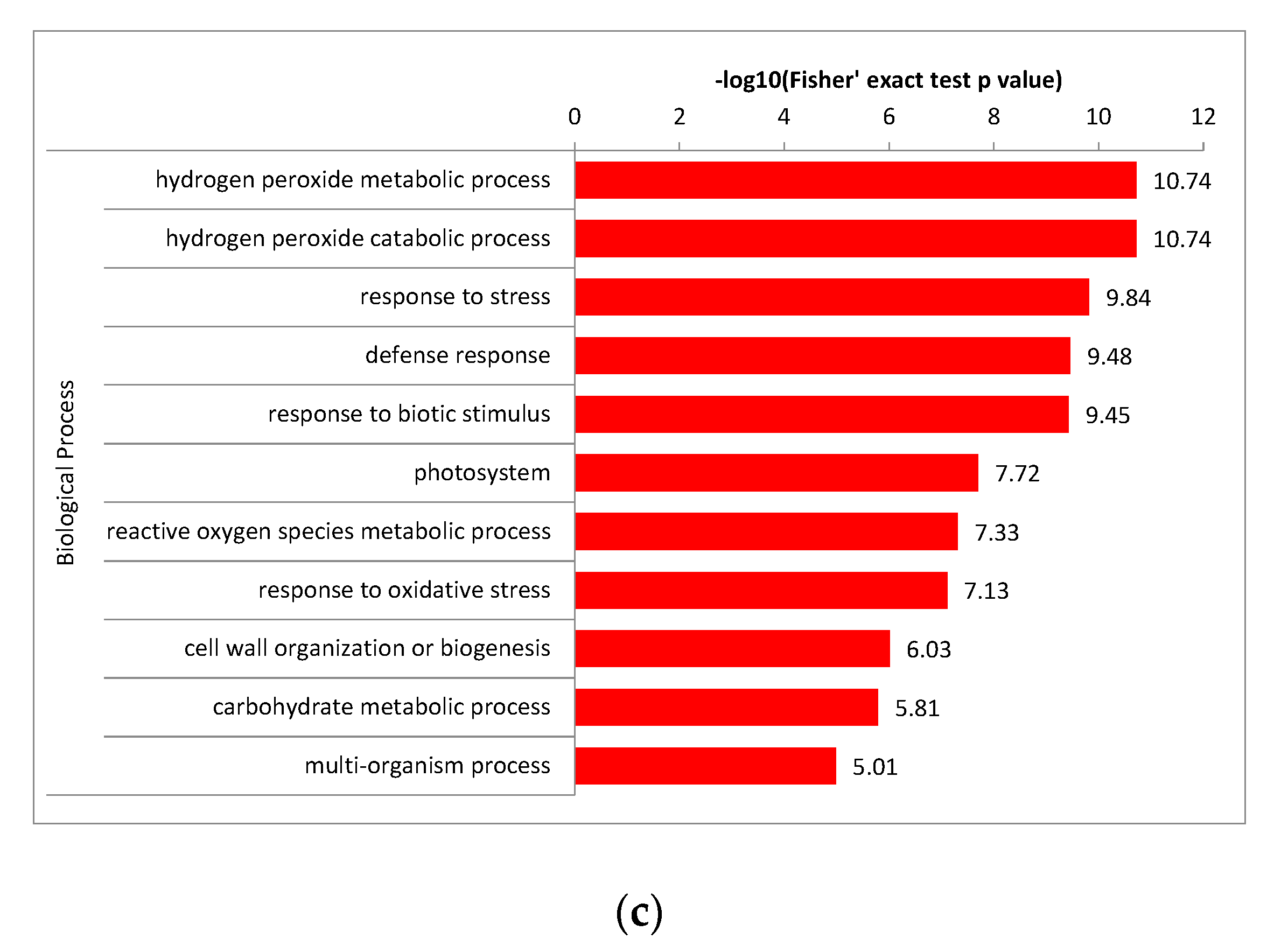
Above all, the significantly enriched GO terms of the biological process in different comparison groups were investigated. In PEC and NEC, the top 5 GO terms were peroxidase-related, further demonstrated the above results that significant enrichment of the phenylpropanoid biosynthesis pathway was observed during the initiation process of SE. In addition, ‘photosynthesis’ term showed significant changes in the PEC differentiation, promoting cotton SE. In GE and PEC, environmental response and photosynthesis related proteins were presented in the top 12 GO terms, indicating that the abundant abiotic stress and photosynthesis responsive proteins might regulate the maturity and development of globular embryos. In GE and NEC, the top 8 GO terms were peroxidase, photosynthesis and environmental response related proteins, being part of an important biological process in the process of cotton somatic embryo transformation. What’s more, glutamate dehydrogenase (GDH) is significantly enriched in the molecular function classification of PEC and GE. GDH is one of the main enzymes of nitrogen metabolism and participates in important biological processes of plant SE. These important biological processes throughout the development process suggested that complex regulatory networks are involved in the cotton SE process (Figure 8).

2.7. Several Major DAPs are Associated with SE Regulation and Modification

SE of cotton is regulated by many factors. The GO and KEGG enrichment analysis showed that peroxidase, photosynthesis-related proteins, stress-responsive proteins, amino acid metabolism-related proteins and other energy metabolism enriched proteins all play an important role in the process of cotton SE. Furthermore, we comprehensively explored and analyzed differentially abundant proteins in cotton SE involving hormone signal response/signaling transduction, transcription/posttranscription and modification regulation (Table 2).

2.8. Comparative and Complementary Proteome of the Candidate DAPs

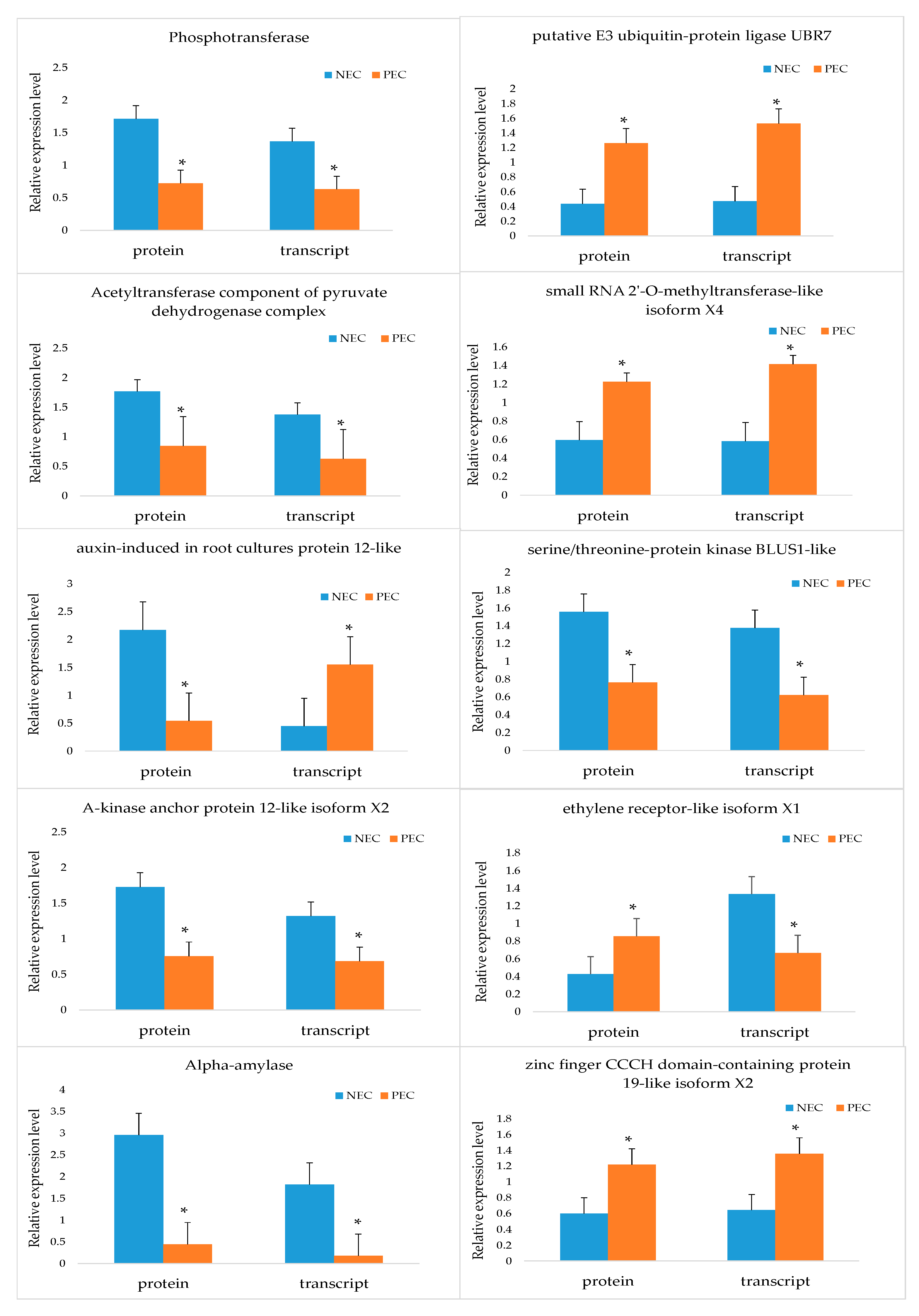
To complement the changes in abundance at the transcriptional level and confirm the authenticity and accuracy of the proteomic analysis, we analyzed ten candidate DAPs in NEC and PEC. Eight out of ten genes under this analysis showed positive correlation between the expression levels of protein and mRNA, indicating that most proteins were regulated directly at the transcription level. For the other two DAPs, negative correlation between their expression levels of protein and mRNA was observed, suggesting that their protein levels might be depended not only on the transcript level but also on the post-translational level (Figure 9).

3. Discussion

Proteomics analyses have long been recognized as a useful tool to dissect the molecular mechanisms of SE. The effectiveness of this technique is strongly dependent on the applied technique of the proteomics analysis system and the experimental system. Proteomics analyses for the somatic embryogenesis in Pinus nigra Arn. [42] and Phoenix dactylifera L. [43] have been previously performed. In this study, we performed quantitative proteomics analysis using the advanced EASY-nLC 1000 UPLC system based on the high-throughput TMT-labeling quantitative detection technique, and we consequently identified 9369 proteins for our samples. Thus, our new study significantly improved the resolution. TMT is advantageous and offers greater sensitivity for the analysis of cotton SE proteomic dynamics than previous methods. Furthermore, most previous studies have focused on the molecular mechanisms of regulation in the late stage of somatic embryogenesis, and little is known about protein regulation and metabolism in the early stage of embryogenesis. In the isobaric tags for relative and absolute quantitation (iTRAQ) proteomics analysis of Ge [11], 6318 proteins were identified in somatic spherical embryo and cotyledon embryo. Subsequently, Zhu [44] identified 5892 proteins related to SE through iTRAQ technology. In our study, a total of 9369 proteins were identified in stages of NEC, PEC and somatic embryos’ initial development period of GEs by TMT-labeling quantitative detection technique. Through identification and annotation of DAPs, we uncovered the key genes/proteins and pathways involved in the critical initial stage of cotton SE. The results generated in this study provide a valuable foundation for further investigation of the roles of DAPs during the expression of totipotency in cotton SE.

3.1. DAPs Enriched in Crucial Biological Processes Associated with Cotton SE

3.1.1. Peroxidase Proteins Involved in Phenylpropanoid Biosynthesis Affect SE

Recently, a proteomic analysis of the somatic embryogenesis induction stage of Medicago truncatula revealed that peroxidase accumulates by day 5 after the induction of somatic embryogenesis and increases four-fold by day 14 [45]. Peroxidase is known to take part in diverse plant processes, such as auxin metabolism, cell wall elongation and stiffening [46]. However, in the present study, our data showed that peroxidase is involved in ‘phenylpropanoid biosynthesis’ pathways (Figure 6a,b; Figure 7), and the expression pattern was different from that of Almeida [45]. We presume that peroxidase might participate in different metabolic pathways with different expression patterns to regulate somatic embryogenesis in different species. Peroxidase is a phenol oxidase and is highly representative in date palm, and polyphenoloxidase are involved in oxidative browning in date palm [47,48]. Abohatem [49] demonstrates that the low rate of successive transfer culture (every 15 or 20 days) reduced the increase in phenolic contents and peroxidase activities in plant tissue leading to an enhancement of tissues/cells browning and then to a decrease in embryonic cell proliferation. Fresh culture medium every 7 days can significantly reduce the oxidative browning of tissue/cells, which is related to the reduction of phenolic compounds and peroxidase activity, thus increasing the proliferation of embryonic protocells. Based on our proteomic profile results, down regulation of many peroxidase DAPs in PEC versus NEC indicates that peroxidase protein activity is weak during SE, which prevents callus browning and promotes embryogenic differentiation.

3.1.2. Photosynthesis in Cotton SE

The photosynthetic potential of cotyledon embryos has been reported in previous studies. For example, in Coffea × arabusta cotyledons photosynthetic capacity and germinated embryos, the cotyledon embryo stage is the earliest photoautotrophic stage to ensure plant development [50]. Rival [51] reported that the maximum photochemical activity of photosystem II is extremely low in proliferating embryos of oil palm and strongly increases at later developmental stages. In this study, the result is that the expression pattern of DAPs in the ‘photosynthetic’ pathway is down- to up regulation. According to our results, ‘Photosystem I P700 chlorophyll an apoprotein’, ‘oxygen-evolving enhancer protein 2’, ‘ATP synthase gamma chain’ and ‘Chlorophyll a–b binding protein’ of the ‘Photosynthesis’ pathway were down regulated in PEC in comparison to GEs (Figure 5b,c; Figure 6a–c). The results indicate that photosynthesis organs and photosynthetic capacity gradually developed from the GE stage for future autotrophy in cotton.

3.1.3. Response to Environment Stresses during SE of Cotton

Stresses are the factors that have been increasingly recognized as having important role in the induction of SE [52]. Embryogenic competence of in vitro cultured somatic cells can be stimulated by various factors, such as phytohormone [53,54], dehydration [55,56], explant wounding [57], heavy metal ions [17,56], high osmotic pressure [58,59], etc. Our data showed that a series of stress responsive biological processes were significantly enriched in PEC and GE, including response to acid chemical, water, inorganic substance, oxygen-containing compound, chemical, abiotic stimulus and biotic stimulus, indicating that callus was protected from the external environmental stress through complex regulatory networks to ensure the embryos development of cotton during the PEC-to-embryos transition of cotton (Figure 5c; Figure 8b). Previous studies have shown that decrease of water availability stimulated a shift from proliferation of cells and early embryos to the production of cotyledonary embryos in the developmental program of the culture [60]. In our study, the water content gradually decreased with the callus culture time increased prompted the tissue cells to respond to dry stress and initiate defense mechanisms to ensure embryo development. In the carrot somatic embryogenesis, 2,4-dichlorophenoxyacetic acid (2,4-D) functions as a stress chemical as well as an auxin [61]. During Arabidopsis somatic embryos induction, cells in the shoot apical meristem (SAM) of wild-type seedlings acquired pluripotency or embryogenic potential under initial stress, and then these cells form somatic embryos on 2,4-D treatment [17]. In our culture system, 2,4-D is also used to induce SE, but stress treatment is necessary before exposure to 2,4-D. From the datas above, we can conclude that stress responses are the indispensable biological processes in plant embryos induction.

3.1.4. Effects of Nitrogen Metabolism Related to SE

Glutamate dehydrogenase (GDH) is one of the enzymes directly related to nitrogen metabolism. Induction of various GDH isoenzymes may suggest their varied anabolic and catabolic functions [62]. GDH, which is directly involved in the oxidation of amino acids, protect tissues from the toxicity of ammonium [63]. Ganced [64,65] strongly suggested that sugar could control the expression of the GDH gene through catabolic inhibition, which has been described in bacteria and yeast. In this study, GDH in nitrogen metabolism was significantly enriched in GE versus PEC and showed a down regulated trend (Figure 6a,c). We speculate that GDH may be more important during the cotton PEC redifferentiation period.

3.2. Other DAPs of Regulatory Factors Associated with Cotton SE

3.2.1. Phytohormone Response Related Proteins

Hormones are the most likely candidates in the regulation of developmental switches [16]. Auxin is the main growth regulator in plants, which is involved in the regulation of cell division and differentiation, as well as the existence of other growth regulators such as abscisic acid [66], ethylene [67], gibberellin [68].
In cotton primary embryogenic calli, the DAPs of ‘Auxin efflux carrier component’ (AP2) and ‘indole-3-acetic acid-amido synthetase GH3. 17-like’ (GH3) were up regulated (Table 2). Auxin-related proteins are essential for initiating dedifferentiation and cell division in already differentiated cells before they can express embryogenic competence [44]. The PIN gene is believed to be the coding element that regulates the auxin efflux mechanism of the polar auxin transport, which is concluded by the polarity localization of the PIN membrane protein and auxin absorption experiment [69]. Blilou [70] pointed to polar auxin transport as a major factor in organ formation. The GH3 gene is one of several sequences screened by differential hybridization of auxin-induced cDNA sequences extracted from auxin-treated soybean tissues [71]. Expression of the GH3 gene has been shown to be rapidly and specifically induced by the application of auxin [72,73]. These conclusions also explained the up regulation of AP2 and GH3 in PEC due to the trend of auxin polar transport organ formation and the key regulation of induced somatic embryo formation.
Gallie [74] identified two ethylene receptor gene families in maize. In developing embryos, the expression levels of members of the two ethylene receptor families were significantly increased, which indicated that embryonic development was involved in ethylene synthesis. In this study, DAPs of ‘ethylene receptor-like isoform X1’ (Table 2) were involved in the ethylene signaling pathway and were up regulated in PEC compared to NEC, suggesting that ethylene receptor may positively regulate SE initiation in cotton.
In the present study, the differentially accumulated ‘GA-stimulated transcript-like protein 1’ (GASL1) was down regulated in PEC and GE (Table 2). Ge [11] demonstrated that, at 10 days after GA treatment, 95% of the embryos showed an aberrant structure, large size, and light-green color. Therefore, we presume that GA negatively affects somatic embryo production and growth via regulation of the GA signaling pathway.
Treatment with ABA improves the efficiency of somatic embryo maturation of Panax ginseng [75] and promotes sugar cane embryo growth [76]. Somatic embryos treated with ABA generate the highest yield of plantlets in Picea abies [77]. In our results, the ‘abscisic acid receptor PYR1-like’ DAP, which is involved in the ABA signal pathway, was up regulated in GE compared to PEC (Table 2); this suggests that the ‘abscisic acid receptor PYR1-like protein’ promotes embryo maturation in GE.

3.2.2. Signal Transduction Related Proteins

In this study, we also found that a large number of important signal regulators, including kinases, calcium signals and GTP-binding related proteins (Table 2), were significantly differential expressed in cotton SE. Ca2+ has an important role in the establishment of cellular polarity during embryogenesis in plants [78]. It as a secondary messenger may trigger various signal transduction pathways [79] in plants SE. Calmodulin or Ca2+-dependent protein kinase may be involved in the regulation of Ca2+ levels in the proembryogenic cell mass (PEM) of sandalwood to promote embryo development [80]. In our TMT profile, the calcium-related proteins ‘calcium-dependent protein kinase 11-like’ and ‘probable calcium-binding protein CML27’ were simultaneously up regulated in PEC versus NEC. We presume that calcium-related proteins involved in signal transduction and regulation of calcium balance, thereby establishing polarity to promote cotton SE.
Phosphoenolpyruvate carboxylase (PEPC), a glycolytic protein and CO2-fixing enzyme [81], participates in TCA cycle under non-photosynthesis conditions [82] and signal transduction of plant embryo development [83]. Therefore, this metabolic pathway maintains the carbon residue pool necessary for oil and storage protein biosynthesis that occurred the later in embryonic development [84]. In our study, the protein ‘phosphoenolpyruvate carboxykinase [ATP]-like’ was up regulated in PEC versus NEC and down regulated in GE versus PEC (Table 2). Thus, we presume that PEPC may not only participate in TCA cycle and fixation of CO2 but also in signal transduction in PEC, providing energy for cotton SE transformation process.
As a result, calcium-related proteins, PEPC and other signal transduction proteins may be involved in a variety of biological processes to promote the transformation process of cotton SE.

3.2.3. SE Associated Proteins of Aquaporins and Fatty Acid Metabolism

In our TMT profile, a large number of aquaporins, including PIP and TIPs, were found to be significantly involved in PEC and GE initiation during SE. Aquaporin was involved in water transport by osmosis to prevent dryness and abortion during embryonic development, which was of great significance in the development of Picea asperata somatic embryos [85]. Here, we found that the aquaporins (PIP210, TIP14 and PIP type) were significantly down regulated during PEC; the aquaporins (PIP2-5, TIP3-2) were significantly up regulated during GE (Table 2). Therefore, we suspect that these proteins are sensitive to water content and light induction during cotton SE initiation process of transdifferentiation. Similarly, previous studies have shown that aquaporin, under stress conditions, forms a ‘tunnel’ to regulate the water transport in the cell membrane [86], and can be induced by light [87]. In addition to reducing the activation energy of water transport, aquaporin also enhanced the permeability of the plasma membrane [88].
Many fatty acid biosynthesis- and metabolism-related proteins, such as ‘cytochrome P450 86B1-like’ and ‘cytochrome P450 86A8-like’, were differentially accumulated. In the fatty acid biosynthesis pathway, they were up regulated in PEC (Table 2). A previous study reported that fatty acids, which affect cell function and growth patterns, appear to be a part of the TDZ action pattern and may play an important role in inducing regeneration [89]. These results implied that aquaporins and perturbations of fatty acid metabolism contribute to the initiation of SE in cotton.

3.2.4. Regulation of SE-related Proteins, Transcription, Posttranscription and Modification

In this study, two types of SE-related proteins and multiple types of transcription factors, zinc finger domains, microRNAs and modification-related proteins were identified (Table 2). Late embryogenesis abundant protein (LEA) was first found in cotton (Gossypium hirsutum) seeds and accumulated in the late stages of embryogenesis, which played a crucial role in cell dehydration tolerance [90]. In current study, two types of SE-related proteins, ‘embryonic protein DC-8-like’ and ‘embryogenesis abundant protein’, were up regulated in GE. It indicating that they may positively regulate somatic embryo maturation and involve in cell dehydration tolerance in cotton.
WUSCHEL (WUS) is a vital transcription factor for labeling embryonic cells [24]. The capability of promoting the vegetative to embryonic transition by WUS, and eventually forming somatic embryos, suggesting that the homeodomain protein also plays a critical role during embryogenesis, in addition to its function in meristem development [91]. In this result, ‘WUSCHEL-related homeobox 9-like’ was up regulated in PEC, indicating that the WUSCHEL-related protein is essential for the initiation of somatic embryogenesis. In addition to WUS, we also found other transcription factors, zinc finger domains related to SE (Table 2).
Members of the Argonaute protein family are key players in the small RNA-directed gene silencing pathway. Various types of small RNA and Argonaute proteins played important roles in embryonic development, cell differentiation and transposon silencing in all higher eukaryotes [92]. In our data, ‘protein argonaute 1-like isoform X2’, ‘protein argonaute 4-like’ and ‘small RNA 2’-O-methyltransferase-like isoform X4’ were significantly up regulated during PEC versus NEC (Table 2), suggesting that RNA-mediated post-transcriptional regulation played an important role in the process of cotton SE transformation as reported in other plants [93,94,95].
In current study, we identified several types of modification-related DAPs, including DNA methylation, chromatin modification, acetylation, ubiquitination and phosphorylation. In the process of carrot SE, the removal of auxin led to the loss of DNA methylation, so that the embryo continued to develop [96]. Six types of methylation-related proteins were identified, which dynamically regulated to cotton SE by different expression patterns. Efficient modification of chromatin structure was crucial in the epigenetic regulation of genes [97]. In our data, ‘chromatin modification-related protein MEAF6-like isoform X3’ promoted maturation and development of cotton GEs by epigenetic regulation. In addition, during the PEC period, we also identified acetylation, ubiquitination and phosphorylation-related proteins with different expression patterns (Table 2), indicating that they played diverse and indispensable functions in regulating cotton SE.
The different expression patterns of SE-related proteins, multitudinal transcription, posttranscription and modification showed that they played a pivotal role in the process of cotton SE [98,99,100,101,102,103]. The results of our high-through put proteomics assay, the large scale of proteins associated with SE, and their complex expression patterns suggest that SE is a concerted process involving multiple molecular pathways controlled by a complicated gene regulatory network.

4. Materials and Methods

4.1. Plant Materials and Culture Conditions

Seeds of cotton cultivar Yuzao 1 (Institute of Cotton Research of CAAS), uncovering the coats, were imbibed in 0.1% (w/v) HgCl2 for 10 min, then rinsed four times by sterile distilled water and germinated on Murashige and Skoog (MS) medium containing 3% (w/v) sucrose and 0.3% (w/v) Phytegel. Hypocotyl explants (0.5–1.0 cm) taken from 5–7 d old seedlings and non-embryogenic callus initiation of Yuzao 1 was done, as described by Wu [104], in MS medium plus B5 vitamins medium (MSB) containing 0.46 µmol L–1 kinetin and 0.45 µmol L–1 2,4-D. Calli were subcultured in MSB medium without any hormone to induce embryogenic calli (EC), as described by Zeng [39]. Samples were collected from the following three stages of SEM (1) NEC from explants cultured in MSB, induced (2) PEC and (3) GEs. The collected samples were immediately frozen in liquid nitrogen, and stored at −80 °C before protein extraction. Each staged sample was prepared for three biological replicates.

4.2. Protein Extraction and Identification

4.2.1. Protein Samples Preparation

The three biological replicates of sample were first ground by liquid nitrogen, then the powder was transferred to 5 cm3 centrifuge tube and sonicated three times on ice using a high intensity ultrasonic processor (Scientz, Ningbo, China) in 4-fold volume phenol extraction buffer (including 10 mM dithiothreitol, 1% Protease Inhibitor Cocktail and 2 mM EDTA). The equal amount of trisaturated phenol (pH 8.0) was added; then, the mixture was further vortexed for 5 min. After centrifugation (4 °C, 10 min, 5000× g), the upper stage of phenol was transferred to a new centrifuge tube. Proteins were precipitated by adding at five volume of 0.1 M ammonium sulfate-saturated methanol to precipitate overnight. After centrifugation at 4 °C for 10 min, the supernatant was discarded. The remaining precipitate was washed with ice-cold methanol once, followed by ice-cold acetone for three times. The protein was redissolved in 8 M urea and the protein concentration was determined with BCA kit (Beyotime Biotechnology, Shanghai, China) according to the manufacturer’s instructions.

4.2.2. Trypsin Digestion

Protein solution with dithiothreitol makes its final concentration of 5 mM, 56 °C for 30 min. After that, acetamide was added to make the final concentration 11 mM and incubated in the dark at room temperature for 15 min. Finally, the sample urea concentration was diluted to less than 2 M. With 1:50 (w/w) quality ratio (trypsin: protein) to join the pancreatic enzyme, 37 °C enzyme solution for the night. Then the trypsin (Promega, Madison, WI, USA) was added at a mass ratio of 1:100 (trypsin: protein), and the enzymatic hydrolysis continued for 4 h.

4.2.3. TMT Labeling

After trypsin digestion, the peptide was desalted by Strata X C18 SPE column (Phenomenex, Torrance, CA, USA) and vacuum-dried. The peptide was reconstituted in 0.5 M TEAB (Sigma, St. Louis, MI, USA) and processed according to the manufacturer’s protocol for TMT kit (Thermo Fisher Scientific, Waltham, MA, USA). Briefly, one unit of TMT reagent was thawed and reconstituted in acetonitrile. The peptide mixtures were then incubated for 2 h at room temperature and pooled, desalted and dried by vacuum centrifugation.

4.2.4. HPLC Fractionation and LC-MS/MS Analysis

The tryptic peptides were fractionated into fractions by high pH reverse-phase HPLC using Agilent 300Extend C18 column (5 μm particles, 4.6 mm ID, 250 mm length, Agilent, Santa Clara, USA). Briefly, peptides were first separated with a gradient of 8% to 32% acetonitrile (pH 9.0) over 60 min into 60 fractions. Then, the peptides were combined into 9 fractions and dried by vacuum centrifuging.
The tryptic peptides were dissolved in 0.1% formic acid (solvent A), directly loaded onto a home-made reversed-phase analytical column (15-cm length, 75 μm i.d.). The gradient was comprised of an increase from 8% to 23% solvent B (0.1% formic acid in 90% acetonitrile) over 38 min, 23% to 35% in 14 min and climbing to 80% in 4 min then holding at 80% for the last 4 min, all at a constant flow rate of 450 nL/min on an EASY-nLC 1000 UPLC system (Thermo Fisher Scientific, Waltham, MA, USA).
The peptides were subjected to NSI source followed by tandem mass spectrometry (MS/MS) in Q ExactiveTM HF-X (Thermo Fisher Scientific, Waltham, MA, USA) coupled online to the UPLC. The electrospray voltage applied was 2.0 kV. The m/z scan range was 350–1600 m/z for a full scan, and intact peptides were detected in the Orbitrap (Thermo Fisher Scientific, Waltham, MA, USA) at a resolution of 60,000. Peptides were then selected for MS/MS using NCE setting as 28 and the fragments were detected in the Orbitrap at a resolution of 17,500. A data-dependent procedure that alternated between one MS scan followed by 20 MS/MS scans with 30 s dynamic exclusion. Automatic gain control (AGC) was set at 1E5. The fixed first mass was set as 100 m/z. The HPLC Fractionation and LC-MS/MS Analysis in our research is supported by Jingjie PTM BioLabs (Hangzhou, China).

4.2.5. Database Search

The resulting MS/MS data were processed using Maxquant search engine (v.1.5.2.8). Tandem mass spectra were searched against the UniProt 14.1 (2009)—Gossypium hirsutum database (78,387 sequences) concatenated with reverse decoy database. Trypsin/P was specified as cleavage enzyme allowing up to 2 missing cleavages. The first search was 20 ppm, the main search was 5 ppm, and the fragment ion mass tolerance was 0.02 Da.

4.3. Bioinformatics

4.3.1. Annotation Methods

GO Annotation

Gene Ontology (GO) annotation proteome was derived from the UniProt-GOA database (http://www.ebi.ac.uk/GOA/). Firstly, converting identified protein ID to UniProt ID and then mapping to GO IDs by protein ID. If some identified proteins were not annotated by UniProt-GOA database, the InterProScan soft would be used to annotated protein’s GO functional based on protein sequence alignment method. Then proteins were annotated according to the biological process, cellular component and molecular function of the three categories of protein Gene Ontology annotation.

Domain Annotation

Identified proteins domain functional description were annotated by InterProScan (a sequence analysis application) based on protein sequence alignment method, and the InterPro domain database was used. InterPro (http://www.ebi.ac.uk/interpro/) is a database that integrates diverse information about protein families, domains and functional sites, and makes it freely available to the public via Web-based interfaces and services. At the heart of the database are diagnostic models, known as signatures, from which protein sequences can be searched for potential functions. InterPro has applications in large-scale genome-wide and metagenomic analyses, and the characterization of individual protein sequences.

KEGG Pathway Annotation

KEGG connects known information on molecular interaction networks, such as pathways and complexes (the “Pathway” database), information about genes and proteins generated by genome projects (including the gene database) and information about biochemical compounds and reactions (including compound and reaction databases). These databases are different networks, known as the “protein network”, respectively, and the “chemical universe”, respectively. Efforts are being made to increase KEGG knowledge, including information on orthographic clustering in the KEGG orthographic database. KEGG Pathways mainly including: Metabolism, Genetic Information Processing, Environmental Information Processing, Cellular Processes, Rat Diseases, Drug Development. Kyoto Encyclopedia of Genes and Genomes (KEGG) database (http://www.genome.jp/kegg/) was used to annotate protein pathway. Firstly, using KEGG online service tools KAAS (http://www.genome.jp/kaas-bin/kaas_main) to annotate protein’s KEGG database description. Then mapping the annotation result on the KEGG pathway database using KEGG online service tools KEGG mapper (http://www.kegg.jp/kegg/mapper.html).

Subcellular Localization Prediction

Eukaryotic cells are elaborately subdivided into membrane-bound chambers with unique functions. Some major constituents of eukaryotic cells are extracellular space, cytoplasm, nucleus, mitochondria, Golgi apparatus, endoplasmic reticulum (ER), peroxisome, vacuoles, cytoskeleton, nucleoplasm, nucleolus, nuclear matrix, and ribosomes. There, we used wolfpsort (http://www.genscript.com/psort/wolf_psort.html) a subcellular localization predication soft to predict subcellular localization. Wolfpsort is an updated version of PSORT/PSORT II for the prediction of eukaryotic sequences.

4.3.2. Functional Enrichment

Enrichment of Gene Ontology Analysis

Through GO annotation, proteins are divided into biological process, cellular compartment, and molecular function. For each class, we used a double-tailed Fisher’s precision test to detect the enrichment of differentially abundant proteins relative to all identified proteins. GO with a revised p value of <0.05 is considered significant.

Enrichment of Pathway Analysis

KEGG database identified enrichment pathways by double-tailed Fisher’s precision test to detect the enrichment of differentially abundant proteins against all identified proteins. The pathway with a corrected p-value < 0.05 was considered significant. According to the KEGG website, these paths are classified into hierarchical categories.

Enrichment of Protein Domain Analysis

For each category protein, InterPro (a resource that provides a functional analysis of protein sequences by classifying them into families and predicting the presence of domains and important sites) database was researched and a two-tailed Fisher’s exact test was employed to test the enrichment of the differentially abundant proteins against all identified proteins. Protein domains with a p-value < 0.05 were considered significant.

Enrichment-Based Clustering

For further hierarchical clustering based on different protein functional classification (such as GO, Domain, Pathway, Complex). We first collated all the categories obtained after enrichment along with their p values, and then filtered for those categories which were at least enriched in one of the clusters with p value <0.05. This filtered P value matrix was transformed by the function x = −log10 (p value). Finally, these x values were z-transformed for each functional category. These z scores were then clustered by one-way hierarchical clustering (Euclidean distance, average linkage clustering) in Genesis. Cluster membership was visualized by a heat map using the “heatmap.2” function from the “gplots” R-package.

Supplementary Materials

Supplementary materials can be found at https://www.mdpi.com/1422-0067/20/7/1691/s1.

Author Contributions

Data curation, H.G. (Haixia Guo), H.G. (Huihui Guo), L.Z., Y.F. (Yijie Fan), Y.F. (Yupeng Fan) and Z.T.; Formal analysis, H.G. (Haixia Guo), H.G. (Huihui Guo), Z.T. and F.Z.; Funding acquisition, F.Z.; Investigation, H.G. (Haixia Guo), H.G. (Huihui Guo), L.Z., Y.F. (Yijie Fan) and Y.F. (Yupeng Fan); Methodology, Z.T.; Project administration, F.Z.; Resources, H.G. (Haixia Guo), H.G. (Huihui Guo), L.Z., Y.F. (Yijie Fan) and Y.F. (Yupeng Fan); Supervision, F.Z.; Writing—original draft, H.G. (Haixia Guo), H.G. (Huihui Guo) and L.Z.; Writing—review & editing, H.G. (Haixia Guo) and F.Z.

Funding

This work was supported by National Key Research and Development Program (2018YFD0100303; 2016YFD0100306), Taishan Scholar Talent Project from PRC (TSQN20161018), the National Natural Science Foundation of China (31401428) and Fok Ying-Tong Foundation (151024).

Acknowledgments

We are grateful for the technique support provided by Jingjie Biotechnology Co., Ltd., Hangzhou, China.

Conflicts of Interest

The authors declare no conflict of interest.

Abbreviations

ABAAbscisic acid
DAPsDifferentially accumulated proteins
ECEmbryogenic calli
GAGibberellin
GEsGlobular embryos
GOGene Ontology
iTRAQIsobaric tags for relative and absolute quantitation
KEGGKyoto encyclopedia of genes and genomes
MSMurashige and Skoog
MSBVitamins medium
NECNonembryogenic calli
PECPrimary embryogenic calli
PEMProembryogenic cell mass
SESomatic embryogenesis
SEMSomatic embryogenesis masses
SSHSuppression subtractive hybridization
TMTThe isobaric labels tandem mass tags

References

  1. Wilkins, T.A.; Rajasekaran, K.; Anderson, D.M. Cotton biotechnology. Crit. Rev. Plant Sci. 2000, 19, 511–550. [Google Scholar] [CrossRef]
  2. Brookes, G.; Barfoot, P. GM Crops: Global Socio-Economic and Environmental Impacts 1996–2010; PG Economics, Ltd.: Dorchester, UK, 2012; Available online: http://www.pgeconomics.co.uk/page/33/global-impact-2012 (accessed on 13 January 2013).
  3. Zhao, X.Y.; Su, Y.H.; Cheng, Z.J.; Zang, X.S. Cell fate switch during in vitro plant organogenesis. J. Integr. Plant Biol. 2008, 50, 816–824. [Google Scholar] [CrossRef] [PubMed]
  4. Mujib, A. Somatic Embryogenesis in Ornamentals and Its Applications; Springer: New Delhi, India, 2016; pp. 67–86. [Google Scholar]
  5. Fehér, A. The initiation phase of somatic embryogenesis: What we know and what we don’t. Acta Biol. Szeged. 2008, 52, 53–56. [Google Scholar]
  6. Fehér, A. Somatic embryogenesis—stress-induced remodeling of plant cell fate. BBA-Gene Regul. Mech. 2015, 1849, 385–402. [Google Scholar]
  7. Stewards, F.C.; Mapbs, M.O.; Mears, K. Growth and organized development of cultured cells I. Organization in cultures grown from freely suspended cells. Am. J. Bot. 1958, 45, 705–708. [Google Scholar] [CrossRef]
  8. Rao, A.Q.; Hussain, S.S.; Shahzad, M.S.; Bokhari, S.Y.; Raza, M.H.; Rakha, A.; Majeed, A.; Shahid, A.A.; Saleem, Z.; Husnain, T.; Riazuddin, S. Somatic embryogenesis in wild relatives of cotton (Gossypium Spp.). J. Zhejiang Univ. Sci. B 2006, 7, 291–298. [Google Scholar] [CrossRef] [PubMed]
  9. Evans, D.A.; Sharp, W.R.; Flick, C.E. Growth and Behavior of Cell Cultures: Embryogenesis and Organogenesis. Plant Tissue Cultures; Trevor, A.T., Ed.; Academic Press: New York, NY, USA, 1981; pp. 45–113. [Google Scholar]
  10. Scowcroft, W.R. Genetic Variability in Tissue Culture: Impact on Germplasm Conservation and Utilization; IBPGR: Roma, Italy, 1984; p. 41. Available online: http://www.sidalc.net/cgi-bin/wxis.exe/?IsisScript=bac.xis&method=post&formato=2&cantidad=1&expresion=mfn=048729 (accessed on 3 April 2019).
  11. Ge, X.; Zhang, C.; Wang, Q.; Yang, Z.; Wang, Y.; Zhang, X.; Wu, Z.; Hou, Y.; Wu, J.; Li, F. iTRAQ protein profile differential analysis between somatic globular and cotyledonary embryos reveals stress, hormone, and respiration involved in increasing plantlet regeneration of Gossypium hirsutum L. J. Proteome Res. 2014, 14, 268–278. [Google Scholar] [CrossRef] [PubMed]
  12. Sakhanokho, H.F.; Rajasekaran, K. Cotton regeneration in vitro. In Fiber Plants; Ramawat, K.G., Ed.; Springer: Cham, Switzerland, 2016; pp. 87–110. [Google Scholar]
  13. Fehér, A. Why somatic plant cells start to form embryos? In Somatic Embryogenesis; Mujib, A., Ed.; Springer: Berlin/Heidelberg, Germany, 2005; pp. 85–101. [Google Scholar]
  14. Karami, O.; Aghavaisi, B.; Pour, A.M. Molecular aspects of somatic-to-embryogenic transition in plants. J. Chem. Biol. 2009, 2, 177–190. [Google Scholar] [CrossRef] [PubMed]
  15. Elhiti, M.; Stasolla, C.; Wang, A. Molecular regulation of plant somatic embryogenesis. In Vitro Cell. Dev. Biol. Plant 2013, 49, 631–642. [Google Scholar] [CrossRef]
  16. Fehér, A.; Pasternak, T.P.; Dudits, D. Transition of somatic plant cells to an embryogenic state. Plant Cell Tissue Org. 2003, 74, 201–228. [Google Scholar] [CrossRef]
  17. Ikeda-Iwai, M.; Umehara, M.; Satoh, S.; Kamada, H. Stress-induced somatic embryogenesis in vegetative tissues of Arabidopsis thaliana. Plant J. 2003, 34, 107–114. [Google Scholar] [CrossRef] [PubMed]
  18. Rose, R.J.; Nolan, K.E. Invited review: Genetic regulation of somatic embryogenesis with particular reference to Arabidopsis thaliana and Medicago truncatula. In Vitro Cell. Dev. Biol. Plant 2006, 42, 473–481. [Google Scholar] [CrossRef]
  19. Reinert, J. Über die Kontrolle der Morphogenese und die Induktion von Adventivembryonen an Gewebekulturen aus Karotten. Planta 1959, 53, 318–333. [Google Scholar] [CrossRef]
  20. Quiroz-Figueroa, F.R.; Rojas-Herrera, R.; Galaz-Avalos, R.M.; Loyola-Vargas, V.M. Embryo production through somatic embryogenesis can be used to study cell differentiation in plants. Plant Cell Tissue Org. 2006, 86, 285. [Google Scholar] [CrossRef]
  21. Mathieu, M.; Lelu-Walter, M.A.; Blervacq, A.S.; David, H.; Hawkins, S.; Neutelings, G. Germin-like genes are expressed during somatic embryogenesis and early development of conifers. Plant Mol. Biol. 2006, 61, 615–627. [Google Scholar] [CrossRef] [PubMed]
  22. Thorpe, T.A. In Vitro Embryogenesis in Plants. Springer Science & Business Media: Dordrecht, The Netherlands, 1995; pp. 267–308. [Google Scholar]
  23. Lakshmanan, P.; Taji, A. Somatic embryogenesis in leguminous plants. Plant Biol. 2000, 2, 136–148. [Google Scholar] [CrossRef]
  24. Elhiti, M.; Tahir, M.; Gulden, R.H.; Khamiss, K.; Stasolla, C. Modulation of embryo-forming capacity in culture through the expression of Brassica genes involved in the regulation of the shoot apical meristem. J. Exp. Bot. 2010, 61, 4069–4085. [Google Scholar] [CrossRef] [PubMed]
  25. Zuo, J.; Niu, Q.W.; Frugis, G.; Chua, N. The WUSCHEL gene promotes vegetative-to-embryonic transition in Arabidopsis. Plant J. 2002, 30, 349–359. [Google Scholar] [CrossRef]
  26. Weijers, D.; Sauer, M.; Meurette, O.; Friml, J.; Ljung, K.; Sandberg, G.; Hooykaas, P.; Offringa, R. Maintenance of embryonic auxin distribution for apical–basal patterning by PIN-FORMED–dependent auxin transport in Arabidopsis. Plant Cell 2005, 17, 2517–2526. [Google Scholar] [CrossRef]
  27. Su, Y.H.; Zhao, X.Y.; Liu, Y.B.; Zhang, C.L.; O’Neill, S.D.; Zhang, X.S. Auxin-induced WUS expression is essential for embryonic stem cell renewal during somatic embryogenesis in Arabidopsis. Plant J. 2009, 59, 448–460. [Google Scholar] [CrossRef]
  28. Vogler, H.; Kuhlemeier, C. Simple hormones but complex signalling. Curr. Opin. Plant Biol. 2003, 6, 51–56. [Google Scholar] [CrossRef]
  29. Braybrook, S.A.; Harada, J.J. LECs go crazy in embryo development. Trends Plant Sci. 2008, 13, 624–630. [Google Scholar] [CrossRef] [PubMed]
  30. Giraudat, J.; Parcy, F.; Bertauche, N.; Gosti, F.; Leung, J.; Morris, P.C.; Bouvier-Durand, M.; Vartanian, N. Current advances in abscisic acid action and signalling. In Signals and Signal Transduction Pathways in Plants; Palme, K., Ed.; Springer: Dordrecht, The Netherlands, 1994; pp. 321–341. [Google Scholar]
  31. Ton, J.; Mauch-Mani, B. β-amino-butyric acid-induced resistance against necrotrophic pathogens is based on ABA-dependent priming for callose. Plant J. 2004, 38, 119–130. [Google Scholar] [CrossRef] [PubMed]
  32. Letarte, J.; Simion, E.; Miner, M.; Kasha, K.J. Arabinogalactans and arabinogalactan-proteins induce embryogenesis in wheat (Triticum aestivum L.) microspore culture. Plant Cell 2006, 24, 691. [Google Scholar] [CrossRef] [PubMed]
  33. Singla, B.; Tyagi, A.K.; Khurana, J.P.; Khurana, P. Analysis of expression profile of selected genes expressed during auxin-induced somatic embryogenesis in leaf base system of wheat (Triticum aestivum) and their possible interactions. Plant Mol. Biol. 2007, 65, 677–692. [Google Scholar] [CrossRef] [PubMed]
  34. Mantiri, F.R.; Kurdyukov, S.; Lohar, D.P.; Sharopova, N.; Saeed, N.A.; Wang, X.D.; Vandenbosch, K.A.; Rose, R.J. The transcription factor MtSERF1 of the ERF subfamily identified by transcriptional profiling is required for somatic embryogenesis induced by auxin plus cytokinin in Medicago truncatula. Plant Physiol. 2008, 146, 1622–1636. [Google Scholar] [CrossRef] [PubMed]
  35. Kulinska-Lukaszek, K.; Tobojka, M.; Adamiok, A.; Kurczynska, E.U. Expression of the BBM gene during somatic embryogenesis of Arabidopsis thaliana. Biol. Plant. 2012, 56, 389–394. [Google Scholar] [CrossRef]
  36. Yang, Z.; Li, C.; Wang, Y.; Zhang, C.; Wu, Z.; Zhang, X.; Liu, C.; Li, F. GhAGL15s, preferentially expressed during somatic embryogenesis, promote embryogenic callus formation in cotton (Gossypium hirsutum L.). Mol. Genet. Genom. 2014, 289, 873–883. [Google Scholar] [CrossRef]
  37. Zheng, Q.; Zheng, Y.; Perry, S.E. AGAMOUS-Like15 Promotes Somatic Embryogenesis in Arabidopsis thaliana and Glycine max in Part by Control of Ethylene Biosynthesis and Response. Plant Physiol. 2013, 161, 2113–2127. [Google Scholar] [CrossRef]
  38. Schmidt, E.D.; Guzzo, F.; Toonen, M.A.; de Vries, S.C. A leucine-rich repeat containing receptor-like kinase marks somatic plant cells competent to form embryos. Development 1997, 124, 2049–2062. [Google Scholar]
  39. Zeng, F.; Zhang, X.; Zhu, L.; Tu, L.; Guo, X.; Nie, Y. Isolation and characterization of genes associated to cotton somatic embryogenesis by suppression subtractive hybridization and macroarray. Plant Mol. Biol. 2006, 60, 167–183. [Google Scholar] [CrossRef] [PubMed]
  40. Thompson, A.; Schäfer, J.; Kuhn, K.; Kienle, S.; Schwarz, J.; Schmidt, G.; Neumann, T.; Johnstone, R.; Mohammed, A.K.; Hamon, C. Tandem mass tags: A novel quantification strategy for comparative analysis of complex protein mixtures by MS/MS. Anal. Chem. 2003, 75, 1895–1904. [Google Scholar] [CrossRef] [PubMed]
  41. Pagel, O.; Loroch, S.; Sickmann, A.; Zahedi, R.P. Current strategies and findings in clinically relevant post-translational modification-specific proteomics. Expert Rev. Proteom. 2015, 12, 235–253. [Google Scholar] [CrossRef] [PubMed]
  42. Klubicová, K.; Uvácková, L.; Danchenko, M.; Nemecek, P.; Skultéty, L.; Salaj, J.; Salaj, T. Insights into the early stage of Pinus nigra Arn. somatic embryogenesis using discovery proteomics. J. Proteom. 2017, 169, 99–111. [Google Scholar] [CrossRef] [PubMed]
  43. Sghaier-Hammami, B.; Drira, N.; Jorrín-Novo, J.V. Comparative 2-DE proteomic analysis of date palm (Phoenix dactylifera L.) somatic and zygotic embryos. J. Proteom. 2009, 73, 161–177. [Google Scholar] [CrossRef] [PubMed]
  44. Zhu, H.G.; Cheng, W.H.; Tian, W.G.; Li, Y.J.; Liu, F.; Xue, F.; Zhu, Q.H.; Sun, Y.Q.; Sun, J. iTRAQ-based comparative proteomic analysis provides insights into somatic embryogenesis in Gossypium hirsutum L. Plant Mol. Biol. 2018, 96, 89–102. [Google Scholar] [CrossRef] [PubMed]
  45. Almeida, A.M.; Parreira, J.R.; Santos, R.; Duque, A.S.; Francisco, R.; Tomé, D.F.; Ricardo, C.P.; Coelho, A.V.; Fevereiro, P. A proteomics study of the induction of somatic embryogenesis in Medicago truncatula using 2DE and MALDI-TOF/TOF. Physiol. Plant. 2012, 146, 236–249. [Google Scholar] [CrossRef]
  46. Passardi, F.; Theiler, G.; Zamocky, M.; Cosioa, C.; Rouhier, N.; Teixera, F.; Margis-Pinheiroe, M.; Ioannidis, V.; Penel, C.; Falquet, L.; Dunand, C. PeroxiBase: The peroxidase database. Phytochemistry 2007, 68, 1605–1611. [Google Scholar] [CrossRef]
  47. Baaziz, M.; Saaidi, M. Preliminary identification of date palm cultivars by esterase isoenzymes and peroxidase activities. Can. J. Bot. 1988, 66, 89–93. [Google Scholar] [CrossRef]
  48. Baaziz, M. The activity and preliminary characterization of peroxidases in leaves of cultivars of date palm, Phoenix dactylifera L. New Phytol. 1989, 111, 403–411. [Google Scholar] [CrossRef]
  49. Abohatem, M.; Zouine, J.; El Hadrami, I. Low concentrations of BAP and high rate of subcultures improve the establishment and multiplication of somatic embryos in date palm suspension cultures by limiting oxidative browning associated with high levels of total phenols and peroxidase activities. Sci. Hortic. Amst. 2011, 130, 344–348. [Google Scholar] [CrossRef]
  50. Afreen, F.; Zobayed, S.M.A.; Kozai, T. Photoautotrophic culture of Coffea arabusta somatic embryos: Photosynthetic ability and growth of different stage embryos. Ann. Bot. 2002, 90, 11–19. [Google Scholar] [CrossRef] [PubMed]
  51. Rival, A.; Beulé, T.; Lavergne, D.; Nato, A.; Havaux, M.; Puard, M. Development of photosynthetic characteristics in oil palm during in vitro micropropagation. J. Plant Physiol. 1997, 150, 520–527. [Google Scholar] [CrossRef]
  52. Karami, O.; Saidi, A. The molecular basis for stress-induced acquisition of somatic embryogenesis. Mol. Biol. Rep. 2010, 37, 2493–2507. [Google Scholar] [CrossRef] [PubMed]
  53. Cui, K.; Xing, G.; Zhou, G.; Liu, X.; Wang, Y. The induced and regulatory effects of plant hormones in somatic embryogenesis. Hereditas 2000, 22, 349–354. [Google Scholar]
  54. Jiménez, V.M. Regulation of in vitro somatic embryogenesis with emphasis on to the role of endogenous hormones. Rev. Bras. Fisiol. Veg. 2001, 13, 196–223. [Google Scholar] [CrossRef]
  55. Kumria, R.; Sunnichan, V.G.; Das, D.K.; Gupta, S.K.; Reddy, V.S.; Bhatnagar, R.K.; Leelavathi, S. High-frequency somatic embryo production and maturation into normal plants in cotton (Gossypium hirsutum) through metabolic stress. Plant Cell Rep. 2003, 21, 635–639. [Google Scholar] [PubMed]
  56. Patnaik, D.; Mahalakshmi, A.; Khurana, P. Effect of water stress and heavy metals on induction of somatic embryogenesis in wheat leaf base cultures. Indian J. Exp. Biol. 2005, 43, 740–745. [Google Scholar] [PubMed]
  57. Cheong, Y.H.; Chang, H.S.; Gupta, R.; Wang, X.; Zhu, T.; Luan, S. Transcriptional profiling reveals novel interactions between wounding, pathogen, abiotic stress, and hormonal responses in Arabidopsis. Plant Physiol. 2002, 129, 661–677. [Google Scholar] [CrossRef]
  58. Karami, O.; Deljou, A.; Esna-Ashari, M.; Ostad-Ahmadi, P. Effect of sucrose concentrations on somatic embryogenesis in carnation (Dianthus caryophyllus L.). Sci. Hortic. 2006, 110, 340–344. [Google Scholar] [CrossRef]
  59. Karami, O.; Deljou, A.; Kordestani, G.K. Secondary somatic embryogenesis of carnation (Dianthus caryophyllus L.). Plant Cell 2008, 92, 273–280. [Google Scholar] [CrossRef]
  60. Klimaszewska, K.; Cyr, D.R.; Sutton, B.C.S. Influence of gelling agents on culture medium gel strength, water availability, tissue water potential, and maturation response in embryogenic cultures of Pinus strobus L. In Vitro Cell Dev. Biol. Plant 2000, 36, 279–286. [Google Scholar] [CrossRef]
  61. Kiyosue, T. Somatic embryogenesis in higher plants. J. Plant Res. 1993, 3, 75–82. [Google Scholar]
  62. Lehmann, T.; Polcyn, W.; Ratajczak, L. Glutamate dehydrogenase isoenzymes in yellow lupine root nodules. III. Affinity for ammonia. Acta Physiol. Plant 1990, 12, 259–263. [Google Scholar]
  63. Yamaya, T.; Oaks, A. Synthesis of glutamate by mitochondria–an anaplerotic function for glutamate dehydrogenase. Physiol. Plant. 1987, 70, 749–756. [Google Scholar] [CrossRef]
  64. Gancedo, J.M. Yeast carbon catabolite repression. Microbiol. Mol. Biol. R. 1998, 62, 334–361. [Google Scholar]
  65. Saier, M.H. Protein phosphorylation and allosteric control of inducer exclusion and catabolite repression by the bacterial phosphoenolpyruvate: Sugar phosphotransferase system. Microbiol. Mol. Biol. Rev. 1989, 53, 109–120. [Google Scholar]
  66. Nishiwaki, M.; Fujino, K.; Koda, Y.; Masuda, K.; Kikuta, Y. Somatic embryogenesis induced by the simple application of abscisic acid to carrot (Daucus carota L.) seedlings in culture. Planta 2000, 211, 756–759. [Google Scholar] [CrossRef]
  67. Jin, L.G.; Li, H.; Liu, J.Y. Molecular characterization of three ethylene responsive element binding factor genes from cotton. J. Integr. Plant Biol. 2010, 52, 485–495. [Google Scholar] [CrossRef]
  68. Ho, S.L.; Huang, L.F.; Lu, C.A.; He, S.L.; Wang, C.C.; Yu, S.P.; Chen, J.; Yu, S.M. Sugar starvation-and GA-inducible calcium-dependent protein kinase 1 feedback regulates GA biosynthesis and activates a 14-3-3 protein to confer drought tolerance in rice seedlings. Plant Mol. Biol. 2013, 81, 347–361. [Google Scholar] [CrossRef]
  69. Friml, J.; Benková, E.; Blilou, I.; Wisniewska, J.; Hamann, T.; Ljung, K.; Woody, S.; Sandberg, G.; Scheres, B.; Jürgens, G.; Palme, K. AtPIN4 mediates sink-driven auxin gradients and root patterning in Arabidopsis. Cell 2002, 108, 661–673. [Google Scholar] [CrossRef]
  70. Blilou, I.; Xu, J.; Wildwater, M.; Willemsen, V.; Paponov, I.; Friml, J.; Heidstra, R.; Aida, M.; Palme, K.; Scheres, B. The PIN auxin efflux facilitator network controls growth and patterning in Arabidopsis roots. Nature 2005, 433, 39. [Google Scholar] [CrossRef] [PubMed]
  71. Hagen, G.; Kleinschmidt, A.; Guilfoyle, T. Auxin-regulated gene expression in intact soybean hypocotyl and excised hypocotyl sections. Planta 1984, 162, 147–153. [Google Scholar] [CrossRef] [PubMed]
  72. Hagen, G.; Guilfoyle, T.J. Rapid induction of selective transcription by auxins. Plant Mol. Biol. 1985, 5, 1197–1203. [Google Scholar] [CrossRef]
  73. Yang, X.Y.; Zhang, X.L. Regulation of somatic embryogenesis in higher plants. Crit. Rev. Plant Sci. 2010, 29, 36–57. [Google Scholar] [CrossRef]
  74. Gallie, D.R.; Young, T.E. The ethylene biosynthetic and perception machinery is differentially expressed during endosperm and embryo development in maize. Mol. Genet. Genom. 2004, 271, 267–281. [Google Scholar] [CrossRef] [PubMed]
  75. Langhansova, L.; Konradova, H.; Vaněk, T. Polyethylene glycol and abscisic acid improve maturation and regeneration of Panax ginseng somatic embryos. Plant Cell Rep. 2004, 22, 725–730. [Google Scholar] [CrossRef]
  76. Nieves, N.; Martinez, M.E.; Castillo, R.; Blanco, M.A.; González-Olmedo, J.L. Effect of abscisic acid and jasmonic acid on partial desiccation of encapsulated somatic embryos of sugarcane. Plant Cell Tissue Org. 2001, 65, 15–21. [Google Scholar] [CrossRef]
  77. Von, A.S.; Hakman, I. Regulation of somatic embryo development in Picea abies by abscisic acid (ABA). J. Plant Physiol. 1988, 132, 164–169. [Google Scholar]
  78. Jürgens, G. Apical–basal pattern formation in Arabidopsis embryogenesis. EMBO J. 2001, 20, 3609–3616. [Google Scholar] [CrossRef]
  79. Zhu, J.K. Abiotic stress signaling and responses in plants. Cell 2016, 167, 313–324. [Google Scholar] [CrossRef] [PubMed]
  80. Anil, V.S.; Rao, K.S. Calcium-mediated signaling during sandalwood somatic embryogenesis. Role for exogenous calcium as second messenger. Plant Physiol. 2000, 123, 1301–1312. [Google Scholar] [CrossRef] [PubMed]
  81. Noah, A.M.; Niemenak, N.; Sunderhaus, S.; Haase, C.; Omokolo, D.N.; Winkelmann, T.; Braun, H.P. Comparative proteomic analysis of early somatic and zygotic embryogenesis in Theobroma cacao L. J. Proteom. 2013, 78, 123–133. [Google Scholar] [CrossRef] [PubMed]
  82. O’Leary, B.; Park, J.; Plaxton, W.C. The remarkable diversity of plant PEPC (phosphoenolpyruvate carboxylase): Recent insights into the physiological functions and post-translational controls of non-photosynthetic PEPCs. Biochem. J. 2011, 436, 15–34. [Google Scholar] [CrossRef]
  83. Aivalakis, G.; Dimou, M.; Flemetakis, E.; Plati, F.; Katinakis, P.; Drossopoulos, J.B. Immunolocalization of carbonic anhydrase and phosphoenolpyruvate carboxylase in developing seeds of Medicago sativa. Plant Physiol. Biochem. 2004, 42, 181–186. [Google Scholar] [CrossRef] [PubMed]
  84. Rolletschek, H.; Borisjuk, L.; Radchuk, R.; Miranda, M.; Heim, U.; Wobus, U.; Weber, H. Seed-specific expression of a bacterial phosphoenolpyruvate carboxylase in Vicia narbonensis increases protein content and improves carbon economy. Plant Biotechnol. J. 2004, 2, 211–219. [Google Scholar] [CrossRef]
  85. Jing, D.; Zhang, J.; Xia, Y.; Kong, L.; OuYang, F.; Zhang, S.; Zhang, H.; Wang, J. Proteomic analysis of stress-related proteins and metabolic pathways in Picea asperata somatic embryos during partial desiccation. Plant Biotechnol. J. 2017, 15, 27–38. [Google Scholar] [CrossRef] [PubMed]
  86. Boursiac, Y.; Chen, S.; Luu, D.T.; Sorieul, M.; van den Dries, N.; Maurel, C. Early effects of salinity on water transport in Arabidopsis roots. Molecular and cellular features of aquaporin expression. Plant Physiol. 2005, 139, 790–805. [Google Scholar] [CrossRef] [PubMed]
  87. Loqué, D.; Ludewig, U.; Yuan, L.; von Wirén, N. Tonoplast intrinsic proteins AtTIP2; 1 and AtTIP2; 3 facilitate NH3 transport into the vacuole. Plant Physiol. 2005, 137, 671–680. [Google Scholar] [CrossRef] [PubMed]
  88. Leitão, L.; Prista, C.; Moura, T.F.; Loureiro-Dias, M.C.; Soveral, G. Grapevine aquaporins: Gating of a tonoplast intrinsic protein (TIP2; 1) by cytosolic pH. PLoS ONE 2012, 7, e33219. [Google Scholar] [CrossRef] [PubMed]
  89. Murch, S.J.; Saxena, P.K. Modulation of mineral and free fatty acid profiles during thidiazuron mediated somatic embryogenesis in peanuts (Arachis hypogeae L.). J. Plant Physiol. 1997, 151, 358–361. [Google Scholar] [CrossRef]
  90. Hundertmark, M.; Hincha, D.K. LEA (late embryogenesis abundant) proteins and their encoding genes in Arabidopsis thaliana. BMC Genom. 2008, 9, 118. [Google Scholar] [CrossRef] [PubMed]
  91. Palovaara, J.; Hakman, I. Conifer WOX-related homeodomain transcription factors, developmental consideration and expression dynamic of WOX2 during Picea abies somatic embryogenesis. Plant Mol. Biol. 2008, 66, 533–549. [Google Scholar] [CrossRef] [PubMed]
  92. Höck, J.; Meister, G. The Argonaute protein family. Genome Biol. 2008, 9, 210. [Google Scholar] [CrossRef] [PubMed]
  93. Szyrajew, K.; Bielewicz, D.; Dolata, J.; Wójcik, A.M.; Nowak, K.; Szczygieł-Sommer, A.; Szweykowska-Kulinska, Z.; Jarmolowski, A.; Gaj, M.D. MicroRNAs are intensively regulated during induction of somatic embryogenesis in Arabidopsis. Front. Plant Sci. 2017, 8, 18. [Google Scholar] [CrossRef]
  94. Takahata, K. Isolation of carrot Argonaute1 from subtractive somatic embryogenesis cDNA library. Biosci. Biotechnol. Biochem. 2008, 72, 900–904. [Google Scholar] [CrossRef] [PubMed]
  95. Tahir, M.; Law, D.A.; Stasolla, C. Molecular characterization of PgAGO, a novel conifer gene of the ARGONAUTE family expressed in apical cells and required for somatic embryo development in spruce. Tree Physiol. 2006, 26, 1257–1270. [Google Scholar] [CrossRef] [PubMed]
  96. Munksgaard, D.; Mattsson, O.; Okkels, F.T. Somatic embryo development in carrot is associated with an increase in levels of S-adenosylmethionine, S-adenosylhomocysteine and DNA methylation. Physiol. Plant. 1995, 93, 5–10. [Google Scholar] [CrossRef]
  97. Jarillo, J.A.; Piñeiro, M.; Cubas, P.; Martínez-Zapater, J.M. Chromatin remodeling in plant development. Int. J. Dev. Biol. 2009, 53, 1581–1596. [Google Scholar] [CrossRef] [PubMed]
  98. Cheng, W.H.; Zhu, H.G.; Tian, W.G.; Zhu, S.H.; Xiong, X.P.; Sun, Y.Q.; Zhu, Q.H.; Sun, J. De novo transcriptome analysis reveals insights into dynamic homeostasis regulation of somatic embryogenesis in upland cotton (G. hirsutum L.). Plant Mol. Biol. 2016, 92, 279–292. [Google Scholar] [CrossRef]
  99. Cao, A.; Zheng, Y.; Yu, Y.; Wang, X.; Shao, D.; Sun, J.; Cui, B. Comparative Transcriptome Analysis of SE initial dedifferentiation in cotton of different SE capability. Sci. Rep. 2017, 7, 8583. [Google Scholar] [CrossRef] [PubMed]
  100. Xiao, Y.; Chen, Y.; Ding, Y.; Wu, J.; Wang, P.; Yu, Y.; Wei, X.; Wang, Y.; Zhang, C.; Li, F.; Ge, X. Effects of GhWUS from upland cotton (Gossypium hirsutum L.) on somatic embryogenesis and shoot regeneration. Plant Sci. 2018, 270, 157–165. [Google Scholar] [CrossRef] [PubMed]
  101. Li, J.; Wang, M.; Li, Y.; Zhang, Q.; Lindsey, K.; Daniell, H.; Jin, S.; Zhang, X. Multi-omics analyses reveal epigenomics basis for cotton somatic embryogenesis through successive regeneration acclimation process. Plant Biotechnol. J. 2019, 17, 435–450. [Google Scholar] [CrossRef] [PubMed]
  102. da Cunha Soares, T.; da Silva, C.R.C.; Chagas Carvalho, J.M.F.; Cavalcanti, J.J.V.; de Lima, L.M.; de Albuquerque Melo Filho, P.; Severino, L.S.; Dos Santos, R.C. Validating a probe from GhSERK1 gene for selection of cotton genotypes with somatic embryogenic capacity. J. Biotechnol. 2018, 270, 44–50. [Google Scholar] [CrossRef] [PubMed]
  103. Xu, J.; Yang, X.; Li, B.; Chen, L.; Min, L.; Zhang, X. GhL1L1 affects cell fate specification by regulating GhPIN1-mediated auxin distribution. Plant Biotechnol. J. 2019, 17, 63–74. [Google Scholar] [CrossRef] [PubMed]
  104. Wu, J.; Zhang, X.; Nie, Y.; Jin, S.X.; Liang, S.G. Factors affecting somatic embryogenesis and plant regeneration from a range of recalcitrant genotypes of Chinese cottons (Gossypium hirsutum L.). In Vitro Cell. Dev. Biol. Plant 2004, 40, 371–375. [Google Scholar] [CrossRef]
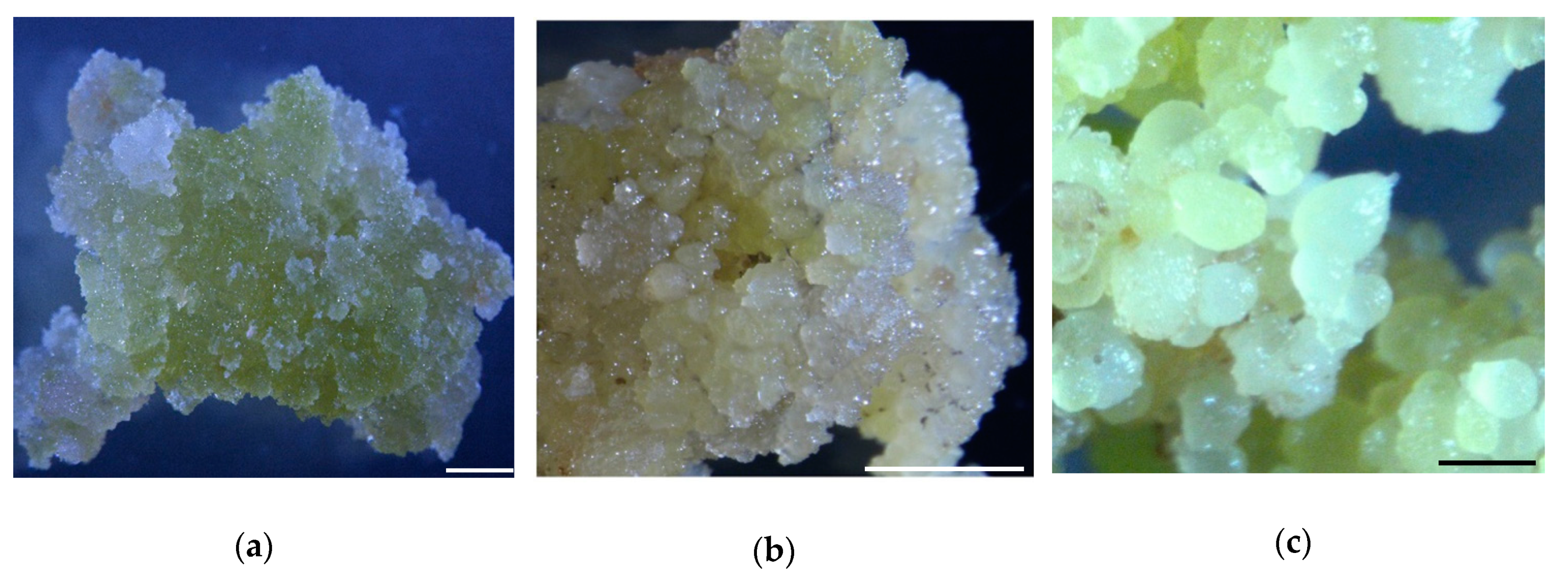
Figure 1. Samples used for proteomic assays: (a) Nonembryogenic calli; (b) Primary embryogenic calli; (c) Globular embryos. Bar (a,b) = 2.5 mm; bar (c) = 0.5 mm.
Ijms 20 01691 g001
Figure 2. Experimental strategy for quantitative proteome analysis and quality control validation of MS data: (a) Mass delta of all identified peptides; (b) Average peptide mass error; (c) Length distribution of all identified peptides. NEC: Nonembryogenic calli; PEC: Primary embryogenic calli; GE: Globular embryos. Each staged sample was prepared for three biological replicates.
Ijms 20 01691 g002
Figure 3. Subcellular functional annotation and GO functional classification of identified proteins. (a) Subcellular locations of identified proteins; (b) GO annotation in terms of cellular component; (c) GO annotation in terms of molecular function; (d) GO annotation in terms of biological process. GO: Gene Ontology.
Ijms 20 01691 g003aIjms 20 01691 g003b
Figure 4. Distribution of differentially accumulated proteins (DAPs): (a) Number of up(red)- and down(green)-regulated DAPs in GE vs. NEC, GE vs. PEC and PEC vs. NEC; (b) Venn diagram to show the distribution of DAPs between PEC vs. NEC (blue circle), GE vs. PEC (yellow circle) and GE vs. NEC (green circle). (c) Expression patterns of DAPs. NEC: Nonembryogenic calli; PEC: Primary embryogenic calli; GE: Globular embryos.
Ijms 20 01691 g004
Figure 5. GO functional cluster of differentially accumulated proteins (DAPs) in PEC vs. NEC, GE vs. PEC and GE vs. NEC: (a) GO functional cluster of DAPs in the molecular function; (b) GO cluster of DAPs in the cellular component; (c) GO functional cluster of DAPs in the biological process. NEC: Nonembryogenic calli; PEC: Primary embryogenic calli; GE: Globular embryos; GO: Gene Ontology.
Ijms 20 01691 g005aIjms 20 01691 g005bIjms 20 01691 g005c
Figure 6. KEGG cluster and pathway enrichment analysis of DAPs: (a) KEGG clusters in PEC vs. NEC, GE vs. PEC and GE vs. NEC; (b) Pathway enrichment in PEC vs. NEC; (c) Pathway enrichment in GE vs. PEC; (d) Pathway enrichment in GE vs. NEC. The pathway enrichment statistical analysis was performed by Fisher’s exact test. The X-axis is folded enrichment; the y-axis is enrichment pathway. The mapping is the protein number. NEC: Nonembryogenic calli; PEC: Primary embryogenic calli; GE: Globular embryos; KEGG: Kyoto encyclopedia of genes and genomes
Ijms 20 01691 g006aIjms 20 01691 g006bIjms 20 01691 g006c
Figure 7. Protein domain enrichment analysis of the differentially accumulated proteins (DAPs) in PEC vs. NEC, GE vs. PEC and GE vs. NEC. NEC: Nonembryogenic calli; PEC: Primary embryogenic calli; GE: Globular embryos.
Ijms 20 01691 g007
Figure 8. GO terms of differentially accumulated proteins (DAPs) in the biological process and the molecular function: (a) The biological process of DAPs in PEC vs. NEC; (b) The biological process and the molecular function of DAPs in GE vs. PEC; (c) The biological process of DAPs in GE vs. NEC. NEC: Nonembryogenic calli; PEC: Primary embryogenic calli; GE: Globular embryos; GO: Gene Ontology.
Ijms 20 01691 g008aIjms 20 01691 g008b
Figure 9. Comparative and complementary proteome of the candidate DAPs in stage of NEC and PEC. Significant differences in expression level were indicated by “*”. DAPs: differentially accumulated proteins; NEC: Nonembryogenic calli; PEC: Primary embryogenic calli; GE: Globular embryos.
Ijms 20 01691 g009
Table 1. MS/MS spectrum database search analysis summary.
Total SpectrumMatched SpectrumPeptidesUnique PeptidesIdentified ProteinsQuantifiable Proteins
360,72074,579 (20.7%)45,06227,67393696730
Table 2. Significantly representative SE regulatory DAPs in PEC vs. NEC, GE vs. PEC and GE vs. NEC.
Gene IDGene NameProtein IDProtein DescriptionPathway AnnotationPEC/NEC RatioGE/PEC RatioGE/NEC Ratio
LOC107907377PIN2A0A120KAE0Auxin efflux carrier componentAuxin signal2.36
LOC107909506GH3.17A0A1U8JQJ4Indole-3-acetic acid-amido synthetase GH3.17-like isoform X2Auxin signal2.14
LOC107948437ETR1A0A1U8NHA4ethylene receptor-like isoform X1Ethylene signal2.023.705
LOC107938108GASL1M1GN42GA-stimulated transcript-like protein 1GA signal0.250.270.068
LOC107955576GASL4M1GMV2GA-stimulated transcript-like protein 4GA signal3.00
LOC107950128PYR1A0A1U8NR07Abscisic acid receptor PYR1-likeABA signal2.312.361
LOC107893363At5g01020A0A1U8I6G4serine/threonine-protein kinase At5g01020-likeSignal transduction0.500.482
LOC107897915A0A1U8IRF6A-kinase anchor protein 12-like isoform X2Signal transduction0.440.459
LOC107909143A0A1U8JP78leucine-rich repeat receptor-like protein kinase PXC2Signal transduction0.450.36
LOC107945188BAM3A0A1U8N9I0leucine-rich repeat receptor-like Ser/Thr -protein kinase BAM3Signal transduction2.03
LOC107935259PCKAA0A1U8M980phosphoenolpyruvate carboxykinase [ATP]-likeSignal transduction2.080.38
LOC107943515At1g56140A0A1U8N4H5probable LRR receptor-like serine/threonine-protein kinase At1g56140Signal transduction0.290.215
LOC107931208TPK1A0A1U8LYM7thiamine pyrophosphokinase 1-like isoform X1Signal transduction0.460.469
LOC107905700PFKA0A1U8JGW8ATP-dependent 6-phosphofructokinaseSignal transduction2.552.504
LOC107943957PV42AA0A1U8N623SNF1-related protein kinase regulatory subunit gamma-like PV42aSignal transduction2.832.757
LOC107937641CPK11A0A1U8MGW7calcium-dependent protein kinase 11-likeSignal transduction2.253.84
LOC107930954CML27A0A1U8LUL1probable calcium-binding protein CML27Signal transduction4.162.88
LOC107916423RHN1A0A1U8KFK5ras-related protein RHN1-likeSignal transduction0.470.333
LOC107889787A0A1U8HV05Embryonic protein DC-8-likeSomatic embryogenesis related proteins3.584.522
LOC107937048Lea2A-AQ03791Embryogenesis abundant proteinSomatic embryogenesis related proteins4.424.746
LOC107941722WOX9A0A1U8MVD7WUSCHEL-related homeobox 9-likeTranscription factor2.58
LOC107905698NFYB6A0A1U8JC47Nuclear transcription factor Y subunit B-6Transcription factor3.222.076.661
bHLH4W5XUY9BHLH4 transcription factorTranscription factor2.16
LOC107920272NFYB9A0A1U8KSD1nuclear transcription factor Y subunit B-9-likeTranscription factor4.072.509
LOC107931333 A0A1U8LVZ2transcription factor HBP-1b (C38)-likeTranscription factor2.40
LOC107924015PHL1A0A1U8L8P3Protein PHR1-LIKE 1-likeTranscription factor0.150.166
LOC107891610At1g07170A0A1U8I119PHD finger-like domain-containing protein 5BZinc finger3.494.21
LOC107909066NERDA0A1U8JUI6zinc finger CCCH domain-containing protein 19-like isoform X2Zinc finger2.03
LOC107927097TAF15BA0A1U8LG36transcription TFIID subunit 15b-likeZinc finger0.44
LOC107962890ZHD5A0A1U8PUW9zinc-finger homeodomain protein 5-likeZinc finger2.935.554
LOC107890886AGO1A0A1U8HY77protein argonaute 1-like isoform X2Posttranscriptional regulation8.446.734
LOC107906203AGO4A0A1U8JEA7protein argonaute 4-likePosttranscriptional regulation2.382.426
LOC107962954HEN1A0A1U8PXD5small RNA 2’-O-methyltransferase-like isoform X4Posttranscriptional regulation2.06
LOC107891032IDM1A0A1U8HYR9increased DNA methylation 1-like isoform X4Modification-related protein2.620.49
LOC107906306MMT1A0A1U8JEK1Methionine S-methyltransferaseModification-related protein0.460.458
LOC107948568SUVH4A0A1U8NHS0histone-lysine N-methyltransferase, H3 lysine-9 specific SUVH4-like isoform X2Modification-related protein0.280.196
LOC107943854CCOAOMTA0A1U8N5R4caffeoyl-CoA O-methyltransferase -likeModification-related protein0.350.363
LOC107953938EMB1691A0A1U8P3T9methyltransferase-like protein 1Modification-related protein2.332.774
LOC107916882IAMT1A0A1U8KGS5indole-3-acetate O-methyltransferase 1Modification-related protein2.32
LOC107958653A0A1U8PI31chromatin modification-related protein MEAF6-like isoform X3Modification-related protein3.485.641
LOC107926365A0A1U8LDK7RNA cytidine acetyltransferaseModification-related protein2.172.333
LOC107960303A0A1U8PLN7Acetyltransferase component of pyruvate dehydrogenase complexModification-related protein0.480.404
LOC107947121UBR7A0A1U8NDR8putative E3 ubiquitin-protein ligase UBR7Modification-related protein2.902.679
LOC107959749RUB2A0A1U8PJK7ubiquitin-NEDD8-like protein RUB2Modification-related protein0.260.286
LOC107938100A0A1U8MII2PhosphotransferaseModification-related protein0.420.476
LOC107898863CYP86B1A0A1U8IP72Cytochrome P450 86B1-likeFatty acid4.512.388
LOC107922796CYP86A8A0A1U8L513Cytochrome P450 86A8-likeFatty acid2.02
LOC107915850PIP2-5A0A1U8KIL6probable aquaporin PIP2-5Aquaporins2.58
LOC107898442TIP3-2A0A1U8IU16probable aquaporin TIP3-2Aquaporins2.299.086
LOC107963873GhPIP2;10D8FSK4Aquaporin PIP210Aquaporins0.140.102
GhTIP1;4D8FSK6Aquaporin TIP14Aquaporins0.200.195
LOC107934987
LOC107944588
PIP1;4G8XV51PIP proteinAquaporins0.220.109
DAPs: differentially accumulated proteins; NEC: Nonembryogenic calli; PEC: Primary embryogenic calli; GE: Globular embryos; GO: Gene Ontology.