Int. J. Mol. Sci. 2020, 21(13), 4620; https://doi.org/10.3390/ijms21134620 - 29 Jun 2020
Cited by 122 | Viewed by 14298
Abstract
Apigenin (4′,5,7-trihydroxyflavone, flavonoid) is a phenolic compound that is known to reduce the risk of chronic disease owing to its low toxicity. The first study on apigenin analyzed its effect on histamine release in the 1950s. Since then, anti-mutation and antitumor properties of
[...] Read more.
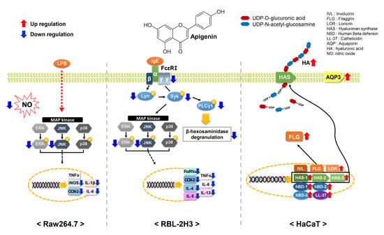
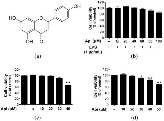
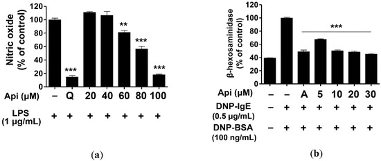
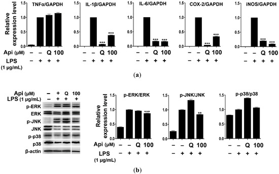
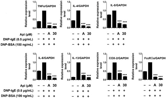
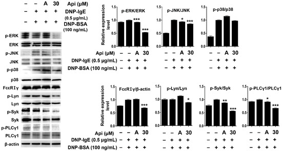
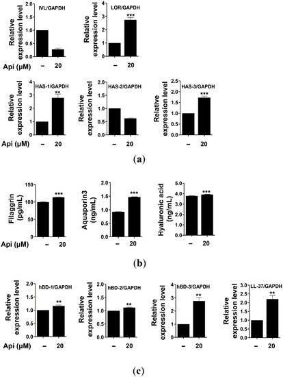
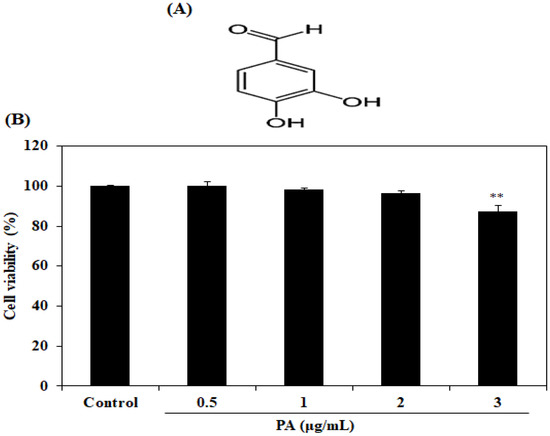
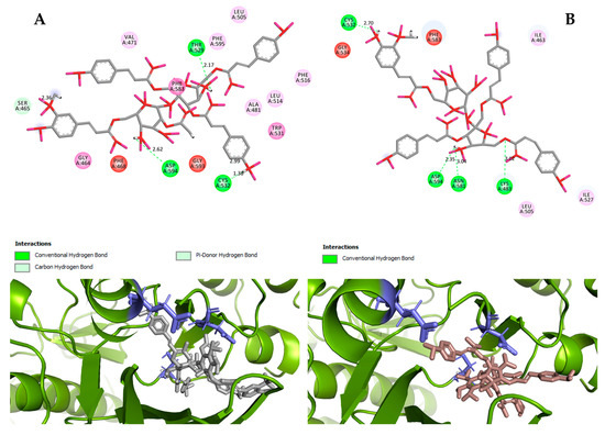
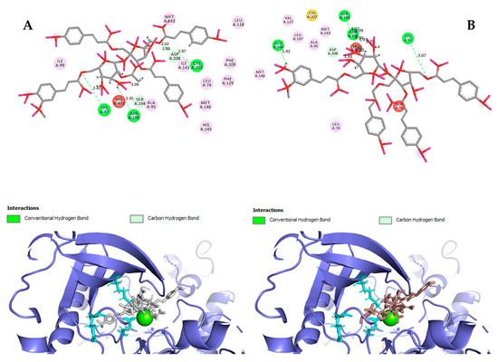
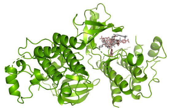
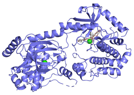
Apigenin (4′,5,7-trihydroxyflavone, flavonoid) is a phenolic compound that is known to reduce the risk of chronic disease owing to its low toxicity. The first study on apigenin analyzed its effect on histamine release in the 1950s. Since then, anti-mutation and antitumor properties of apigenin have been widely reported. In the present study, we evaluated the apigenin-mediated amelioration of skin disease and investigated its applicability as a functional ingredient, especially in cosmetics. The effect of apigenin on RAW264.7 (murine macrophage), RBL-2H3 (rat basophilic leukemia), and HaCaT (human immortalized keratinocyte) cells were analyzed. Apigenin (100 μM) significantly inhibited nitric oxide (NO) production, cytokine expression (interleukin (IL)-1β, IL6, cyclooxygenase (COX)-2, and inducible nitric oxide synthase [iNOS]), and phosphorylation of mitogen-activated protein kinase (MAPK) signal molecules, including extracellular signal-regulated kinase (ERK) and c-Jun N-terminal protein kinase (JNK) in RAW264.7 cells. Apigenin (30 μM) also inhibited the phosphorylation of signaling molecules (Lyn, Syk, phospholipase Cγ1, ERK, and JNK) and the expression of high-affinity IgE receptor FcεRIα and cytokines (tumor necrosis factor (TNF)-α, IL-4, IL-5, IL-6, IL-13, and COX-2) that are known to induce inflammation and allergic responses in RBL-2H3 cells. Further, apigenin (20 μM) significantly induced the expression of filaggrin, loricrin, aquaporin-3, hyaluronic acid, hyaluronic acid synthase (HAS)-1, HAS-2, and HAS-3 in HaCaT cells that are the main components of the physical barrier of the skin. Moreover, it promoted the expression of human β-defensin (HBD)-1, HBD-2, HBD-3, and cathelicidin (LL-37) in HaCaT cells. These antimicrobial peptides are known to play an important role in the skin as chemical barriers. Apigenin significantly suppressed the inflammatory and allergic responses of RAW264.7 and RBL cells, respectively, and would, therefore, serve as a potential prophylactic and therapeutic agent for immune-related diseases. Apigenin could also be used to improve the functions of the physical and chemical skin barriers and to alleviate psoriasis, acne, and atopic dermatitis.
Full article
(This article belongs to the Special Issue Bioactive Phenolics and Polyphenols 2020)
►
Show Figures
Graphical abstract


