Corneal UV Protective Effects of a Topical Antioxidant Formulation: A Pilot Study on In Vivo Rabbits

 , , , and
Abstract
:1. Introduction
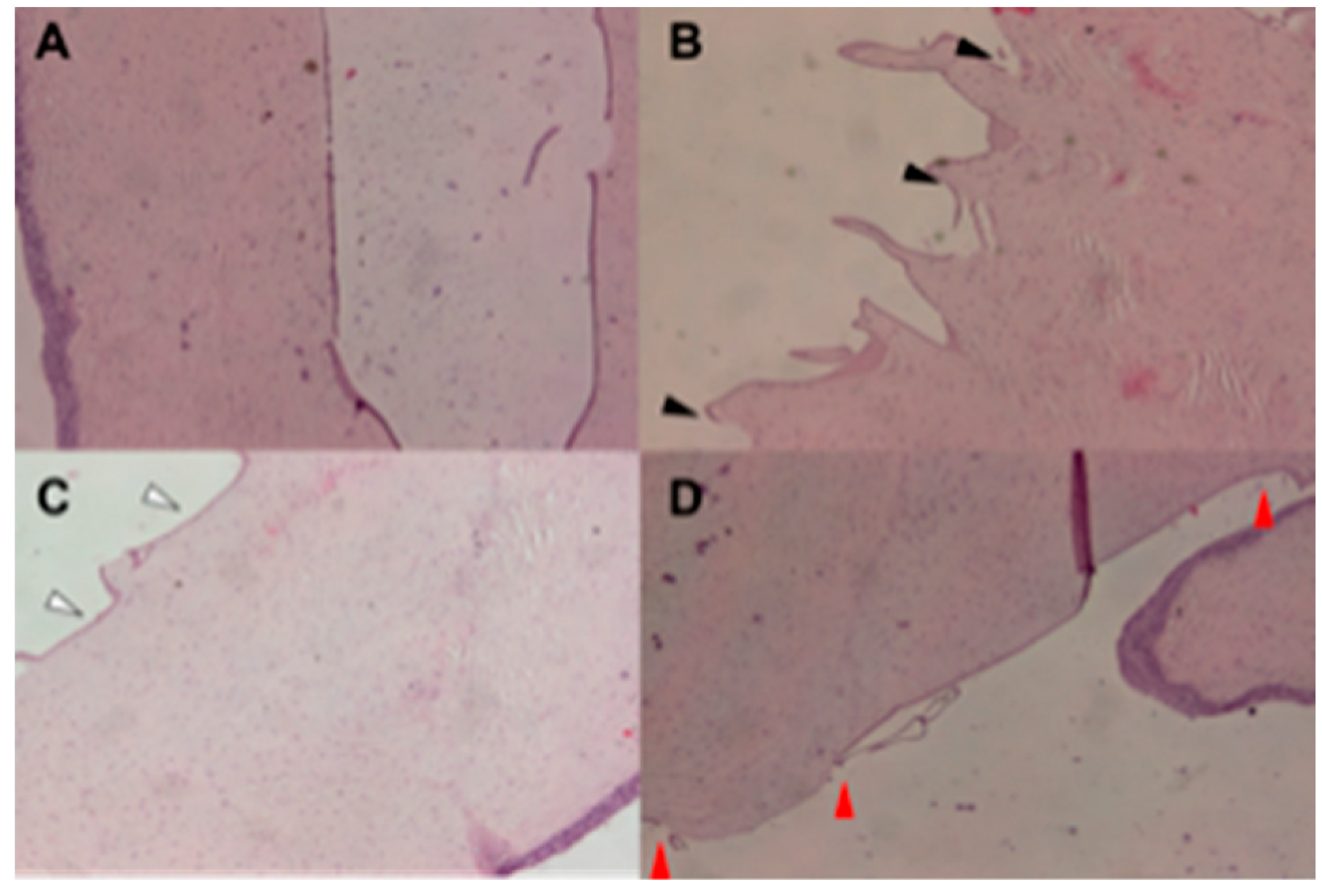
2. Results
3. Discussion
4. Materials and Methods
4.1. Animals
4.2. UV Irradiation
4.3. Ophthalmic Preparation
4.4. Histochemistry
4.5. Statistical Analysis
5. Conclusions
Author Contributions
Funding
Acknowledgments
Conflicts of Interest
References
- Cejka, C.; Luyckx, J.; Cejková, J. Central corneal thickness considered an index of corneal hydration of the UVB irradiated rabbit cornea as influenced by UVB absorber. Physiol. Res. 2012, 61, 299–306. [Google Scholar] [CrossRef] [PubMed]
- Wang, F.; Gao, Q.; Hu, L.; Gao, N.; Ge, T.; Yu, J.; Liu, Y. Risk of eye damage from the wavelength-dependent biologically effective UVB spectrum irradiances. PLoS ONE 2012, 7, e52259. [Google Scholar] [CrossRef] [PubMed]
- Abengózar-Vela, A.; Arroyo, C.; Reinoso, R.; Enríquez-de-Salamanca, A.; Corell, A.; González-García, M.J. In vitro model for predicting the protective effect of ultraviolet-blocking contact lens in human corneal epithelial cells. Curr. Eye Res. 2015, 40, 792–799. [Google Scholar] [CrossRef] [PubMed]
- Chaney, E.K.; Sliney, D.H. Re-evaluation of the ultraviolet hazard action spectrum—The impact of spectral bandwidth. Health Phys. 2005, 89, 322–332. [Google Scholar] [CrossRef] [PubMed]
- Chalam, K.V.; Khetpal, V.; Rusovici, R.; Balaiya, S. A review: Role of ultraviolet radiation in age-related macular degeneration. Eye Contact Lens 2011, 37, 225–232. [Google Scholar] [CrossRef]
- Coroneo, M. Ultraviolet radiation and the anterior eye. Eye Contact Lens Sci. Clin. Pract. 2011, 37, 214–224. [Google Scholar] [CrossRef]
- Saccà, S.; Cutolo, C.; Ferrari, D.; Corazza, P.; Traverso, C. The eye, oxidative damage and polyunsaturated fatty acids. Nutrients 2018, 10, 668. [Google Scholar] [CrossRef] [Green Version]
- Bashir, H.; Seykora, J.; Lee, V. Invisible shield: Review of the corneal epithelium as a barrier to UV radiation, pathogens, and other environmental stimuli. J. Ophthalmic Vis. Res. 2017, 12, 305. [Google Scholar]
- Zigman, S. Ocular light damage. Photochem. Photobiol. 1993, 57, 1060–1068. [Google Scholar] [CrossRef]
- Tenkate, T.D. Occupational exposure to ultraviolet radiation: A health risk assessment. Rev. Environ. Health 1999, 14, 187–209. [Google Scholar] [CrossRef]
- Kabuyama, Y.; Homma, M.K.; Kurosaki, T.; Homma, Y. Early signaling events induced by 280-nm UV irradiation. Eur. J. Biochem. 2002, 269, 664–670. [Google Scholar] [CrossRef] [PubMed]
- Ma, W.; Wlaschek, M.; Tantcheva-Poór, I.; Schneider, L.A.; Naderi, L.; Razi-Wolf, Z.; Schüller, J.; Scharffetter-Kochanek, K. Chronological ageing and photoageing of the fibroblasts and the dermal connective tissue. Clin. Exp. Dermatol. 2001, 26, 592–599. [Google Scholar] [CrossRef] [PubMed]
- Kochevar, I.E. Cytotoxicity and mutagenicity of excimer laser radiation. Lasers Surg. Med. 1989, 9, 440–445. [Google Scholar] [CrossRef]
- Tanaka, Y.; Nakayama, J. Upregulated epidermal growth factor receptor expression following near-infrared irradiation simulating solar radiation in a three-dimensional reconstructed human corneal epithelial tissue culture model. Clin. Interv. Aging 2016, 11, 1027–1033. [Google Scholar] [CrossRef] [PubMed] [Green Version]
- Mesa, R.; Bassnett, S. UV-B-Induced DNA damage and repair in the mouse lens. Invest. Ophthalmol. Vis. Sci. 2013, 54, 6789. [Google Scholar] [CrossRef] [Green Version]
- Behar-Cohen, F.; Baillet, G.; de Ayguavives, T.; Garcia, P.O.; Krutmann, J.; Peña-García, P.; Reme, C.; Wolffsohn, J.S. Ultraviolet damage to the eye revisited: Eye-sun protection factor (E-SPF®), a new ultraviolet protection label for eyewear. Clin. Ophthalmol. 2014, 8, 87–104. [Google Scholar] [CrossRef] [Green Version]
- Youssef, P.N.; Sheibani, N.; Albert, D.M. Retinal light toxicity. Eye (Lond) 2011, 25, 1–14. [Google Scholar] [CrossRef] [Green Version]
- Ortwerth, B.J.; Bhattacharyya, J.; Shipova, E. Tryptophan metabolites from young human lenses and the photooxidation of ascorbic acid by UVA light. Invest. Ophthalmol. Vis. Sci. 2009, 50, 3311–3319. [Google Scholar] [CrossRef]
- An, M.-J.; Kim, C.-H.; Nam, G.-Y.; Kim, D.-H.; Rhee, S.; Cho, S.-J.; Kim, J.-W. Transcriptome analysis for UVB-induced phototoxicity in mouse retina. Environ. Toxicol. 2018, 33, 52–62. [Google Scholar] [CrossRef]
- Gaillard, E.R.; Zheng, L.; Merriam, J.C.; Dillon, J. Age-related changes in the absorption characteristics of the primate lens. Invest. Ophthalmol. Vis. Sci. 2000, 41, 1454–1459. [Google Scholar]
- Gaillard, E.R.; Merriam, J.; Zheng, L.; Dillon, J. Transmission of light to the young primate retina: Possible implications for the formation of lipofuscin. Photochem. Photobiol. 2011, 87, 18–21. [Google Scholar] [CrossRef] [PubMed]
- Cejková, J.; Stípek, S.; Crkovská, J.; Ardan, T.; Pláteník, J.; Cejka, C.; Midelfart, A. UV Rays, the prooxidant/antioxidant imbalance in the cornea and oxidative eye damage. Physiol. Res. 2004, 53, 1–10. [Google Scholar] [PubMed]
- Cejka, C.; Pláteník, J.; Guryca, V.; Sirc, J.; Michálek, J.; Brůnová, B.; Cejková, J. Light absorption properties of the rabbit cornea repeatedly irradiated with UVB rays. Photochem. Photobiol. 2007, 83, 652–657. [Google Scholar] [CrossRef]
- Podskochy, A.; Gan, L.; Fagerholm, P. Apoptosis in UV-exposed rabbit corneas. Cornea 2000, 19, 99–103. [Google Scholar] [CrossRef]
- Sidjanin, D.; Zigman, S.; Reddan, J. DNA damage and repair in rabbit lens epithelial cells following UVA radiation. Curr. Eye Res. 1993, 12, 773–781. [Google Scholar] [CrossRef] [PubMed]
- Hammond, B.R., Jr.; Renzi-Hammond, L. Individual variation in the transmission of UVB radiation in the young adult eye. PLoS ONE 2018, 13, e0199940. [Google Scholar] [CrossRef]
- Cejková, J.; Stípek, S.; Crkovská, J.; Ardan, T.; Midelfart, A. Reactive oxygen species (ROS)-generating oxidases in the normal rabbit cornea and their involvement in the corneal damage evoked by UVB rays. Histol. Histopathol. 2001, 16, 523–533. [Google Scholar]
- Cejková, J.; Ardan, T.; Filipec, M.; Midelfart, A. Xanthine oxidoreductase and xanthine oxidase in human cornea. Histol. Histopathol. 2002, 17, 755–760. [Google Scholar]
- Buddi, R.; Lin, B.; Atilano, S.R.; Zorapapel, N.C.; Kenney, M.C.; Brown, D.J. Evidence of oxidative stress in human corneal diseases. J. Histochem. Cytochem. 2002, 50, 341–351. [Google Scholar] [CrossRef]
- Najjar, D.M.; Awwad, S.T.; Zein, W.M.; Haddad, W.F. Assessment of the corneal endothelium in acute ultraviolet keratitis. Med. Sci. Monit. 2006, 12, MT23–MT25. [Google Scholar]
- Vizzarri, F.; Palazzo, M.; Bartollino, S.; Casamassima, D.; Parolini, B.; Troiano, P.; Caruso, C.; Costagliola, C. Effects of an antioxidant protective topical formulation on eye exposed to ultraviolet-irradiation: A study in rabbit animal model. Physiol. Res. 2018, 67, 457–464. [Google Scholar] [CrossRef]
- Cejka, C.; Cejkova, J. Oxidative stress to the cornea, changes in corneal optical properties, and advances in treatment of corneal oxidative injuries. Oxid. Med. Cell. Longev. 2015, 2015, 1–10. [Google Scholar] [CrossRef] [PubMed] [Green Version]
- Wakamatsu, T.H.; Dogru, M.; Ayako, I.; Takano, Y.; Matsumoto, Y.; Ibrahim, O.M.A.; Okada, N.; Satake, Y.; Fukagawa, K.; Shimazaki, J.; et al. Evaluation of lipid oxidative stress status and inflammation in atopic ocular surface disease. Mol. Vis. 2010, 16, 2465–2475. [Google Scholar] [PubMed]
- Seen, S.; Tong, L. Dry eye disease and oxidative stress. Acta Ophthalmol. 2018, 96, e412–e420. [Google Scholar] [CrossRef] [PubMed] [Green Version]
- Zigman, S.; McDaniel, T.; Schultz, J.B.; Reddan, J.; Meydani, M. Damage to cultured lens epithelial cells of squirrels and rabbits by UV-A (99.9%) plus UV-B (0.1%) radiation and alpha tocopherol protection. Mol. Cell. Biochem. 1995, 143, 35–46. [Google Scholar] [CrossRef]
- Yam, J.C.S.; Kwok, A.K.H. Ultraviolet light and ocular diseases. Int. Ophthalmol. 2014, 34, 383–400. [Google Scholar] [CrossRef]
- Norval, M.; Cullen, A.P.; de Gruijl, F.R.; Longstreth, J.; Takizawa, Y.; Lucas, R.M.; Noonan, F.P.; van der Leun, J.C. The effects on human health from stratospheric ozone depletion and its interactions with climate change. Photochem. Photobiol. Sci. 2007, 6, 232. [Google Scholar] [CrossRef]
- Hepel, M.; Stobiecka, M.; Peachey, J.; Miller, J. Intervention of glutathione in pre-mutagenic catechol-mediated DNA damage in the presence of copper(II) ions. Mutat. Res. Fundam. Mol. Mech. Mutagen. 2012, 735, 1–11. [Google Scholar] [CrossRef]
- Gwon, A. The rabbit in cataract/IOL surgery. In Animal Models in Eye Research; Academic Press: Cambridge, MA, USA, 2008; pp. 184–204. [Google Scholar]
- Delic, N.C.; Lyons, J.G.; Di Girolamo, N.; Halliday, G.M. Damaging effects of ultraviolet radiation on the cornea. Photochem. Photobiol. 2017, 93, 920–929. [Google Scholar] [CrossRef] [Green Version]
- Kaye, G.I. Studies on the cornea. III. The fine structure of the frog cornea and the uptake and transport of colloidal particles by the cornea in vivo. J. Cell Biol. 1962, 15, 241–258. [Google Scholar] [CrossRef]
- Bartollino, S.; Palazzo, M.; Semeraro, F.; Parolini, B.; Caruso, C.; Merolla, F.; Guerra, G.; Costagliola, C. Effects of an antioxidant protective topical formulation on retinal tissue of UV-exposed rabbits. Int. Ophthalmol. 2020, 40, 925–933. [Google Scholar] [CrossRef] [PubMed]
- Giblin, F.J.; Lin, L.-R.; Simpanya, M.F.; Leverenz, V.R.; Fick, C.E. A class I UV-blocking (senofilcon A) soft contact lens prevents UVA-induced yellow fluorescence and NADH loss in the rabbit lens nucleus in vivo. Exp. Eye Res. 2012, 102, 17–27. [Google Scholar] [CrossRef] [PubMed] [Green Version]
- Rubinfeld, R.S.; Caruso, C.; Ostacolo, C. Corneal cross-linking: The science beyond the myths and misconceptions. Cornea 2019, 38, 780–790. [Google Scholar] [CrossRef] [PubMed]
- Caruso, C.; Epstein, R.L.; Ostacolo, C.; Pacente, L.; Troisi, S.; Barbaro, G. Customized corneal cross-linking-a mathematical model. Cornea 2017, 36, 600–604. [Google Scholar] [CrossRef] [Green Version]
- Caruso, C.; Epstein, R.L.; Troiano, P.; Ostacolo, C.; Barbaro, G.; Pacente, L.; Bartollino, S.; Costagliola, C. Topography and pachymetry guided, rapid epi-on corneal cross-linking for keratoconus. Cornea 2019, 39, 56–62. [Google Scholar] [CrossRef]
- Wollensak, G.; Aurich, H.; Wirbelauer, C.; Sel, S. Significance of the riboflavin film in corneal collagen crosslinking. J. Cataract Refract. Surg. 2010, 36, 114–120. [Google Scholar] [CrossRef]
- Chapter 3—Meteorological Data. Available online: http://www.fao.org/3/X0490E/x0490e07.htm (accessed on 16 July 2019).
- Caruso, C.; Barbaro, G.; Epstein, R.L.; Tronino, D.; Ostacolo, C.; Sacchi, A.; Pacente, L.; Del Prete, A.; Sala, M.; Troisi, S. Corneal cross-linking: Evaluating the potential for a lower power, shorter duration treatment. Cornea 2016, 35, 659–662. [Google Scholar] [CrossRef] [Green Version]
- Schumacher, S.; Mrochen, M.; Wernli, J.; Bueeler, M.; Seiler, T. Optimization model for UV-riboflavin corneal cross-linking. Invest. Ophthalmol. Vis. Sci. 2012, 53, 762. [Google Scholar] [CrossRef] [Green Version]
- United States Environment Protection Agency. Sun Safety. Available online: https://www.epa.gov/sunsafety (accessed on 15 October 2019).
- Hwang, H.S.; Kim, M.S. Ultraviolet-visible light spectral transmittance of rabbit corneas after riboflavin/ultraviolet-A (365 nm) corneal collagen cross-linking. Mol. Vis. 2013, 19, 2113–2123. [Google Scholar]
- Horiuchi, S.; Hirano, H.; Ono, S. Reduced and oxidized glutathione concentrations in the lenses of riboflavin-deficient rats. J. Nutr. Sci. Vitaminol. 1984, 30, 401–403. [Google Scholar] [CrossRef]
- Constantinides, P.P.; Han, J.; Davis, S.S. Advances in the use of tocols as drug delivery vehicles. Pharm. Res. 2006, 23, 243–255. [Google Scholar] [CrossRef]
- Costagliola, C.; Libondi, T.; Menzione, M.; Rinaldi, E.; Auricchio, G. Vitamin E and red blood cell glutathione. Metabolism 1985, 34, 712–714. [Google Scholar] [CrossRef]
- Costagliola, C.; Iuliano, G.; Menzione, M.; Rinaldi, E.; Vito, P.; Auricchio, G. Effect of vitamin E on glutathione content in red blood cells, aqueous humor and lens of humans and other species. Exp. Eye Res. 1986, 43, 905–914. [Google Scholar] [CrossRef]
- Costagliola, C.; Menzione, M. Effect of vitamin E on the oxidative state of glutathione in plasma. Clin. Physiol. Biochem. 1990, 8, 140–143. [Google Scholar]
- Caruso, C.; Porta, A.; Tosco, A.; Eletto, D.; Pacente, L.; Bartollino, S.; Costagliola, C. A Novel vitamin E TPGS-based formulation enhances chlorhexidine bioavailability in corneal layers. Pharmaceutics 2020, 12, 642. [Google Scholar] [CrossRef] [PubMed]
- Hajibabaei, K. Antioxidant properties of vitamin E. Ann. Res. Antioxidants 2016, 1, 2–3. [Google Scholar]
- Tessem, M.B.; Bathen, T.F.; Čejková, J.; Midelfart, A. Effect of UV-A and UV-B irradiation on the metabolic profile of aqueous humor in rabbits analyzed by 1H NMR spectroscopy. Investig. Ophthalmol. Vis. Sci. 2005, 46, 776–781. [Google Scholar] [CrossRef] [PubMed] [Green Version]
- Fukuda, M.; Sasaki, K. Changes in the antibacterial activity of melanin-bound drugs. Ophthalmic Res. 1990, 22, 123–127. [Google Scholar] [CrossRef]
- Fukuda, M.; Sasaki, K. Differences between albino and pigmented rabbit eyes in the intraocular pharmacokinetics of sparfloxacin. Drugs 1995, 49, 314–316. [Google Scholar] [CrossRef]
- Toledo, C.R.; Pereira, V.V.; Dourado, L.F.N.; Paiva, M.R.B.; Silva-Cunha, A. Corosolic acid: Antiangiogenic activity and safety of intravitreal injection in rats eyes. Doc. Ophthalmol. 2019, 138, 181–194. [Google Scholar] [CrossRef]
- Nappi, C.; Di Spiezio Sardo, A.; Guerra, G.; Di Carlo, C.; Bifulco, G.; Acunzo, G.; Sammartino, A.; Galli, V. Comparison of intranasal and transdermal estradiol on nasal mucosa in postmenopausal women. Menopause 2004, 11, 447–455. [Google Scholar] [CrossRef]
- Nappi, C.; Di Spiezio Sardo, A.; Guerra, G.; Bifulco, G.; Testa, D.; Di Carlo, C. Functional and morphologic evaluation of the nasal mucosa before and after hormone therapy in postmenopausal women with nasal symptoms. Fertil. Steril. 2003, 80, 669–671. [Google Scholar] [CrossRef]
- Rossi, A.; Pace, S.; Tedesco, F.; Pagano, E.; Guerra, G.; Troisi, F.; Werner, M.; Roviezzo, F.; Zjawiony, J.K.; Werz, O.; et al. The hallucinogenic diterpene salvinorin A inhibits leukotriene synthesis in experimental models of inflammation. Pharmacol. Res. 2016, 106, 64–71. [Google Scholar] [CrossRef] [PubMed]

| CTRL | IG | G30 | G60 | |
|---|---|---|---|---|
| Fold increase vs. CTRL | 1 | 1.768667 | 1.40292 | 1.676709 |
| Mean | 423,600 | 749,207 | 594,276 | 710,254 |
| Median | 427,500 | 759,386 | 600,771 | 714,339 |
| MIN | 387,000 | 564,455 | 536,799 | 633,306 |
| MAX | 456,000 | 841,418 | 623,610 | 751,708 |
| RANGE | 69,000 | 276,963 | 86,811 | 118,402 |
| Student’s t Test p Value | |
|---|---|
| 0.0000000005 | CTRL vs. IG |
| 0.00000000001 | CTRL vs. G30 |
| 0.000000000000012 | CTRL vs. G60 |
| 0.00003 | IG vs. G30 |
| 0.19 | IG vs. G60 |
| Cell Count. | ||||
|---|---|---|---|---|
| CTRL | IG | G30 | G60 | |
| Count | 33 | 143 | 76 | 141 |
| Area | 24,393 | 49,504 | 34,611 | 49,504 |
| Density | 0.001352847 | 0.002889 | 0.002196 | 0.002848 |
| Relative Cell Density | 1 | 2.14 | 1.62 | 2.11 |
© 2020 by the authors. Licensee MDPI, Basel, Switzerland. This article is an open access article distributed under the terms and conditions of the Creative Commons Attribution (CC BY) license (http://creativecommons.org/licenses/by/4.0/).
Share and Cite
Palazzo, M.; Vizzarri, F.; Ondruška, L.; Rinaldi, M.; Pacente, L.; Guerra, G.; Merolla, F.; Caruso, C.; Costagliola, C. Corneal UV Protective Effects of a Topical Antioxidant Formulation: A Pilot Study on In Vivo Rabbits. Int. J. Mol. Sci. 2020, 21, 5426. https://doi.org/10.3390/ijms21155426
Palazzo M, Vizzarri F, Ondruška L, Rinaldi M, Pacente L, Guerra G, Merolla F, Caruso C, Costagliola C. Corneal UV Protective Effects of a Topical Antioxidant Formulation: A Pilot Study on In Vivo Rabbits. International Journal of Molecular Sciences. 2020; 21(15):5426. https://doi.org/10.3390/ijms21155426
Chicago/Turabian StylePalazzo, Marisa, Francesco Vizzarri, Lubomir Ondruška, Michele Rinaldi, Luigi Pacente, Germano Guerra, Francesco Merolla, Ciro Caruso, and Ciro Costagliola. 2020. "Corneal UV Protective Effects of a Topical Antioxidant Formulation: A Pilot Study on In Vivo Rabbits" International Journal of Molecular Sciences 21, no. 15: 5426. https://doi.org/10.3390/ijms21155426